Báo cáo khoa học: Human telomeric G-quadruplex: structures of DNA and RNA sequences ppt
Bạn đang xem bản rút gọn của tài liệu. Xem và tải ngay bản đầy đủ của tài liệu tại đây (547.04 KB, 11 trang )
MINIREVIEW
Human telomeric G-quadruplex: structures of DNA and
RNA sequences
Anh Tua
ˆ
n Phan
School of Physical & Mathematical Sciences, Nanyang Technological University, Singapore
Introduction
Human telomeric DNA contains thousands of tandem
repeats of the G-rich (GGGTTA)
n
sequence [1], with a
3¢-end overhang of 100–200 nucleotides [2]. Telomeres
can be transcribed by DNA-dependent RNA polymer-
ase II, and telomeric-repeat-containing RNA mole-
cules, ranging from 100 to 9000 nucleotides, have been
detected in nuclear fractions [3–5]. Under physiological
ionic conditions, human telomeric DNA and RNA
G-rich sequences are capable of forming a four-
stranded helical structure, known as the G-quadruplex
[6–14], based on the stacking of multiple G•G•G•G
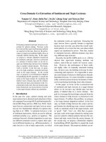
tetrads (or G-tetrads) [15] (Fig. 1A). Such a structure
might be important for telomere biology [5–14,16–19]
and a good target for drug design [5–14,19–21].
G-quadruplex structures formed by various G-rich
sequences have been reviewed previously [6–14]. This
minireview focuses on a simple topological and struc-
tural description of different DNA and RNA G-quad-
ruplexes formed by short and long human telomeric
sequences. Structural views on targeting these G-quad-
ruplexes by small molecules are also discussed. Finally,
the minireview highlights some future challenges for
structural studies of human telomeric G-quadruplexes.
The interactions of proteins with G-quadruplexes have
been studied [16–19], but the structural knowledge on
these interactions is still limited and is not covered
here. Accompanying minireviews in this series [22–24]
discuss the thermodynamic and kinetic properties of
human telomeric G-quadruplexes and the current
status of their targeting by small molecules.
Basics of G-quadruplex topologies
Oligonucleotides containing G-stretches can form
monomeric, dimeric or tetrameric G-quadruplexes by
folding ⁄ assembling one, two or four separate strands
Keywords
DNA; G-q uadru plex; G-quadruplex structure;
G-quadruplex topology; G-tetrad; G-tetrad
core; grooves in G-quadruplexes; human
telomere; loops in G-quadruplexes; RNA
Correspondence
A. T. Phan, Division of Physics & Applied
Physics, School of Physical & Mathematical
Sciences, Nanyang Technological University,
Singapore 637371
Fax: +65 6795 7981
Tel: +65 6514 1915
E-mail:
(Received 25 June 2009, revised
14 September 2009, accepted 6 October
2009)
doi:10.1111/j.1742-4658.2009.07464.x
Telomeres play an important role in cellular aging and cancer. Human
telomeric DNA and RNA G-rich sequences are capable of forming a four-
stranded structure, known as the G-quadruplex. Such a structure might be
important for telomere biology and a good target for drug design. This
minireview describes the structural diversity or conservation of DNA and
RNA human telomeric G-quadruplexes, discusses structural views on
targeting these G-quadruplexes and presents some future challenges for
structural studies.
FEBS Journal 277 (2010) 1107–1117 ª 2009 The Author Journal compilation ª 2009 FEBS 1107
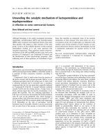
(see below). A G-quadruplex contains a G-tetrad core
(Fig. 2A–D), formed by the stacking of several tetrads
and supported by four backbone strands (or columns).
Linkers connecting these strands are called loops
(Fig. 2E–G). G-quadruplex structures are polymorphic
regarding the G-tetrad core and loops (Fig. 2).
Cations, such as K
+
and Na
+
, stabilize G-quadruplex-
es by coordinating the carbonyl groups of guanines at
the center of the G-tetrad core, and the preferred
G-quadruplex structures adopted by a G-rich sequence
depend on the nature of cations.
The G-tetrad core can be classified with regard to
two mutually related factors, the relative orientations
of the strands and the glycosidic conformations [anti
H
H
H
N
N
N
N
N
H
O
H
H
H
N
N
N
N
N
H
O
H
H
H
N
N
N
N
N
H
O
H
H
H
N
N
N
N
N
H
O
G
G
G
G
O
H
H
N
N
N
N
N
O
H
H
G
O
H
H
N
N
N
N
N
O
H
H
G
A
DE
BC
Fig. 1. (A) G-tetrad alignment. (B,C) Guanine
in (B) anti and (C) syn glycosidic conforma-
tions. (D,E) Schematic presentation of a
G-tetrad (D) used in Figs 3–6 with each
guanine shown as a rectangular and (E)
used in Fig. 2 with the G-tetrad shown as a
square.
AB CD
EF G
Fig. 2. (A–D) Four types of G-tetrad cores: (A) parallel G-tetrad core, (B) (3 + 1) G-tetrad core, (C) antiparallel G-tetrad core (up–up–down–
down) and (D) antiparallel G-tetrad core (up–down–up–down). (E–G) Three types of loops (colored red): (E) diagonal loop, (F) edgewise loop
and (G) double-chain-reversal loop. Arrows indicate the strand orientations, from 5¢ to 3¢ direction.
Human telomeric G-quadruplex structures A. T. Phan
1108 FEBS Journal 277 (2010) 1107–1117 ª 2009 The Author Journal compilation ª 2009 FEBS
(Fig. 1B) or syn (Fig. 1C)] of guanines, which in turn
define specific patterns of groove dimensions. There
are four different possibilities for the relative strand
orientations in the G-tetrad core (Fig. 2A–D): (a) four
strands are oriented in the same direction (designated
a parallel-stranded core) (Fig. 2A); (b) three strands
are oriented in one direction and the fourth in the
opposite direction [designated a (3 + 1) core, also
called a hybrid core in the literature] (Fig. 2B); (c) two
neighboring strands are oriented in one direction and
the two remaining strands in the opposite direction
(designated an up–up–down–down core, also called an
antiparallel-stranded core in the literature) (Fig. 2C);
and (d) two strands across one diagonal are oriented
in one direction and the two remaining strands across
the other diagonal in the opposite direction (designated
an up–down–up–down core, also called an antiparal-
lel-stranded core in the literature) (Fig. 2D). The
glycosidic conformations of guanines within a G-tetrad
are geometrically associated with the relative strand
orientations, being respectively: (a) anti•anti•anti•anti
or syn•syn•syn•syn, (b) syn•anti•anti•anti or anti•syn•
syn•syn, (c) syn•syn•anti•anti and (d) syn•anti•syn•
anti. The hydrogen-bond directionality of a G-tetrad
in the core can be clockwise or anticlockwise, and this
is directly related to the glycosidic conformations of
guanines for each type of strand orientations (e.g.
Fig. 3). The stacking patterns between adjacent G-tetr-
ads of the same hydrogen-bond directionality differ
from those between adjacent G-tetrads of opposite
hydrogen-bond directionalities.
There are three major loop types: (a) diagonal loop
connecting two opposing antiparallel strands across
the diagonal (Fig. 2E); (b) edgewise loop (also called
lateral loop) connecting two adjacent antiparallel
strands (Fig. 2F); and (c) double-chain-reversal loop
(also called propeller loop or side loop) connecting
two adjacent parallel strands (Fig. 2G). The latter
shares some features with another loop type called
V-shaped loop [14].
5′
5′
5′
5′
3′
3′
3′
3′
A
M
M
M
M
3′
5′
5′
3′
B
M
M
M
M
G
5′
3′
M
M
M
M
F
5′
3′
N
M
M
W
H
5′
3′
W
N
M
M
5′
5′
3′
3′
C
W
W
N
N
5′
5′
3′
3′
E
N
W
M
M
5′
5′
3′
3′
D
N
W
M
M
I
5′
3′
N
W
M
M
J
5′
3′
N
W
M
M
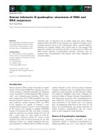
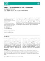
Fig. 3. Schematic structure of human telomeric G-quadruplexes. (A) Tetrameric parallel-stranded G-quadruplex observed for the single-repeat
human telomeric sequences d(TTAGGG) and d(TTAGGGT) in K
+
solution [25]. (B) Dimeric parallel-stranded G-quadruplex observed for the
two-repeat human telomeric sequence d(TAGGGTTAGGGT) in a K
+
-containing crystal [27] and in K
+
solution [28]. (C) Dimeric antiparallel-
stranded G-quadruplex observed for two-repeat human telomeric sequence d(TAGGGTTAGGGT) in K
+
solution [28]. (D) Asymmetric dimeric
(3 + 1) G-quadruplex observed for the three-repeat human telomeric sequence d(GGGTTAGGGTTAGGGT) in Na
+
solution [30]. (E) Asymmet-
ric dimeric (3 + 1) G-quadruplex association observed for the three-repeat human telomeric sequence d(GGGTTAGGGTTAGGGT) and the sin-
gle-repeat human telomeric sequence d(TAGGGT) in Na
+
solution [30] and in K
+
solution (unpublished results). (F) Basket-type form
observed for d[A(GGGTTA)
3
GGG] in Na
+
solution [31]. (G) Propeller-type form observed for d[A(GGGTTA)
3
GGG] in a K
+
-containing crystal
[27]. (H) (3 + 1) Form 1 observed for d[TA(GGGTTA)
3
GGG] in K
+
solution [39–44,46]. (I) (3 + 1) Form 2 observed for d[TA(GGGTTA)
3
GGGTT]
in K
+
solution [41,45,46]. (J) Basket-type form observed for d[(GGGTTA)
3
GGGT] in K
+
solution [47]. anti guanines are colored cyan; syn gua-
nines are colored magenta; loops are colored red. M, N and W represent medium, narrow and wide grooves, respectively.
A. T. Phan Human telomeric G-quadruplex structures
FEBS Journal 277 (2010) 1107–1117 ª 2009 The Author Journal compilation ª 2009 FEBS 1109
Short human telomeric DNA sequences
Short human telomeric sequences often serve as
models for high-resolution structural studies of the
telomere. Various G-quadruplex structures have been
observed for human telomeric DNA sequences con-
taining one, two, three or four repeats under different
experimental conditions [25–54]. The number of
G-tracts is often taken as the number of repeats, when
the studied sequences do not contain exact multiples of
the TTAGGG repeat. For example, the sequence
d[AGGG(TTAGGG)
3
] is usually considered as a four-
repeat human telomeric sequence [31]. Multiple
G-quadruplex conformations can be observed for a
given sequence, making structural elucidation difficult
[28,41]. This conformational heterogeneity can be over-
come by judicious choices of the flanking nucleotides
and ⁄ or base-analogue substitutions [28,39–47].
In K
+
solution, the single-repeat human telomeric
sequences d(TTAGGG) and d(TTAGGGT) form a
tetrameric parallel-stranded G-quadruplex containing
three G-tetrad layers in which all guanines adopt the
anti glycosidic conformation [25] (Fig. 3A), indicating
that this structure is preferred in the absence of loop-
ing constraints. There are four medium-size grooves in
such a structure. For the d(TTAGGG) sequence, high
concentrations of K
+
and ⁄ or DNA favor the 3¢-end
stacking of two such G-quadruplex blocks into a struc-
ture containing six G-tetrad layers [25,26] (Fig. 4A).
In a K
+
-containing crystal, the two-repeat human
telomeric sequence d(TAGGGTTAGGGT) forms a
dimeric parallel-stranded propeller-type G-quadruplex
[27] (Fig. 3B). In this structure, all guanines are anti,
the four grooves are of medium size and the two loops
are double-chain-reversal (or propeller). In K
+
solu-
tion, the same sequence interconverts between parallel-
and antiparallel-stranded dimeric G-quadruplexes [28]
(Fig. 3B,C), whose folding and unfolding rates are
distinct [28]. The parallel form is symmetric (Fig. 3B)
and similar to the propeller-type structure observed in
the crystalline state. The antiparallel form (up–down–
up–down core) is asymmetric (Fig. 3C): glycosidic con-
formations of guanines along two consecutive G-tracts
are 5¢-syn-anti-anti-3¢ and 5¢-syn-syn-anti-3¢ for one
strand and 5¢-syn-syn-anti-3¢ and 5¢-syn-anti-anti-3¢ for
the other strand of the dimer; glycosidic conformations
of guanines around G-tetrads are syn•anti•syn•anti;
there are two wide and two narrow grooves; the struc-
ture has two edgewise loops at the two ends of the G-
tetrad core that span across the wide grooves. In Na
+
solution, CD spectra suggest that two-repeat human
telomeric sequences adopt antiparallel-stranded
G-quadruplex(es) [29].
The three-repeat human telomeric sequence
d(GGGTTAGGGTTAGGGT) forms in Na
+
solution
an asymmetric dimeric G-quadruplex, whose G-tetrad
core involves all three G-tracts of one strand and only
the 3¢-end G-tract of the other strand [30] (Fig. 3D).
The core of this structure, called the (3 + 1) core, has
three strands oriented in one direction and one strand
oriented in the opposite direction; glycosidic conforma-
tions of guanines along G-tracts are 5¢-syn-anti-anti-3¢
and 5¢-syn-syn-anti-3¢; there are two syn•anti•anti•anti
G-tetrads and one anti•syn•syn•syn G-tetrad; there are
one narrow, one wide and two medium grooves. The
two edgewise grooves span the neighboring wide and
narrow grooves, respectively. The three-repeat human
telomeric sequence d(GGGTTAGGGTTAGGGT) can
also associate with the single-repeat human telomeric
sequence d(TAGGGT) in Na
+
solution [30] and K
+
solution (unpublished results) to form the same
G-quadruplex topology (Fig. 3E).
Extensive research has been dedicated to the
structures formed by sequences containing four human
telomeric TTAGGG repeats, because this is considered
the minimum length required for intramolecular
G-quadruplex folding. Several G-quadruplex folding
topologies have been proposed [27,31–51], with high-
resolution structures reported for five intramolecular
G-quadruplexes [27,31,42–47].
In Na
+
solution, the four-repeat human telomeric
sequence d[AGGG(TTAGGG)
3
] forms an antiparallel-
stranded basket-type G-quadruplex [31] (Fig. 3F). The
core (up–up–down–down type) of this structure con-
sists of three syn•syn•anti•anti G-tetrads, which occur
with 5¢-syn-anti-syn-3¢ or 5¢-anti-syn-anti-3¢ along the
G-tracts; there are one narrow, one wide and two
5′
3′
5′
5′
5′
5′
5′
5′
5′
5′
5′
5′
5′
3′
3′
3′
3′
3′
3′
3′3′
3′
3′
3′
AB
Fig. 4. Schematic structure of the stacking between two human
telomeric G-quadruplex blocks, each involving three G-tetrads. (A)
3¢-3¢ stacking observed for the human telomeric DNA sequence
d(TTAGGG) in K
+
solution [25,26] and (B) 5¢-5¢ stacking observed
for human telomeric RNA sequence r(GGGUUAGGGU) in K
+
solution [66].
Human telomeric G-quadruplex structures A. T. Phan
1110 FEBS Journal 277 (2010) 1107–1117 ª 2009 The Author Journal compilation ª 2009 FEBS
medium grooves. Loops are consecutively edgewise–
diagonal–edgewise.
In a K
+
-containing crystal, the same sequence forms a
propeller-type parallel-stranded G-quadruplex involving
three G-tetrad layers [27] (Fig. 3G): all guanines are
anti; loops are double-chain-reversal; four grooves are of
medium size, three of which are occupied by loops.
In K
+
solution, multiple G-quadruplex conforma-
tions have been observed for four-repeat human telo-
meric sequences [28,29,32–54]. The d[TAGGG(TTA
GGG)
3
] and d[TAGGG(TTAGGG)
3
TT] sequences
form predominantly intramolecular (3 + 1) G-quadru-
plexes Form 1 [39–44,46] (Fig. 3H) and Form 2
[41,45,46] (Fig. 3I), respectively. These structures con-
tain a three-G-tetrad (3 + 1) core with one double-
chain-reversal and two edgewise loops, but they differ
in the order of loop arraignments: in Form 1 the first
linker adopts the double-chain-reversal loop configura-
tion, whereas in Form 2 the third linker adopts the
double-chain-reversal loop configuration. Form 1 and
Form 2 are also the predominant conformations of the
d[TTAGGG(TTAGGG)
3
] [41,46] and d[TTAGGG(T
TAGGG)
3
TT] [41,45,46] sequences, respectively. The
human telomeric sequence d[TAGGG(TTAGGG)
3
T]
adopts both Form 1 and Form 2 with comparable pro-
portions in K
+
solution ([46] and unpublished results).
In K
+
solution, the human telomeric sequence
d[GGG(TTAGGG)
3
T] predominantly forms an intra-
molecular basket-type G-quadruplex involving only
two G-tetrads, designated Form 3 [47] (Fig. 3J):
the antiparallel-stranded core is of the up–up–down–
down type; the two G-tetrads are syn•syn•anti•anti
G-tetrads; there are one narrow, one wide and two
medium grooves; loops are consecutively edgewise–
diagonal–edgewise. Several other four-repeat human
telomeric sequences, which start with a G (e.g. d[GGG
(TTAGGG)
3
], d[GGG(TTAGGG)
3
TT] and d[GGG(T
TAGGG)
3
TTA]), also adopt Form 3 in K
+
solution
[47]. Despite the presence of only two G-tetrad layers,
Form 3 adopted by d[GGG(TTAGGG)
3
T] is more
stable than Form 1 and Form 2 adopted by d[TAG
GG(TTAGGG)
3
] and d[TAGGG(TTAGGG)
3
TT] in
K
+
solution, respectively. With extensive base pairing
and stacking in the loops, Form 3 is made up totally
of four to six base pair ⁄ triple ⁄ tetrad layers, which
might explain the high stability of this structure. The
folding principle of Form 3 indicates that the overall
G-quadruplex topology of a G-rich sequence is defined
not only by maximizing the number of G-tetrads, but
also by maximizing all possible base pairing and
stacking in the loops.
G-quadruplex structures determined for human telo-
meric DNA sequences show different configurations
for TTA loops in various contexts involving three
G-tetrads [27,31,42–47,55–60], as well as GTTA and
GTTAG loops in Form 3 involving two G-tetrads
[47]. Base pairing and stacking are generally observed
in these loops [27,31,42–47,55–60]. It has been
suggested that these loops are dynamic and may be
good targets for specific ligand recognitions [46,47,55–
59].
Different patterns of the G-tetrad hydrogen-bond
directionalities are observed for the structures
described above (Fig. 3). For example, the hydrogen-
bond directionality alternates clockwise–anticlockwise
for adjacent G-tetrads in the Na
+
solution basket-type
G-quadruplex (Fig. 3F), whereas it remains the same
for all G-tetrads in the parallel-stranded G-quadruplexes
(Fig. 3A,B,G).
Other G-quadruplex folds have been proposed
for DNA sequences containing human telomeric
TTAGGG repeats under different experimental condi-
tions [39,48–51]. It has been reported that molecular
crowding conditions can favor parallel-stranded
G-quadruplex conformation(s) [54].
Human telomeres, which encompass thousands of
canonical TTAGGG repeats, can be interspersed with
some sequence-variant repeats [61,62]. In particular,
short contiguous arrays of variant
CTAGGG repeats
in the human telomere (variation is underlined) are
unstable in the male germline and somatic cells [63].
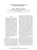
In K
+
solution, DNA sequences containing four
human telomeric variant
CTAGGG repeats (e.g. d[AG
GG(
CTAGGG)
3
]) form a new antiparallel intramolec-
ular G-quadruplex involving two G-tetrads and a
G•C•G•C tetrad (Fig. 5) [64].
Short human telomeric RNA sequences
The two-repeat human telomeric RNA sequence r(UA-
GGGUUAGGGU) forms, in both Na
+
solution [65]
and K
+
solution [66], a propeller-type parallel-stranded
dimeric G-quadruplex, the same folding topology
observed for the DNA counterpart in a K
+
-containing
crystal (Fig. 3B). However, unlike the propeller-type
DNA G-quadruplex [27] in which DNA residues prefer
the C2¢-endo sugar puckering conformation, the high-
definition structure of the propeller-type RNA G-quad-
ruplex in K
+
solution [66] shows both C2 ¢-endo and
C3¢-endo conformations (residues in the loops adopt
C2¢-endo conformation; residues in the central G-tetrad
adopt C3¢-endo conformation; residues in the external
G-tetrad can adopt both C2¢-endo and C3¢-endo
conformations).
In K
+
solution, the human telomeric RNA sequence
r(GGGUUAGGGU) forms a structure involving
A. T. Phan Human telomeric G-quadruplex structures
FEBS Journal 277 (2010) 1107–1117 ª 2009 The Author Journal compilation ª 2009 FEBS 1111
5¢-end stacking of two propeller-type three-layer
G-quadruplex blocks [66] (Fig. 4B). The lack of two
residues UA at the 5¢-end might favor this stacking
structure [66].
Data suggest that the lack of U at the 3¢-end of the
human telomeric RNA sequence r(GGGUUAGGG)
might favor further stacking of G-quadruplexes at this
end to form a higher order structure [66]. CD spectra
suggest that the four-repeat human telomeric RNA
sequences also form parallel-stranded structure in Na
+
and K
+
solution [65,66]. The conservation of the
G-quadruplex folding topology for human telomeric
RNA sequences in Na
+
and K
+
solution [65,66]
contrasts to the situation for human telomeric DNA
counterparts in which multiple conformations are
observed [25–54].
Long human telomeric DNA and RNA
sequences
The next step toward understanding the structure of
the ‘real’ telomeres is to address the question on the
structure of long human telomeric DNA sequences
[27,37,67,68]. The problem is the same for the long
human telomeric RNA sequences [66,69]. Data
[37,67,69] suggested that the structures of long human
telomeric DNA and RNA sequences are based on multi-
ple G-quadruplex blocks, each formed by a four-repeat
5′
3′
N
N
W
W
H
H
H
H
H
H
N
N
G
C
N
N
N
N
H
H
N
N
O
O
H
H
H
H
H
H
N
N
G
K
+
C
N
N
N
N
H
H
N
N
O
O
AB
Fig. 5. Schematic structure of (A) the
chair-type form G-quadruplex formed by
variant human telomeric sequence
d[A(GGGCTA)
3
GGG] in K
+
solution, which
contains two G-tetrads and (B) a G•C•G•C
tetrad [64]. anti guanines are colored
cyan; syn guanines are colored magenta;
cytosines are colored brown; loops are
colored red. M, N and W represent medium,
narrow and wide grooves, respectively.
5′
5′
3′
5′–5′ stacking
5′–5′ stacking
3′–3′ stacking
3′
5′–3′ stacking
5′–3′ stacking
5′–3′ stacking
5′–5′ stacking
5′–3′ stacking
3′
5′
5′
3′
ABCD
Fig. 6. Models for arrangements G-quadruplexes in long human telomeric DNA and RNA sequences. (A) ‘Beads-on-a-string’ [67], (B) ‘same-
direction stacking’ [27], (C) ‘alternate-direction stacking’ [66] and (D) coexistence of all the three modes (A, B and C) for connection between
G-quadruplex blocks. Linkers connecting consecutive G-quadruplex blocks are colored red.
Human telomeric G-quadruplex structures A. T. Phan
1112 FEBS Journal 277 (2010) 1107–1117 ª 2009 The Author Journal compilation ª 2009 FEBS
segment (see above). Several models have been proposed
regarding the arrangements of these G-quadruplex
blocks [27,66,67]. In one model, G-quadruplex blocks
are arranged like ‘beads-on-a-string’ [67], i.e. they can
move relatively independently of each other and are
constrained only by the connecting linkers (Fig. 6A).
Alternatively, G-quadruplex blocks can stack on
each other to form a higher order structure. There
may be three possible stacking modes between two
parallel-stranded G-quadruplex blocks: (a) 5¢-to-5¢,in
which the stacking interface is formed between the
5¢-end of each block; (b) 3¢-to-3¢, in which the stacking
interface is formed between the 3¢-end of each block;
and (c) 5¢-to-3¢, in which the stacking interface is
formed between the 5¢-end of one block and the 3¢-end
of the other. In the ‘same-direction stacking’ model
proposed for long human telomeric DNA, successive
propeller-type parallel-stranded G-quadruplex blocks,
which are oriented in the same direction, stack 5¢-to-3¢
continuously (Fig. 6B) [27]. It has been suggested that
a 200-nucleotide human telomeric DNA sequence, if
folded into a stack of G-quadruplex, would form a
rod of 60 A
˚
(compared with a 680 A
˚
-long B-DNA
helix) [27]. Successive (3 + 1) G-quadruplex blocks
can also stack continuously according to this model
[39,40,42]. In the ‘alternate-direction stacking’ model
proposed for long human telomeric RNA (or DNA),
the successive propeller-type G-quadruplex blocks
stack according to 5¢-to-5¢ and 3¢-to-3¢ modes (Fig. 6C)
[66]. In a model built for the 12-repeat human telomer-
ic RNA r[UAGGG(UUAGGG)
11
] sequence (Fig. 7),
the linkers that connect two consecutive G-quadruplex
blocks match well with the connecting distances,
thereby resulting in these linkers being nicely packed
in the grooves [66]. This type of linker arrangement
can also connect G-quadruplex blocks of different
folding topologies without generating knots. It is also
possible that all these arrangements of G-quadruplexes
coexist in the contexts of long telomeric DNA (or
RNA). Figure 6D shows an example of the coexistence
of three different connecting interfaces between consec-
utive G-quadruplex blocks.
A structural model for the eight-repeat human telo-
meric DNA sequence, built to satisfy various biophysi-
cal measurements, shows the stacking of two (3 + 1)
G-quadruplex blocks (Form 1 [39–44] and Form 2)
through bases in the loops [60].
Biochemical data on DNA sequences containing up
to seven human telomeric repeats suggested that
G-quadruplex preferentially forms at the 3¢-end [70].
The dimeric (3 + 1) G-quadruplex assembly was
proposed to be formed in the so-called T-loop [30],
where the 3¢-end overhang invades the preceding dou-
ble-stranded part of the telomere [71]. This looping
configuration of the telomere was illustrated in a stable
lariat, in which the connection point was a (3 + 1)
G-quadruplex [72].
Targeting human telomeric sequences
by small molecules: structural views
The formation of G-quadruplexes by the telomeric
G-rich DNA overhang has been shown to inhibit the
activity of telomerase [73], an enzyme [74] required for
the proliferation of most cancer cells [75]. Therefore,
G-quadruplexes formed by human telomeric DNA are
promising anticancer targets [20,21]. Human telomeric
RNAs might also be potential drug targets based on
their biological importance [5].
A desired ligand would recognize a G-quadruplex
structure formed by human telomeric sequences with
high affinity and specificity. Different G-quadruplex
recognition modes are possible: (a) stacking on the
ends of the G-tetrad core, (b) groove binding, (c)
taking place of one or more strands in the core, (d)
interacting with the backbone (core and loops), and (e)
interacting with the loop bases. A ligand that uses
several recognition modes may have an enhanced
binding affinity and specificity.
5
′
3′
Fig. 7. A model for the high-order structure of the long human telo-
meric RNA sequence r[UAGGG(UUAGGG)
11
]. Bases of guanines
are colored cyan; O4¢ of guanines yellow; UUA linkers connecting
consecutive G-quadruplex blocks are colored red. Figure adapted
from Martadinata & Phan [66].
A. T. Phan Human telomeric G-quadruplex structures
FEBS Journal 277 (2010) 1107–1117 ª 2009 The Author Journal compilation ª 2009 FEBS 1113
Many of the reported G-quadruplex ligands
[56–59,76–91] contain planar aromaric rings, which
can interact with human telomeric G-quadruplex by
stacking on the terminal G-tetrads [56–59,76,78–
80,90,91]. To date, there is no conclusive evidence
supporting the intercalation of a planar ligand between
G-tetrad layers. In addition to the end-stacking
binding mode of the aromatic rings, some ligands also
contain other moieties that can recognize loops by
stacking with loop bases or forming intermolecular
hydrogen bonds [57–59,79] or recognize the backbone
with electrostatic interactions [90,91]. The grooves in
G-quadruplexes can also be recognized through hydro-
gen bonds [92] or hydrophobic interactions [93]. Alter-
natively, the G-rich human telomeric DNA (or RNA)
strand can be trapped in a G-quadruplex structure
with a linear guanine-containing molecule [30] based
on a different backbone, such as PNA [94,95]. In the
context of long human telomeric sequences, ligands
can be designed to position between consecutive
G-quadruplex blocks [96]. Finally, fluorescent ligands
can be designed to probe the formation and the
ligand-induced stabilization of telomeric G-quadru-
plexes in the cell [97,98].
Future challenges and prospects for
structural studies
Despite a wealth of current knowledge about human
telomeric G-quadruplexes, there remain many
challenges associated with the structure and molecular
recognition in the human telomeres. These include: (a)
the structure and dynamics of all possible DNA, RNA
and DNA ⁄ RNA hybrid G-quadruplexes formed by
short and long human telomeric sequences; (b) the
structural basis for molecular recognition of human
telomeric G-quadruplexes by different small molecules
and proteins; and (c) the detection of G-quadruplex
structures and conformational transitions in the human
telomeres in living cells.
Acknowledgements
This research was supported by Singapore Biomedical
Research Council grant 07 ⁄ 1 ⁄ 22 ⁄ 19 ⁄ 542, Singapore
Ministry of Education grants (ARC30 ⁄ 07 and
RG62 ⁄ 07) and Nanyang Technological University
start-up grants (SUG5 ⁄ 06 and RG138 ⁄ 06) to ATP.
References
1 Moyzis RK, Buckingham JM, Cram LS, Dani M,
Deaven LL, Jones MD, Meyne J, Ratliff RL & Wu JR
(1988) A highly conserved repetitive DNA sequence,
(TTAGGG)
n
, present at the telomeres of human chro-
mosomes. Proc Natl Acad Sci USA 85, 6622–6626.
2 Makarov VL, Hirose Y & Langmore JP (1997) Long
G tails at both ends of human chromosomes suggest a
C strand degradation mechanism for telomere shorten-
ing. Cell 88, 657–666.
3 Azzalin CM, Reichenbach P, Khoriauli L, Giulotto E
& Lingner J (2007) Telomeric repeat containing RNA
and RNA surveillance factors at mammalian chromo-
some ends. Science 318, 798–801.
4 Schoeftner S & Blasco MA (2008) Developmentally
regulated transcription of mammalian telomeres by
DNA-dependent RNA polymerase II. Nat Cell Biol 10,
228–236.
5 Horard B & Gilson E (2008) Telomeric RNA enters the
game. Nat Cell Biol 10, 113–115.
6 Williamson JR (1994) G-quartet structures in telomeric
DNA. Annu Rev Biophys Biomol Struct 23, 703–730.
7 Gilbert DE & Feigon J (1999) Multistranded DNA
structures. Curr Opin Struct Biol 9, 305–314.
8 Simonsson T (2001) G-quadruplex DNA structures –
variations on a theme. Biol Chem 382, 621–628.
9 Neidle S & Parkinson GN (2003) The structure of telo-
meric DNA. Curr Opin Struct Biol 13, 275–283.
10 Davis JT (2004) G-quartets 40 years later: from
5¢-GMP to molecular biology and supramolecular
chemistry. Angew Chem Int Ed Engl 43, 668–698.
11 Phan AT, Kuryavyi V & Patel DJ (2006) DNA
architecture: from G to Z. Curr Opin Struct Biol 16,
288–298.
12 Burge S, Parkinson GN, Hazel P, Todd AK & Neidle S
(2006) Quadruplex DNA: sequence, topology and struc-
ture. Nucleic Acids Res 34 , 5402–5415.
13 Phan AT, Kuryavyi V, Luu KN & Patel DJ (2007)
Structural diversity of G-quadruplex scaffolds. In
Quadruplex Nucleic Acids (Neidle S & Balasubramanian
S, eds), pp. 81–99. Royal Society of Chemistry,
Cambridge.
14 Patel DJ, Phan AT & Kuryavyi V (2007) Human telo-
mere, oncogenic promoter and 5¢-UTR G-quadruplexes:
diverse higher order DNA and RNA targets for cancer
therapeutics. Nucleic Acids Res 35, 7429–7455.
15 Gellert MN, Lipsett MN & Davies DR (1962) Helix
formation by guanylic acid. Proc Natl Acad Sci USA
48, 2013–2018.
16 Paeschke K, Simonsson T, Postberg J, Rhodes D &
Lipps HJ (2005) Telomere end-binding proteins control
the formation of G-quadruplex DNA structures in vivo.
Nat Struct Mol Biol 12, 847–854.
17 Maizels N (2006) Dynamic roles for G4 DNA in the
biology of eukaryotic cells. Nat Struct Mol Biol 13,
1055–1059.
18 Fry M (2007) Tetraplex DNA and its interacting pro-
teins. Front Biosci 12
, 4336–4351.
Human telomeric G-quadruplex structures A. T. Phan
1114 FEBS Journal 277 (2010) 1107–1117 ª 2009 The Author Journal compilation ª 2009 FEBS
19 Oganesian L & Bryan TM (2007) Physiological rele-
vance of telomeric G-quadruplex formation: a potential
drug target. BioEssays 29, 155–165.
20 Sun D, Thompson B, Cathers BE, Salazar M, Kerwin
SM, Trent JO, Jenkins TC, Neidle S & Hurley LH
(1997) Inhibition of human telomerase by a G-quadru-
plex-interactive compound. J Med Chem 40, 2113–
2116.
21 Mergny JL & He
´
le
`
ne C (1998) G-quadruplex DNA: a
target for drug design. Nat Med 4, 1366–1367.
22 Chaires JB (2009) Human telomeric G-quadruplex:
thermodynamic and kinetic studies of telomeric quadru-
plex stability. FEBS J 277, 1098–1106.
23 Neidle S (2009) Human telomeric G-quadruplex: The
current status of telomeric G-quadruplexes as therapeu-
tic targets in human cancer. FEBS J 277, 1118–1125.
24 Arora A, Kumar N, Agarwal T & Maiti S (2009)
Human telomeric G-quadruplex: targeting with small
molecules. FEBS J 277, 1345.
25 Wang Y & Patel DJ (1992) Guanine residues in
d(T
2
AG
3
) and d(T
2
G
4
) form parallel-stranded potas-
sium cation stabilized G-quadruplexes with anti
glycosidic torsion angles in solution. Biochemistry 31,
8112–8119.
26 Kato Y, Ohyama T, Mita H & Yamamoto Y (2005)
Dynamics and thermodynamics of dimerization of par-
allel G-quadruplexed DNA formed from d(TTAG
n
)
(n = 3-5). J Am Chem Soc 127, 9980–9981.
27 Parkinson GN, Lee MPH & Neidle S (2002) Crystal
structure of parallel quadruplexes from human
telomeric DNA. Nature 417, 876–880.
28 Phan AT & Patel DJ (2003) Two-repeat human telo-
meric d(TAGGGTTAGGGT) sequence forms intercon-
verting parallel and antiparallel G-quadruplexes in
solution: distinct topologies, thermodynamic properties,
and folding ⁄ unfolding kinetics. J Am Chem Soc 125,
15021–15027.
29 Rujan IN, Meleney JC & Bolton PH (2005) Vertebrate
telomere repeat DNAs favor external loop propeller
quadruplex structures in the presence of high
concentrations of potassium. Nucleic Acids Res 33,
2022–2031.
30 Zhang N, Phan AT & Patel DJ (2005) (3 + 1) assembly
of three human telomeric repeats into an asymmetric
dimeric G-quadruplex. J Am Chem Soc 127, 17277–
17285.
31 Wang Y & Patel DJ (1993) Solution structure of the
human telomeric repeat d[AG
3
(T
2
AG
3
)
3
] G-tetraplex.
Structure 1, 263–282.
32 Redon S, Bombard S, Elizondo-Riojas MA &
Chottard JC (2003) Platinum cross-linking of adenines
and guanines on the quadruplex structures of the
AG
3
(T2AG
3
)
3
and (T
2
AG
3
)
4
human telomere
sequences in Na
+
and K
+
solutions. Nucleic Acids
Res 31, 1605–1613.
33 He Y, Neumann RD & Panyutin IG (2004) Intra-
molecular quadruplex conformation of human telomeric
DNA assessed with
125
I-radioprobing. Nucleic Acids
Res 32, 5359–5367.
34 Risitano A & Fox KR (2005) Inosine substitutions
demonstrate that intramolecular DNA quadruplexes
adopt different conformations in the presence of
sodium and potassium. Bioorg Med Chem Lett 15,
2047–2050.
35 Rezler EM, Seenisamy J, Bashyam S, Kim MY, White
E, Wilson WD & Hurley LH (2005) Telomestatin and
diseleno sapphyrin bind selectively to two different
forms of the human telomeric G-quadruplex structure.
J Am Chem Soc 127, 9439–9447.
36 Qi J & Shafer RH (2005) Covalent ligation studies on
the human telomere quadruplex. Nucleic Acids Res 33,
3185–3192.
37 Vorlickova M, Chladkova J, Kejnovska I, Fialova M &
Kypr J (2005) Guanine tetraplex topology of human
telomere DNA is governed by the number of
(TTAGGG) repeats. Nucleic Acids Res 33, 5851–5860.
38 Li J, Correia JJ, Wang L, Trent JO & Chaires JB
(2005) Not so crystal clear: the structure of the
human telomere G-quadruplex in solution differs from
that present in a crystal. Nucleic Acids Res 33, 4649–
4659.
39 Xu Y, Noguchi Y & Sugiyama H (2006) The new
models of the human telomere d[AGGG(TTAGGG)
3
]
in K
+
solution. Bioorg Med Chem 14, 5584–5591.
40 Ambrus A, Chen D, Dai J, Bialis T, Jones RA & Yang
D (2006) Human telomeric sequence forms a hybrid-
type intramolecular G-quadruplex structure with mixed
parallel ⁄ antiparallel strands in potassium solution.
Nucleic Acids Res 34, 2723–2735.
41 Phan AT, Luu KN & Patel DJ (2006) Different loop
arrangements of intramolecular human telomeric
(3 + 1) G-quadruplexes in K
+
solution. Nucleic Acids
Res 34, 5715–5719.
42 Luu KN, Phan AT, Kuryavyi V, Lacroix L & Patel DJ
(2006) Structure of the human telomere in K
+
solution:
an intramolecular (3 + 1) G quadruplex scaffold.
J Am Chem Soc 128, 9963–9970.
43 Dai J, Punchihewa C, Ambrus A, Chen D, Jones RA &
Yang D (2007) Structure of the intramolecular human
telomeric G-quadruplex in potassium solution: a novel
adenine triple formation. Nucleic Acids Res 35, 2240–
2250.
44 Matsugami A, Xu Y, Noguchi Y, Sugiyama H &
Katahira M (2007) Structure of a human telomeric
DNA sequence stabilized by 8-bromoguanosine
substitutions, as determined by NMR in a K
+
solution.
FEBS J 274, 3545–3556.
45 Dai J, Carver M, Punchihewa C, Jones RA & Yang D
(2007) Structure of the hybrid-2 type intramolecular
human telomeric G-quadruplex in K
+
solution: insights
A. T. Phan Human telomeric G-quadruplex structures
FEBS Journal 277 (2010) 1107–1117 ª 2009 The Author Journal compilation ª 2009 FEBS 1115
into structure polymorphism of the human telomeric
sequence. Nucleic Acids Res 35, 4927–4940.
46 Phan AT, Kuryavyi V, Luu KN & Patel DJ (2007)
Structure of two intramolecular G-quadruplexes formed
by natural human telomere sequences in K
+
solution.
Nucleic Acids Res 35, 6517–6525.
47 Lim KW, Amrane S, Bouaziz S, Xu W, Mu Y, Patel
DJ, Luu KN & Phan AT (2009) Structure of the human
telomere in K
+
solution: a stable basket-type G-quad-
ruplex with only two G-tetrad layers. J Am Chem Soc
131, 4301–4309.
48 Pedroso IM, Duarte LF, Yanez G, Burkewitz K &
Fletcher TM (2007) Sequence specificity of inter- and
intramolecular G-quadruplex formation by human telo-
meric DNA. Biopolymers 87, 74–84.
49 Gaynutdinov TI, Neumann RD & Panyutin IG (2008)
Structural polymorphism of intramolecular quadruplex
of human telomeric DNA: effect of cations, quadru-
plex-binding drugs and flanking sequences. Nucleic
Acids Res 36, 4079–4087.
50 Okamoto K, Sannohe Y, Mashimo T, Sugiyama H &
Terazima M (2008) G-quadruplex structures of human
telomere DNA examined by single molecule FRET and
Br
G-substitution. Bioorg Med Chem 16, 6873–6879.
51 Amrane S, Ang RW, Tan ZM, Li C, Lim JK, Lim JM,
Lim KW & Phan AT (2009) A novel chair-type
G-quadruplex formed by a Bombyx mori telomeric
sequence. Nucleic Acids Res 37, 931–938.
52 Ying L, Green JJ, Li H, Klenerman D & Balasubrama-
nian S (2003) Studies on the structure and dynamics of
the human telomeric G quadruplex by single-molecule
fluorescence resonance energy transfer. Proc Natl Acad
Sci USA 100, 14629–14634.
53 Lee JY, Okumus B, Kim DS & Ha T (2005) Extreme
conformational diversity in human telomeric DNA.
Proc Natl Acad Sci USA 102 , 18938–18943.
54 Xue Y, Kan ZY, Wang Q, Yao Y, Liu J, Hao YH &
Tan Z (2007) Human telomeric DNA forms parallel-
stranded intramolecular G-quadruplex in K
+
solution
under molecular crowding condition. J Am Chem Soc
129, 11185–11191.
55 Haider S, Parkinson GN & Neidle S (2008) Molecular
dynamics and principal components analysis of human
telomeric quadruplex multimers. Biophys J 95, 296–311.
56 Parkinson GN, Ghosh R & Neidle S (2007) Structural
basis for binding of porphyrin to human telomeres. Bio-
chemistry 46, 2390–2397.
57 Campbell NH, Parkinson GN, Reszka AP & Neidle S
(2008) Structural basis of DNA quadruplex recognition
by an acridine drug. J Am Chem Soc 130, 6722–6724.
58 Parkinson GN, Cuenca F & Neidle S (2008) Topology
conservation and loop flexibility in quadruplex–drug
recognition: crystal structures of inter- and intramolecu-
lar telomeric DNA quadruplex–drug complexes. J Mol
Biol 381, 1145–1156.
59 Campbell NH, Patel M, Tofa AB, Ghosh R, Parkinson
GN & Neidle S (2009) Selectivity in ligand recognition
of G-quadruplex loops. Biochemistry 48, 1675–1680.
60 Petraccone L, Trent JO & Chaires JB (2008) The tail of
the telomere. J Am Chem Soc 130, 16530–16532.
61 Allshire RC, Dempster M & Hastie ND (1989) Human
telomeres contain at least 3 types of G-rich repeat distrib-
uted non-randomly. Nucleic Acids Res 17, 4611–4627.
62 Baird DM, Jeffreys AJ & Royle NJ (1995)
Mechanisms underlying telomere repeat turnover,
revealed by hypervariable variant repeat distribution
patterns in the human Xp ⁄ Yp telomere. EMBO J 14,
5433–5443.
63 Mendez-Bermudez A, Hills M, Pickett HA, Phan AT,
Mergny JL, Riou JF & Royle NJ (2009) Human telo-
meres that contain (CTAGGG)
n
repeats show replica-
tion dependent instability in somatic cells and the male
germline. Nucleic Acids Res 37, 6225–6238.
64 Lim KW, Alberti P, Guedin A, Lacroix L, Riou JF,
Royle NJ, Mergny JL & Phan AT (2009) Sequence
variant (CTAGGG)
n
in the human telomere favors a
G-quadruplex structure containing a G•C •G•C tetrad.
Nucleic Acids Res 37, 6239–6248.
65 Xu Y, Kaminaga K & Komiyama M (2008) G-quadru-
plex formation by human telomeric repeats containing
RNA in Na
+
solution. J Am Chem Soc 130, 11179–
11184.
66 Martadinata H & Phan AT (2009) Structure of propel-
ler-type parallel-stranded RNA G-quadruplexes, formed
by human telomeric RNA sequences in K
+
solution.
J Am Chem Soc 131, 2570–2578.
67 Yu HQ, Miyoshi D & Sugimoto N (2006) Characteriza-
tion of structure and stability of long telomeric DNA
G-quadruplexes. J Am Chem Soc 128, 15461–15468.
68 Pomerantz AK, Moerner WE & Kool ET (2008)
Visualization of long human telomere mimics by single-
molecule fluorescence imaging. J Phys Chem B 112,
13184–13187.
69 Randall A & Griffith JD (2009) Structure of long telo-
meric RNA transcripts: the G-rich RNA forms a com-
pact repeating structure containing G-quartets. J Biol
Chem 284, 13980–13986.
70 Tang J, Kan ZY, Yao Y, Wang Q, Hao YH & Tan Z
(2008) G-quadruplex preferentially forms at the very
3¢-end of vertebrate telomeric DNA. Nucleic Acids Res
36, 1200–1208.
71 Griffith JD, Comeau L, Rosenfield S, Stansel RM,
Bianchi A, Moss H & de Lange T (1999) Mammalian
telomeres end in a large duplex loop. Cell 97, 503–514.
72 Xu Y, Sato H, Sannohe Y, Shinohara KI & Sugiyama
H (2008) Stable lariat formation based on a G-quadru-
plex scaffold. J Am Chem Soc 130, 16470–16471.
73 Zahler AM, Williamson JR, Cech TR & Prescott DM
(1991) Inhibition of telomerase by G-quartet DNA
structures. Nature, 350, 718–720.
Human telomeric G-quadruplex structures A. T. Phan
1116 FEBS Journal 277 (2010) 1107–1117 ª 2009 The Author Journal compilation ª 2009 FEBS
74 Greider CW & Blackburn EH (1985) Identification of a
specific telomere terminal transferase activity in Tetra-
hymena extracts. Cell 43, 405–413.
75 Kim NW, Piatyszek MA, Prowse KR, Harley CB, West
MD, Ho PL, Coviello GM, Wright WE, Weinrich SL
& Shay JW (1994) Specific association of human telo-
merase activity with immortal cells and cancer. Science
266, 2011–2015.
76 Searle MS & Balkwill GD (2006) DNA quadruplex–
ligand recognition: structure and dynamics. In Quadru-
plex Nucleic Acids (Neidle S & Balasubramanian S, eds),
pp. 131–153. Royal Society of Chemistry, Cambridge.
77 De Cian A, Lacroix L, Douarre C, Temime-Smaali N,
Trentesaux C, Riou JF & Mergny JL (2008) Targeting
telomeres and telomerase. Biochimie 90, 131–155.
78 Clark GR, Pytel PD, Squire CJ & Neidle S (2003)
Structure of the first parallel DNA quadruplex–drug
complex. J Am Chem Soc 125, 4066–4067.
79 Haider SM, Parkinson GN & Neidle S (2003) Structure
of a G-quadruplex–ligand complex. J Mol Biol 326,
117–125.
80 Cocco MJ, Hanakahi LA, Huber MD & Maizels N
(2003) Specific interactions of distamycin with G-quad-
ruplex DNA. Nucleic Acids Res 31, 2944–2951.
81 De Cian A, Delemos E, Mergny JL, Teulade-Fichou
MP & Monchaud D (2007) Highly efficient G-quadru-
plex recognition by bisquinolinium compounds. JAm
Chem Soc 129, 1856–1857.
82 Barbieri CM, Srinivasan AR, Rzuczek SG, Rice JE,
LaVoie EJ & Pilch DS (2007) Defining the mode,
energetics and specificity with which a macrocyclic
hexaoxazole binds to human telomeric G-quadruplex
DNA. Nucleic Acids Res 35, 3272–3286.
83 Bejugam M, Sewitz S, Shirude PS, Rodriguez R, Shahid
R & Balasubramanian S (2007) Trisubstituted isoalloxa-
zines as a new class of G-quadruplex binding ligands:
small molecule regulation of c-kit oncogene expression.
J Am Chem Soc 129, 12926–12927.
84 Shirude PS, Gillies ER, Ladame S, Godde F, Shin-Ya
K, Huc I & Balasubramanian S (2007) Macrocyclic and
helical oligoamides as a new class of G-quadruplex
ligands. J Am Chem Soc 129, 11890–11891.
85 Cetinkol OP, Engelhart AE, Nanjunda RK, Wilson
WD & Hud NV (2008) Submicromolar, selective
G-quadruplex ligands from one pot: thermodynamic
and structural studies of human telomeric DNA binding
by azacyanines. Chembiochem 9, 1889–1892.
86 Kieltyka R, Englebienne P, Fakhoury J, Autexier C,
Moitessier N & Sleiman HF (2008) A platinum supra-
molecular square as an effective G-quadruplex binder
and telomerase inhibitor. J Am Chem Soc 130, 10040–
10041.
87 Kieltyka R, Fakhoury J, Moitessier N & Sleiman HF
(2008) Platinum phenanthroimidazole complexes as
G-quadruplex DNA selective binders. Chemistry 14,
1145–1154.
88 Tera M, Ishizuka H, Takagi M, Suganuma M, Shin-ya
K & Nagasawa K (2008) Macrocyclic hexaoxazoles as
sequence- and mode-selective G-quadruplex binders.
Angew Chem Int Ed Engl 47, 5557–5560.
89 Tera M, Iida K, Ishizuka H, Takagi M, Suganuma M,
Doi T, Shin-ya K & Nagasawa K (2009) Synthesis of a
potent G-quadruplex-binding macrocyclic heptaoxazole.
Chembiochem 10, 431–435.
90 Phan AT, Kuryavyi V, Gaw HY & Patel DJ (2005)
Small-molecule interaction with a five-guanine-tract
G-quadruplex structure from the human MYC
promoter. Nat Chem Biol 1, 167–173.
91 Dixon IM, Lopez F, Tejera AM, Este
`
ve JP, Blasco
MA, Pratviel G & Meunier B (2007) A G-quadruplex
ligand with 10000-fold selectivity over duplex DNA.
J Am Chem Soc 129, 1502–1503.
92 Kettani A, Gorin A, Majumdar A, Hermann T, Skrip-
kin E, Zhao H, Jones R & Patel DJ (2000) A dimeric
DNA interface stabilized by stacked A•(G•G•G•G)•A
hexads and coordinated monovalent cations. J Mol Biol
297, 627–644.
93 Martino L, Virno A, Pagano B, Virgilio A, Di Micco S,
Galeone A, Giancola C, Bifulco G, Mayol L & Randa-
zzo A (2007) Structural and thermodynamic studies of
the interaction of distamycin A with the parallel quad-
ruplex structure [d(TGGGGT)]
4
. J Am Chem Soc 129,
16048–16056.
94 Datta B, Schmitt C & Armitage BA (2003) Formation
of a PNA2–DNA2 hybrid quadruplex. J Am Chem Soc
125, 4111–4118.
95 Paul A, Sengupta P, Krishnan Y & Ladame S (2008)
Combining G-quadruplex targeting motifs on a single
peptide nucleic acid scaffold: a hybrid (3 + 1) PNA–
DNA bimolecular quadruplex. Chemistry 14, 8682–
8689.
96 Xu Y, Yamazaki S, Osuga H & Sugiyama H (2006)
The recognition of higher-order G-quadruplex by chiral
cyclic-helicene molecules. Nucleic Acids Symp Ser 50,
183–184.
97 Chang CC, Kuo IC, Ling IF, Chen CT, Chen HC, Lou
PJ, Lin JJ & Chang TC (2004) Detection of quadruplex
DNA structures in human telomeres by a fluorescent
carbazole derivative. Anal Chem 76, 4490–4494.
98 Yang P, De Cian A, Teulade-Fichou MP, Mergny JL &
Monchaud D (2009) Engineering bisquinolinium ⁄
thiazole orange conjugates for fluorescent sensing of
G-quadruplex DNA. Angew Chem Int Ed Engl 48,
2188–2191.
A. T. Phan Human telomeric G-quadruplex structures
FEBS Journal 277 (2010) 1107–1117 ª 2009 The Author Journal compilation ª 2009 FEBS 1117