Báo cáo khoa học: Estrogen-related receptor a and PGC-1-related coactivator constitute a novel complex mediating the biogenesis of functional mitochondria potx
Bạn đang xem bản rút gọn của tài liệu. Xem và tải ngay bản đầy đủ của tài liệu tại đây (478.29 KB, 13 trang )
Estrogen-related receptor a and PGC-1-related coactivator
constitute a novel complex mediating the biogenesis of
functional mitochondria
Delphine Mirebeau-Prunier1–3, Soazig Le Pennec1,2, Caroline Jacques1,2, Naig Gueguen3, Julie
´ ´
Poirier1,2, Yves Malthiery1–3 and Frederique Savagner1–3
1 INSERM, UMR694, Angers, France
´
2 Universite d’Angers, France
´
3 CHU Angers, Laboratoire de Biochimie et Biologie moleculaire, France
Keywords
cell proliferation; estrogen-related receptor a;
mitochondrial biogenesis; PGC-1-related
coactivator; respiratory chain
Correspondence
D. Mirebeau-Prunier, INSERM, UMR 694,
CHU, 4 rue Larrey, 49033 Angers, France
Fax: +33 241 35 40 17
Tel: +33 241 35 33 14
E-mail:
(Received 17 September 2009, revised 10
November 2009, accepted 25 November
2009)
doi:10.1111/j.1742-4658.2009.07516.x
Mitochondrial biogenesis, which depends on nuclear as well as mitochondrial genes, occurs in response to increased cellular ATP demand. The
nuclear transcriptional factors, estrogen-related receptor a (ERRa) and
nuclear respiratory factors 1 and 2, are associated with the coordination of
the transcriptional machinery governing mitochondrial biogenesis, whereas
coactivators of the peroxisome proliferator-activated receptor c coactivator-1 (PGC-1) family serve as mediators between the environment and this
machinery. In the context of proliferating cells, PGC-1-related coactivator
(PRC) is a member of the PGC-1 family, which is known to act in partnership with nuclear respiratory factors, but no functional interference
between PRC and ERRa has been described so far. We explored three thyroid cell lines, FTC-133, XTC.UC1 and RO 82 W-1, each characterized by
a different mitochondrial content, and studied their behavior towards PRC
and ERRa in terms of respiratory efficiency. Overexpression of PRC and
ERRa led to increased respiratory chain capacity and mitochondrial mass.
The inhibition of ERRa decreased cell growth and respiratory chain capacity in all three cell lines. However, the inhibition of PRC and ERRa produced a greater effect in the oxidative cell model, decreasing the
mitochondrial mass and the phosphorylating respiration, whereas the nonphosphorylating respiration remained unchanged. We therefore hypothesize
that the ERRa–PRC complex plays a role in arresting the cell cycle
through the regulation of oxidative phosphorylation in oxidative cells, and
through some other pathway in glycolytic cells.
Introduction
Mitochondrial biogenesis depends on nuclear transcriptional factors to coordinate the transcriptional
machinery, and on transcriptional coactivators to inte-
grate environmental signals into this program of mitochondrial biogenesis. Most studies to date have
focused on changes in energy metabolic pathways that
Abbreviations
COX, cytochrome c oxidase; CS, citrate synthase; Cyt c, cytochrome c somatic; ERE, estrogen response element; ERR, estrogen-related
receptor; ERRE, estrogen-related receptor response element; ERa, estrogen receptor a; FCCP, carbonyl cyanide p-trifluoromethoxyphenylhydrazone; HIF, hypoxia-inducible factor; LDH, lactate dehydrogenase; mtDNA, mitochondrial DNA; NRF, nuclear respiratory factor; PGC-1,
peroxisome proliferator-activated receptor c coactivator-1; PPAR, peroxisome proliferator-activated receptor; PRC, PGC-1-related coactivator;
siRNA, short interfering RNA.
FEBS Journal 277 (2010) 713–725 ª 2010 The Authors Journal compilation ª 2010 FEBS
713
ERRa-PRC complex and mitochondrial biogenesis
D. Mirebeau-Prunier et al.
enable the organism to adapt to its fluctuating nutritional status or to varying environmental conditions.
However, the identification of the key factors of mitochondrial biogenesis in the context of proliferating
cells should open up promising new lines of research
in this field.
The nuclear respiratory factors NRF-1 and NRF-2
and the estrogen-related receptor a (ERRa) are the
main nuclear transcriptional factors associated with
the expression levels of the majority of respiratory
chain genes [1]. Peroxisome proliferator-activated
receptor c coactivator-1a (PGC-1a) is the founding
member of the family of transcriptional coactivators,
including peroxisome proliferator-activated receptor
c coactivator-1b (PGC-1b) and PGC-1-related coactivator (PRC) [2]. Each of these coactivators induces
mitochondrial biogenesis in a specific context. PGC-1a
and PGC-1b have been mainly associated with the
modulation of metabolic pathways in tissues that
require high oxidative energy production, such as heart
and skeletal muscle [3]. Unlike PGC-1a and PGC-1b,
PRC is ubiquitous and more abundantly expressed in
proliferating cells than in growth-arrested cells. PRC is
known to interact with NRF-1 and NRF-2 to increase
the gene expression of several subunits of respiratory
chain complexes [4–6]. However, a subset of respiratory chain subunits does not appear to be regulated by
NRF-1 or NRF-2, indicating that other regulatory factors are implicated in the coordination of the expression of the nuclear and mitochondrial genomes.
ERRa is an orphan nuclear receptor that binds to
the ERR response element (ERRE) as either a monomer or a dimer, depending on the ERRE sequence.
ERRa heterodimers with member 1 and 3 of the signal
transducers and activators of transcription family,
NRF-1 and cAMP responsive element binding protein
have been found in heart cells in vitro [7]. ERRa interacts with different coactivators, such as PGC-1a, to
regulate cellular energy metabolism [8]. The interference between ERRa and PRC has been reported
recently, but its effect on mitochondrial biogenesis has
not been explored [6]. Involved in mitochondrial functions, ERRa participates in mitochondrial biogenesis,
oxidative phosphorylation and oxidative stress defense,
as well as in mitochondrial dynamics [8–12]. Clinical
studies and investigations into the molecular mechanisms of ERRa function have revealed the different
roles played by this receptor in tumor proliferation and
prognosis. In terms of structure, ERRa, which is similar to estrogen receptor a (ERa), can interfere with
estrogen signaling and serve as a prognosticator in
breast, ovarian and endometrial cancers [13–16]. In
colorectal cancer, ERRa mRNA levels are significantly
714
higher in tumoral tissue relative to normal tissue, and
associated with tumor stage as well as histological
grade [17]. In all of these highly proliferative tumors,
the cell metabolism is forced to shift to anaerobic glycolysis because of the hypoxic environment of the
tumor. In this context, ERRs have been found recently
to serve as essential cofactors of hypoxia-inducible factor (HIF) in cancer cell lines [18]. In contrast, in muscle
cells, ERRa and PGC-1a operate either independently
of HIF in response to hypoxia, or as regulators of
intracellular oxygen availability in a manner dependent
on HIF under physiological conditions [19,20]. Thus,
ERRa can promote either cell growth or mitochondrial
biogenesis according to the status of cellular oxygen.
Our study investigates tumor models in which we
determine the interference between PRC and ERRa in
the integrative regulation of metabolism involved in
mitochondrial and cellular proliferation. Thyroid oncocytic tumors and the cellular XTC.UC1 model have a
high rate of mitochondrial biogenesis and oxidative
cellular metabolism because of the increased expression
of PRC, ERRa and NRF-1 [21–23]. Moreover, in thyroid tissue, PGC-1a was not induced [23]. In this context, we compared the metabolic status of three
thyroid cell lines – FTC-133, XTC.UC1 and RO 82
W-1 – derived from follicular cell carcinoma. We characterized the basal mitochondrial status of these cell
lines according to respiratory chain functionality and
gene expression. In two of these lines, selected for their
different behavior towards ERRa, we explored the regulation of mitochondrial biogenesis and cell proliferation through the ERRa–PRC pathway via the
overexpression or inhibition of the two genes.
Results
Mitochondrial status of FTC-133, XTC.UC1 and
RO 82 W-1
Quantitative PCR was used to evaluate the mitochondrial DNA (mtDNA) level in each cell line (Fig. 1A).
mtDNA levels in FTC-133 and XTC.UC1 were 3.9
and 2.4 times higher, respectively, than in RO 82 W-1.
Similarly, the expression of ND5 mRNA, encoded by
mtDNA, and cytochrome c somatic (Cyt c) mRNA,
encoded by nuclear DNA, was 3.2 and 3.1 times
greater, respectively, in FTC-133, and 1.8 and 2.8
times greater, respectively, in XTC.UC1 than in RO 82
W-1 (Fig. 1B).
Quantitative PCR was used to determine the mRNA
levels of the main transcriptional factors (ERRa,
NRF-1 and NRF-2) and coactivators (PGC-1a, PGC1b, PRC) required for the biogenesis and function of
FEBS Journal 277 (2010) 713–725 ª 2010 The Authors Journal compilation ª 2010 FEBS
D. Mirebeau-Prunier et al.
A
ERRa-PRC complex and mitochondrial biogenesis
Relative mRNA level
60
50
40
30
20
10
FTC-133
XTC.UC1 RO 82 W-1
30
Relative mRNA level
300
250
200
150
100
50
0
Respiration rate
(nmolO2·min–1·(mg protein)–1
C
10
20
15
10
5
8
XTC.UC1
PRC
RO 82 W-1
6
4
2
0
COX activity
20
10
150
100
PGC-1β
0
FTC-133 XTC.UC1 RO 82 W-1
NRF-1
NRF-2
Oligomycin-insensitive
4
3
2
1
0
CS activity
1000
0.5
COX/CS
0.4
800
0.3
600
0.2
400
0.1
200
50
ERRα
Oligomycin-sensitive
5
1200
U·mg–1 of protein
200
RO 82 W-1
FTC-133 XTC.UC1 RO 82 W-1
300
U·mg–1 of protein
XTC.UC1
30
6
Maximal
250
PGC-1α
FTC-133
40
0
Cyt C
FTC-133
350
25
0
ND5
Basal
D
50
Respiration rate
(nmolO2·min–1·(mg protein)–1
Relative mRNA level
B 350
Relative mRNA level
0
0
FTC-133 XTC.UC1 RO 82 W-1
0.0
FTC-133 XTC.UC1 RO 82 W-1
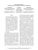
Fig. 1. Mitochondrial status for FTC-133, XTC.UC1 and RO 82 W-1 cells. (A) Relative levels of mtDNA were determined by quantitative realtime PCR and normalized to b-globin DNA levels (B) Relative expression levels of several genes were determined by quantitative real-time
PCR and were normalized against b-globin cDNA levels (C) Oxygen consumption was defined in the basal respiratory condition (basal respiratory), the maximal stimulation condition by the uncoupling of oxidative phosphorylation with FCCP (maximal respiratory) and the nonphosphorylating respiratory condition with oligomycin (oligomycin-insensitive). Phosphorylating respiration (oligomycin-sensitive) was calculated by
subtracting nonphosphorylating respiration from basal respiration. (D) Enzymatic activity of COX and CS, and the ratio of COX activity to CS
activity. Results are the mean values ± SD of six experiments.
the mitochondria (Fig. 1B). In our three cell lines,
ERRa and PRC were predominantly expressed relative
to the other factors, and the expression was signifi-
cantly higher for FTC-133 and XTC.UC1 than for RO
82 W-1.We checked ERRa protein expression in our
three cell lines, but not for PRC, because no commer-
FEBS Journal 277 (2010) 713–725 ª 2010 The Authors Journal compilation ª 2010 FEBS
715
ERRa-PRC complex and mitochondrial biogenesis
D. Mirebeau-Prunier et al.
cial antibody is currently available for this protein. We
confirmed that the ERRa protein levels were higher
for FTC-133 and XTC.UC1 than for RO 82 W-1 (data
not shown).
We determined the mitochondrial respiratory rate by
means of the cellular oxygen consumption in the different cell lines (Fig. 1C). The basal cellular oxygen
consumption for FTC-133 and XTC.UC1 was three
times higher than that for RO 82 W-1. Mitochondrial
complexes I and III were inhibited by rotenone and
antimycin, respectively, to check for nonmitochondrial
respiration. Relative to the maximal respiration rate,
the nonmitochondrial respiration rates amounted to
10 ± 2% in FTC-133, 19 ± 2% in XTC.UC1 and
14 ± 5% in RO 82 W-1. This indicates the predominant (80%) contribution of mitochondria to the total
cellular oxygen consumption in our three cell lines.
Mitochondrial respiration comprises phosphorylating
respiration, which represents the fraction used for ATP
synthesis, and nonphosphorylating respiration. The
nonphosphorylating respiration rate, i.e. the oligomycin-insensitive fraction, was recorded after the inhibition of ATP synthase with oligomycin, and the
phosphorylating respiration rate, i.e. the oligomycinsensitive fraction, was calculated by subtracting the
nonphosphorylating respiration rate from the basal
respiration rate. The evaluation of the oligomycin-sensitive oxygen consumption rate showed that FTC-133
and XTC.UC1 used much more oxygen (nearly 40%)
for ATP synthesis than did RO 82 W-1 (10%).
To evaluate mitochondrial function, we stimulated
cellular oxygen consumption with the uncoupler
carbonyl cyanide p-trifluoromethoxyphenylhydrazone
(FCCP) to produce maximal mitochondrial respiration. We observed a 40–60% increase in oxygen consumption in the three cell lines (Fig. 1C). We
measured the enzymatic activity of mitochondrial
complex IV (cytochrome c oxidase, COX) to evaluate the direct mitochondrial function and assayed the
citrate synthase (CS) level to evaluate the mitochondrial mass. COX activities were three times higher
for FTC-133 and XTC.UC1 than for RO 82 W-1,
and CS activities were twice as high for FTC-133
and XTC.UC1 than for RO 82 W-1 (Fig. 1D). Comparing the COX activity with the mitochondrial mass
using the COX ⁄ CS ratio, we found that FTC-133
and XTC.UC1 cells presented twice as much COX
activity for the same mitochondrial mass as did RO
82 W-1.
Lastly, we evaluated the glycolytic metabolism by
measuring the lactate dehydrogenase (LDH) activity.
We measured the LDH activity in FTC-133,
XTC.UC1 and RO 82 W-1. Comparing the LDH
716
activity with the mitochondrial mass using the
LDH ⁄ CS ratio, we found that RO 82 W-1 cells presented at least 40% more LDH activity than did
FTC-133 and XTC.UC1.
Our results show that FTC-133 and XTC.UC1 cells
undergo oxidative metabolism with a high content of
efficient mitochondria, whereas RO 82 W-1 metabolism is mainly glycolytic, with mitochondria using little
electron transport for phosphorylation.
ERRa is involved in the metabolic regulation of
the three thyroid cell lines
We investigated the effects of XCT790, a specific
inverse agonist of ERRa. As controls of the inhibitory
effect of XCT790 on ERRa, we used the expression of
ERRa-validated target genes, such as Cyt c and ATP
synthase subunit b [8]. Quantitative PCR was used to
evaluate the levels of these genes after treatment with
5 lm XCT790 for 10 days. The expression of both
genes was downregulated by treatment with XCT790
by at least 40% relative to untreated controls. Treatment with 5 lm XCT790 for 10 days inhibited cell proliferation in the three cell lines (Fig. 2A). This
inhibition began earlier – in less than 4 days – for RO
82 W-1 than for the other two cell lines. Similarly, the
inhibition of cell proliferation after 10 days was greater
for RO 82 W-1 (60.3%) than for XTC.UC1 (44.2%)
or FTC-133 (25.8%). The three cell lines grew differently and, after 10 days, there were four times as many
FTC-133 cells as RO 82 W-1 cells. The level of inhibition was probably related to the different proliferative
statuses of the cells. Nevertheless, the inhibition of
ERRa with XCT790 decreased significantly the basal
oxygen consumption and the maximal respiration only
in FTC-133 cells (Fig. 2B). Moreover, COX and CS
activities were reduced in FTC-133 cells, whereas the
COX ⁄ CS ratio remained unaltered. In the other two
cell lines, XCT790 had no significant effect on cellular
oxygen consumption; COX activity decreased significantly for RO 82 W-1 (P < 0.05) and consistently for
XTC.UC1 (P = 0.07), whereas the CS activity was
unchanged (Fig. 2C).
In all three cell lines, cell growth and mitochondrial complex IV activity decreased when ERRa was
inhibited. ERRa may affect cell growth by a mechanism independent of its effect on mitochondrial respiration in our three cell lines. However, the greatest
ERRa regulation of oxidative phosphorylation was
observed for FTC-133 cells, with decreased basal
oxygen consumption and reduced maximal mitochondrial respiration. We therefore postulated that ERRa
influences cell growth through the control of respira-
FEBS Journal 277 (2010) 713–725 ª 2010 The Authors Journal compilation ª 2010 FEBS
D. Mirebeau-Prunier et al.
ERRa-PRC complex and mitochondrial biogenesis
A
XCT790
Vehicle
FTC-133
Cell number
60
40
20
0
0
3
7
30
25
20
XTC.UC1
20
15
10
5
0
0
10
3
Days
Vehicle
FTC-133
6
5
*
4
*
3
2
1
0
Basal
Maximal
Respiration rate
(nmolO2·min–1·(mg protein)–1
Respiration rate
(nmolO2·min–1·(mg protein)–1
7
7
5
4
3
2
1
0
Basal
Maximal
*
150
100
*
0
U·mg–1 of protein
250
1000
800
XTC.UC1 RO 82 W-1
7
10
7
RO 82 W-1
6
5
4
3
2
1
0
Basal
Maximal
COX/CS
0.4
0.3
*
*
0.2
600
400
0.1
200
0.0
0
FTC-133
Days
XCT790
CS activity
300
3
XCT790
1200
50
0
6
Vehicle
200
5
0
10
XTC.UC1
COX activity
C 350
10
Days
B
U·mg–1 of protein
7
RO 82 W-1
15
Respiration rate
(nmolO2·min–1·(mg protein)–1
Cell number
80
Cell number
100
FTC-133 XTC.UC1 RO 82 W-1
FTC-133
XTC.UC1 RO 82 W-1
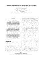
Fig. 2. Inhibition of ERRa with inverse agonist XCT790 in FTC-133, XTC.UC1 and RO 82 W-1 cells. (A) Analysis of proliferation by direct cell
counting in the presence (filled triangles) or absence (open triangles) of 5 lM XCT790 for 10 days. (B) Basal and maximal mitochondrial respiratory rate in the presence (filled bars) or absence (open bars) of 5 lM XCT790 for 10 days. (C) COX and CS activity for FTC-133, XTC.UC1
and RO 82 W-1 cells in the presence (filled bars) or absence (open bars) of 5 lM XCT790 for 10 days. Results are the mean values ± SD.
*P < 0.05 versus cells in the absence of XCT790.
tory capacity in cells with preferential oxidative
metabolism.
The PRC–ERRa complex activates transcription
directly through a consensus estrogen response
element (ERE)
To determine whether PRC can function as a coactivator of ERRa, transient transfections into RO 82
W-1 cells were performed using the 3X ERE TATA
luc reporter construction (Fig. 3A). The reporter plasmid contains three copies of the vitellogenin authentic
promoter ERE that have been demonstrated to bind
to ERRa and the complex ERRa–PGC1a [24,25]. No
effect on reporter activity was observed after transfection with PRC alone. Forced overexpression of
ERRa, without PRC transfection, probably stimulated reporter construction because of the presence of
endogenous ERRa coactivators in these cells.
However, 3X ERE TATA luc reporter activity was
stimulated to a greater extent when ERRa and PRC
were coexpressed. This activation was reduced by at
least 50% when transfected cells were incubated for
48 h with XCT790 (Fig. 3B). These findings suggest
that ERRa interacts with PRC to induce gene
transcription.
FEBS Journal 277 (2010) 713–725 ª 2010 The Authors Journal compilation ª 2010 FEBS
717
ERRa-PRC complex and mitochondrial biogenesis
D. Mirebeau-Prunier et al.
A
B
Fig. 3. The ERRa–PRC complex activates transcription directly. RO
82 W-1 cells were transfected with reporter plasmid 3X ERE TATA
luc (1 lg), together with the indicated amount of the expression
plasmids of ERRa and PRC. Luciferase activity was determined
48 h after transfection and normalized against renilla luciferase
activity. The results are presented in relative LUC units (RLU).
(A) In normal medium. (B) In the presence (filled bars) or absence
(open bars) of 5 lM XCT790 for 48 h. The same amounts of expression plasmids of ERRa and PRC were used in (A) and (B). a, control
0 ng ERRa plasmid with 0 ng PRC; b, 0 ng ERRa plasmid with
500 ng PRC; c, 50 ng ERRa plasmid with 500 ng PRC; d, 250 ng
ERRa plasmid with 500 ng PRC. The results are the mean
values ± SD of three experiments performed in duplicate.
ERRa requires PRC to induce mitochondrial
biogenesis
To investigate the functional relationship between
ERRa and PRC, we overexpressed both genes in RO
82 W-1 thyroid cancer cells, which have low mitochondrial mass and poor expression of ERRa and PRC. As
we have shown (Fig. 3), transfection with 50 ng of
ERRa plasmid and 50 ng of PRC plasmid induces
gene transcription. Overexpression of these genes was
verified by quantitative PCR, and was at least
100-fold. We then evaluated the consequence on direct
mitochondrial function by measuring the protein level
and enzymatic activity of mitochondrial complex IV
(COX activities), and on mitochondrial mass by
718
measuring the CS activity and mtDNA level. Transfection with PRC or ERRa alone had no significant
effect, whereas the coexpression of PRC and ERRa
led to increased COX activity (P = 0.05), higher protein level of the complex IV subunit (P £ 0.05) and
greater CS activity (P = 0.07), but no increase in
mtDNA (data not shown) (Fig. 4). However, the
COX ⁄ CS activity ratio remained stable. The overexpression of ERRa and PRC showed that the two factors act together to coordinate COX and CS activities.
We investigated the consequence of ERRa and PRC
inhibition using FTC-133 cells, which are strongly regulated by ERRa. FTC-133 cells were treated for 10 days
with XCT790 or vehicle and, on the sixth day, the cells
were transfected with PRC short interfering RNA
(siRNA) or a negative control (scrambled siRNA). We
measured the cellular oxygen consumption rates and the
COX and CS activities. In the presence of PRC siRNA
or XCT790, the basal cellular oxygen consumption was
reduced by about 35% and 20%, respectively. When
PRC siRNA and XCT790 were placed together in the
same flask, the basal cellular oxygen consumption
decreased to 50% (Fig. 5A). Oxygen consumption measured in the presence of the uncoupler FCCP (i.e. the
maximal respiratory rate) increased to 30% without
inhibition of ERRa and PRC, but to only 15% with
cells treated with XCT790 and transfected with PRC
siRNA. The oxygen fraction used for ATP synthesis, i.e.
the oligomycin-sensitive oxygen consumption rate,
represented 50% of the basal respiration without inhibition of ERRa and PRC, but only 10% when the cells
were treated with XCT790 and transfected with PRC
siRNA. These findings showed that, when ERRa and
PRC were inhibited, the phosphorylating respiration
efficiency decreased (Fig. 5A). COX and CS activities
were measured in the same experiments (Fig. 5B). Both
activities decreased after the addition of XCT790, but
no additional effect was recorded when ERRa and PRC
were jointly inhibited. The decrease in COX activity, CS
activity and cellular oxygen consumption following the
inhibition of ERRa confirmed the effect of this factor
on the mitochondrial respiratory chain. Inhibition of
both members of the ERRa–PRC complex decreased
the cellular oxygen consumption more significantly, but
produced no additional effect on COX and CS activities. These findings suggest the involvement of both factors in the regulation of the mitochondrial respiratory
chain, independent of COX and CS activities.
Discussion
Mitochondria contribute to the generation of energy
through oxidative phosphorylation. The biogenesis of
FEBS Journal 277 (2010) 713–725 ª 2010 The Authors Journal compilation ª 2010 FEBS
D. Mirebeau-Prunier et al.
2500
COX activity
200
U·mg–1 of protein
U·mg–1 of protein
A 250
ERRa-PRC complex and mitochondrial biogenesis
150
100
50
0
CT
PRC
ERR
0.15
CS activity
2000
0.10
1500
1000
0.05
500
0.00
0
ERR
PRC
COX/CS
CT
PRC
ERR
ERR
PRC
CT
PRC
ERR
ERR
PRC
P ≤ 0.05
B
P ≤ 0.05
2.5
Fold changes
2
1.5
1
0.5
0
ERR
PRC
ERR
PRC
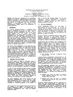
Fig. 4. ERRa–PRC complex-induced mitochondrial function. RO 82 W-1 cells were transfected with 50 ng ERRa and ⁄ or 50 ng PRC. Controls
were transfected with empty vectors. (A) COX activity, CS activity and the ratio of COX activity to CS activity were determined 48 h after
transfection (B) Protein levels of complex IV subunit were determined by western blot and presented relative to the control which was
assigned a value of unity. The results are the mean values ± SD of three experiments performed in duplicate.
functional mitochondria requires the expression of a
large number of genes encoded by the nuclear and
mitochondrial genetic systems. The coordination of
mitochondrial biogenesis depends mainly on a small
number of transcription factors (NRF-1, NRF-2 and
ERRa) and coactivators (PGC-1a, PGC-1b and PRC).
There is no unique system controlling oxidative phosphorylation, and the choice of these inducible coactivators is determined at different levels in response to
environmental or hormonal stimuli. In this work, we
focused on the integration of the regulation of the
mitochondrial respiratory apparatus with the genetic
program controlling cell proliferation. PRC is induced
rapidly by mitogenic signals and stimulates mitochondrial biogenesis through its specific interaction with
NRF-1 or NRF-2 [4–6]. The functional interference
between ERRa and PRC has not yet been investigated.
Nevertheless, ERRa, known to be involved in cellular
metabolic regulation, also interacts with key factors of
cell growth, such as the tumor suppressor p53 or HIF
involved in the transcriptional response to hypoxia
[7,18].
Our earlier work on thyroid oncocytic tumors, rich
in functional mitochondria, demonstrated a high
expression of PRC, NRF-1 and ERRa relative to
normal thyroid tissues [21–23]. We have shown that
the thyroid oncocytic cell line, XTC.UC1, is a good
model for the study of the PRC-dependent regulation
of mitochondrial and cell proliferation. In this study,
we show that follicular thyroid tumors represent models in which PRC and ERRa interfere to induce mitochondrial biogenesis. In the three thyroid cell lines
used here, i.e. FTC-133, XTC.UC1 and RO 82 W-1,
the expression of PRC and ERRa was correlated with
the mitochondrial mass, the expression of mitochondrial genes and the activity of the COX and CS
enzymes. ERRa has already been shown to regulate
COX and CS enzymes [8,12]. To investigate the functional relationship between ERRa and PRC, we modulated the expression and activity of each of these
factors: we overexpressed ERRa and PRC by transient
transfection, underexpressed PRC with siRNA and
inhibited ERRa with an inverse agonist, XCT790.
XCT790, an artificial synthetic compound, is known to
interfere specifically with the ligand-binding domain of
ERRa without affecting estrogen receptor signalling
[26], and to induce the degradation of ERRa [27]. In
our cell lines, we verified the effects of XCT790 on validated ERRa target genes, such as Cyt c and ATP synthase subunit b. As we could not exclude the action of
FEBS Journal 277 (2010) 713–725 ª 2010 The Authors Journal compilation ª 2010 FEBS
719
Respiration rate
(nmolO2·min–1·(mg protein)–1
Respiration rate
(nmolO2·min–1·(mg protein)–1
A
D. Mirebeau-Prunier et al.
Respiration rate
(nmolO2·min–1·(mg protein)–1
ERRa-PRC complex and mitochondrial biogenesis
Fig. 5. Dependence of mitochondrial
function on the ERRa–PRC complex. FTC133 cells were treated for 10 days with
XCT790 or vehicle. On the sixth day, the
cells were transfected with control or PRC
siRNA. (A) Oxygen consumption defined in
the basal condition (basal respiratory), the
maximal stimulation condition by the uncoupling of oxidative phosphorylation with FCCP
(maximal respiratory) and the nonphosphorylating respiratory condition with oligomycin
(oligomycin-insensitive). Phosphorylating respiration (oligomycin-sensitive) was calculated by subtracting the nonphosphorylating
respiration from the basal respiration.
(B) Enzymatic activity of COX and CS, and
the ratio of COX activity to CS activity. The
results are the mean values ± SD.
*P £ 0.05 versus control siRNA-expressing
cells in the absence of XCT790; P £ 0.05
versus control siRNA-expressing cells in the
presence of XCT790; , P £ 0.05 versus
PRC siRNA-expressing cells in the absence
of XCT790.
B
XCT790 on other proteins, these results need to be
confirmed by further ERRa siRNA experiments. We
explored the effect of ERRa through the regulation of
target gene expression via ERREs [7,8,28]. In glycolytic
RO 82 W-1 cells, we observed an increase in COX and
CS activity when PRC and ERRa were both overexpressed, whereas there was no effect when only one of these
factors was overexpressed. This phenomenon has been
described previously for the ERRa–PGC-1a complex,
with the inhibition of ERRa impairing the ability of
PGC-1a to enhance mitochondrial gene expression [9].
Thus, as in the case of PGC-1a, ERRa may be consid720
ered as a PRC effector, mediating cell metabolism
through direct and indirect action on several gene promoters. In the thyroid model, the action of ERRa,
together with PRC, on other transcription factors, such
as NRF-1 and NRF-2, may be suspected. Indeed, NRF1 expression was proportional to ERRa and PRC levels
(Fig. 1), the inhibition of ERRa drastically decreased
NRF-1 expression (data not shown), and it was necessary to overexpress PRC as well as ERRa in cells to
increase COX and CS activity. In this context, the transcription of NRF-1 seems to be dependent on the
expression level of the ERRa–PRC complex.
FEBS Journal 277 (2010) 713–725 ª 2010 The Authors Journal compilation ª 2010 FEBS
D. Mirebeau-Prunier et al.
Surprisingly, the overexpression of PRC and ERRa
in glycolytic RO 82 W-1 had no effect on the mtDNA
copy number. The lack of correlation between CS and
COX activities and mtDNA copy number, described
here, is consistent with the apparent independence of
the mtDNA copy number and expression of the respiratory chain subunits reported by Vercauteren et al.
[29]. They modulated the expression of PRC and
found the regulation of three mitochondrial transcripts
(COX, ND6 and cytochrome b), but no change in the
mtDNA copy number. This indicates that mtDNA
replication is not dependent directly on the ERRa–
PRC complex. In our study, we looked for the effect
of the ERRa–PRC complex 48 h after overexpression
of PRC and ERRa. We suspect that a further period
of treatment may be required to reveal the effect of
the complex on mtDNA levels.
With regard to the enzymatic and respiratory parameters, we showed that the expression of ERRa and PRC
was related to the respiratory capacity and phosphorylating respiration. Inhibition of ERRa and PRC in the
oxidative FTC-133 model led to a decrease in respiratory chain capacity (COX activity) and mitochondrial
mass (CS activity) in a coordinated manner, as the
COX ⁄ CS ratio remained stable. The consequence was a
diminution in phosphorylating respiration without any
change in nonphosphorylating respiration. However,
this was not true for the XTC.UC1 model, which presented a greater proportion of nonphosphorylating
basal respiration. In this model, the inhibition of ERRa
led to a significant decrease in the COX ⁄ CS ratio as a
result of the diminution of the respiratory chain capacity
(COX activity), but not of the mitochondrial mass (CS
activity), and without affecting the respiratory parameters. Other studies support the concept of independent
pathways for the regulation of CS, COX and mitochondrial respiratory activity. Indeed, serum induction in
BALB ⁄ 3T3 fibroblasts increases mitochondrial respiration, but not CS activity [30]. Moreover, during
myogenesis, CS has been shown to be regulated by
a phosphatidylinositol 3-kinase-dependent pathway,
which is not the case for COX [31]. ERRa is not a
unique factor controlling oxidative phosphorylation. As
described elsewhere, mice lacking ERRa are viable
[10,32] and the inhibition of ERRa in other cell models
decreases the respiratory parameter only partially [9].
This suggests that other factors are involved in the control of oxidative phosphorylation, with ERRa playing a
role in the regulation of mitochondrial quality through
the modification of phosphorylating respiration, rather
than in mitochondrial biogenesis.
With regard to the effect of the ERRa–PRC complex on cell proliferation, we found that cell growth
ERRa-PRC complex and mitochondrial biogenesis
slowed down in each of the three thyroid cell lines
investigated when ERRa was inhibited. The involvement of ERRs in the regulation of the cell cycle has
been demonstrated previously [7]. Our work suggests
that this effect is dependent on the metabolic status of
the cell line. In the case of the glycolytic cell line, RO
82 W-1, ERRa inhibition led to an arrest in growth
without affecting the respiratory parameter. However,
the cells were quiescent, suggesting that the ERRa–
PRC complex is involved in the control of the early
phase of the cell cycle. This is in accordance with the
role played by PRC and ERRa in the transition from
the G1 to the S phase of the cell cycle [29,33]. When
the cells are mostly involved in an oxidative process,
as in the case of the FTC-133 thyroid cell line, the
inhibition of ERRa may lead to a slowing down of cell
growth, partly by decreasing the respiratory capacity
and phosphorylating respiration.
In conclusion, the ERRa–PRC transcriptional complex plays a consistent role in increasing the coupling
efficiency of mitochondria in the cell proliferative pathway. Interestingly, ERRa is preferentially used,
according to the cellular metabolic status, either to
control the cell cycle or to promote the efficiency of
oxidative phosphorylation. For cells using the glycolytic pathway, the ERRa–PRC complex plays a role in
cell cycle arrest, whereas it acts on the cell cycle as well
as on oxidative phosphorylation in the case of oxidative cells. Thus, ERRa should be considered as one of
the key targets in the therapy of solid tumors.
Materials and methods
Cell lines and growth conditions
Three human follicular thyroid carcinoma cell lines were
used: the XTC.UC1 cells were oncocytic variants kindly
provided by O. Clark (Mt. Zion Medical Center of the University of California, San Francisco, CA, USA) [21,34]; the
other cell lines, FTC-133 and RO 82 W-1, were obtained
from the Interlab Cell Line Collection (National Institute
for Cancer Research, Genoa, Italy).
FTC-133 and XTC.UC1 cells were grown in Dulbecco’s
modified medium (Invitrogen Corporation, Carlsbad, CA,
USA), supplemented with 10% fetal bovine serum
(Seromed, Biochrom AG, Berlin, Germany), 1% l-glutamine (Invitrogen) and 1% penicillin ⁄ streptomycin (Invitrogen). We added 10 mmL)1 thyroid-stimulating hormone
(Sigma-Aldrich, St Louis, MO, USA) for XTC.UC1.
RO 82 W-1 cells were grown in 60% Dulbecco’s modified
medium and 30% endothelial basal medium (both from
PAA, Pasching, Austria) supplemented with 10% fetal bovine
serum, 1% l-glutamine and 1% penicillin ⁄ streptomycin.
FEBS Journal 277 (2010) 713–725 ª 2010 The Authors Journal compilation ª 2010 FEBS
721
ERRa-PRC complex and mitochondrial biogenesis
D. Mirebeau-Prunier et al.
In all experiments, XCT790 (Sigma-Aldrich) was used at
a final concentration of 5 lm for a 10 day treatment,
replaced with fresh medium every 3 days.
Transient transfections and luciferase assay
Cells were plated 2 days before transfection. We performed
transient transfection with lipofectamine (Invitrogen), as
described by the manufacturer. Cells were collected and
assayed 48 h later.
For experimentation with luciferase activity, each well
was transfected with 1 lg of reporter plasmid 3X ERE
TATA luc (Addgene, Cambridge, MA, USA), 0.05–0.5 lg
of plasmid PRC (Origene Technologies, Rockville, MD,
USA), 0.05–0.5 lg of plasmid ERRa (Addgene) and 0.5 lg
of pRL-CMV (Promega, Madison, WI, USA) as internal
control of transfection efficiency. After 48 h, cells were
harvested for luciferase reporter assay using the Dual-Luciferase Reporter Assay System (Promega). The luciferase
activity was normalized to that of the internal control renilla luciferase as relative luciferase units. All assays were performed in duplicate in three separate experiments.
siRNA
To knock down PRC expression, three predesigned PRC
siRNAs (Applied Biosystems, Foster City, CA, USA)
were tested in comparison with a scrambled negative control siRNA (scrambled siRNA, #4635). The PRC siRNA
(#121729) was chosen on at least 50% of PRC mRNA
expression knockdown. For this study, 30 nm of this
PRC siRNA was transfected using siPORT NeoFX, as
recommended by the manufacturer’s manual (all from
Applied Biosystems). After 48 h, the cells were harvested
for assay.
In vitro cell growth assay
Cells were plated at 105 cells per 25 cm2 flask and cultured
in growth medium for 10 days, replaced with fresh medium
every 3 days. The cells were counted every 3 days using a
Z1 Coulter Particle Counter (Beckman Coulter, Fullerton,
CA, USA). All counts were performed in duplicate and
repeated in two independent experiments.
Quantitative PCR analysis
Total RNA was isolated from cultured cells using an
RNeasy kit (Qiagen, Hilden, Germany). RNA integrity was
determined using a Bio-Analyzer 2100 (Agilent Technologies, Waldbronn, Germany).
Reverse transcription was performed on 1 lg of RNA
with an Advantage RT-for-PCR kit (Clontech, Palo Alto,
CA, USA) following the manufacturer’s recommendations.
722
DNA was isolated using the High Pure PCR Template
Preparation Kit as recommended by the manufacturer
(Roche Applied Science, Mannheim, Germany).
Real-time quantification was performed in a 96-well plate
using IQ SYBR Green supermix and Chromo4 (Biorad,
Hercules, CA, USA). Data were normalized to b-globin.
The sequences of the primers used in this study were as follows: ERRa: 5¢-AAGACAGCAGCCCCAGTGAA-3¢ and
5¢-ACACCCAGCACCAGCACCT-3¢; PRC: 5¢-CACTGG
TTGACCCTGTTCCT-3¢ and 5¢-GTGTTTCAGGGCTTC
TCTGC-3¢; Cyt c: 5¢-CCAGTGCCACACCGTTGAA-3¢
and 5¢-TCCCCAGATGATGCCTTTGTT-3¢; ATP synthase
subunit b: 5¢-CCTTCTGCTGTGGGCTATCA-3¢ and 5¢TCAAGTCATCAGCAGGCACA-3¢; ND5: 5¢-TAACCCC
ACCCTACTAAACC-3¢ and 5¢-GATTATGGGCGTTGA
TTAGTAG-3¢; b-globin: 5¢-CAACTTCATCCACGTTCA
CC-3¢ and 5¢-ACACAACTGTGTTCACTAGC-3¢.
Western blot
Cells were rinsed in NaCl ⁄ Pi, trypsinized and collected in
centrifuge tubes. Proteins (20 lg) were separated by SDSPAGE and transferred to poly(vinylidene difluoride) membranes (Hybond-P, Amersham International plc, Little
Chalfont, Buckinghamshire, UK) by electroblotting. The
membranes were incubated in 5% nonfat milk in TBSTween (Tris-buffered saline with 0.1% Tween-20). The
membranes were incubated with dilutions of the following
antibodies: monoclonal anti-tubulin (Abcam, Cambridge,
UK), monoclonal anti-complex-IV (Mitosciences, Eugene,
OR, USA) and polyclonal anti-ERRa (Abcam), overnight.
After several washes in TBS-Tween, the membranes were
incubated with an appropriate chemiluminescent-labelled
horseradish peroxidase-conjugated secondary antibody
(Jackson ImmunoResearch, WestGrove, PA, USA). The
blots were developed using the enhanced chemiluminescence
method (ECLplus, Amersham Pharmacia Biotech, Little
Chalfont, Buckinghamshire, UK). Signal quantification was
performed by nonsaturating picture scanning by a gel Doc
1000 Molecular Analyst apparatus (Biorad).
Respiratory parameters and respiratory ratio in
intact cells
Respiratory parameters and the coupling state were investigated in intact cells by polarography using a high-resolution Oroboros O2k oxygraph (Oroboros Instruments,
Innsbruck, Austria), as described elsewhere [35,36].
The basal respiration rate, defined as respiration in the
cell culture medium without additional substrates or
effectors, was determined by measuring the linear rate of
oxygen flux in intact cells (3 · 106 cells placed at 37 °C
in 2 mL Dulbecco’s modified medium). Mitochondrial
respiration comprises coupled and uncoupled respiration,
FEBS Journal 277 (2010) 713–725 ª 2010 The Authors Journal compilation ª 2010 FEBS
D. Mirebeau-Prunier et al.
ERRa-PRC complex and mitochondrial biogenesis
determined using the ATP synthase inhibitor oligomycin.
The ATP synthase was then inhibited with oligomycin
(4 lgỈmL)1) and the nonphosphorylating respiration rate
was recorded (oligomycin-insensitive). The phosphorylating respiration rate (oligomycin-sensitive) was calculated
by subtracting the nonphosphorylating respiration rate
from the basal respiration rate. The maximal respiration
was recorded by the uncoupling of oxidative phosphorylation by stepwise titration of FCCP (0.2–2.0 lm) up to
the optimum. Finally, respiration was inhibited by the
sequential addition of 5 lm rotenone and 2 lgỈmL)1 antimycin (complex I and III inhibitors, respectively) to
check for nonmitochondrial respiration (all from SigmaAldrich).
Enzymatic activities
The activities of CS, COX and LDH were measured on cell lysates at 37 °C in a cell buffer [250 mm saccharose, 20 mm
tris(hydroxymethyl)aminomethane, 2 mm EGTA, 1 mgỈmL)1
bovine serum albumin, pH 7.2] using a Beckman DU 640
spectrophotometer (Beckman Coulter).
COX activity was measured in 50 mm KH2PO4 buffer,
using 15 lm reduced cytochrome c and 2.5 mm b-d-dodecylmaltoside [37]. The CS activity was measured in a
reaction medium consisting of 0.1 mm 5,5¢-dithiobis(2-nitrobenzoic acid), 1 mm oxaloacetic acid, 0.3 mm acetyl-CoA
and Triton X-100 (4%), and LDH [35] was assayed by
standard procedures. Specific enzymatic activities were
expressed in mIU [i.e. nanomoles of cytochrome c, 5,5¢dithiobis(2-nitrobenzoic acid) or NADH per minute per
milligram of protein, respectively]. The cellular protein content was determined using the bicinchoninic assay kit
(Uptima, Interchim, Montlucon, France) with bovine serum
¸
albumin as standard (all from Sigma-Aldrich, except Tris
from Eurobio, Les Ulis, France).
Statistical analysis
The results were expressed as the mean values ± standard
deviation (SD). The statistical significance of the observed
variations was assessed using the Wilcoxon signed-rank
test. Differences were considered to be significant at
P < 0.05. All analyses were performed using statview
version 5.0 (SAS Institute, Gary, NC, USA).
Acknowledgements
This work was supported by grants from
We thank D. Couturier and C. Wetterwald
cal assistance, and K. Malkani for critical
the manuscript. We thank J.M. Vanacker
providing reporter plasmids and O. Clark
providing XTC.UC1 cells.
INSERM.
for technireading of
for kindly
for kindly
References
1 Scarpulla RC (2006) Nuclear control of respiratory gene
expression in mammalian cells. J Cell Biochem 97,
673–683.
2 Puigserver P, Wu Z, Park CW, Graves R, Wright M &
Spiegelman BM (1998) A cold-inducible coactivator of
nuclear receptors linked to adaptive thermogenesis. Cell
92, 829–839.
3 Handschin C & Spiegelman BM (2006) Peroxisome
proliferator-activated receptor gamma coactivator 1
coactivators, energy homeostasis, and metabolism.
Endocr Rev 27, 728–735.
4 Andersson U & Scarpulla RC (2001) Pgc-1-related
coactivator, a novel, serum-inducible coactivator of
nuclear respiratory factor 1-dependent transcription in
mammalian cells. Mol Cell Biol 21, 3738–3749.
5 Gleyzer N, Vercauteren K & Scarpulla RC (2005)
Control of mitochondrial transcription specificity
factors (TFB1M and TFB2M) by nuclear respiratory
factors (NRF-1 and NRF-2) and PGC-1 family
coactivators. Mol Cell Biol 25, 1354–1366.
6 Vercauteren K, Gleyzer N & Scarpulla RC (2008)
PGC-1-related coactivator complexes with HCF-1 and
NRF-2b in mediating NRF-2(GABP)-dependent
respiratory gene expression. J Biol Chem 283, 12102–
12111.
7 Dufour CR, Wilson BJ, Huss JM, Kelly DP, Alaynick
WA, Downes M, Evans RM, Blanchette M & Giguere
V (2007) Genome-wide orchestration of cardiac functions by the orphan nuclear receptors ERRa and c.
Cell Metab 5, 345–356.
8 Schreiber SN, Emter R, Hock MB, Knutti D, Cardenas
J, Podvinec M, Oakeley EJ & Kralli A (2004) The
estrogen-related receptor a (ERRa) functions in PPARc
coactivator 1a (PGC-1a)-induced mitochondrial biogenesis. Proc Natl Acad Sci USA 101, 6472–6477.
9 Mootha VK, Handschin C, Arlow D, Xie X, St Pierre
J, Sihag S, Yang W, Altshuler D, Puigserver P,
Patterson N et al. (2004) Erralpha and C ⁄ b specify
PGC-1a-dependent oxidative phosphorylation gene
expression that is altered in diabetic muscle. Proc Natl
Acad Sci USA 101, 6570–6575.
10 Villena JA, Hock MB, Chang WY, Barcas JE, Giguere
V & Kralli A (2007) Orphan nuclear receptor estrogenrelated receptor a is essential for adaptive thermogenesis. Proc Natl Acad Sci USA 104, 1418–1423.
11 Cartoni R, Leger B, Hock MB, Praz M, Crettenand A,
Pich S, Ziltener JL, Luthi F, Deriaz O, Zorzano A et al.
(2005) Mitofusins 1 ⁄ 2 and ERRa expression are
increased in human skeletal muscle after physical
exercise. J Physiol 567, 349–358.
12 Rangwala SM, Li X, Lindsley L, Wang X, Shaughnessy
S, Daniels TG, Szustakowski J, Nirmala NR, Wu Z &
Stevenson SC (2007) Estrogen-related receptor a is
FEBS Journal 277 (2010) 713–725 ª 2010 The Authors Journal compilation ª 2010 FEBS
723
ERRa-PRC complex and mitochondrial biogenesis
13
14
15
16
17
18
19
20
21
22
23
724
D. Mirebeau-Prunier et al.
essential for the expression of antioxidant protection
genes and mitochondrial function. Biochem Biophys Res
Commun 357, 231–236.
Ariazi EA, Clark GM & Mertz JE (2002) Estrogenrelated receptor a and estrogen-related receptor c
associate with unfavorable and favorable biomarkers,
respectively, in human breast cancer. Cancer Res 62,
6510–6518.
Suzuki T, Miki Y, Moriya T, Shimada N, Ishida T,
Hirakawa H, Ohuchi N & Sasano H (2004) Estrogenrelated receptor a in human breast carcinoma as a
potent prognostic factor. Cancer Res 64, 4670–4676.
Sun P, Sehouli J, Denkert C, Mustea A, Konsgen D,
Koch I, Wei L & Lichtenegger W (2005) Expression of
estrogen receptor-related receptors, a subfamily of
orphan nuclear receptors, as new tumor biomarkers in
ovarian cancer cells. J Mol Med 83, 457–467.
Watanabe A, Kinoshita Y, Hosokawa K, Mori T,
Yamaguchi T & Honjo H (2006) Function of estrogenrelated receptor a in human endometrial cancer. J Clin
Endocrinol Metab 91, 1573–1577.
Cavallini A, Notarnicola M, Giannini R, Montemurro
S, Lorusso D, Visconti A, Minervini F & Caruso MG
(2005) Oestrogen receptor-related receptor a (ERRa)
and oestrogen receptors (ERa and ERb) exhibit
different gene expression in human colorectal tumour
progression. Eur J Cancer 41, 1487–1494.
Ao A, Wang H, Kamarajugadda S & Lu J (2008)
Involvement of estrogen-related receptors in transcriptional response to hypoxia and growth of solid tumors.
Proc Natl Acad Sci USA 105, 7821–7826.
Arany Z, Foo SY, Ma Y, Ruas JL, Bommi-Reddy A,
Girnun G, Cooper M, Laznik D, Chinsomboon J,
Rangwala SM et al. (2008) HIF-independent regulation
of VEGF and angiogenesis by the transcriptional
coactivator PGC-1a. Nature 451, 1008–1012.
O’Hagan KA, Cocchiglia S, Zhdanov AV, Tambawala
MM, Cummins EP, Monfared M, Agbor TA, Garvey
JF, Papkovsky DB, Taylor CT et al. (2009) PGC-1a is
coupled to HIF-1a-dependent gene expression by
increasing mitochondrial oxygen consumption in
skeletal muscle cells. Proc Natl Acad Sci USA 106,
2188–2193.
Savagner F, Chevrollier A, Loiseau D, Morgan C,
Reynier P, Clark O, Stepien G & Malthiery Y (2001)
Mitochondrial activity in XTC.UC1 cells derived from
thyroid oncocytoma. Thyroid 11, 327–333.
Savagner F, Franc B, Guyetant S, Rodien P, Reynier P
& Malthiery Y (2001) Defective mitochondrial ATP
synthesis in oxyphilic thyroid tumors. J Clin Endocrinol
Metab 86, 4920–4925.
Savagner F, Mirebeau D, Jacques C, Guyetant S,
Morgan C, Franc B, Reynier P & Malthiery Y (2003)
PGC-1-related coactivator and targets are upregulated
24
25
26
27
28
29
30
31
32
33
34
in thyroid oncocytoma. Biochem Biophys Res Commun
310, 779–784.
Johnston SD, Liu X, Zuo F, Eisenbraun TL, Wiley SR,
Kraus RJ & Mertz JE (1997) Estrogen-related receptor a
1 functionally binds as a monomer to extended half-site
sequences including ones contained within estrogenresponse elements. Mol Endocrinol 11, 342–352.
Huss JM, Kopp RP & Kelly DP (2002) Peroxisome
proliferator-activated receptor coactivator-1a (PGC-1a)
coactivates the cardiac-enriched nuclear receptors
estrogen-related receptor-a and –c. Identification of
novel leucine-rich interaction motif within PGC-1a.
J Biol Chem 277, 40265–40274.
Willy PJ, Murray IR, Qian J, Busch BB, Stevens WC
Jr, Martin R, Mohan R, Zhou S, Ordentlich P, Wei P
et al. (2004) Regulation of PPARc coactivator 1a
(PGC-1a) signaling by an estrogen-related receptor a
(ERRa) ligand. Proc Natl Acad Sci USA 101, 8912–
8917.
Lanvin O, Bianco S, Kersual N, Chalbos D & Vanacker
JM (2007) Potentiation of ICI182,780 (Fulvestrant)induced estrogen receptor-a degradation by the estrogen
receptor-related receptor-a inverse agonist XCT790.
J Biol Chem 282, 28328–28334.
Huss JM, Torra IP, Staels B, Giguere V & Kelly DP
(2004) Estrogen-related receptor a directs peroxisome
proliferator-activated receptor a signaling in the transcriptional control of energy metabolism in cardiac and
skeletal muscle. Mol Cell Biol 24, 9079–9091.
Vercauteren K, Gleyzer N & Scarpulla RC (2009) Short
hairpin RNA-mediated silencing of PRC (PGC1-related coactivator) results in a severe respiratory
chain deficiency associated with the proliferation of
aberrant mitochondria. J Biol Chem 284, 2307–2319.
Herzig RP, Scacco S & Scarpulla RC (2000) Sequential
serum-dependent activation of CREB and NRF-1 leads
to enhanced mitochondrial respiration through the
induction of cytochrome c. J Biol Chem 275,
13134–13141.
Kraft CS, LeMoine CM, Lyons CN, Michaud D,
Mueller CR & Moyes CD (2006) Control of mitochondrial biogenesis during myogenesis. Am J Physiol Cell
Physiol 290, C1119–C1127.
Luo J, Sladek R, Carrier J, Bader JA, Richard D &
Giguere V (2003) Reduced fat mass in mice lacking
orphan nuclear receptor estrogen-related receptor a.
Mol Cell Biol 23, 7947–7956.
Bianco S, Lanvin O, Tribollet V, Macari C, North S &
Vanacker JM (2009) Modulating ERRa activity inhibits
cell proliferation. J Biol Chem 284, 23286–23292.
Zielke A, Tezelman S, Jossart GH, Wong M, Siperstein
AE, Duh QY & Clark OH (1998) Establishment of a
highly differentiated thyroid cancer cell line of Hurthle
cell origin. Thyroid 8, 475–483.
FEBS Journal 277 (2010) 713–725 ª 2010 The Authors Journal compilation ª 2010 FEBS
D. Mirebeau-Prunier et al.
35 Hutter E, Renner K, Pfister G, Stockl P, Jansen-Durr P
& Gnaiger E (2004) Senescence-associated changes in
respiration and oxidative phosphorylation in primary
human fibroblasts. Biochem J 380, 919–928.
36 Loiseau D, Chevrollier A, Verny C, Guillet V, Gueguen
N, Pou de Crescenzo MA, Ferre M, Malinge MC,
Guichet A, Nicolas G et al. (2007) Mitochondrial
ERRa-PRC complex and mitochondrial biogenesis
coupling defect in Charcot–Marie–Tooth type 2A
disease. Ann Neurol 61, 315–323.
37 Rustin P, Lebidois J, Chretien D, Bourgeron T,
Piechaud JF, Rotig A, Munnich A & Sidi D (1994)
Endomyocardial biopsies for early detection of mitochondrial disorders in hypertrophic cardiomyopathies.
J Pediatr 124, 224–228.
FEBS Journal 277 (2010) 713–725 ª 2010 The Authors Journal compilation ª 2010 FEBS
725