Báo cáo khoa học: Cysteine residues exposed on protein surfaces are the dominant intramitochondrial thiol and may protect against oxidative damage docx
Bạn đang xem bản rút gọn của tài liệu. Xem và tải ngay bản đầy đủ của tài liệu tại đây (642.03 KB, 16 trang )
Cysteine residues exposed on protein surfaces are the
dominant intramitochondrial thiol and may protect
against oxidative damage
Raquel Requejo, Thomas R. Hurd, Nikola J. Costa and Michael P. Murphy
MRC Mitochondrial Biology Unit, Wellcome Trust ⁄ MRC Building, Cambridge, UK
Introduction
The thiol functional group plays a major role in intra-
cellular antioxidant defences. Cysteine residues in the
active sites of proteins such as thioredoxin (Trx), glut-
aredoxin (Grx) and peroxiredoxin (Prx) detoxify reac-
tive oxygen species (ROS) and reactive nitrogen species
and reduce oxidized protein thiols [1,2]. The low
molecular weight thiol glutathione (GSH) acts in
conjunction with GSH peroxidases, Grxs and
glutathione S-transferases to detoxify ROS and
electrophiles and to recycle oxidized protein thiols [3].
In addition to these enzyme-catalysed reactions, thiols
can also react directly with some ROS and reactive
nitrogen species; therefore, solvent-exposed thiols
within cells may contribute to endogenous antioxidant
defences [1,4,5]. Consequently, cysteine residues
exposed on the surface of proteins without a clear
functional or structural role may still make an impor-
tant contribution to antioxidant defences [2]. However,
Keywords
cysteine; glutathione; mitochondria;
peroxynitrite; protein thiol
Correspondence
M. P. Murphy, MRC Mitochondrial Biology
Unit, Wellcome Trust ⁄ MRC Building, Hills
Road, Cambridge CB2 0XY, UK
Fax: +44 0 1223 252905
Tel: +44 0 1223 252900
E-mail:
Re-use of this article is permitted in
accordance with the Terms and Conditions
set out at ey.
com/authorresources/onlineopen.html
(Received 17 November 2009, revised 1
January 2010, accepted 8 January 2010)
doi:10.1111/j.1742-4658.2010.07576.x
Cysteine plays a number of important roles in protecting the cell from
oxidative damage through its thiol functional group. These defensive func-
tions are generally considered to be carried out by the low molecular
weight thiol glutathione and by cysteine residues in the active sites of pro-
teins such as thioredoxin and peroxiredoxin. In addition, there are thiols
exposed on protein surfaces that are not directly involved with protein
function, although they can interact with the intracellular environment. In
the present study, in subcellular fractions prepared from rat liver or heart,
we show that the quantitatively dominant free thiols are those of cysteine
residues exposed on protein surfaces and not those carried by glutathione.
Within the mitochondrial matrix, the concentration of exposed protein
thiols is 60–90 mm, which is approximately 26-fold higher than the gluta-
thione concentration in that compartment. This suggests that exposed pro-
tein thiols are of greater importance than glutathione for nonenzyme
catalysed reactions of thiols with reactive oxygen and nitrogen species and
with electrophiles within the cell. One such antioxidant role for exposed
protein thiols may be to prevent protein oxidative damage. In the present
study, in mitochondrial membranes and in complex I, we show that
exposed protein thiols protect against tyrosine nitration and protein
dysfunction caused by peroxynitrite. Therefore, exposed protein thiols
are the dominant free thiol within the cell and may play a critical role in
intracellular antioxidant defences against oxidative damage.
Abbreviations
ACA, e-amino-n-caproic acid; AMS, 4-acetamido-4¢-maleimidylstilbene-2,2¢-disulfonic acid; BN-PAGE, blue native gel-PAGE; DDM, n-dodecyl-
b-
D-maltopyranoside; DMPO, 5,5-dimethyl-1-pyrroline-N-oxide; DTNB, 5,5¢-dithiobis(2-nitrobenzoic acid); Grx, glutaredoxin; GSH, glutathione;
GSSG, glutathione disulfide; HAR, hexa-ammineruthenium (III) chloride; MnSOD, manganese superoxide dismutase; ONOO
),
peroxynitrite;
Prx, peroxiredoxin; ROS, reactive oxygen species; tBHP, tert-butyl hydrogen peroxide; Trx, thioredoxin; TrxR, thioredoxin reductase.
FEBS Journal 277 (2010) 1465–1480 ª 2010 The Authors Journal compilation ª 2010 FEBS 1465
this possibility is not widely recognized and there is lit-
tle experimental evidence to support a protective role
for exposed protein thiols. One factor impeding pro-
gress is the assumption that GSH is the quantitatively
dominant intracellular thiol. Although a number of
studies have investigated the intracellular abundance of
protein thiols [2,5–8], little is known about the amount
of exposed protein thiols within cells in comparison to
GSH, or whether they are important in cellular defence.
To determine the contribution of exposed protein thiols
to the intracellular redox environment, we have mea-
sured their abundance on native proteins from tissue
subfractions relative to the amount of GSH, quantified
exposed protein thiols within isolated mitochondria
and determined whether these protein thiols can pro-
tect against oxidative damage caused by peroxynitrite
(ONOO
)
). These findings indicate that the cysteine
residues exposed on the surface of proteins are the
dominant intracellular thiol and that they may play an
important role in intracellular antioxidant defences.
Results
Quantification of exposed protein thiols and GSH
in tissue subfractions
To assess the importance for antioxidant defence of
exposed thiols on the surfaces of proteins in their
native conformations, we quantified exposed and total
protein thiols in tissue subfractions (Fig. 1). Tissue
homogenates from rat liver and heart were fractionated
by sequential differential centrifugation to give super-
natants from the 3000 g (crude homogenate), 10 000 g
(cytosol and microsomes) and 100 000 g centrifuga-
tions (soluble cytosol fraction) and a mitochondrial
fraction (pellet from the 10 000 g centrifugation). To
measure exposed protein thiols, we used the mild deter-
gent n-dodecyl-b-d-maltopyranoside (DDM) to solubi-
lize membrane proteins with minimal disruption to
protein conformation. The suspensions were then trea-
ted with dithiothreitol to reduce thiols that had become
reversibly oxidized during fractionation. The dith-
iothreitol and low molecular weight thiols such as
GSH were then removed by centrifugal gel filtration
and exposed protein thiols were measured using
5,5¢-dithiobis(2-nitrobenzoic acid) (DTNB). Control
experiments showed that lysing mitochondria by
freeze ⁄ thawing instead of with DDM treatment gave
similar levels of exposed thiols (data not shown). Total
protein thiols were measured after complete denatur-
ation of the proteins with SDS. Exposed and total pro-
tein thiols for each fraction are shown in Fig. 1A,B,
for liver and heart, respectively. The total protein
thiols in the fractions were in the range 50–225
nmolÆmg protein
)1
. Allowing for variation in cysteine
content between different tissues and subcellular
fractions, these values are consistent with the known
cysteine content of mammalian proteins of approxi-
mately 2% of amino acid residues. On average,
approximately 70% of total protein thiols were
exposed to the solvent (range 56–84%).
We next measured GSH and glutathione disulfide
(GSSG) in each fraction prior to dithiothreitol treat-
ment or centrifugal filtration (Fig. 1C, D). Most of the
GSH pool was present as GSH and the total GSH con-
tent varied in the range 2–80 nmolÆmg protein
)1
(Fig. 1C, D). The total amounts of GSH equivalents in
each fraction as a percentage of exposed protein thiols
are also shown above the data bars (Fig. 1C, D). In all
fractions, the GSH content was substantially less that
that of exposed protein thiols, in the range 3–51%.
Because GSH is by far the most abundant intracellular
low molecular thiol, this demonstrates that exposed
protein thiols are the quantitatively dominant intra-
cellular thiol and, in some cases, are present at a 20–30-
fold higher concentration than GSH. This finding is
consistent with exposed protein thiols playing a role in
intracellular antioxidant defences.
Quantification of exposed protein thiols and GSH
within mitochondria
To further analyse the potential role of surface protein
thiols in antioxidant defences, we next focussed on
their role within mitochondria. This was carried out
because: mitochondria are a major source of ROS
within the cell [9] and, consequently, have extensive
antioxidant defences; the pH in the mitochondrial
matrix ( 7.8) is higher than in the cytosol (7.2), ren-
dering protein thiols (typical pK
a
8–9) more reactive
for processes requiring the thiolate; and, finally, mito-
chondria have experimental advantages because they
are discrete, closed systems with their own GSH, Trx,
thioredoxin reductase (TrxR), NADPH and Grx sys-
tems that can be investigated under conditions that are
physiologically relevant.
First, we quantified exposed and total protein thiols
in membrane and soluble fractions from liver and heart
mitochondria (Fig. 1E, F). Approximately 70% of total
protein thiols were exposed to the solvent (range
55–85%) (Fig. 1E, F). However, these measurements
cannot distinguish exposed protein thiols on the
mitochondrial outer membrane, the intermembrane
space and on the outer face of the inner membrane from
those within the mitochondrial matrix. Because matrix
protein thiols are of the greatest interest as a result of
Protein thiols R. Requejo et al.
1466 FEBS Journal 277 (2010) 1465–1480 ª 2010 The Authors Journal compilation ª 2010 FEBS
0
50
100
150
200
250
300
0
20
40
60
80
100
120
Protein thiols
(nmol·mg prot
–1
)
**
**
**
**
**
**
**
**
Exposed thiols
Total thiols
GSH equivalents
(nmol·mg prot
–1
)
Supernatants Mitos
> 3K > 10K > 100K
Supernatants Mitos
> 3K > 10K > 100K
0
5
10
15
20
25
30
0
12
8
4
60
70
80
90
Liver Heart
Total GSH
GSH
GSSG
14%
23%
28%
3%
21%
5%
51%
3%
0
10
20
30
40
50
60
70
80
Membrane
fraction
Soluble
fraction
0
5
10
15
20
25
30
Exposed thiols
Total thiols
Membrane
Fraction
Soluble
Fraction
Protein thiols
(nmol·mg prot
–1
)
Liver
Mitochondria
Heart
Mitochondria
0
10
20
30
40
50
60
70
Liver Heart
*
*
0
0.5
1
1.5
2
2.5
Liver Heart
GSH equivalents
(nmol·mg prot
–1
)
Protein thiols
(nmol·mg prot
–1
)
=–7nmol
(–12%)
Δ
Δ
=–11nmol
(–26%)
Control
+AMS
Protein thiols
(nmol·mg prot
–1
)
GSH equivalents
(nmol·mg prot
–1
)
Protein thiols
(nmol·mg prot
–1
)
Liver
Heart
Total GSH
GSH
GSSG
AB
CD
EF
GH
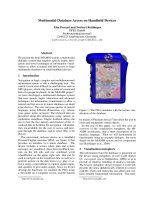
Fig. 1. Total and exposed protein thiols and GSH in liver and heart tissue homogenates and mitochondria. (A, B) Total and exposed protein thi-
ols in sequential supernatants from 3000 g, 10 000 g and 100 000 g centrifugations, and from a mitochondrial fraction, isolated from liver (A)
and heart (B) tissue homogenates. **P < 0.01 for comparison of total and exposed thiols by Student’s t-test. (C, D) Total GSH equivalents,
GSH and 2· GSSG, in sequential supernatants from 3000 g, 10 000 g and 100 000 g centrifugations, and from a mitochondrial fraction, iso-
lated from liver (C) and heart (D) tissue homogenates. The percentages above the data bars indicate the total GSH content of the fraction as a
percentage of its exposed protein thiol content. (E, F) Total and exposed thiols in membrane and matrix fractions from liver (E) or heart (F) mito-
chondria. Mitochondria (5 mgÆmL
)1
protein) were suspended in KCl buffer, pelleted by centrifugation and separated into membrane and matrix
fractions and then exposed and total protein thiols were measured. (G) Exposed mitochondrial protein thiols ± the thiol alkylating agent AMS.
Mitochondria (5 mgÆmL
)1
protein) were incubated in KCl buffer ± AMS (100 lM) for 10 min at 30 °C. Samples were then centrifuged and
exposed protein thiols were measured. (H) GSH content of rat liver and heart mitochondria. Mitochondria (5 mgÆmL
)1
protein) were incubated
in KCl buffer for 10 min at 30 °C and the GSH and GSSG contents measured. All data are the mean ± SD of three independent experiments.
R. Requejo et al. Protein thiols
FEBS Journal 277 (2010) 1465–1480 ª 2010 The Authors Journal compilation ª 2010 FEBS 1467
the elevated oxidative stress of that compartment, we
measured these by blocking nonmatrix protein thiols
with the membrane impermeant thiol alkylating agent
4-acetamido-4¢-maleimidylstilbene-2,2¢-disulfonic acid
(AMS) (Fig. 1G). AMS decreased the total amount of
exposed protein thiols by 7 nmolÆmg protein
)1
()12%)
in liver mitochondria and by 11 nmolÆmg protein
)1
()26%) in heart mitochondria (Fig. 1G). Thus, the
amount of exposed protein thiols is approximately 48
and 31 nmolÆmg protein
)1
within the matrices of liver
and heart mitochondria, respectively. This is 25–30-fold
higher than their GSH contents of 1–2 nmolÆmg
protein
)1
(Fig. 1H). The mitochondrial matrix volume
under these conditions is approximately 0.5 llÆmg
protein
)1
[10], giving a concentration of GSH of
approximately 3 mm, which contrasts with the matrix
concentration for exposed protein thiols of 60–90 mm.
Therefore, within the mitochondrial matrix, exposed
cysteine residues on the surface of proteins are by far
the dominant free thiol.
Response of exposed mitochondrial protein thiols
to oxidative stress
The high concentration of exposed protein thiols within
the mitochondrial matrix is consistent with them play-
ing a role in antioxidant defence. If this is the case, then
their redox state should respond to mitochondrial oxi-
dative stress. Treatment of liver or heart mitochondria
with diamide oxidized the matrix GSH pool, decreased
the GSH content by 1–1.5 nmolÆmg protein
)1
and led
to the formation of GSSG and up to 0.4 nmolÆmg
protein
)1
of protein mixed disulfides (Fig. 2A, B).
Under these conditions, there was a loss of 9–19
nmolÆmg protein
)1
of exposed protein thiols, corre-
sponding to 15–32% of the total present (Fig. 2C, D).
Similarly, treatment of liver mitochondria with tert-
butyl hydrogen peroxide (tBHP) or ONOO
)
oxidized
14–18 nmolÆmg protein
)1
exposed protein thiols, corre-
sponding to 24–31% of the total present (Fig. 2E). Oxi-
dation of exposed protein thiols by tBHP was fully
reversed by dithiothreitol, whereas that by ONOO
)
was
partially reversed and that by diamide was not reversed
(Fig. 2E), presumably as a result of the formation of
higher thiol oxidation states such as sulfinic and
sulfonic acids that are not reduced by dithiothreitol
[11]. When stressed mitochondria were washed to
remove the oxidant and reincubated, the oxidation of
exposed protein thiols was partially restored by intra-
mitochondrial reduction processes (Fig. 2F). Therefore,
during oxidative stress, the extent of thiol modification
of exposed protein thiols is ten to 20-fold greater in
magnitude than that of the entire GSH pool, and a
proportion of the changes to exposed protein thiols can
be reversed. These findings are consistent with exposed
protein thiols within mitochondria playing an antioxi-
dant role during their response to oxidative stress.
Protection against ONOO
)
-induced tyrosine
nitration by exposed protein thiols
The data shown in Figs 1 and 2 reveal that there is a
high concentration of exposed protein thiols within
mitochondria that respond to oxidative stress. To
determine whether these exposed protein thiols could
protect mitochondrial proteins against oxidative dam-
age, we next investigated isolated mitochondrial mem-
branes. This system contains an active respiratory
chain and has a large number of exposed thiols that
are easily accessible and measureable [12–14]. As an
oxidant, we chose ONOO
)
because it contributes to
mitochondrial oxidative damage in a range of patholo-
gies [15] and is known to react with protein thiols [16].
An important mode of damage caused by ONOO
)
is
the specific oxidation of protein tyrosine residues to
3-nitrotyrosine by a two step process involving the
initial formation of a tyrosyl radical, which then goes
on to react with a
•
NO
2
radical to form nitrotyrosine
[15,17]. Because the formation of 3-nitrotyrosine can
be measured using a specific antibody [17], the deter-
mination of the effect of exposed protein thiols on
tyrosine nitration in mitochondrial membranes serves
to indicate whether exposed protein thiols can be
involved in antioxidant defences.
There were approximately 85 nmolÆmg protein
)1
total protein thiols in mitochondrial membranes and
approximately 70 nmolÆmg protein
)1
of these were
exposed to the solvent (Fig. 3A). There was a dose-
dependent decrease in exposed protein thiols on
reaction with ONOO
)
that was largely reversed by
dithiothreitol, consistent with the oxidation of protein
thiols by ONOO
)
to thiyl radicals and sulfenic acids
[16] (Fig. 3A). The reaction of ONOO
)
with mitochon-
drial membranes also formed 3-nitrotyrosine from
tyrosine residues, as indicated by immunoblotting
with a specific antibody (Fig. 3B). The formation of
3-nitrotyrosine was dependent on the concentration of
ONOO
)
(Fig. 3C). To determine whether exposed pro-
tein thiols decreased 3-nitrotyrosine formation, we pre-
treated membranes with N-ethylmaleimide to block all
exposed thiols. This rendered tyrosine residues in the
membranes far more susceptible to nitration on expo-
sure to ONOO
)
(Fig. 3B, C). To determine whether
thiyl radicals were formed on the cysteine residues of
membrane proteins during exposure to ONOO
)
,we
added the spin trap 5,5-dimethyl-1-pyrroline-N-oxide
Protein thiols R. Requejo et al.
1468 FEBS Journal 277 (2010) 1465–1480 ª 2010 The Authors Journal compilation ª 2010 FEBS
(DMPO), which forms stable protein adducts with
thiyl radicals that can be detected on immunoblots
[18]. This experiment demonstrated the N-ethylmalei-
mide-sensitive formation of DMPO-protein adducts,
which is consistent with protein thiol oxidation by
ONOO
)
(Fig. 3D).
0
0.5
1
1.5
2
2.5
GSH equivalent
(nmol
·
mg prot
–1
)
GSH equivalent
(nmol
·
mg prot
–1
)
Protein thiols
(nmol
·
mg prot
–1
)
Protein thiols
(nmol
·
mg prot
–1
)
Protein thiols
(nmol
·
mg prot
–1
)
0
0.2
0.4
0.6
0.8
1
1.2
1.4
1.6
Total GSH
GSH
GSSG
Pr-SSG
*
*
*
*
*
*
*
*
*
*
*
*
*
*
0 0.5 5
Diamide [m
M
]
Diamide [m
M
]
0 0.5 5
**
**
**
**
Liver Heart
0
10
20
30
40
50
60
70
80
0
10
20
30
40
50
60
70
80
= –16 nmol
(–27%)
= –19 nmol
(–32%)
= –9.4 nmol
(–15%)
= –11 nmol
(–19%)
Control Diamide tBHP
ONOO
–
–DTT
+DTT
Δ
= –18 nmol
(–31%)
Time (min)
*
*
0 0.5 5
Diamide [m
M
]
0 0.5 5
Diamide [m
M
]
Liver Heart
Liver Liver
tBHP
ONOO
–
Diamide
Exposed protein thiols
(% Control)
0
10
20
30
40
50
60
70
*
40
50
60
70
80
90
100
110
120
–30 –10 10 30 50 70
+ Oxidant
Resuspension
0
Δ
= –16 nmol
(–27%)
Δ
= –14 nmol
(–24%)
Δ
Δ
Δ
Δ
AB
CD
EF
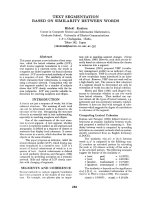
Fig. 2. Exposed protein thiols and GSH in oxidatively stressed mitochondria. (A–D) Effect of diamide on exposed protein thiols, protein-GSH
mixed disulfides and GSH. Mitochondria (5 mgÆmL
)1
protein) from the liver (A, C) or heart (B, D) were incubated with diamide for 5 min at
37 °C. The values after the D in (C) and (D) are the actual and the percentage changes in protein thiols relative to controls. (E) Effects of oxi-
dants and dithiothreitol on exposed mitochondrial protein thiols. Liver mitochondria (5 mgÆmL
)1
protein) were incubated for 5 min with
0.5 m
M ONOO
)
, tBHP or diamide and exposed protein thiols measured. For some incubations, the mitochondria were incubated with 1 mM
dithiothreitol before measurement of protein thiols. The values after the D in (C) and (D) are the actual and the percentage changes in pro-
tein thiols relative to controls. (F) Reduction of mitochondrial thiols after oxidative stress. Liver mitochondria (5 mgÆmL
)1
protein) were incu-
bated with either carrier, 0.5 m
M tBHP, ONOO
)
or diamide for 10 min. Next, mitochondria were pelleted by centrifugation and resuspended
in medium without oxidant. The exposed protein thiols were measured as a percentage of parallel control incubations that had undergone
the same isolation and resuspension procedures but without exposure to oxidant. All data are the mean ± SD of three experiments:
*P < 0.05, **P < 0.01 relative to controls by Student’s t-test. DTT, dithiothreitol.
R. Requejo et al. Protein thiols
FEBS Journal 277 (2010) 1465–1480 ª 2010 The Authors Journal compilation ª 2010 FEBS 1469
The data shown in Fig. 3B, C indicate that blocking
exposed protein thiols with N-ethylmaleimide renders
membrane proteins more susceptible to nitration by
ONOO
)
. We suggest that this occurs because N-ethyl-
maleimide blocks thiols, thereby preventing cysteine
residues from protecting tyrosine residues from nitra-
tion. However, an alternative interpretation is that
exposed protein thiols react rapidly with ONOO
)
to
0
10
20
30
40
50
60
70
80
90
Exposed thiols
Exposed thiols + DTT
Protein thiols
(nmol
·
mg prot
–1
)
0
0.5
1
2
[ONOO
–
] (mM)
25
20
250
150
100
75
50
37
m (kDa)
m
(kDa)
m (kDa)
ONOO
–
–+–+
NEM – – + +
α–nitrotyrosine
0 0.5 1
+
00.51 2
–––
–
2
++ +
ONOO
–
(mM)
NEM
220
120
100
80
60
50
40
30
20
α–nitrotyrosine
220
120
100
80
60
50
40
30
20
ONOO
–
–+–+
NEM – – + +
α–DMPO
– NEM
+NEM
0
0.04
k (s
–1
)
0.08
0.12
0.16
–Membranes +Membranes
–NEM
+NEM
ΔA
302
= 0.5
10 s
ONOO
–
***
***
***
AB
CD
EF
Fig. 3. Effect of blocking exposed protein thiols with N-ethylmaleimide on nitration by ONOO
)
of mitochondrial membrane proteins. (A–D)
Mitochondrial membranes were incubated (20 mgÆmL
)1
protein) at 37 °C in membrane buffer with either no additions or after pre-treatment
with 1 m
M N-ethylmaleimide for 10 min. Next, the membranes were exposed to different doses of ONOO
)
, or decomposed ONOO
)
for the
control incubations, for 5 min. (A) After incubation exposed protein thiols were measured. Data are the mean ± SD of three experiments.
***P < 0.001 by Student’s t-test. (B) After incubation mitochondrial membranes (75 lg of protein) was separated by SDS-PAGE and
immunblotted to detect 3-nitrotyrosine residues. (C) After incubation, mitochondrial membranes (50 lg of protein) was separated by
SDS-PAGE and immunoblotted to detect 3-nitrotyrosine residues. (D) Membranes were incubated as above but in the presence of DMPO
(100 m
M). After incubation, mitochondrial membranes (75 lg of protein) was separated by SDS-PAGE and immunoblotted to detect DMPO
protein adducts. (E, F) Rate of decay of ONOO
)
. (E) The decomposition of ONOO
)
(1 mM) was monitored by measuring A
302
after its
addition to a rapidly stirred suspension of membranes (1 mg
Æ
mL
)1
protein) incubated as described above in presence or absence of 1 mM
N-ethylmaleimide. (F) Rate constants for decomposition of ONOO
)
in controls or samples containing mitochondrial membranes, with or
without N-ethylmaleimide. Data are the mean ± SD of three experiments. DTT, dithiothreitol; NEM, N-ethylmaleimide.
Protein thiols R. Requejo et al.
1470 FEBS Journal 277 (2010) 1465–1480 ª 2010 The Authors Journal compilation ª 2010 FEBS
accelerate its degradation, and that N-ethylmaleimide
treatment may slow this process, thereby enhancing
nitration by increasing the bulk exposure of tyrosines to
ONOO
)
. To determine whether this could be the case,
we investigated the effect of N-ethylmaleimide treat-
ment on the rate of decay of ONOO
)
. Accordingly,
ONOO
)
was injected into a rapidly stirred membrane
suspension ± N-ethylmaleimide and the absorption of
ONOO
)
was measured over time (Fig. 3E). The first-
order decay process was analysed to generate rate con-
stants for the decay of ONOO
)
(Fig. 3F). In the
absence of membranes, the ONOO
)
t
1 ⁄ 2
was approxi-
mately 5 s and, in the presence of membranes, the t
1 ⁄ 2
increased to approximately 13 s (Fig. 3F), probably as
a result of permeation of ONOO
)
into the hydrophobic
membrane core [19]. In the presence or absence of mem-
branes, the rate of decay of ONOO
)
was unaffected by
N-ethylmaleimide (Fig. 3E, F). Therefore, N-ethylma-
leimide treatment does not alter membrane exposure to
the bulk of the added ONOO
)
and the increased mem-
brane nitration in the presence of N-ethylmaleimide is a
result of cysteine residues blocking tyrosine nitration by
ONOO
)
by local interactions and not a result of the
effects on the overall concentration of ONOO
)
added
to the suspension.
Exposed protein thiols protect complex I against
damage by ONOO
)
Having shown that exposed protein thiols decreased
tyrosine nitration in mitochondrial membranes, we
next investigated whether the prevention of nitration
had functional consequences for the proteins affected.
Accordingly, we investigated whether exposed protein
thiols could protect mitochondrial complex I from
ONOO
)
damage. Complex I was chosen because it is
a major component of the mitochondrial respiratory
chain and is known to be readily nitrated and inacti-
vated by ONOO
)
both in vitro and in vivo [20–22].
Furthermore, complex I has a large number of redox-
active exposed thiols on its surface that interact with
the GSH pool and have been suggested to play a role
in protecting the complex from oxidative damage
[14,23].
First, the effects of ONOO
)
on complex I nitration
in mitochondrial membranes were examined (Fig. 4).
Accordingly, we exposed membranes to ONOO
)
, then
isolated complex I by blue native-PAGE (BN-PAGE)
and further separated the complex into its constituent
subunits by SDS-PAGE in the second dimension [23]
(Fig. 4A). This process isolated complex I, as con-
firmed by re-probing the immunoblots for the com-
plex I 75 kDa, 51 kDa and 23 kDa subunits (Fig. 4A).
This process revealed that there was extensive nitration
of complex I subunits in membranes exposed to
ONOO
)
and that this nitration was increased by
N-ethylmaleimide pre-treatment (Fig. 4A). When iso-
lated complex I was incubated with ONOO
)
, this also
led to tyrosine nitration that was greatly enhanced by
pre-treatment of complex I with N-ethylmaleimide
(Fig. 4B).
To determine whether the increased nitration of
complex I by ONOO
)
in the presence of N-ethylmalei-
mide had any functional impact, we next assessed the
effect of ONOO
)
on complex I activity. Because alky-
lating complex I thiols with N-ethylmaleimide inhibits
its NADH-ubiquinone oxidoreductase activity, we
instead investigated the NADH-dependent reduction
of hexa-ammineruthenium (III) chloride (HAR) by
complex I [24]. This assay measures the activity of the
flavin mononucleotide site of complex I and is not
inhibited by N-ethylmaleimide. Furthermore, the flavin
mononucleotide binding site is on the 51 kDa subunit
of complex I, and the data in Fig. 4A,B suggest that
this subunit is likely to be extensively nitrated by
ONOO
)
and that N-ethylmaleimide renders the
51 kDa subunit more susceptible to nitration. The
HAR activity of complex I in membranes and in the
isolated complex were measured in N-ethylmaleimide-
treated membranes or isolated complex I as a percent-
age of the activities in non N-ethylmaleimide-treated
controls (Fig. 4C). N-ethylmaleimide treatment did not
affect the HAR activity of either the complex in mem-
branes or of the isolated complex. However, in the
presence of N-ethylmaleimide, ONOO
)
inhibition of
HAR activity was significantly enhanced (Fig. 4C).
This is consistent with exposed thiols on complex I
protecting it from oxidative damage and indicates that
the inhibition of complex I HAR activity by ONOO
)
correlates with the extent of tyrosine nitration.
Recycling of oxidized protein thiols by GSH
The data obtained so far support a role for surface
protein thiols in protecting protein tyrosine residues
from nitration by ONOO
)
. However, through this
reaction, an exposed protein thiol will be converted to
a sulfenic acid or a thiyl radical [16], which may react
with O
2
to become irreversibly oxidized to a sulfinic or
sulfonic acid, damaging the protein and preventing the
thiol from protecting tyrosine residues any further. For
exposed cysteine residues to be effective antioxidants,
it is important for the sulfenic acid or thiyl radical to
be rapidly recycled back to a thiol. One way in which
this may occur is by the sulfenic acid ⁄ thiyl radical
reacting with GSH to generate a mixed disulfide, or a
R. Requejo et al. Protein thiols
FEBS Journal 277 (2010) 1465–1480 ª 2010 The Authors Journal compilation ª 2010 FEBS 1471
radical anion mixed disulfide, respectively. The radical
anion mixed disulfide would then lose its electron to
O
2
by the Winterbourn reaction [25]. These reactions
would convert the partially oxidized thiols to protein
GSH mixed disulfides, which are rapidly reduced to a
thiol by the GSH pool and Grx in mitochondria and
on complex I [13,14].
To determine whether this recycling pathway is pos-
sible, we investigated the reaction of GSH with par-
tially oxidized protein thiols in our system. In doing
so, we could not add GSH and ONOO
)
to mitochon-
drial membranes at the same time because it would
not be possible to distinguish the reaction of a protein
sulfenic acid ⁄ thiyl radical with GSH from that of a
protein thiol with GSH that had been directly oxidized
by reaction with ONOO
)
. To overcome this, we gener-
ated protein sulfenic acid ⁄ thiyl radicals on the mito-
chondrial membranes that persisted after the added
ONOO
)
had decayed. Accordingly, we incubated
mitochondrial membranes in a rapidly stirred, closed
chamber with the respiratory substrate succinate
(Fig. 5A). The rapid respiration by the membranes
eliminated O
2
and kept the system anaerobic (Fig. 5A),
thereby extending the lifetime of any protein thiols
partially oxidized to thiyl radicals or sulfenic acids.
Addition of ONOO
)
to the anaerobic system led to its
complete decay after 20 s (Fig. 5B). To determine
whether any partially oxidized protein thiols generated
by ONOO
)
persisted after the ONOO
)
had decayed,
we next added excess DMPO 10, 30 and 60 s after
ONOO
)
and measured DMPO-protein adduct forma-
tion (Fig. 5C). This revealed that there was still signifi-
cant N-ethylmaleimide-sensitive DMPO-protein adduct
formation even when DMPO was added 30 or 60 s
after ONOO
)
(Fig. 5C), by which time the added
ONOO
)
had decayed (Fig. 5B). To determine whether
these partially oxidized protein thiols could react with
GSH, we next added ONOO
)
to an anaerobic mem-
brane suspension and, after 30 s, when all of the
ONOO
)
would have decayed, we added [
3
H]GSH.
ONOO
–
0 0.5 1 2
NEM
––––
(mM)
0 0.5 1 2
+++ +
αCI75
αCI23
αCI51
α-nitrotyrosine
120
100
80
60
50
40
30
20
m (kDa)
m (kDa)
A
ONOO
–
0 0.5 1 2
NEM
–– – –
(mM)
0 0.5 1 2
++ ++
αCI75
αCI51
α-nitrotyrosine
120
100
80
60
50
40
30
20
B
HAR activity post NEM
(% No NEM Control)
0 0.5 1 2
0
20
40
60
80
100
120
Membranes + NEM
Isolated CI + NEM
**
*
*
*
*
***
[ONOO
–
] (mM)
C
Fig. 4. Nitration and inhibition of complex I by ONOO
)
. (A) Mitochondrial membranes were incubated as described in Fig. 3A–D with the
indicated concentrations of ONOO
)
or of decomposed ONOO
)
for controls. Next, membrane samples ( 150 lg of protein per lane) were
separated by BN-PAGE, the complex I band excised and further separated by SDS-PAGE and then immunoblotted using an antibody against 3-
nitrotyrosine. The blot was reprobed using antisera against the 75 kDa, 51 kDa or 23 kDa complex I subunits and one lane of this is shown. (B)
Isolated complex I (25 lg) was incubated in 50 lL of KCl buffer at 37 °C for 10 min ± N-ethylmaleimide, then the indicated concentrations of
ONOO
)
, or decomposed ONOO
)
for controls, were added and the samples were processed 5 min later. For this, 300 lL of ethanol was added
and, after overnight incubation at )20 °C, protein was pelleted, suspended in loading buffer, and 10 lg of protein was separated by SDS-
PAGE and immunblotted using an antibody against 3-nitrotyrosine. The blot was reprobed using antisera against the 75 kDa and 51 kDa
complex I subunits and one lane of this is shown. (C) The activity of complex I measured as NADH:HAR oxidoreductase activity in mem-
branes and isolated complex I. Data are the mean ± SD of three independent measurements and are the percentage of parallel control
measurements. Data are the mean ± SD of three measurements (*P < 0.05, **P < 0.01, ***P < 0.001). NEM, N-ethylmaleimide.
Protein thiols R. Requejo et al.
1472 FEBS Journal 277 (2010) 1465–1480 ª 2010 The Authors Journal compilation ª 2010 FEBS
Two minutes later, the mitochondrial membranes were
isolated and processed ± dithiothreitol to quantify the
amount of [
3
H]GSH bound to the membranes by a
disulfide bond (Fig. 5D). This demonstrated that there
was a dose-dependent increase in dithiothreitol-sensi-
tive binding of [
3
H]GSH to membranes on exposure to
ONOO
)
, which is consistent with the formation of a
mixed disulfide between a partially oxidized protein
thiol and GSH. Such mixed disulfides in membranes
and complex I will be rapidly recycled to thiols by Grx
and GSH [13,14], suggesting that this is one mecha-
nism by which oxidized protein thiols can be recycled
by the GSH pool.
Discussion
In the present study, we have demonstrated that
exposed thiols on protein surfaces are the most abun-
dant class of thiol within the cell. The content of
exposed protein thiols was significantly higher than
that of the predominant low molecular thiol GSH in
all fractions investigated. These findings are consistent
with an important role for protein thiols in intracellu-
lar redox homeostasis [7,8]. Focussing on mitochon-
dria, it was found that the concentration of exposed
protein thiols within the mitochondrial matrix was apr-
poximately 60–90 mm, which is 20–25-fold greater than
that of GSH in the same compartment. Therefore,
within mitochondria, the non-enzymatic reactions of
thiols will be dominated by those of exposed protein
thiols, and not by those of GSH.
Maintaining a high cysteine content on the surface
of proteins, where the cysteine residue is not involved
in any enzymatic or structural activity, is a significant
cost to the organism compared to using nonsulfur
amino acids [26], suggesting that surface cysteine resi-
ONOO
–
–+ + + + – + + ++
NEM – – –
– –++ + ++
Time (s)
0 0 10 30 60 0 0 10 30 60
220
120
100
80
60
50
40
30
20
m (kDa)
0
0.2
0.4
0.6
0.8
1
–DTT
+DTT
GSH equivalent
(nmol
·
mg prot
–1
)
0 0.5 1 2
**
*
α-DMPO
ONOO
–
ΔA
302
= 0.7
10 s
Time (min)
[O
2
] (% Saturation)
ONOO
–
[
3
H]GSH
30 s 2 min
0
50
3 min
Remove
sample
[ONOO
–
]
(mM)
A
C
B
D
Fig. 5. Glutathionylation of exposed protein thiyl radicals. Mitochondrial membranes (1 mgÆmL
)1
protein) were incubated in 1 mL of mem-
brane buffer containing 2 m
M succinate and 4 lg of rotenone within a rapidly stirred O
2
electrode chamber. (A) Anaerobic incubation of mito-
chondrial membranes. An O
2
electrode trace of a typical mitochondrial membrane experiment is shown. Respiration by the membranes
consumes all of the O
2
, causing the incubation to become anaerobic. The points corresponding to those at which ONOO
)
and [
3
H]GSH were
added and where the samples were taken for analysis for the experiment in (D) are indicated on the trace. (B) Time course of ONOO
)
decay. Mitochondrial membranes (1 mgÆmL
)1
protein) were stirred rapidly in a 3 mL sealed, anaerobic cuvette and, where indicated, 1 mM
ONOO
)
was added and its decay followed at 302 nm. (C) Stability of protein radicals produced by the treatment with ONOO
)
. Mitochondrial
membranes were incubated at room temperature ± N-ethylmaleimide (NEM) (1 m
M) for 10 min under anaerobic conditions. Next, ONOO
)
(0.5 mM) was added at various times, and then DMPO (100 mM) was added. The membrane proteins (150 lg of protein) were separated by
SDS-PAGE and immunoblotted to detect DMPO-protein adducts. (D) Mitochondrial membranes were incubated anaerobically as above. Next,
the indicated concentrations of ONOO
)
were added, 30 s later 100 lM [
3
H]GSH was added and, 2 min later, membranes were isolated,
treated ± dithithreitol and the content of [
3
H]GSH determined by scintillation counting. Data are the mean ± SD of three experiments.
*P < 0.05 by Student’s t-test. DTT, dithiothreitol.
R. Requejo et al. Protein thiols
FEBS Journal 277 (2010) 1465–1480 ª 2010 The Authors Journal compilation ª 2010 FEBS 1473
dues may have a beneficial role. A proportion of these
thiols are likely to be involved in redox regulation, and
may exist in local environments that favour this. How-
ever, the proportion of exposed protein thiols in this
category is likely to be small; for example, < 1% of
exposed mitochondrial thiols are modified by S-nitro-
sation [27]. We suggest that the high concentration of
exposed thiols within mitochondria plays a role in pro-
tection from nonspecific damage. This can occur
because of the rapid reaction of thiols with many of
the damaging species present in biological systems.
Furthermore, because many of these potentially pro-
tective thiol reactions occur through the thiolate form,
the higher pH in the mitochondrial matrix compared
to the cytosol (7.8 versus 7.2) will make these thiols
approximately five-fold more reactive than elsewhere
in the cell as a result of the typical pK
a
of protein
thiols being approximately 8.5. The reaction rates of
thiols on the surface of proteins will vary widely
depending on local environment [28]. Even so, esti-
mates of the rates of some of these potentially protec-
tive reactions can be made. The reaction of
•
NO
2
with
the thiols of GSH or cysteine is fast ( 3–
5 · 10
7
m
)1
Æs
)1
) [29] and the rate of reaction of
ONOO
)
with the exposed thiol of BSA is 2–
3 · 10
2
m
)1
Æs
)1
[16]. Thiols can also react with electro-
philes such as the reactive aldehyde products of lipid
oxidative damage [30]. For example, the rate of reac-
tion of cysteine residues in small peptides with 4-hy-
droxynonenal is 1.2 m
)1
Æs
)1
[31]. Exposed protein
thiols can also react with carbohydrate breakdown
products such as glyoxal [32]. Although exposed pro-
tein thiols will react with H
2
O
2
, the rate is likely to be
similar to that for the thiolate of cysteine ( 22–
26 m
)1
Æs
)1
) [4], which is far slower than that of H
2
O
2
with mitochondrial PrxIII ( 2 · 10
7
m
)1
Æs
)1
) [33].
Similarly, the direct reaction of thiols with superoxide
is possible; however, because the rate is in the range
30–1000 m
)1
Æs
)1
[4], it is negligible compared to that of
manganese superoxide dismutase (MnSOD) ( 2 · 10
9
m
)1
Æs
)1
) [9]. Exposed protein thiols will also react very
rapidly (2–4 · 10
10
m
)1
Æs
)1
) [34] with the hydroxyl rad-
ical but, because this species reacts with similarly
rapidity with most other biological molecules, there
will be little selectivity for the thiol. Therefore, we sug-
gest that the high concentration of cysteine residues
exposed on protein surfaces may play an important
antioxidant role within mitochondria by reacting
with some, but not all, damaging species within
mitochondria.
These protective reactions of exposed protein thiols
will act to block further damage, generating a modified
protein thiol. In some cases, it may be acceptable to
sacrifice the protein thiol; however, if this mechanism
is to be effective as antioxidant process, then the oxi-
dized protein thiols will have to be recycled. The cyste-
ine residues along with those of methionine are the
only ones that can be reversibly oxidized and reduced
by biological processes. How this may occur is well
established. Exposed thiols on protein surfaces will
often react with ROS by one or two electron oxidation
to a thiyl radical or a sulfenic acid, respectively
(Fig. 6A). However, these products are unstable in the
presence of O
2
, leading to further irreversible oxidation
to sulfinic or sulfonic acids. To avoid this, both thiyl
radicals and sulfenic acids can be rapidly recycled by
reaction with other thiols. The thiyl radical will react
with GSH, or with an adjacent cysteine residue, to
form a disulfide radical anion, which can then react
with O
2
to form superoxide by the Winterborn reac-
tion to regenerate a disulfide [25]. This effectively
exports the radical to the mitochondrial matrix where
it will be converted to H
2
O
2
by the action of MnSOD
and then degraded by PrxIII [25]. Similarly, a sulfenic
acid will also react with GSH or an adjacent protein
thiol to form a disulfide. These reactions with GSH
generate mixed disulfides that can persist, or rapidly
rearrange to form an intraprotein disulfide [14]. The
intraprotein disulfides would be reduced by Trx, or by
Grx and GSH, whereas the persistent mixed disulfides
will be reduced by GSH catalysed by Grx [6,35–37].
The resultant GSSG or oxidized Trx will then be
reduced using NADPH via TrxR or glutathione reduc-
tase. This cycle may operate in a similar way for other
reversible thiol modifications such as by reactive alde-
hydes or carbohydrate derivatives, although it is
unclear whether there are specific mechanisms to
recycle all such thiol modifications. Thus, it is possible
to construct an antioxidant cycle for exposed surface
protein thiols that extends an earlier proposal of Tho-
mas et al. [2] (Fig. 6A). The vital role of glutathione
and Grx in this cycle is supported by the fact that
Grx2 is essential in preventing mitochondrial oxidative
damage [38,39].
In addition to being part of a general antioxidant
cycle within the mitochondrial matrix, the location of
the exposed thiols on the surface of proteins may also
prevent oxidative damage to the proteins on which
they are located. To do this, the exposed thiol will
preferentially sustain the oxidative damage, rather
than another amino acid, as a result of its greater
reactivity with most damaging species, thereby acting
as a local antioxidant on the protein surface. Accord-
ingly, the damage to the exposed thiol will be recy-
cled through the mechanisms outlined in Fig. 6A.
This mechanism would enable oxidative damage to
Protein thiols R. Requejo et al.
1474 FEBS Journal 277 (2010) 1465–1480 ª 2010 The Authors Journal compilation ª 2010 FEBS
the protein to be continually repaired, and is similar
to a proposal by which methionine residues can pro-
tect adjacent amino acid residues by being preferen-
tially oxidized to methionine sulfoxide, which is then
recycled by methionine sulfoxide reductases [40,41]. In
addition to direct reaction of a surface thiol with a
damaging species, it is also possible that thiols on the
protein surface can funnel damage away from other
amino acid residues after they have been oxidized,
thereby repairing them. This is based on the work by
Zhang et al. [42] showing that tyrosyl radicals can be
reduced and repaired by an intramolecular reaction
with an adjacent cysteine residue. Such an electron
transfer reaction would convert the thiol to a thiyl
SH
SH
S
S
SH
SOH
SH
SO
n
H
SH
SSG
SH
SH
YH
SH
S
YH
SH
SSG
YH
S
S
YH
SH
SH
SH
SNO
SH
SSG
S
S
SH
SO
n
H
O
2
O
2
GSH
GSH
Grx
GSSG
O
2
O
2
•-
O
2
•-
O
2
GSH
Tr x
Tr x
O
2
O
2
•-
O
2
O
2
•-
Grx
GSH
GSSG
NO
SH
SH
GSH
NO
–
Tr x
Grx
GSH
SH
S
•
•-
S
S
SH
SSG
•-
Y
SH
SH
•
•
S
S
YH
•-
SH
S
•
SH
SSG
YH
•-
GSSG
SH
SH
YH
GSH
SH
SO
n
H
O
2
ONOO
–
ONOO
–
2 electron
oxidation
1 electron
oxidation
SOD/PrxIII radical sink
SOD/PrxIII radical sink
ONOO
–
YNO
2
SH
SH
•
NO
2
ONOO
–
1 electron
oxidation
SOD/PrxIII radical sink
SOD/PrxIII radical sink
A
B
C
Fig. 6. Modes of protection against oxidative damage by exposed protein thiols. The three panels show the various ways in which exposed
protein thiols can protect against oxidative damage. (A) Modes of recycling of exposed protein thiols after oxidation. A schematic protein
(shaded) is shown with two exposed thiols. Oxidation by ROS can generate a sulfenic acid (RSOH) or a thiyl radical. These can be irrevers-
ibly oxidized to higher thiol oxidation states (RSO
n
H). The sulfenic acid can be converted to an intramolecular disulfide, or form a mixed disul-
fide with GSH. The thiyl radical can form a radical anion intramolecular disulfide, or a mixed disulfide with GSH. These can lose an electron
to O
2
to form superoxide. The mixed disulfide thus formed can be recycled to a thiol by the action of GSH and Grx, whereas an intramolecu-
lar disulfide can be recycled by Trx. (B) Intramolecular electron transfer from a thiol to a tyrosyl radical. ROS generates a tyrosyl radical on a
tyrosine residue, which is then is reduced by an adjacent thiol to generate a thiyl radical. The thiyl radical can be recycled back to a thiol by
the mechanisms outlined in (A). (C) Role of NO in preventing protein oxidative damage. The thiyl radical generated by ROS can react rapidly
with NO to generate a S-nitrosothiol. This will decrease the extent of irreversible oxidation of the thiol. The S-nitrosothiol can then be recy-
cled back to a disulfide as shown.
R. Requejo et al. Protein thiols
FEBS Journal 277 (2010) 1465–1480 ª 2010 The Authors Journal compilation ª 2010 FEBS 1475
radical, which would be recycled by the pathways
outlined in Fig. 6A. This possibility is illustrated in
Fig. 6B. In effect, this mechanism enables protein oxi-
dative damage to be funnelled to a cysteine residue
and then exported from the protein into the mito-
chondrial matrix to be dealt with by MnSOD and
PrxIII. The rate of intramolecular electron transfer
from the tyrosyl radical to the cysteine residue is fast
(10
3
–10
4
m
)1
Æs
)1
) within simple peptides [42]. Similar
reactions may occur to enable cysteine residues to
reduce radicals generated on other aromatic amino
acid residues such as phenylalanine and tryptophan
[43], and oxidative damage could also be funnelled to
methionine residues, which could then be recycled by
methionine suphoxide reductases [40,41].
In the present study, we have shown that exposed
protein thiols on the surface of complex I and in mito-
chondrial membranes decrease protein nitration by
ONOO
)
. This may occur by either of the mechanisms
discussed above. Thiols in the vicinity of tyrosine resi-
dues may preferentially react with the local ONOO
)
pool. However, the rate of reaction for ONOO
)
with
protein thiols (2–3 · 10
2
m
)1
Æs
)1
) [16] is only moderate,
and blocking protein thiols with N-ethylmaleimide had
no effect on the degrdation of ONOO
)
(Fig. 3E),
suggesting that cysteine residues are unlikely to be
completely effective at diverting ONOO
)
from this
reaction. Instead, we suggest that the intramolecular
electron transfer mechanism shown in Fig. 6B and dis-
cussed above may explain much of the protection
against tyrosine nitration by ONOO
)
in our experi-
ments. In this scenario, the initial formation of a
tyrosyl radical by ONOO
)
is quenched by its intramo-
lecular reaction with a nearby thiol (Fig. 6B) before it
can react further with the
•
NO
2
radical to form 3-ni-
trotyrosine. In addition, the fact that blocking thiols
with N-ethylmaleimide accentuated the loss of com-
plex I HAR activity on ONOO
)
treatment also sug-
gests that the thiols protect against damage to protein
function. One further extension of the intramolecular
electron transfer between tyrosyl radicals and cysteine
suggested by Zhang et al. [42] is that it may enable the
selective S-nitrosation of thiols under oxidative condi-
tions, by generating thiyl radicals that then react with
NO to give an S-nitrosothiol [41]. We suggest that this
mechanism could also contribute to an antioxidant
cycle by recycling thiyl radicals, thereby preventing
their irreversible oxidation. This may occur because
NO reacts rapidly with thiyl radicals (2–3 · 10
9
m
)1
Æs
)1
) [42] and the S-nitrosothiol thus formed can be
rapidly recycled back to a thiol (Fig. 6C). Thus, the
formation of S-nitrosothiols may be part of a protein
antioxidant defence cycle.
To summarize, we have shown that the quantita-
tively dominant thiol within cells comprises cysteine
residues exposed on the surface of proteins. One rea-
son for this may be to protect proteins from damage
and we propose that exposed surface protein thiols are
part of an important antioxidant cycle within mito-
chondria. Future work aiming to test this hypothesis
should identify surface cysteine residues that do not
affect the activity of a protein but which are involved
in preventing oxidative damage. These findings suggest
that more attention should be paid to the role of thiols
exposed on the surface of proteins in the defence of
cells and mitochondria against oxidative damage.
Experimental procedures
Preparation of tissue and mitochondrial fractions
To prepare tissue homogenates, rats were killed by cervical
dislocation and the heart and liver removed to ice-cold STE
(250 mm sucrose, 5 mm Tris-HCl, 1 mm EGTA, pH 7.4).
The liver was homogenized using a dounce homogenizer
and the heart using an Ultra-Turrax homogenizer (IKA
Works, Inc., Wilmington, NC, USA) followed by dounce
homogenization. The homogenates were centrifuged (3000 g
for 3 min at 4 °C) giving the cell lysate as the supernatant.
The cell lysate was further centrifuged (10 000 g for 10 min
at 4 °C) to generate the mitochondrial fraction as the pellet,
which was washed twice in STE. Portions of the superna-
tant from the 10 000 g centrifugation were further centri-
fuged (100 000 g for 15 min at 4 °C) to generate a post
100 000 g supernatant. These fractions were stored on ice
for up to 3–4 h before analysis. Mitochondria for other
incubations were prepared by homogenization followed by
differential centrifugation in STE, or in STE containing
0.1% (w ⁄ v) fat-free BSA, for liver and heart, respectively
[44], and protein contents were determined by the biuret
assay. Bovine heart mitochondrial membranes were pre-
pared by disruption of bovine heart mitochondria in a blen-
der, followed by collection and washing by centrifugation
[45]. These preparations had negligible matrix contamina-
tion, as indicated by the lack of MnSOD detected by
immunoblotting, and were open fragments of mitochondrial
membranes [12]. Complex I was prepared by solubilization
of membranes with DDM (Anatrace Inc., Maumee, OH,
USA) followed by ion-exchange chromatography [46].
Pooled fractions were further purified by gel filtration and
the purified complex I was stored in 20 mm Tris-HCl (pH
7.5), 150 mm NaCl, containing 0.03% DDM, 2 mm
tris(2-carboxyethyl)phosphine and 10% glycerol at )80 °C.
Immediately prior to experiments, this buffer was replaced
with one lacking tris(2-carboxyethyl)phosphine by centri-
fugation in a Micro Bio-Spin 6 (Bio-Rad, Hercules, CA,
USA) chromatography column.
Protein thiols R. Requejo et al.
1476 FEBS Journal 277 (2010) 1465–1480 ª 2010 The Authors Journal compilation ª 2010 FEBS
Mitochondrial incubations
Mitochondria were incubated at 1–5 mgÆmL
)1
protein in
KCl buffer (120 mm KCl, 10 mm Hepes, 1 mm EGTA, pH
7.4) supplemented with 5 mm succinate and 4 lgÆmL
)1
rote-
none at 37 °C. To separate mitochondria into membrane
and soluble fractions, mitochondria were pelleted by centri-
fugation (10 000 g for 5 min) resuspended in STE containing
1% DDM and centrifuged (100 000 g for 15 min at 4 ° C)
giving a supernatant of soluble mitochondrial proteins and a
mitochondrial membrane pellet. To determine the exposed
protein thiol content in the mitochondrial matrix, mitochon-
dria were incubated with 0.1 mm of the membrane-
impermeant thiol alkylating agent AMS at 30 °C for 10 min.
To ensure AMS was not accessing matrix-facing protein
thiols, a control experiment was carried out that measured
the effect of a range of AMS concentrations on the mito-
chondrial matrix GSH pool. Incubation of heart or liver
mitochondria with concentrations of AMS up to 0.1 mm did
not deplete mitochondrial GSH, whereas AMS concentra-
tions of 1 mm and above did and, consequently, 0.1 mm
AMS was used for these experiments. Similar results to those
obtained using AMS were obtained with the alternative
membrane-impermeant thiol alkylating agents ([2-methylam-
monium)ethyl]methanio sulfonate bromide) or sodium
(S-sulfonatopropyl)methyl thiosulfonate (both from Pierce,
Rockford, IL, USA). For these experiments, the alkyating
agents were incubated at 100 or 250 lm for 10 min with liver
or heart mitochondria at 30 °C, and then the mitochondria
were pelleted by centrifugation, lysed by freeze ⁄ thawing (·3)
and the exposed protein thiols measured. For experiments
with bovine heart mitochondrial membranes, the membranes
were preincubated at 20 mgÆmL
)1
protein with 1 mm dithio-
threitol in membrane buffer (20 mm Tris, 1 mm EDTA, pH
7.3) for 10 min at 37 °C. The membranes were then pelleted
by centrifugation (10 000 g for 5 min) and washed twice in
membrane buffer before use.
Protein thiol assays
Each tissue subfraction was treated with dithiothreitol
(1 mm) followed by gel filtration on a spin column
(Micro Bio-Spin 6; Bio-Rad) pre-equilibrated in the
appropriate buffer before measurement of exposed protein
thiols in the presence of DDM (1%), or total protein thi-
ols in the presence of SDS (2%) using the DTNB assay
[47]. For this, fractions were diluted (1 : 17) with DTNB
buffer (10 mm DTNB, 0.1 mm NaH
2
PO
4
, pH 8), incu-
bated for 30 min at room temperature and A
412
was mea-
sured using a plate reader (SpectraMax Plus 384;
Molecular Devices, Sunnyvale, CA, USA), relative to a
standard curve of 0–250 lm GSH. Protein concentration
was measured by the bicinchoninic acid assay using BSA
as standard [48]. Pre-treatment of tissue or mitochondrial
fractions with 50 mm N-ethylmaleimide for 10 min prior
to isolation led to loss of > 93% of exposed thiols and
> 95% of total thiols.
Tissue fractions that had been incubated with 1 mm
dithiothreitol, for 30 min at 30 °C, followed by dialysis
(3 · 1 h, then overnight) under argon against 0.1 mm
NaH
2
PO
4
(pH 8) contained levels of exposed and total pro-
tein thiols that were similar to those obtained by centrifugal
gel filtration (data not shown). The absence of dithiothrei-
tol treatment decreased surface protein thiols in liver and
heart mitochondria by 5–6%. Separating mitochondria into
membrane and soluble fractions by suspending mitochon-
dria (1 mg of protein) in 100 lLof80mm NaH
2
PO
4
(pH
8) followed by freeze ⁄ thawing (·3) gave levels of exposed
and total protein thiols similar to that shown in Fig. 1
(data not shown). Denaturing proteins by incubation with
8 m urea gave a total protein thiol content similar to that
obtained using 2% SDS (data not shown).
Peroxynitrite synthesis
Peroxynitrite was synthesized from sodium nitrite and acidi-
fied H
2
O
2
followed by quenching with NaOH in a simple
flow reactor as described previously [49,50]. The final solu-
tion was treated with MnO
2
to remove excess H
2
O
2
and
then filtered and aliquots were stored at –20 °C. The con-
centration was determined from e
302
= 1670 m
)1
Æcm
)1
[51].
Control experiments were carried out using decomposed
ONOO
)
, which was obtained by allowing the ONOO
)
to
decompose for 5 min before the NaOH was added.
Electrophoresis and immunoblotting
For SDS-PAGE, samples in loading buffer were separated
on 5–20% gradient gels run using a Bio-Rad Mini Protean
System and then transferred to nitrocellulose. The blot was
incubated with the appropriate primary antibodies followed
by a secondary antibody-horseradish peroxidase conjugate
and visualized by enhanced chemiluminescence (ECL Plus;
GE Healthcare, Milwaukee, WI, USA). The antibodies used
were mouse monoclonal raised against 3-nitrotyrosine conju-
gated to keyhole limpet haemocyanin (Sigma, St Louis, MO,
USA); rabbit polyclonal serum raised against 5,5-dimethyl-2-
(8-octanoic acid)-1-pyrroline N-oxide coupled to ovalbumin
(Cayman Chemical Company, Ann Arbor, MI, USA); and
rabbit polyclonal sera raised against the bovine complex I
75 kDa, 51 kDa or 23 kDa subunits (provided by J. E.
Walker, MRC, MBU, Cambridge, UK).
BN-PAGE was used to isolate mitochondrial complex I
from mitochondrial membranes [23]. Pelleted mitochondrial
membranes (0.5 mg of protein) were resuspended in 60 lL
of extraction buffer [1% DDM, 0.75 m e-amino-n-caproic
acid (ACA), 50 mm Bis-Tris-HCl, pH 7.0] and incubated
on ice for 15 min and then clarified by centrifugation in an
AirfugeÔ (Beckman Coulter, Fullerton, CA, USA) at
17 psi ( 100 000 g) for 15 min. To 50 lL of supernatant,
R. Requejo et al. Protein thiols
FEBS Journal 277 (2010) 1465–1480 ª 2010 The Authors Journal compilation ª 2010 FEBS 1477
3.75 lL of BN gel loading buffer [0.5 m ACA, 5% (w ⁄ v)
Serva Blue G250] was added, and then 20 lL of sample
was resolved on a 1 mm thick 5–12% acrylamide gradient
gel containing 0–0.2% (w ⁄ v) glycerol, 1.5 m ACA, 150 mm
Bis-Tris-HCl pH 7.0 (4 °C), overlaid with a 3.9% acrylam-
ide stacking gel in the same buffer. The anode buffer was
50 mm Bis-Tris (pH 7.0) and the cathode buffer was 0.02%
(w ⁄ v) Coomassie blue G250, 50 mm tricine, 15 mm Bis-Tris
(pH 7.0). The gel was run at 4 °C for 1 h at 100 V and then
overnight at 40 V in cathode buffer without coomassie
blue. Bands corresponding to complex I were excised and
incubated in denaturing alkylating buffer (2% SDS, 50 mm
N-ethylmaleimide, 125 mm Tris, pH 7.0) at room tempera-
ture for 5 min and then, resolved on a 12% SDS-PAGE
gel, transferred to nitrocellulose using a Bio-Rad Trans-
Blot Semi-Dry transfer cell and then probed with antibod-
ies. For re-probing, blots were incubated in stripping buffer
[62.5 mm Tris-HCl, 2% (w ⁄ v) SDS, 100 mm 2-mercaptoeth-
anol, pH 6.8] for 30 min at 50 °C with constant agitation,
washed (·5) with PBST [137 mm NaCl, 10.2 mm NaHPO
4
,
2.7 mm KCl, 1.8 mm KH
2
PO
4
, 0.05% (v ⁄ v) Tween 20, pH
7.4] before blocking the membrane and repeating the immu-
noblotting procedure with a different antibody.
Other assays
Complex I activity was assessed as the NADH:HAR activ-
ity measured by the decrease in A
340
[24]. Accordingly,
1.5 mL of 125 lm NADH and 1 mm HAR in 50 mm KCl,
10 mm Tris, 1 mm EDTA (pH 7.4) was equilibrated at
30 °C with stirring in an Aminco DW2000 spectrophotome-
ter (Aminco International Inc., Lake Forest, CA, USA).
The reaction was started by addition of mitochondrial mem-
branes (20 lg of protein) or isolated complex I (1 lg of pro-
tein). To measure GSH, protein was precipitated by the
addition of 5% sulfosalicylic acid followed by centrifugation
(10 000 g for 5 min) and the supernatants taken for mea-
surement of GSH and GSSG using the recycling assay [11].
Glutathione protein mixed disulfides were determined by
reducing the protein pellet with sodium borohydride and mea-
suring the released GSH using the glutathione recycling assay
[11]. To measure the binding of [
3
H]GSH to mitochondrial
membranes, membranes (1 mgÆmL
)1
protein) were incubated
in membrane buffer at room temperature ( 23 °C) in the stir-
red 3 mL chamber of an oxygen electrode (Rank Brothers,
Bottisham, UK) in the presence of 2 mm succinate and
4 lgÆmL
)1
rotenone. [
3
H]GSH (100 lm, 19 246
BqÆmmol
)1
, 37 MBqÆmL
)1
; American Radiolabeled Chemi-
cals Inc., St Louis, MO, USA) was diluted 1 : 1 with 20 mm
unlabelled GSH to make a 10 mm [
3
H]GSH stock solution.
To assess binding of [
3
H]GSH to the membranes, the
membrane suspension was divided in two and one half was
incubated with 1 mm dithiothreitol and the other with
carrier for 2 min. Next, the membranes were pelleted by
centrifugation (10 000 g for 5 min), the protein pellets
dissolved in 50 lL of 20% Triton X-100 and suspended in
3 mL of Fluoran-Safe 2 scintillant and the [
3
H]GSH content
measured using a Tri-Carb 2 800 TR Perkin Elmer scintilla-
tion counter (Perkin Elmer, Boston, MA, USA) with appro-
priate quench correction. Samples of the [
3
H] GSH stock
solution were measured to determine its specific activity.
Acknowledgements
This work was supported by the Medical Research
Council (MRC, UK) and in part by the Spanish Min-
istry of Science and Technology. We thank Judy Hirst
and Martin King for providing isolated bovine heart
complex I.
References
1 Winterbourn CC (2008) Reconciling the chemistry and
biology of reactive oxygen species. Nat Chem Biol 4,
278–287.
2 Thomas JA, Poland B & Honzatko R (1995) Protein
sulfhydryls and their role in the antioxidant function
of protein S-thiolation. Arch Biochem Biophys 319,
1–9.
3 Reed D (1990) Glutathione: toxicological implications.
Annu Rev Pharmacol Toxicol 30, 603–631.
4 Winterbourn CC & Metodiewa D (1999) Reactivity of
biologically important thiol compounds with superoxide
and hydrogen peroxide. Free Radic Biol Med 27, 322–
328.
5 Schafer FQ & Buettner GR (2001) Redox environment
of the cell as viewed through the redox state of the glu-
tathione disulfide ⁄ glutathione couple. Free Radic Biol
Med 30, 1191–1212.
6 Gilbert HF (1990) Molecular and cellular aspects of
thiol-disulfide exchange. Adv Enzymol Relat Areas Mol
Biol 63, 69–172.
7 Hansen RE, Roth D & Winther JR (2009) Quantifying
the global cellular thiol-disulfide status. Proc Natl Acad
Sci USA 106, 422–427.
8 Di Simplicio P, Cacace MG, Lusini L, Giannerini F,
Giustarini D & Rossi R (1998) Role of protein -SH
groups in redox homeostasis – the erythrocyte as a
model system. Arch Biochem Biophys 355, 145–152.
9 Murphy MP (2009) How mitochondria produce reactive
oxygen species. Biochem J 417, 1–13.
10 Ross MF, Prime TA, Abakumova I, James AM,
Porteous CM, Smith RAJ & Murphy MP (2008) Rapid
and extensive uptake and activation of hydrophobic
triphenylphosphonium cations within cells. Biochem J
411, 633–645.
11 Scarlett JL, Packer MA, Porteous CM & Murphy MP
(1996) Alterations to glutathione and nicotinamide nu-
cleotides during the mitochondrial permeability transi-
Protein thiols R. Requejo et al.
1478 FEBS Journal 277 (2010) 1465–1480 ª 2010 The Authors Journal compilation ª 2010 FEBS
tion induced by peroxymitrite. Biochem Pharmacol 52,
1047–1055.
12 Dahm CC, Moore K & Murphy MP (2006) Persistent
S-nitrosation of complex I and other mitochondrial
membrane proteins by S-nitrosothiols but not nitric
oxide or peroxynitrite: implications for the interaction
of nitric oxide with mitochondria. J Biol Chem 281,
10056–10065.
13 Taylor ER, Hurrell F, Shannon RJ, Lin TK, Hirst J &
Murphy MP (2003) Reversible glutathionylation of
complex I increases mitochondrial superoxide forma-
tion. J Biol Chem 278, 19603–19610.
14 Beer SM, Taylor ER, Brown SE, Dahm CC, Costa NJ,
Runswick MJ & Murphy MP (2004) Glutaredoxin 2
catalyzes the reversible oxidation and glutathionylation
of mitochondrial membrane thiol proteins: implications
for mitochondrial redox regulation and antioxidant
defense. J Biol Chem 279, 47939–47951.
15 Szabo C, Ischiropoulos H & Radi R (2007) Peroxyni-
trite: biochemistry, pathophysiology and development
of therapeutics. Nat Rev 6, 662–680.
16 Radi R, Beckman JS, Bush KM & Freeman BA (1991)
Peroxynitrite oxidation of sulfhydryls. J Biol Chem 266,
4244–4250.
17 Beckman JS, Ye YZ, Anderson PG, Chen J, Accavitti
MA, Tarpey MM & White CR (1994) Extensive nitra-
tion of protein tyrosines in human atherosclerosis
detected by immunohistochemistry. Biol Chem Hoppe-
Seyler 375, 81–88.
18 Mason RP (2004) Using anti-5,5-dimethyl-1-pyrroline
N-oxide (anti-DMPO) to detect protein radicals in time
and space with immuno-spin trapping. Free Radic Biol
Med 36, 1214–1223.
19 Marla SS, Lee J & Groves JT (1997) Peroxynitrite rap-
idly permeates phospholipid bilayers. Proc Natl Acad
Sci USA 94, 14243–14248.
20 Bharath S & Andersen JK (2005) Glutathione depletion
in a midbrain-derived immortalized dopaminergic cell
line results in limited tyrosine nitration of mitochondrial
complex I subunits: implications for Parkinson’s
disease. Antioxid Redox Signal 7, 900–910.
21 Yamamoto T, Maruyama W, Kato Y, Yi H,
Shamoto-Nagai M, Tanaka M, Sato Y & Naoi M
(2002) Selective nitration of mitochondrial complex I by
peroxynitrite: involvement in mitochondria dysfunction
and cell death of dopaminergic SH-SY5Y cells. J Neural
Transm 109, 1–13.
22 Liu B, Tewari AK, Zhang L, Green-Church KB, Zweier
JL, Chen YR & He G (2009) Proteomic analysis of pro-
tein tyrosine nitration after ischemia reperfusion injury:
mitochondria as the major target. Biochim Biophys Acta
1794, 476–485.
23 Hurd TR, Requejo R, Filipovska A, Brown S, Prime
TA, Robinson AJ, Fearnley IM & Murphy MP (2008)
Complex I within oxidatively stressed bovine heart
mitochondria is glutathionylated on Cys-531 and Cys-
704 of the 75-kDa subunit: potential role of CYS resi-
dues in decreasing oxidative damage. J Biol Chem 283,
24801–24815.
24 Sled VD & Vinogradov AD (1993) Kinetics of the mito-
chondrial NADH-ubiquinone oxidoreductase interac-
tion with hexammineruthenium(III). Biochim Biophys
Acta 1141, 262–268.
25 Winterbourn CC (1993) Superoxide as an intracellular
radical sink. Free Radic Biol Med 14, 85–90.
26 Craig CL & Weber RS (1998) Selection costs of amino
acid substitutions in ColE1 and ColIa gene clusters har-
bored by Escherichia coli. Mol Biol Evol 15, 774–776.
27 Prime TA, Blaikie FH, Evans C, Nadtochiy SM, James
AM, Dahm CC, Vitturi DA, Patel RP, Hiley CR,
Abakumova I et al. (2009) A mitochondria-targeted S-
nitrosothiol modulates respiration, nitrosates thiols, and
protects against ischemia-reperfusion injury. Proc Natl
Acad Sci USA 106, 10764–10769.
28 Jacob MH, Amir D, Ratner V, Gussakowsky E & Haas
E (2005) Predicting reactivities of protein surface cyste-
ines as part of a strategy for selective multiple labeling.
Biochemistry 44, 13664–13672.
29 Ford E, Hughes MN & Wardman P (2002) Kinetics of
the reactions of nitrogen dioxide with glutathione, cys-
teine, and uric acid at physiological pH. Free Radic Biol
Med 32, 1314–1323.
30 Levonen AL, Landar A, Ramachandran A, Ceaser EK,
Dickinson DA, Zanoni G, Morrow JD & Darley-
Usmar VM (2004) Cellular mechanisms of redox cell
signalling: role of cysteine modification in controlling
antioxidant defences in response to electrophilic lipid
oxidation products. Biochem J 378, 373–382.
31 Doorn JA & Petersen DR (2003) Covalent adduction of
nucleophilic amino acids by 4-hydroxynonenal and
4-oxononenal. Chem Biol Interact 143–144, 93–100.
32 Zeng J, Dunlop RA, Rodgers KJ & Davies MJ (2006)
Evidence for inactivation of cysteine proteases by reac-
tive carbonyls via glycation of active site thiols. Biochem
J 398, 197–206.
33 Cox AG, Peskin AV, Paton LN, Winterbourn CC &
Hampton MB (2009) Redox potential and peroxide
reactivity of human peroxiredoxin 3. Biochemistry 48,
6495–6501.
34 Bonini MG & Augusto O (2001) Carbon dioxide stimu-
lates the production of thiyl, sulfinyl, and disulfide radi-
cal anion from thiol oxidation by peroxynitrite. J Biol
Chem 276, 9749–9754.
35 Gallogly MM, Starke DW & Mieyal JJ (2009) Mecha-
nistic and kinetic details of thiol-disulfide exchange by
glutaredoxins and potential mechanisms of regulation.
Antioxid Redox Signal 11, 1059–1081.
36 Lundberg M, Johansson C, Chandra J, Enoksson M,
Jacobsson G, Ljung J, Johansson M & Holmgren A
(2001) Cloning and expression of a novel human
R. Requejo et al. Protein thiols
FEBS Journal 277 (2010) 1465–1480 ª 2010 The Authors Journal compilation ª 2010 FEBS 1479
glutaredoxin (Grx2) with mitochondrial and nuclear
isoforms. J Biol Chem 276, 26269–26275.
37 Gladyshev VN, Liu A, Novoselov SV, Krysan K, Sun
QA, Kryukov VM, Kryukov GV & Lou MF (2001)
Identification and characterization of a new mammalian
glutaredoxin (thioltransferase), Grx2. J Biol Chem 276,
30374–30380.
38 Enoksson M, Fernandes AP, Prast S, Lillig CH,
Holmgren A & Orrenius S (2005) Overexpression of
glutaredoxin 2 attenuates apoptosis by preventing
cytochrome c release. Biochem Biophys Res Commun
327, 774–779.
39 Lillig CH, Lonn ME, Enoksson M, Fernandes AP &
Holmgren A (2004) Short interfering RNA-mediated
silencing of glutaredoxin 2 increases the sensitivity of
HeLa cells toward doxorubicin and phenylarsine oxide.
Proc Natl Acad Sci USA 101, 13227–13232.
40 Levine RL, Berlett BS, Moskovitz J, Mosoni L &
Stadtman ER (1999) Methionine residues may protect
proteins from critical oxidative damage. Mech Ageing
Dev 107, 323–332.
41 Luo S & Levine RL (2009) Methionine in proteins
protects against oxidative stress. FASEB J 23, 464–
472.
42 Zhang H, Xu Y, Joseph J & Kalyanaraman B (2005)
Intramolecular electron transfer between tyrosyl radical
and cysteine residue inhibits tyrosine nitration and
induces thiyl radical formation in model peptides trea-
ted with myeloperoxidase, H
2
O
2
, and NO
2
–
: EPR SPIN
trapping studies. J Biol Chem 280, 40684–40698.
43 Madej E, Folkes LK, Wardman P, Czapski G &
Goldstein S (2008) Thiyl radicals react with nitric oxide
to form S-nitrosothiols with rate constants near the
diffusion-controlled limit. Free Radic Biol Med 44,
2013–2018.
44 Chappell JB & Hansford RG (1972) Preparation of
mitochondria from animal tissues and yeasts. In Subcel-
lular components: preparation and fractionation (Birnie
GD ed), pp 77–91. Butterworths, London.
45 Walker JE, Skehel JM & Buchanan SK (1995) Struc-
tural analysis of NADH: ubiquinone oxidoreductase
from bovine heart mitochondria. Methods Enzymol 260,
14–34.
46 Sharpley MS, Shannon RJ, Draghi F & Hirst J (2006)
Interactions between phospholipids and NADH:ubiqui-
none oxidoreductase (complex I) from bovine mito-
chondria. Biochemistry 45, 241–248.
47 Ellman G & Lysko H (1979) A precise method for the
determination of whole blood and plasma sulfhydryl
groups. Anal Biochem 93, 98–102.
48 Smith PK, Krohn RI, Hermanson GT, Mallia AK,
Gartner FH, Provenzano MD, Fujimoto EK, Goeke
NM, Olson BJ & Klenk DC (1985) Measurement of
protein using bicinchoninic acid. Anal Biochem 150,
76–85.
49 Packer MA & Murphy MP (1994) Peroxynitrite causes
calcium efflux from mitochondria which is prevented by
Cyclosporin A. FEBS Lett 345, 237–240.
50 Beckman JS, Beckman TW, Chen J, Marshall PA &
Freeman BA (1990) Apparent hydroxyl radical produc-
tion by peroxynitrite: implications for endothelial injury
from nitric oxide and superoxide. Proc Natl Acad Sci
USA 87, 1620–1624.
51 Hughes MN & Nicklin HG (1968) The chemistry of
pernitrites. Part 1. Kinetics of decomposition of perni-
trous acid. J Am Chem Soc A, 450–452.
Protein thiols R. Requejo et al.
1480 FEBS Journal 277 (2010) 1465–1480 ª 2010 The Authors Journal compilation ª 2010 FEBS