Báo cáo khoa học: Peptides from purified soybean b-conglycinin inhibit fatty acid synthase by interaction with the thioesterase catalytic domain pot
Bạn đang xem bản rút gọn của tài liệu. Xem và tải ngay bản đầy đủ của tài liệu tại đây (831.84 KB, 13 trang )
Peptides from purified soybean b-conglycinin inhibit fatty
acid synthase by interaction with the thioesterase
catalytic domain
Cristina Martinez-Villaluenga
1
, Sanjeewa G. Rupasinghe
2
, Mary A. Schuler
2
and Elvira Gonzalez de
Mejia
1
1 Department of Food Science and Human Nutrition, University of Illinois at Urbana-Champaign, IL, USA
2 Department of Cell and Developmental Biology, University of Illinois at Urbana-Champaign, IL, USA
Keywords
b-conglycinin-derived peptides; fatty acid
synthase; inhibitors; soybean; thioesterase
Correspondence
E. Gonzalez de Mejia, 1201 West Gregory
Drive, 228 ERML, MC-051, Urbana,
IL 61801, USA
Fax: +1 217 265 0925
Tel: +1 217 244 3196
E-mail:
(Received 19 August 2009, revised 7
January 2010, accepted 8 January
2010)
doi:10.1111/j.1742-4658.2010.07577.x
Fatty acid synthase (FAS) is uniquely expressed at high levels in cancer
cells and adipose tissue. The objectives of this study were to identify, purify
and validate soy FAS inhibitory peptides and to predict their binding
modes. Soy peptides were isolated from hydrolysates of purified b-conglyci-
nin by co-immunoprecipitation and identified using LC-MS ⁄ MS. Three
peptides, KNPQLR, EITPEKNPQLR and RKQEEDEDEEQQRE, inhib-
ited FAS. The biological activity of these peptides was confirmed by their
inhibitory activity against purified chicken FAS (IC
50
= 79, 27 and 16 lm,
respectively) and a high correlation (r = )0.7) with lipid accumulation in
3T3-L1 adipocytes. The FAS inhibitory potency of soy peptides also corre-
lated with their molecular mass, pI value and the number of negatively
charged and hydrophilic residues. Molecular modeling predicted that the
large FAS inhibitory peptides (EITPEKNPQLR and RKQEEDE-
DEEQQRE) bond to the thioesterase domain of human FAS with lower
interaction energies ()442 and )353 kcalÆmol
)1
, respectively) than classical
thioesterase inhibitors (Orlistat, )91 kcalÆmol
)1
and C75, )51 kcalÆmol
)1
).
Docking studies suggested that soy peptides blocked the active site through
interactions within the catalytic triad, the interface cavity and the hydro-
phobic groove in the human FAS thioesterase domain. FAS thioesterase
inhibitory activities displayed by the synthetic soy peptides
EITPEKNPQLR and RKQEEDEDEEQQRE (IC
50
= 10.1 ± 1.6 and
10.7 ± 4.4 lm, respectively) were higher than C75 (58.7 lm) but lower
than Orlistat (0.9 lm). This is the first study to identify FAS inhibitory
peptides from purified b-conglycinin hydrolysates and predict their binding
modes at the molecular level, leading to their possible use as nutraceuticals.
Structured digital abstract
l
MINT-7544766, MINT-7546418, MINT-7546830: Beta-conglycinin (uniprotkb:P25974) binds
(
MI:0407)toAlpha subunit of BC (uniprotkb:P13916)bypull down (MI:0096)
l
MINT-7547140, MINT-7547249: Beta-conglycinin (uniprotkb:P25974) binds (MI:0407)to
Beta subunit of BC (uniprotkb:
P25974)bypull down (MI:0096)
Abbreviations
ACP, acyl carrier protein; CIP, co-immunoprecipitation; DMEM, Dulbecco’s modified Eagle’s medium; ER, b-enoyl reductase; FAS, fatty acid
synthase; PDB, Protein Data Bank; SBC, soybean b-conglycinin; TBST, Tris-buffered saline containing 0.1% Tween 20; TE, thioesterase.
FEBS Journal 277 (2010) 1481–1493 ª 2010 The Authors Journal compilation ª 2010 FEBS 1481
Introduction
Fatty acid synthase (FAS, EC 3.2.1.85) is a multicom-
ponent enzyme that catalyzes the de novo biosynthesis
of long-chain fatty acids from acetyl-CoA and malo-
nyl-CoA through a NADPH-dependent cyclic reaction
[1]. FAS is homodimeric and each polypeptide chain
(270 kDa) carries seven catalytic domains integrating
all the steps needed for fatty acid synthesis [2,3]. The
growing fatty acid is covalently attached to an acyl
carrier protein (ACP), which transports it through the
active sites where each reaction is catalyzed. Once the
fatty acid reaches 16–18 carbon atoms in length, it is
released by the thioestherase (TE) domain [1].
Human FAS is downregulated in most normal
human tissues but is highly expressed in adipose and
malignant tissues [4,5]. Because of FAS overexpression
in certain chronic diseases, it has become an important
molecular target for chemoprevention and therapeutic
intervention [6,7]. The discovery and development of
pharmacologic FAS inhibitors promise the prevention
of obesity, related metabolic disorders and cancer [5,8].
Inhibition of FAS in the central nervous system mark-
edly reduces food intake and body weight in animal
models [9]. In particular, inhibition of FAS in the
hypothalamus and pancreatic b cells protects mice
against high fat diet-induced metabolic syndrome [10].
Pharmacological inhibition of FAS results in a 7–10%
longer survival time in mice with gastrointestinal
cancer [11].
Cerulenin and C75 represent two synthetic com-
pounds reported to inhibit FAS [12] but their use has
been limited by several drawbacks, including their
irreversible behavior, low specificity, high chemical
reactivity, interference with other cellular processes
and controversial toxic effects [13–15]. Orlistat (tetra-
hydrolipstatin) is an anti-obesity drug intended to
inhibit gastric and pancreatic lipases [16] but it has
been found to inhibit FAS by interacting with its TE
domain; however, it has shown poor systemic stability
and bioavailability [17]. The selective FAS inhibitor
GSK837149A discovered by Vazquez et al. [18] was
shown to have very low cell permeability in cell cul-
ture. Additional plant-derived compounds have also
been discovered as potential FAS inhibitors [19–22].
Our previous in vitro studies have shown that soybean
b-conglycinin (SBC) contains active peptides that
inhibit FAS [23] and fatty acid biosynthesis in adipo-
cytes [24]. The objectives of this study were to iden-
tify SBC-derived peptides with FAS inhibitory
activity using co-immunoprecipitation (CIP) and pro-
teomic techniques. The FAS inhibitory activity of the
identified peptides was established in both biochemi-
cal assays and cell-based models of 3T3-L1 adipo-
cytes. The relationship between the chemical
characteristics of these peptides and their FAS inhibi-
tory potency was defined, and their binding modes
were predicted by docking simulations using the crys-
tal structures of mammalian FAS (PBD ID code:
2VZ8) [25] and human FAS (PBD ID code: 2PX6)
[17]. This study provides valuable information for the
rational design of new FAS inhibitors and the prepa-
ration of natural compounds potentially preventing
the development of cancer, obesity and related meta-
bolic disorders.
Results
Identification of FAS inhibitory peptides from
purified SBC hydrolysate
Previous work in our laboratory demonstrated that
hydrolysates from b-conglycinin with alcalase (Bacil-
lus licheniformis) exert a potent inhibitory effect
(IC
50
=30lm) on FAS activity [23]. In this study,
amino acid sequences, identified by LC-MS ⁄ MS, of
FAS inhibitory peptides co-immunoprecipitated from
purified SBC hydrolysate were found to be KNPQLR
(758 Da), EITPEKNPQLR (1324 Da) and RKQEEDE-
DEEQQRE (1847 Da) (Table 1). blast results indicated
that the peptides KNPQLR and EITPEKNPQLR
matched sequences present in both the a and b subunits
of SBC, whereas RKQEEDEDEEQQRE matched
sequences in the a subunit.
Table 1. Identification of co-immunoprecipitated soybean b-conglycinin-derived peptides by LC-MS ⁄ MS. The peptide sequences listed were
found with a confidence of at least 95% (P < 0.05).
Experimental mass (Da) Theoretical mass (Da) Putative sequence Protein fragment Protein source Accession no.
754.18 754.44 KNPQLR f (428–434) a subunit of b-conglycinin P13916
754.18 754.44 KNPQLR f (263–268) b subunit of b-conglycinin P25974
1323.79 1323.72 EITPEKNPQLR f (424–434) a subunit of b-conglycinin P13916
1323.79 1323.72 EITPEKNPQLR f (258–268) b subunit of b-conglycinin P25974
1846.88 1846.79 RKQEEDEDEEQQRE f (165–178) a subunit of b-conglycinin P13916
b-conglycinin peptides inhibit fatty acid synthase C. Martinez-Villaluenga et al.
1482 FEBS Journal 277 (2010) 1481–1493 ª 2010 The Authors Journal compilation ª 2010 FEBS
Confirmation of the FAS inhibitory activity of the
identified peptides
To confirm that the identified peptides (KNPQLR,
EITPEKNPQLR and RKQEEDEDEEQQRE) inhib-
ited FAS activity, their amino acid sequences were
custom synthesized and their FAS inhibitory activities
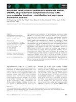
tested. Figure 1 shows the FAS inhibitory activity of
the three synthetic peptides and the C75 positive con-
trol. The FAS inhibitory responses for each of these
peptides were dose dependent, reaching 38.2, 76.5
and 79.4% inhibition at 50 lm for KNPQLR,
EITPEKNPQLR and RKQEEDEDEEQQRE, respec-
tively compared with 77.8% inhibition at 150 lm for
C75. Interestingly, although the three synthetic pep-
tides showed FAS inhibitory activity, their potency
was different. RKQEEDEDEEQQRE exerted a strong
inhibitory activity ($ 40% inhibition) at doses as low
as 12 lm. To compare the potency of the FAS inhibi-
tory response across all of the peptides and the C75
positive control, classical sigmoidal dose–response
curves were plotted and used to calculate the IC
50
val-
ues listed in Table 2. In these, the larger peptides,
RKQEEDEDEEQQRE (IC
50
= 16.5 lm) and EI-
TPEKNPQLR (IC
50
= 27.4 lm), showed significantly
higher (P < 0.05) potency (lower IC
50
value) than the
C75 positive control (IC
50
= 80.3 lm). The smaller
KNPQLR peptide had a higher IC
50
value (79.9 lm)
(P < 0.05) more comparable with the positive control
C75 (P > 0.05).
Structure–potency relationship of FAS inhibitory
peptides
To further understand the structure–potency relation-
ship of the FAS inhibitory peptides, we examined the
relationships between the physicochemical and
biochemical features of these peptides and their respec-
tive inhibitory potency (Table 2). Positive correlations
were observed between their potency (lower IC
50
value,
a
a
a
abc
bcd
cde
def
def
defg
efg
fg
f
12 25 30 50 60 150
[Compound] (µ
M)
120
100
80
60
40
20
0
FAS activity inhibition (%)
KNPQLR
EITPEKNPQLR
RKQEEDEDEEQQRE
C75
Fig. 1. Fatty acid synthase (FAS) inhibitory activity of synthetic pep-
tides and C75. Synthetic peptides KNPQLR, EITPEKNPQLR and
RKQEEDEDEEQQRE inhibited FAS in a dose-dependent manner
which was similar to the positive control C75. Evaluation of FAS
activity was performed after 20-min preincubation with different
concentrations of each compound. Values were expressed as per-
cent inhibition of FAS activity compared with a negative control that
included no inhibitors. Each dataset corresponds to the mean of
three independent replicates with error bars indicating the standard
deviations. Different letters indicate significant differences at
P < 0.05 in one-way ANOVA analysis.
Table 2. Fatty acid synthase (FAS) inhibitory potency and physicochemical and biochemical characteristics of synthetic peptides. MM, pep-
tide molecular mass; pI, theoretical isoelectric point of each peptide; GRAVY, grand average of hydropathicity index. Parameters were
obtained using the Protparam tool in the ExPASY Proteomic Server.
Physicochemical properties
Biological activity
No. of charged
residues
Hydrophilic
amino
acids (%) GRAVY
Aliphatic
index
IC
50
value of FAS inhibitory
activity (l
M)
a
MM pI Negative Positive
KNPQLR 79.9 ± 15.3
b
754.8 11 0 2 3 )2.2 65.0
EITPEKNPQLR 27.4 ± 7.8
c
1324.5 6.2 2 2 6 )1.63 70.9
RKQEEDEDEEQQRE 16.5 ± 2.9
c
1847.8 4.3 8 3 13 )3.67 0
C75 (positive control) 80.3 ± 19.5
b
254.3 – – – – –
Correlation coefficient (r)
d
+0.89 )0.99 +0.64 +0.40 +0.69 )0.16 )0.33
a
Data represent the mean ± SD of three independent experiments;
b,c
Different letters (b,c) in the column indicate statistical difference
(P < 0.05, in one-way ANOVA analysis).
d
Statistical correlations were carried out between the indicated parameter with the FAS inhibitory
potency (the lower the IC
50
value the higher the potency) of each peptide.
C. Martinez-Villaluenga et al. b-conglycinin peptides inhibit fatty acid synthase
FEBS Journal 277 (2010) 1481–1493 ª 2010 The Authors Journal compilation ª 2010 FEBS 1483
higher potency), their molecular masses (r=+0.89)
and the number of negatively charged (r = +0.64)
and hydrophilic (r = +0.69) residues. By contrast, a
strong negative correlation was observed between their
potency and their pI values (r = )0.99). Together,
these results suggest that peptides with higher inhibi-
tory potency are larger and have more negatively
charged and hydrophilic residues. No correlations were
found between their IC
50
values and other physico-
chemical and biochemical parameters, such as the
number of positively charged residues, grand average
of hydropathicity index and aliphatic index.
Identification of the potential binding site of FAS
and inhibitory peptides from SBC
Peptides EITPEKNPQLR and RKQEEDEDEEQ-
QRE were selected for use in the ligand–enzyme
docking simulations because they displayed higher
FAS inhibitory potency. To identify potential binding
sites for these peptide inhibitors, the multidomain
porcine FAS crystal structure (PBD ID code: 2VZ8)
[25] was searched for cavities near the identified active
site residues in each domain. Because this structure
lacked the ACP and TE domains, the human ACP
structure (PBD ID code: 2CG5) and human TE
domain structure (PBD ID code: 1XKT) lacking three
short loop regions were also included in this search.
Modeling of these regions using the moe (Chemical
Computing Group, Montreal, Canada) program
allowed for cavity identification in the complete
assembled structure, excluding openings extending
into the structurally undefined interdomain regions.
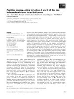
Of the seven crystallographically defined domains, the
TE domain had the largest cavity closest to the active
site (3959 A
˚
3
) and the b-enoyl reductase (ER) domain
had the second largest cavity (3697 A
˚
3
) (Fig. 2).
Docking of the EITPEKNPQLR inhibitory peptide in
both sites predicted interaction energies in the ER
domain higher than those in the TE domain
(Table 3). Docking of the RKQEEDEDEEQQRE
inhibitory peptide predicted near equivalent interac-
tion energies in the ER and TE domains. In both
sites, the predicted energies of the protein–ligand
complexes were much higher for the larger RKQEE-
DEDEEQQRE peptide than for the smaller EI-
TPEKNPQLR peptide. These results suggest that the
ER domain is not flexible enough to accommodate
relatively large inhibitors and that the TE domain is
a better target for these types of peptide inhibitors.
To confirm the binding of these peptide inhibitors in
the TE domain, a biochemical enzyme inhibition assay
was performed using a recombinant human FAS TE.
The TE inhibitory activity displayed by peptides
EITPEKNPQLR and RKQEEDEDEEQQRE was
compared with C75 and Orlistat (Table 3). Soy pep-
tides were more potent (10 lm) than C75 (58.7 lm),
however, their potency was $ 10-fold lower
(P < 0.05) than Orlistat (0.9 lm). Similar to C75, soy
peptides blocked > 50% of the TE activity; however,
this inhibition was lower (P < 0.05) than Orlistat
(77.3%) at 100 lm.
Binding and interaction modes of FAS inhibitory
peptides
To evaluate in more detail the binding modes of
these peptide inhibitors in the TE domain, they were
KS
DH
KR
ER
ACP
TE
MAT
Fig. 2. Identification of active-site cavities in
fatty acid synthase (FAS). In this representa-
tion, the multidomain FAS is compiled in
MOE from the swine FAS crystal structure
(PDB ID code: 2VZ8) [25], the human ACP
structure (PBD ID code: 2CG5) [41] and the
human thioesterase (TE) domain structure
(PBD ID code: 1XKT) [1]. The protein back-
bone is represented as an orange line,
active-site cavities are shown as blue
spheres and catalytic residues are shown in
space-filling format. MAT, malonyl-CoA
transacylase domain; KS, b-ketoacyl
synthase domain; KR, b-ketoacyl reductase
domain; DH, dehydratase domain; ER,
b-enoyl reductase domain; ACP, acyl-carrier
protein domain; TE, thioestherase domain.
b-conglycinin peptides inhibit fatty acid synthase C. Martinez-Villaluenga et al.
1484 FEBS Journal 277 (2010) 1481–1493 ª 2010 The Authors Journal compilation ª 2010 FEBS
docked individually within the predicted binding site
using the DOCK function within moe and compared
with the docking modes predicted for C75 and Orli-
stat [14,26]. The predicted lowest energy conforma-
tions of these peptides, C75 and Orlistat inhibitors, in
the human FAS TE domain model are shown in
His 2481
Ser 2308
Asp 2338
His 2481
Ser 2308
Asp 2338
His 2481
Ser 2308
Asp 2338
His 2481
Ser 2308
Asp 2338
His 2481
Ser 2308
Asp 2338
His 2481
Ser 2308
Asp 2338
His 2481
Ser 2308
Asp 2338 His 2481
Ser 2308
Asp 2338
Subdomain A
Subdomain B
N
C
Loop II
Loop III
Loop I
A
C
B
D
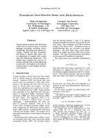
Fig. 3. Predicted overall fold of the thioes-
therase (TE) domain with inhibitors bound.
The lowest-energy binding mode for
EITPEKNPQLR (A), RKQEEDEDEEQQRE
(B), C75 (C) and Orlistat (D) rendered in ball-
and-stick format in the human TE domain
model (backbone in tube format) is shown
with catalytic triad residues Ser2308,
His2481 and Asp 2338 in space-filling for-
mat. Details regarding docking simulations
are summarized in Table 3.
Table 3. Molecular docking within the fatty acid synthase (FAS) potential binding site and human FAS thioesterase (TE) inhibitory potency
(IC
50
) of soybean b-conglycinin-derived peptides, C75 and Orlistat.
EITPEKNPQLR RKQEEDEDEEQQRE C75
a
Orlistat
a
b-Enoyl reductase
Predicted interaction energy (kcalÆmol
)1
) )279.1 )438.0 – –
Predicted energy of protein–ligand complex (kcalÆmol
)1
) )1750.9 )144.1 – –
Thioesterase
Predicted interaction energy (kcalÆmol
)1
) )353.0 )442.3 )51.2 )90.4
Predicted energy of protein–ligand complex (kcalÆmol
)1
) )1180.3 )955.5 )980.44 )1020.5
Distance to the catalytic triad (A
˚
)
Ser2308 4.93 2.40 4.00 3.14
Asp2338 2.98 3.98 4.14 4.81
His2481 2.39 2.16 2.12 2.22
Inhibition (%) of human FAS TE
b
55.46 ± 1.45
c
52.90 ± 4.34
c
55.53 ± 2.43
c
77.89 ± 0.81
d
Human FAS TE inhibitory potency (IC
50
, lM)
b
10.05 ± 1.60
c
10.71 ± 4.36
c
58.71 ± 6.74
e
0.93 ± 0.13
d
a
C75 and Orlistat were docked only in the active site of TE domain because they are known to target the TE domain [Cheng et al. [31]].
b
Compounds were tested at a concentration of 100 lM. Values indicate the mean ± SD of at least two independent experiments.
c,d,e
Dif-
ferent letters (c, d, e) in the same row indicate significant difference at P<0.05 in one-way ANOVA analysis.
C. Martinez-Villaluenga et al. b-conglycinin peptides inhibit fatty acid synthase
FEBS Journal 277 (2010) 1481–1493 ª 2010 The Authors Journal compilation ª 2010 FEBS 1485
Fig. 3A–D; interaction energies and the distances
between inhibitor atoms and the catalytic triad are
presented in Table 3. In this docking mode, the EI-
TPEKNPQLR peptide is predicted to be positioned
at a distance of 4.93, 2.39 and 2.98 A
˚
from Ser2308,
His2481 and Asp2338, respectively, in the TE domain
(Fig. 3A), with a low interaction energy ()353.0
kcalÆmol
)1
) (Table 3). By comparison, the larger
RKQEEDEDEEQQRE peptide docked in the TE
domain (Fig. 3B) at distances of 2.40, 2.16 and
3.98 A
˚
from the Ser2308, His2481 and Asp2338,
respectively, with the lowest interaction energy
()442.3 kcalÆmol
)1
), suggesting that this peptide is a
better inhibitor than EITPEKNPQLR; this is in
agreement with IC
50
values listed in Table 2. In their
docking modes, C75 and Orlistat were positioned at
greater distances from both the Asp2338 and Ser2308
residue in the TE domain than was the RKQEEDE-
DEEQQRE peptide and were predicted to interact
more weakly ()51.2 and )90.4 kcalÆmol
)1
, respec-
tively) with the TE domain than was the smaller
SBC-derived peptide (Table 3). In addition, correla-
tion analyses between the inhibitory potency of pep-
tides EITPEKNPQLR, RKQEEDEDEEQQRE and
C75 and their interaction energies with the TE
domain showed a strong correlation (r = 0.99).
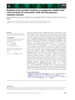
Close-up views of the binding modes of SBC-derived
peptides and Orlistat with the TE active site (Fig. 4)
suggest that the palmitic core of Orlistat is bound
almost exclusively to a hydrophobic groove generated
by subdomain B, and its peptidyl moiety is bound in
the interface cavity, whereas the hexanoil tail digs into
the short chain pocket where the catalytic triad exists.
These views also suggest that the larger EI-
TPEKNPQLR and RKQEEDEDEEQQRE peptides
bind throughout the long hydrophobic groove of the
TE domain in a orientation similar to that of Orlistat
with their amino acid side chains also extending into
the interface cavity and short chain pocket. The poten-
tial interaction modes of these peptides with the TE
domain suggest that EITPEKNPQLR (Fig. 5A) and
RKQEEDEDEEQQRE (Fig. 5B) bind mainly via
hydrophilic interactions (hydrogen-bonding and elec-
trostatic interactions) with active site residues. By con-
trast, only the hydrophilic peptidyl group of Orlistat
participates in hydrogen bonding with catalytic triad
residues Tyr2307, His2481 and Arg2482 located in the
interface cavity.
FAS inhibitory activity of synthetic peptides in
3T3-L1 adipocytes
In a cell-based model, FAS inhibition was measured
by monitoring the inhibition of lipid accumulation in
3T3-L1 adipocytes compared with the C75 positive
control compound. As shown in Fig. 6, synthetic pep-
tides displayed dose-dependent inhibition of lipid drop-
let accumulation in adipocytes. The highest inhibition
percentages for KNPQLR, EITPEKNPQLR and
RKQEEDEDEEQQRE were observed after cell treat-
ment at 100 lm (30.1, 29.6 and 34.2%, respectively).
In these assays, KNPQLR and EITPEKNPQLR pep-
tides showed similar (P > 0.05) inhibitory potency;
however, significantly lower (P < 0.05) than C75 at
50 lm (38.8%) and 100 lm (46.3%). By contrast, the
RKQEEDEDEEQQRE peptide showed an inhibitory
activity similar to C75 at all concentrations tested with
the exception of 50 lm, which displayed only 27.3%
Fig. 4. Molecular surface representation of
the thioestherase (TE) domain with inhibi-
tory peptides EITPEKNPQLR (orange ball-
and-stick format), RKQEEDEDEEQQRE
(blue ball-and-stick format) and Orlistat (red
ball-and-stick format). The potential surface
is colored to reflect hydrogen bonding (pink),
strong hydrophilic (green) and mild
hydrophilic (blue) regions.
b-conglycinin peptides inhibit fatty acid synthase C. Martinez-Villaluenga et al.
1486 FEBS Journal 277 (2010) 1481–1493 ª 2010 The Authors Journal compilation ª 2010 FEBS
inhibition (P > 0.05). Inhibition of lipid accumulation
by these peptides correlated with FAS inhibition
(r = 0.70) (Fig. 6) even though the magnitudes of inhi-
bition in these peptides in the cell-based model were
lower than the magnitude of FAS inhibition measured
in biochemical assays; this is probably because of the
A
B
C
Fig. 5. Detailed 2D interactions between
inhibitors and the thioestherase (TE) domain.
Calculated using the
MOE program following
the method of Clark & Labute [44], residues
in the TE domain that contribute to the bind-
ing of EITPEKNPQLR (A), RKQEEDE-
DEEQQRE (B) and Orlistat (C) are shown
with green circles indicating residues with
no polar or charged side chains and light
mauve circles indicating polar side chains
that are either acidic (red ring) or basic (blue
ring). Arrows indicate hydrogen bonds to
side chain (green) and backbone (blue)
residues.
C. Martinez-Villaluenga et al. b-conglycinin peptides inhibit fatty acid synthase
FEBS Journal 277 (2010) 1481–1493 ª 2010 The Authors Journal compilation ª 2010 FEBS 1487
low permeability of cell to these longer peptides. No
effect on cell viability of 3T3-L1 adipocytes was
observed with any of the treatments used in this study,
indicating no cellular toxicity (data not shown).
Discussion
FAS is an important target for prevention and thera-
peutic interventions because multiple lines of evidence
have shown high levels of FAS expression in cancer,
obesity and metabolic disorders [27]. The discovery
and development of agents that block FAS activity
highlight the potential for the prevention and treat-
ment of those chronic diseases. Our previous work
demonstrated that SBC contains FAS inhibitory pep-
tides that may be released by enzymatic hydrolysis
with alcalase [23]. This study has identified the FAS
inhibitory peptides in the SBC hydrolysate using CIP,
taking advantage of the specific affinity between FAS
and its inhibitory peptides. This CIP approach has
identified for the first time three peptide fragments
from the a and b subunits of SBC (KNPQLR,
EITPEKNPQLR and RKQEEDEDEEQQRE) as
potential inhibitors of FAS activity and their activities
were confirmed using their custom synthesized pep-
tides. Our results have indicated that the inhibitory
potency of these peptides (16.5–79.9 lm) is within the
range found for purified SBC hydrolysates (IC
50
=30
lm) and soybean hydrolysates (50.4–175.1 lm) [23]. In
comparison with other natural inhibitors, the inhibi-
tory potency of these peptides is within the range
found for flavonoids from green tea (2.3–111.7 lm)
[28,29] and tannins from Geum japonicum var. chinense
(0.2–41.4 lm) [22], which has been evaluated in pre-
clinical studies [22,30].
Molecular modeling has identified the TE domain as
the potential binding site for the FAS inhibitory pep-
tides from SBC. Molecular docking has shown that
soy peptides displayed a different inhibitory mecha-
nism than C75. Soy peptides are selective inhibitors of
the FAS TE domain, whereas C75 has been shown to
interact at several sites in FAS [14]. The predicted
binding energy of C75 in the FAS b-ketoacyl synthase
domain was )53.9 kcalÆmol
)1
, similar to that observed
in the TE domain ()51.2 kcalÆmol
)1
). These results
indicate that C75 is not a selective inhibitor for a
particular FAS domain, in agreement with previous
findings [14]. We also confirmed that the synthetic pep-
tides EITPEKNPQLR and RKQEEDEDEEQQRE
inhibited 4-methylumbelliferone heptanoate hydrolysis
by TE in in vitro experiments. Therefore, these
peptides are antagonists of TE under near physiologic
conditions, meaning that they bind to the unoccupied
enzyme and reduce substrate turnover. The TE domain
coordinates the terminal step of fatty acid synthesis by
hydrolyzing palmitate from the 4¢-phosphopanteine
arm of the ACP domain [1]. Its active site is comprised
of a hydrophobic groove with a distal pocket at the
interface of subdomains A and B and a hydrophilic
catalytic triad (Ser2308, His2481 and Asp2338) at the
proximal end of the groove [17]. Palmitate, the main
biological product of FAS, binds in the hydrophobic
groove, its hydrophilic carboxyl group interacting with
the catalytic triad, and its hydrophobic, hydrocarbon
chain extending away from the triad [31]. From the
binding modes that we have predicted, inhibitory
peptides appear to block the catalytic activity of TE
through hydrophilic interactions with enzyme residues
located in the catalytic triad, the hydrophobic groove
and the interface cavity. The biochemical parameters
of these peptides suggest that the numbers of nega-
tively charged and hydrophylic residues are important
predictors of their potency, in agreement with the fact
that hydrophilic interactions are important to block
the catalytic activity of the TE domain [31]. The high
number of charged and hydrophilic groups in these
inhibitors provides for strong hydrogen bonding and
electrostatic interactions with the catalytic residues of
the TE domain.
Analysis of the TE domain in a variety of species
indicates that catalytic triad residues are completely
conserved from insects to mammals and all other
[compound] (µM)
0
10
20
30
40
50
60
1 10 50 100
Inhibition (%) lipid accumulation
KNPQLR
EITPEKNPQLR
RKQEEDEDEEQQRE
C75
i
ghi
hi
fgh
ghi
fghi
fgh
def
fg
ef
cde
ab
bc
bcd
abc
a
Fig. 6. Inhibition of lipid accumulation in 3T3-L1 adipocytes by syn-
thetic peptides. 3T3-L1 adipocyte cells were treated with the syn-
thetic KNPQLR, EITPEKNPQLR and RKQEEDEDEEQQRE peptides
at concentrations ranging from 0 to 100 l
M on days 3, 5 and 7, and
lipid accumulation was measured on day 10 using the Oil Red O
assay as outlined in Experimental procedures. Each dataset corre-
sponds to the average of three independent replicates with error
bars indicating the standard deviation. Different letters indicate sig-
nificant differences at P < 0.05 in ANOVA analysis.
b-conglycinin peptides inhibit fatty acid synthase C. Martinez-Villaluenga et al.
1488 FEBS Journal 277 (2010) 1481–1493 ª 2010 The Authors Journal compilation ª 2010 FEBS
residues are conserved from birds to mammals, with
the exception at Phe2370 which is changed to Ala2370
in chickens [1]. This suggests that our current predic-
tions on the binding mode of SBC-derived peptides in
the human FAS TE domain can validly explain inhibi-
tion of catalytic activity in the chicken FAS.
Some evidence has shown that dietary soy protein
may promote satiety and weight loss [32,33] and
protect against certain types of cancer [34]. The
obesity-preventive effects of soybean protein have
been associated with its ability to decrease lipid syn-
thesis, adipogenesis and thermogenesis by regulating
gene expression [32]. Dietary intake of soy protein
has also been reported to reduce tumor incidence in a
rat model of chemically induced colon cancer by
attenuating FAS expression [34]. Our results provide
additional insight into the preventive mechanisms of
soy components in showing that SBC peptide frag-
ments inhibit FAS activity in adipose cells in the
same way as C75. Schmid et al. [35] reported that
FAS inhibition by C75 prevented adipogenesis in a
cell-based model. FAS inhibitory activity of SBC pep-
tide fragments may potentially be found in cancer
cells, liver or hypothalamus, as shown previously for
C75. Inactivation of hypothalamic FAS by C75 is
linked to satiety and dramatic weight loss [15]
because accumulation of the substrate malonyl CoA
through the inhibition of FAS appears to inhibit the
expression of neuropeptide Y which promotes inges-
tion [9]. Moreover, the FAS inhibitory activity of
C75 induced apoptosis and prevented the growth of
multiple tumor xenografts in vivo [36,37]. Our findings
clarify the mechanism linking FAS inhibition with the
anti-obesity effects of soy protein-derived peptides. In
conclusion, the soy peptides EITPEKNPQLR and
RKQEEDEDEEQQRE inhibited the TE domain and
de novo fatty acid synthesis in adipocytes. The bind-
ing mode of these peptides in the large palmitate-
binding pocket is of particular interest and will guide
future research. These FAS inhibitory peptides can
serve as lead compounds to design peptoid analogs
(oligomers of N-subtituted glycine) with equivalent
biological activity, enhanced systemic stability and
bioavailability than standard peptides [38]. The rele-
vance of the identification of these SBC-derived pep-
tides is noticeable because of the novelty of their
biological activity and chemical nature. Molecular
docking has allowed us to predict binding modes for
SBC-derived peptides (EITPEKNPQLR and RKQEE-
DEDEEQQRE) in the TE domain. Based on our
data, it is likely that the consumption of soy high in
b-conglycinin represents a preventive alternative to
improve health and wellness.
Experimental procedures
Materials
b-Conglycinin was purified from soybean defatted flour as
described in Wang et al. [39]. FAS inhibitory peptides were
produced from SBC hydrolysis with alcalase from Bacil-
lus licheniformis, as detailed in Martinez-Villaluenga et al.
[24]. The identified FAS inhibitory peptides (> 95% purity)
were custom synthesized by GenScript (Piscataway, NJ,
USA). FAS was isolated from chicken liver and purified
(70% purity) as described by Tian et al. [40]. Human
recombinant FAS TE (residues 2010–2509) was kindly pro-
vided by J.W. Smith (Burnham Institute for Medical
Research, CA, USA). 3T3-L1 (also designated ATCC
CCL-92.1) preadipocytes from Swiss albino mouse and
Dulbecco’s modified Eagle’s medium (DMEM) were pur-
chased from the American Type Culture Collection (Rock-
ville, MD, USA). Calf bovine serum, fetal bovine serum
and Dulbecco’s phosphate buffer saline were from Invitro-
gen (Rockville, MD, USA). Alcalase from B. licheniformis
(EC 3.4.21.62) and C75 were purchased from Sigma-
Aldrich (St. Louis, MO, USA). Protein A ⁄ G beads, non-
specific goat IgG and goat polyclonal IgG against a peptide
mapping at the C-terminus of FAS were from Santa Cruz
Biotechnology (Santa Cruz, CA, USA). Unless otherwise
stated, all chemical reagents were from Sigma-Aldrich.
FAS activity assay
FAS activity was assayed by a spectrophotometric method
using a Synergy 2 Microplate Reader System equipped with
temperature controller (Biotek Instruments, Winooksi, VA,
USA). NADPH oxidation was followed at 37 °C by mea-
suring the decrease in absorbance at 340 nm in a 96-well
clear-bottomed polysterene plate (Corning, NY, USA).
Reactions were performed in a final volume of 150 lL con-
taining 3 lm acetyl-CoA, 10 lm malonyl-CoA and 35 lm
NADPH and 0.3 lm FAS in 0.1 m potassium phosphate
buffer. Initial rates were calculated for the slope of the pro-
gress curves during the first 5 min.
FAS inhibition studies
Synthetic peptides and the C75 positive control compound
were used for FAS inhibition studies with stock solutions
of the synthetic peptides and C75 dissolved in deionized
water and dimetylsulfoxide, respectively, and serial dilutions
made in 0.1 m potassium phosphate buffer (pH 7.0). Inhibi-
tion studies were performed by measuring the residual FAS
activity after enzyme preincubation with inhibitors for
20 min at 37 °C. Potency was determined by dose–response
curves in which the range of concentrations was distributed
in a logarithmic scale and the IC
50
values were calculated
C. Martinez-Villaluenga et al. b-conglycinin peptides inhibit fatty acid synthase
FEBS Journal 277 (2010) 1481–1493 ª 2010 The Authors Journal compilation ª 2010 FEBS 1489
using nonlinear regression sigmoidal curve fit functions in
GraphPad prism 4.00 (Graphpad Software Inc., San Diego,
CA, USA).
Inhibition of FAS TE enzymatic activity was performed
using a fluorescence method described by Richardson &
Smith [41]. Peptides were added to yield a final concentra-
tion of 100 lm; in this assay the ability of the recombinant
TE to cleave 4-methylumbelliferone heptanoate and hydro-
lyzed it to the fluorescent 4-methylumbelliferone was
followed over time at 360 ⁄ 435 nm.
Co-immunoprecipitation
To purify FAS inhibitory peptides a CIP approach was
performed. Briefly, 200 lLofb-conglycinin hydrolysate
(2.5 mgÆmL
)1
in 0.1 m potassium phosphate buffer,
pH 7.0) were added with 2 lL of goat IgG and 25 lLof
protein A ⁄ G beads to preclear nonspecific peptides binding
to IgG and ⁄ or agarose beads. These samples were mixed
on an end-over-end mixer for 60 min at 4 °C. After pre-
clearing, samples were centrifuged at 1000 g for 5 min at
4 °C. The supernatant (80 lL) was added to 120 lLof
2 lm FAS in 0.1 m potassium phosphate buffer (pH 7.0).
The negative control consisted of 80 lL of 0.1 m potas-
sium phosphate buffer (pH 7.0) added to 120 lL FAS.
The blank consisted of 200 lL of 0.1 m potassium phos-
phate buffer (pH 7.0). These samples were incubated for
40 min at 37 °C. For CIP, each sample was incubated with
10 lL goat polyclonal antibody (FAS IgG) for 60 min at
4 °C and then 30 lL protein A ⁄ G beads were added and
mixed on an end-over-end mixer overnight at 4 °C. After
incubation with the antibody, samples were centrifuged at
1000 g for 5 min at 4 °C and the pellet washed three times
with radioimmunoprecipitation buffer. The sediment was
resuspended in HPLC-grade water and boiled for 3 min to
release proteins from the beads. Then, 20 lL acetonitrile
containing 0.8 lL formic acid were added to extract the
peptides and proteins, the beads were removed by centrifu-
gation at 1000 g for 5 min at 4 °C, and the final superna-
tant was stored at )20 °C before identification of FAS
inhibitory peptides.
Western blot analysis
To confirm the CIP of FAS, western blot analysis was
carried out using goat polyclonal antibody (FAS IgG). Pro-
teins released from the beads were resuspended in Laemmli
loading buffer (BioRad, Hercules, CA, USA) containing
5% 2-mercaptoethanol. Samples (20 lg soluble protein)
were loaded onto 15% Tris ⁄ HCl ready gels and run
through a mini-electrophoresis kit at 200 V constant for
40 min. Further, proteins were transferred to poly(vinyli-
dene diflouride) membrane (BioRad) in blotting buffer
(25 mm Tris, 192 mm glycine pH 8.3, 0.1% SDS) using
western sandwich assembly for 1 h at 4 °C using 125 V.
After the transfer, membrane was blocked with 5% non-fat
dry milk in Tris-buffered saline containing 0.1% Tween 20
(TBST) for 1 h, followed by an overnight incubation with
goat polyclonal anti-(FAS IgG) (1 : 200) at 4 °C. Further,
membrane was washed with TBST four times and was incu-
bated with bovine anti-(goat IgG) horseradish peroxidase
conjugates (1 : 1000) for 1 h at room temperature. The
membrane was washed again in TBST for four times and
signals were visualized using chemiluminescence reagent
(GE Healthcare, Chalfont St Giles, UK) and a Kodak
Image Station 440 CF (Eastman Kodak Co., New Haven,
CT, USA).
LC-MS
⁄
MS
Samples were injected (10 lL) onto a dC
18
Atlantis nano-
Acquity column (75 · 150 mm, 3 lm particle size; Waters,
Milford, MA, USA) using 0.1% aqueous formic acid as
solvent A and acetonitrile with 0.1% formic acid as sol-
vent B. A linear gradient from 1 to 60% B was run for
60 min and back to 1% B for 10 min with the flow rate
maintained at 0.25 mLÆmin
)1
. MS analysis was carried out
in a Q-Tof API-US nanoAcquity LC (Waters) mass spec-
trometer equipped with an electron spray ion source. The
Q-Tof instrument was operated in positive ion mode. Spec-
tra were recorded over the m ⁄ z range 100–1500. Using
MASCOT the m⁄ z spectral data were processed and used
for de novo peptide sequencing and database searching.
Peptide identification was carried out by searching against
the NCBI or SWISS-PROT database [taxonomy = viridi-
plantae (green plants)]. Only peptides identified with a con-
fidence of at least 95% were considered to be correct calls
(P < 0.05).
Cell culture and treatments
The 3T3-L1 preadipocytes were seeded at 3 · 10
4
cellsÆ-
well
)1
in 24-well plates and cultured in DMEM growth
medium containing 1% sodium pyruvate, 1% penicil-
lin ⁄ streptomycin and 10% calf bovine serum (days 1 and
2). After reaching 100% confluence, the cells were stimu-
lated with DMEM growth medium containing 1%
sodium pyruvate, 1% penicillin ⁄ streptomycin, 10% fetal
bovine serum, 0.5 mm isobutylmethylxanthine, 1 lm dexa-
methasone and 1.7 lm insulin (days 3 and 4). Cells were
then maintained in fetal bovine serum ⁄ DMEM with
1.7 lm insulin for another 2 days (days 5 and 6), fol-
lowed by culturing with fetal bovine serum ⁄ DMEM for
an additional 4 days (days 7–10), at which time > 90%
of cells were mature adipocytes with fat droplets. Cells
were treated on days 3, 5 and 7 of the differentiation
process with synthetic peptides dissolved in Dulbecco’s
phosphate buffer saline at a concentration ranging from
0to50lm and incubated at 37 °Cina5%CO
2
atmo-
sphere for 48 h.
b-conglycinin peptides inhibit fatty acid synthase C. Martinez-Villaluenga et al.
1490 FEBS Journal 277 (2010) 1481–1493 ª 2010 The Authors Journal compilation ª 2010 FEBS
Viability assay
The cell proliferation assay was conducted using CellTiter
96 Aqueous One Solution Proliferation assay kit (Promega
Corp., Madison, WI, USA) using 3-(4,5-dimethylthiazol-
2-yl)-5-(3-carboxymethoxyphenyl)-2-(4-sulfophenyl)-2H-tet-
razolium, inner salt, and an electron coupling reagent,
phenazine ethosulfate. Briefly, 5 · 10
3
preadipocytesÆwell
)1
were seeded in a 96-well plate and the total volume was
adjusted to 200 lL with growth medium. Cells were treated
on days 3, 5 and 7 of the differentiation process with differ-
ent concentrations of synthetic peptides dissolved in Dul-
becco’s phosphate buffer saline. On day 10, the growth
medium was replaced by 100 lL fresh growth medium
and 20 lL 3-(4,5-dimethylthiazol-2-yl)-5-(3-carboxymethoxy-
phenyl)-2-(4-sulfophenyl)-2H-tetrazolium bromide⁄ ph enazine
ethosulfat e was ad d ed t o eac h well. T he p late wa s i ncub ated
for2hat37°C and the a bsorbance (A) was read at 515 nm
in a microplate reader. The percentage of viable cells was calcu-
lated with respect to cells treated with Dulbecc o’s phosphate
buffer saline u sing the following equation:
A
treatment; 515 nm
=A
control; 515 nm
 100 ¼ % cell viability
Lipid quantification in 3T3-L1 adipocytes by Oil
Red O assay
Adipocytes were washed twice with cold Dulbecco’s phos-
phate buffer saline and fixed with 10% formaldehyde for
1 h. Then, cells were washed with 60% isopropanol and air
dried. Oil Red O stock solution (0.2 g in 60% isopropanol)
was filtered through a 0.22-lm membrane and added to
lipid droplets for 30 min. After Oil Red O lipid staining,
cells were washed with water four times and air dried. Oil
Red O dye was eluted by adding 100% isopropanol. After
10 min incubation at room temperature, the absorbance (A)
at 510 nm was measured using a microplate reader. Percent
inhibition of lipid accumulation was calculated using the
following equation:
A
control; 510 nm
À A
treatment; 510 nm
=
A
control; 510 nm  100 ¼ % inhibition of lipid content
Molecular modeling
To identify potential binding sites for the peptide inhibitors
the multi domain porcine fatty acid synthase crystal struc-
ture (PBD ID code: 2VZ8) [25] was searched for cavities
near the identified active site residues in each domain.
Because this structure lacked the ACP and TE domains,
the human ACP structure (PBD ID code: 2CG5) [42] was
used in the search. Parts of identified cavities extending to
the interdomain regions were excluded because they could
not be accounted for with the single domain structures. The
volumes of each of the identified cavities were calculated
using the SITE VOLUME SCRIPT function within moe.
The human TE domain structure available (PBD ID code:
1XKT) [1] lacked three loop regions: loop I (residues 2326–
2328 missing in chain A only) that connects a helix 4 (a4)
to b strand 5 (b5) and forms a surface loop on the under-
side of the a ⁄ b domain; loop II (residues 2344–2360) that
bridges subdomain A and subdomain B; and loop III (resi-
dues 2450–2460) that occurs near the catalytic triad linking
b6tob7. The missing loops were modeled using the
HOMOLOGY function in moe 2008.10 and the aligned
sequences of the TE domain of swine FAS (GenBank
accession no. NP_001093400) and human FAS (GenBank
accession no. AAB35516.1).
Inhibitors were docked using the DOCK function of
moe. The initial 3D structures of the ligands were con-
structed using the BUILDER function in the moe program.
The initial positions of these compounds were set within
the catalytic site and docking simulations were carried out
by using the CHARMM27 force field [43] and the simu-
lated annealing conformation search method within the
DOCK function. One hundred conformations were gener-
ated for each ligand tested while keeping protein side chains
fixed, and these were sorted in ascending order according
to their total energy. Binding modes with the lowest total
energies and extended conformation were chosen for sec-
ond-round energy minimizations during which all protein
side chains were allowed to move freely. Protein–ligand
interactions were established using the LIGAND INTER-
ACTION function in moe which follows the method
described by Clark & Lebute [44].
Statistical analysis
Data were expressed as means of at least two independent
replicates. Results were compared using one-way analysis
of variance (ANOVA) using the GLM procedure of sas
(SAS Institute, Cary, NC, USA). Group means were con-
sidered to be significantly different, as determined by the
technique of protective least-significant differences (LSD),
when ANOVA indicated an overall significant treatment
effect (P < 0.05).
Acknowledgements
This research was supported by the USDA Coopera-
tive State Research, Education and Extension Service
(CSREES), AG 2007-34505-15767 Future Foods IL;
Illinois Soybean Association; the European Commis-
sion, Marie Curie IOF grant (PIOF-GA-2008-219860)
for Career Development (to CM-V). Special acknowl-
edgements to Drs J. W. Smith and R. D. Richardson
C. Martinez-Villaluenga et al. b-conglycinin peptides inhibit fatty acid synthase
FEBS Journal 277 (2010) 1481–1493 ª 2010 The Authors Journal compilation ª 2010 FEBS 1491
from the Burnham Institute for Medical Research,
California for providing us with the human recombi-
nant FAS TE.
References
1 Chakravarty B, Gu ZW, Chirala SS, Wakil SJ &
Quiocho FA (2004) Human fatty acid synthase:
structure and substrate selectivity of the thioester-
ase domain. Proc Natl Acad Sci USA 101, 15567–
15572.
2 Smith S & Tsai SC (2007) The type I fatty acid and
polyketide synthases: a tale of two megasynthases. Nat
Prod Rep 24, 1041–1072.
3 Maier T, Jenni S & Ben N (2006) Architecture of mam-
malian fatty acid synthase at 4.5 A
˚
resolution. Science
311, 1258–1262.
4 Berndt J, Kovacs P, Ruschke K, Kloting N, Fasshauer
M, Schou MR, Korner A, Stumvoll M & Bluher M
(2007) Fatty acid synthase gene expression in human
adipose tissue: association with obesity and type 2
diabetes. Diabetologia 50, 1472–1480.
5 Sheng H, Niu B & Sun H (2009) Metabolic targeting of
cancers: from molecular mechanisms to therapeutic
strategies. Curr Med Chem 16, 1561–1587.
6 Buettner C (2007) Does FASing out new fat in the
hypothalamus make you slim? Cell Metab 6, 249–251.
7 Chakravarthy MV, Zhu Y, Lopez M, Yin L, Wozniak
DF, Coleman T, Hu Z, Wolfgang M, Vidal-Puig A,
Lane MD et al. (2007) Brain fatty acid synthase acti-
vates PPARa to maintain energy homeostasis. J Clin
Invest 117, 2539–2552.
8 Ronnett GV, Kim E-K, Landree LE & Tu Y (2005)
Fatty acid metabolism as a target for obesity treatment.
Physiol Behav 85, 25–35.
9 Loftus TM, Jaworsky DE, Frehywot GL, Townsend
CA, Ronnett GV, Lane MD & Kuhajda FP (2000)
Reduced food intake and body weight in mice treated
with fatty acid synthase inhibitors. Science 288, 2379–
2381.
10 Chakravarthy MV, Zhu Y, Yin L, Coleman T, Pappan
KL, Marshall CA, McDaniel ML & Sememkovich CF
(2009) Inactivation of hypothalamic FAS protects mice
from diet-induced obesity and inflammation. J Lipid
Res 50, 630–640.
11 Dowling S, Cox J & Cenedella RJ (2009) Inhibition
of fatty acid synthase by Orlistat accelerates gastric
tumor cell apoptosis in culture and increases survival
rates in gastric tumor-bearing mice in vivo. Lipids 44,
489–498.
12 Ho TS, Ho YP, Wong WY, Chiu LCM, Wong YS &
Ooi VEC (2007) Fatty acid synthase inhibitors cerulenin
and C75 retard growth and induce caspase-dependent
apoptosis in human melanoma A-375 cells. Biomed
Pharmacother 61, 578–587.
13 Yang N, Kays JS, Skillman TR, Burris L, Seng TW &
Hammond C (2004) C75 activates carnitine palmitoyl-
tranferase-1 in isolated mitochondria and intact cells
without displacement of bound malonyl CoA. J
Pharmacol Exp Ther 312, 127–133.
14 Rendina AR & Chend D (2005) Characterization of the
inactivation of rat fatty acid synthase by C-75: inhibi-
tion of partial reactions and protection by substrates.
Biochem J 388, 895–903.
15 Lopez M & Dieguez C (2007) C75, a fatty acid synthase
(FAS) inhibitor. Recent Pat Endocr Metab Immune
Drug Discovery 1, 53–62.
16 Tiss A, Lengsfeld H, Carriere F & Verger R (2009)
Inhibition of human pancreatic lipase by tetrahydro-
lipstatin: further kinetic studies showing its reversibility.
J Mol Catal B-Enzym 58, 41–47.
17 Pemble CW, Johnson LC, Kridel SJ & Lowther WT
(2007) Crystal structure of the thioesterase domain of
human fatty acid synthase inhibited by Orlistat. Nat
Struct Mol Biol 14
, 704–709.
18 Vazquez MJ, Leavens W, Liu R, Rodriguez B, Read
M, Richards S, Winegar D & Dominguez JM (2008)
Discovery of GSK837149A, an inhibitor of human fatty
acid synthase targeting the b-ketoacyl reductase reac-
tion. FEBS J 275, 1556–1567.
19 Brusselmans K, Vrolix R, Verhoeven G & Swinnen JV
(2005) Induction of cancer cell apoptosis by flavonoids
is associated with their ability to inhibit fatty acid syn-
thase activity. J Biol Chem 280, 5636–5645.
20 Rivkin A, Kim YR, Goulet MT, Bays N, Hill AD,
Kariv I, Krauss S, Ginanni N, Strack PR, Kohl NE
et al. (2006) 3-Aryl-4-hydroxyquinolin-2(1H)-one deriv-
atives as type I fatty acid synthase inhibitors. Bioorg
Med Chem Lett 16, 4620–4623.
21 Tian WX (2006) Inhibition of fatty acid synthase by
polyphenols. Curr Med Chem 13, 967–977.
22 Liu H, Li J, Zhao W, Bao L, Song X, Xia Y, Wang X,
Zhang C, Wang X, Yao X et al. (2009) Fatty acid syn-
thase inhibitors from Geum japonicum Thunb. var. chin-
ense. Chem Biodivers 6, 402–410.
23 Gonzalez de Mejia E, Martinez-Villaluenga C, Roman
M & Bring NA (2009) Fatty acid synthase and in vitro
adipogenic response of human adipocytes inhibited by a
and a’ subunits of soybean b-conglycinin hydrolysates.
Food Chem 119, 1571–1577.
24 Martinez-Villaluenga C, Bringe NA, Berhow MA &
Gonzalez de Mejia E (2008) b-Conglycinin embeds active
peptides that inhibit lipid accumulation in 3T3-L1 adipo-
cytes in vitro. J Agric Food Chem 56, 10533–10543.
25 Maier T, Leibundgut M & Ban N (2008) The crystal
structure of a mammalian fatty acid synthase. Science
321, 1315–1322.
26 Kridel SJ, Axelrod F, Rozenkrantz N & Smith JW
(2004) Orlistat is a novel inhibitor of fatty acid synthase
with antitumor activity. Cancer Res 64, 2070–2075.
b-conglycinin peptides inhibit fatty acid synthase C. Martinez-Villaluenga et al.
1492 FEBS Journal 277 (2010) 1481–1493 ª 2010 The Authors Journal compilation ª 2010 FEBS
27 Menendez JA, Vazquez-Martin A, Ortega FJ & Fernan-
dez-Real JM (2009) Fatty acid synthase: association
with insulin resistance, type 2 diabetes, and cancer. Clin
Chem 55, 425–438.
28 Wang X, Song KS, Guo QX & Tian WX (2003) The
galloyl moiety of green tea catechins is the critical struc-
tural feature to inhibit fatty-acid synthase. Biochem
Pharmacol 66, 2039–2047.
29 Li BH & Tian WX (2004) Inhibitory effects of flavo-
noids on animal fatty acid synthase. J Biochem 135, 85–
91.
30 Puig T, Relat J, Marrero PF, Haro D, Brunet J &
Colomer R (2008) Green tea catechin inhibits fatty acid
synthase without stimulating carnitine palmitoyltrans-
ferase-1 or inducing weight loss in experimental ani-
mals. Anticancer Res 28, 3671–3676.
31 Cheng F, Wang Q, Chen M, Quiocho FA & Ma J
(2008) Molecular docking study of the interactions
between the thioesterase domain of human fatty acid
synthase and its ligands. Proteins 70, 1228–
1234.
32 Velasquez MT & Bhathena SJ (2007) Role of dietary
soy protein in obesity. Int J Med Sci 4, 72–82.
33 Vaughn N, Rizzo A, Doane D, Beverly JL & Gonzalez
de Mejia E (2008) Intracerebroventricular administra-
tion of soy protein hydrolysates reduces body weight
without affecting food intake in rats. Plant Foods Hum
Nutr 63, 41–46.
34 Xiao R, Su Y, Simmen RCM & Simmen FA (2008)
Dietary soy protein inhibits DNA damage and cell sur-
vival of colon epithelial cells through attenuated expres-
sion of fatty acid synthase. Am J Physiol Gastrointest
Liver Physiol 294, G868–G876.
35 Schmid B, Rippmann JF, Tadayyon M & Hamilton BS
(2005) Inhibition of fatty acid synthase prevents prea-
dipocyte differentiation. Biochem Biophys Res Commun
328, 1073–1082.
36 Gabrielson EW, Pinn ML, Testa JR & Kuhajda FP
(2001) Increased fatty acid synthase is a therapeutic
target in mesothelioma. Clin Cancer Res 7, 153–157.
37 Wang HQ, Altomare DA, Skele KL, Poulikakos PI,
Kuhajda FP, di Cristofano A & Testa JR (2005)
Positive feedback regulation between AKT activation
and fatty acid synthase expression in ovarian carcinoma
cells. Oncogene 24, 3574–3582.
38 Fowler SA & Blackwell HE (2009) Structure–function
relationships in peptoids: recent advances toward deci-
phering the structural requirements for biological func-
tion. Org Biomol Chem 7, 1508–1524.
39 Wang W, Bringe NA, Berhow MA & Gonzalez de
Mejia E (2008) b-Conglycinin among sources of bioac-
tives in hydrolysates of different soybean varieties that
inhibit leukemia cells in vitro. J Agric Food Chem 56,
4012–4020.
40 Tian WX, Hsu RY & Wang YS (1985) Studies on the
reactivity of the essential sulfydryl group as a confor-
mational probe for the fatty acid synthetase of chicken
liver. Inactivation by 5,5¢-dithiobis-(2-nitrobenzoic acid)
and intersubunit cross-linking of the inactivated
enzyme. J Biol Chem 260, 11375–11387.
41 Richardson RD & Smith JW (2007) Novel antagonists
of the thioesterase domain of human fatty acid syn-
thase. Mol Cancer Ther 6, 2120–2126.
42 Bunkoczi G, Pasta S, Joshi A, Wu X, Kavanagh KL,
Smith S & Oppermann U (2007) Mechanism and sub-
strate recognition of human holo ACP synthase. Chem
Biol 14, 1243–1253.
43 MacKerell AD, Bashford D, Bellott M, Dunbrack RL,
Evanseck JD, Field MJ, Fischer S, Gao J, Guo H,
Ha S et al. (1998) All-atom empirical potential for
molecular modeling and dynamics studies of proteins. J
Phys Chem B 102, 3586–3616.
44 Clark AM & Labute P (2007) 2D depiction of protein–
ligand complexes. J Chem Inf Model 47, 1933–1944.
C. Martinez-Villaluenga et al. b-conglycinin peptides inhibit fatty acid synthase
FEBS Journal 277 (2010) 1481–1493 ª 2010 The Authors Journal compilation ª 2010 FEBS 1493