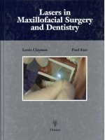
Lasers in Maxillofacial Surgery and Dentistry potx
Bạn đang xem bản rút gọn của tài liệu. Xem và tải ngay bản đầy đủ của tài liệu tại đây (9.31 MB, 168 trang )
Contents
Contributors ix
Preface xi
Foreword xiii
1. Physical Considerations of Surgical Lasers 1
Terry A. Fuller
2. Practical Laser Safety in Oral and Maxillofacial Surgery 11
Lawrence M. Elson
3. Specific Guide to the Use of Lasers 19
Lewis dayman, Richard Reid
4. Preneoplasia of the Oral Cavity 37
Lewis dayman
5. Papillomas and Human Papillomavirus 55
Richard Reid. Myron Slrasser
6. Soft Tissue Excision Techniques 63
Lewis dayman. Paul Kuo
7. Transoral Resection of Oral Cancer 85
Lewis dayman
8. Outpatient Treatment of Snoring and Sleep Apnea Syndrome
with C0
2
Laser: Laser-Assisted Uvulopalatoplasty 111
Yves-Victor Kamami. James W. Woolen
9. The Carbon Dioxide Laser in Laryngeal Surgery 121
Robert J. Meleca
10. Uses of Lasers in Dentistry 127
Harvey Wigdor
viii Contents
11. Phototherap y wit h Laser s and Dye s 13 7
Dan J. Castro. Romaine E. Saxlon, Jacques Soudanl
12. Laser Phototherma l Therap y for Cancer Treatmen t 14 3
Dan J. Castro. Romaine E. Saxlon. Jacques Soudant
13. Laser-Assiste d Temporomandibula r Join t Surger y 15 1
Steven J. Butler
14. Endoscopi c Sinu s Surgery : A Significan t Adjunc t to
Maxillofacia l Surger y 15 7
Jeffrey J. Moses. Claus R. l^ange
15. Lase r Biostimulation : Photobioactivation , a Modulatio n of
Biologi c Processe s by Low-Intensit y Lase r Radiatio n 16 5
Joseph S. Rosenshein
16. Lase r Tissu e Fusio n 17 5
PaulKuo
17. Lase r Applicatio n in Microgravity , Aerospace , an d
Militar y Operation s 17 9
I'aul Kuo. Michael D. Colvard
Appendi x 18 1
Glossar y 18 3
Inde x 18 5
1 Physical Considerations of Surgical Lasers
• f
Terry A. Fuller
HISTORY
A laser, an acronym lor light amplification by stimulated
emission of radiation, is a device for generating a high-in-
tensity, ostensibly parallel beam of monochromatic (single
wavelength) electromagnetic radiation. The possibility of
stimulated emission was predicted by Einstein in I9I7;
based on the work of Gordon in 1955 and Schawlow and
Townes in 1958, Maiman created the first operational laser
in 1960, a ruby laser emitting a brilliant red beam of light.
This was followed within 3 years by the development of the
argon, carbon dioxide (C0
2
), and neodymium:yttrium-alu-
minum-garnet (Nd:YAG) lasers, which remain the most
widely used lasers in medicine.
In 1963 the ruby laser was employed in the treatment of
pigmented dermatologic lesions and for photocoagulation
of the retina. Early applications of lasers in oral and max-
illofacial surgery began to appear in the mid- to late 1970s.
Potential advantages of surgical lasers were clear from the
beginning, but the cost, unreliability, and operational com-
plexity of the early machines greatly limited the actual use
of lasers, except in the fields of ophthalmology and derma-
tology, until the past 15 to 18 years. In recent years im-
proved understanding of light-tissue interactions and, of
greatest importance to the surgeon, new technologies for
delivering laser light to (he tissue, has transformed lasers
into versatile and valuable surgical instruments. This chap-
ter presents the fundamentals of laser physics and intro-
duces the reader to the interactions between light and tissue.
Full appreciation of the uses, limitations, benefits, and
risks of surgical lasers requires a basic understanding of
laser physics and the biologic action of light.
LIGHT
Electromagnetic radiation is energy transmitted through
space. It can be viewed either as propagated waves of char-
acteristic energies, or as discrete (and the smallest) parcels
of energy called photons. Electromagnetic radiation is
quantified in terms of two reciprocal forms of measure-
ment: frequency (v), expressed in Hertz (Hz) or cycles per
second, and wavelength (\), expressed in metric units of
length. Which units are employed in any particular applica-
tion is largely a matter of convention. The wavelength of
the radiation in the visible region of the spectrum (Fig. 1-1)
defines the color of the light.
Atoms (ions or molecules) at their lowest energy or
ground state possess an intrinsic amount of energy. When
excited through the process of absorption by the input of
thermal, electromagnetic, or other forms of energy, they are
raised to one of several distinct higher energy levels. The
absorbed energy is subsequently and spontaneously re-
leased (spontaneous emission) in the form of a quantum of
energy corresponding to the difference between the ground
and excited states (E, — E
2
= E
a
). All particles making the
transition between the same two energy levels will emit
light of identical energy and wavelength (Fig. 1-2).
Ordinary sunlight or lamplight consists of many wave-
lengths; even light, colored from passing through a filter,
represents a broad spectrum of many wavelengths. Such
light emanates in all directions from its source. The inten-
sity diminishes as the inverse square of the distance from
the source. As discussed below, a laser uses the principle of
stimulated emission to produce light of a markedly different
quality.
The spontaneous emission of photons from an excited
atom may occur at any time and in any direction. If, how-
ever, a photon of E
A
strikes an atom already in an upper en-
ergy stale E
2
, it stimulates the emission of a second photon
of light. This second photon has precisely the same energy
or wavelength and is spatially and temporally synchronous
with and traveling in exactly the same direction as the ini-
tial photon. If these two photons strike additional atoms in
the excited state E-j, they will yield an amplifying cascade
of photons—laser light—that is monochromatic (a single
wavelength), coherent (synchronous waves), and collimated
(parallel rays).
THE LASER
Lasers consist of a small number of basic components as
shown in Figure 1-3. An active lasing medium, which can
be a solid, liquid, or gas, is enclosed within a laser cavity
bounded by two perfectly parallel reflectors (mirrors).
High-energy radiation is pumped into the active medium by
means of a pump source. The pump source is energy gener-
ally provided by an intense optical or electrical discharge.
The energy from the pump source is absorbed by the active
1
2 Lasers in Maxillofacial Surgery and Dentistry
Figure 1-1. Electromagnetic spectrum.
Figure 1-2. Energy slate diagram.
medium until the majority of atoms, ions, or molecules are
raised to their upper energy state. This is a condition known
as a population inversion and is a necessary condition to
generate laser light. The two parallel reflectors are situated
at the ends of the laser cavity and act to constrain the light
along and within the axis of the cavity. Thus, the light is re-
peatedly bounced between the reflectors. This will stimu-
late the emission of even more photons (amplification) in
that axial direction. Light traveling in other directions es-
capes the cavity and is lost as heal. One of the mirrors is
only partially reflective, enabling some of the light to es-
cape the cavity as a beam of laser light.
Different lasing media, because of their particular
atomic, molecular, or ionic structure and energy levels, emit
light of characteristic wavelengths. The properties of the
most common surgical lasers are listed in Table 1-1.
CO? Laser
Carbon dioxide lasers employ carbon dioxide gas (in addi-
tion to other gases required for sustained stimulated emis-
sion of radiation) as a lasing or active medium. The gases
are either sealed in a tube or are circulated from a tank.
When excited by direct current (DC) or radio-frequency
(RF) voltage, the carbon dioxide absorbs a portion of this
energy and raises the CO> molecule to an upper energy
state. The excited C0
2
molecule spontaneously decays and
emits mid-infrared photons at a wavelength of 10.6(H) nm
Physical Considerations of Surgical Lasers 3
Figure 1-3. Basic laser components.
Table 1-1. Characteristics of Surgical Lasers
LASER TYPE
WAVELENGTH
SPECTRAL
REGION
MODE
TYPICAL MAX
POWER
C0
2
10.600 nm
Mid-Infrared
CW & Gated & Superpulsed
I00W CW
Holmium
2.100 nm
Near Infrared
Pulsed
l5Wavg.
Nd:YAG
1,064 nm
Near Infrared CW & Pulsed
IO0W CW
Diode
800-890 nm Near Infrared
CW
> 50W
KTP/KDP
532 nm
Visible
Pulsed
25Wavg.
Argon
488/514 nm.
Visible
CW
20W
Excimer ArF
190
nm
Ultraviolet Pulsed SSOmJ
-XcCI
308 nm
Ultraviolet Pulsed 250mJ
Erbium: YAG(Er: YAG)
Reproduced with permission of T.A.F modified from Fuller TA. Thermal Surgical Lasers. Philadelphia: Surgical Laser Technologies. Inc 1992.
(10.6 pm). Power (measured in units of watts, W) is the
lime function of energy (measured in joules, J) and can be
delivered either continuously (continuous wave, CW) or in
a train of pulses. The carbon dioxide laser can be pulsed in
a manner thai results in high energy, rapidly repeating
pulses typically referred to as superpulses. In contrast to
CW surgical lasers, which generate power up to 100 W, the
superpulsed C0
2
laser generates power up to 10,000 W in a
repeating train of pulses. There are substantial differences
in clinical effect between CW, conventional pulsing, and
superpulsed modes of operation (see Chapter 3).
Infrared light is in a region of the electromagnetic spec-
trum that is not visible to the human eye. Therefore, a sec-
ond low-power visible laser [typically a red beam from a
helium-neon (HeNe) laser or visible diode laser) beam is
precisely aligned and coaxial with the C0
2
laser beam for
aiming purposes. The delivery system used to carry Ihe laser
light to the lissue is of critical importance to the surgeon.
The C0
2
laser generally uses an articulated arm as its prin-
cipal delivery system. An articulated arm is a series of hol-
low tubes connected together through a series of six to eight
articulating mirrors. This is in contrast to very thin, continu-
4 Lasers in Maxillofacial Surgery and Dentistry
ously flexible, glass (fused silica) fiber optics generally
used for near infrared and visible lasers. Glass is opaque to
10,600 nm light and thus is not suitable for CO
:
laser trans-
mission. The CO
:
laser is primarily used for cutting and va-
porizing tissue in open procedures or in procedures where
rigid endoscopy is acceptable.
Argon and Frequency-Doubled Nd.YAG Lasers
Argon and frequency-doubled Nd:YAG laser (also referred
to as a KTP laser), although technologically very different
from each other, are devices that generate laser energy in
the green region of the electromagnetic spectrum. The
argon laser employs an electrically excited ionized argon
gas as a lasing medium. The high heat transfer requires a
water-jacketed cooling system, which permits power out-
puts of up to 25 W. More portable, air-cooled units arc lim-
ited to power outputs of 5 to 10 W. This laser emits blue-
green light at 488 and 514 nm. The KTP laser uses a
Nd:YAG laser in combination with a potassium titanyl sul-
fate (KTP) crystal. The Nd:YAG portion of this laser sys-
tem generates a wavelength of 1064-nm energy whose fre-
quency is doubled (wavelength is halved) on passing
through the KTP crystal. The result is a beam of green light
at 530 nm. The emission from both the argon and KTP
lasers can be transmitted through flexible glass fiber optics
thai can carry the light to the surgical site. Since the light is
visible, no secondary aiming beam is required. Safety
glasses are required to protect the patient and operating
room personnel from the therapeutic beam of all surgical
lasers. However, glasses used for the green lasers necessar-
ily block green light and thus tend to obscure the overall vi-
sualization of the surgical field.
Nd.YAG Laser
The neodymium.yttrium-aluminum-garne t (Nd:YAG) laser
is a solid-state device that generates light in the near in-
frared region of the spectrum at 1064 nm. The active
medium of this laser is the neodymium atoms doped into a
matrix of yttrium, aluminum, and garnet. The neodymium
atoms are optically excited by way of a bright arc lamp.
This relatively efficient laser generates a wavelength of
1064 nm and is outside the visible region of the spectrum.
Therefore, the Nd:YAG laser requires an aiming beam sim-
ilar to that used by C0
2
lasers. Safety glasses for this laser
are transparent to visible light and do not obscure the sur-
geon's surgical view. The surgical Nd:YAG lasers com-
monly deliver continuous (CW) power up to KM) W and can
be passed easily through inexpensive flexible fiber optics.
In addition to the CW mode of operation, the Nd:YAG laser
can be configured to operate in a special pulsed mode re-
ferred to as Q-switched. The Q-switched laser emits pulses
of pico- to nanoseconds in duration. This mode is often
used in ophthalmology to disrupt the posterior capsule in
secondary cataracts or in shock-wave lithotripsy.
Holmium: YAG
The holmium:YAG laser is technologically associated with
the Nd:YAG laser. This solid-state laser uses holmium as
its active medium doped into a matrix of yttrium, alu-
minum, and garnet. Due to its inherently inefficient opera-
tion and certain thermal design considerations, this laser is
pulsed. It emits rapid pulses of energy at 2100 nm in the
mid-infrared part of the spectrum. Like the NdiYAG laser,
this laser requires an aiming beam. The holmium:YAG
beam can be delivered through fiber optics. However, such
fibers must be made of low OH (hydroxyl radical) glass
due to the high absorption of this wavelength to water.
Diode Laser
In contrast to the gas and solid-state lasers discussed thus
far. diode lasers are in a category of devices that emit light
from semiconductor materials. They are operated in a man-
ner similar to a transistor in which an electric potential is
applied to dissimilar semiconductor materials. In contrast to
gas, solid state, and liquid lasers, semiconductor lasers re-
quire no high voltages or currents, no arc lamps or optical
pump sources, and have no required moving parts. They are
very efficient (typically >30—35%), but arc capable of gen-
erating only relatively low power levels. Individual "high-
power" laser diodes typically generate only 1 to 1^ W per
diode. To gain useful power from the laser, multiple de-
vices must be used in concert. Linear (one-dimensional) ar-
rays or two-dimensional arrays are being developed to gain
sufficient power for surgery. Additionally, ganging individ-
ual diodes in various optical configurations are being ex-
plored: each approach carries its own benefits and draw-
backs. Both commercial and prototype surgical diode laser
systems are able to deliver 20 to 50 W. There are currently
severe fiber optic size and maximum power limitations as
well as diode and system warranty and lifetime issues. Cur-
rently, the most popular diode lasers emit light in the 800-
to 890-nm range. Lasers in the shorter wavelength range
provide biologic effects similar to those of the Nd:YAG
lasers. Longer wavelengths have higher tissue absorption
characteristics.
The technological specifications of a given laser type and
model indicate how much power (or in the case of a pulsed
laser, energy) can be practically delivered to tissue and the
means by which the power can be conveyed to tissue. When
laser energy interacts with tissue its output power is distrib-
uted over the area of an illuminated spot. This distribution
or power density or fluence (power/area. W/cnr) is inti-
mately related to the tissue effect. The power density can be
altered by changing either the power of the laser or spot size
Physical Considerations of Surgical Lasers 5
of the laser beam. The effect that a particular laser emission
has on tissue, and thus the surgeon's ability to effectively
utilize that emission, depends upon power density and other
specifications as well as the characteristics of tissue. Only
by matching the characteristics of the laser beam and the
tissue can one begin to accurately predict the effect that the
laser will have in surgery.
THERMAL LASER—TISSUE EFFECTS
The focus of this book is on the interactions of laser energy
and tissue that result in an elevation of the tissue tempera-
ture. These so-called thermal lasers represent the majority
of all applications of lasers in medicine. Thus, lasers that
are Q-switched or lasers that operate at low powers for
biostimulation or photodynamic therapy (PDT) interactions
are excluded herein from discussion. This section presents
an outline of the principal variables affecting the clinical
end point.
The utility of the thermal laser resides with its capability
of providing the surgeon the ability to accurately predict the
nature and extent of a thermally induced laser lesion in tis-
sue. The goal of laser surgery is thus to create a tempera-
ture gradient (Fig. 1-4) or profile in tissue that will result in
coagulation or vaporization of tissue. Coagulation provides
hemostasis and. if desired, necrosis of tissue. Vaporization
(the conversion of solid and liquid phase tissue components
into gaseous phase components) provides the ability to cut,
incise, excise, resect or ablate tissue.
Coagulation and vaporization are two different effects
created by the same process: heating of tissue. Coagulation
generally occurs when the temperature is elevated from
60°C to <100°C. Obvious changes occur in the tissue at
these temperatures resulting from the thermal denaturation
of tissue protein, and include blanching and shrinkage as
well as puckering due to dehydration. When the tempera-
ture is elevated near and above the boiling point of water
(100°C), vaporization of liquid and solid components occur.
A frank defect is left that includes a zone of char (carbon, as
a result of the combustion of tissue) surrounded by coagu-
lated tissue. The extent of the area of vaporization, char,
and coagulation (as well as a heat-affected zone surround-
ing the coagulation, which can cause edema) is defined by
the temperature gradient. Thus, by altering the gradient, the
surgical effect can be altered. There are several variables
that determine the gradient. They include the laser parame-
ters such as power density, duration of exposure, wave-
length, and method of delivery of laser energy as well as
tissue parameters.
Light can be absorbed, transmitted, scattered, or reflected
by tissue (Fig. 1-5). Only light that has been absorbed can
yield a therapeutic result. Light that is transmitted through
or reflected from tissue yields no effect until and unless it is
absorbed. The measure of the degree to which tissue ab-
sorbs light is the absorption coefficient, a (measured in
units of cm
-1
). It is a measure of the amount of energy ab-
Figure 1-4. Temperature gradients in tissue.
6 Lasers in Maxillofacia l Surgery and Dentistry
OW?3SLT.INC./T.A .r
Figure 1-5. Interactions of light and tissue.
sorbed through a distance of the absorbing material. The
penetration depth of the laser in a given tissue is propor -
tional to the inverse of the absorption coefficient «. The
more highly absorbed the light (high a), the shallower the
penetration. As can be seen in Figure I -6, this results in the
light energy being converted to heat energy within a shal-
low layer of tissue, and therefore results in intense surface
heat. A tissue with a high « will create a steep temperature
gradient.
Figure 1-7 illustrates the absorption of light by tissue at
different wavelengths. The y-axis indicates greater absorp-
tion (less penetration) and thus higher resulting tempera-
tures. It can be readily seen that the C0
2
and erbium
(Er).YAG lasers would create high surface temperature and
very steep temperature gradients in the tissue. Both the C0
2
and EnYAG laser beams are preferentially absorbed by
water, and because water is by far the largest component of
most tissue, this results in the rapid transformation of light
into heal within about 0.2 to 1.0 mm of the tissue surface.
The intense thermal response quickly evaporates the water
and vaporizes tissue. The temperature gradient is so steep
that it has relatively poor coagulation properties. The dura-
tion of exposure is another key variable in determining the
extent of a laser-induced lesion. Long exposure times result
in conduction of heat into surrounding lissue and thus im-
prove hemostasis and increase coagulation necrosis. In con-
trast, techniques exist to diminish coagulation necrosis. The
superpulse CO
:
laser is one such example. This laser uses
rapidly repeating, high peak power pulses with pulse energy
in the range of 50 to 120 mJ/pulse. The result of application
of the superpulsed laser is the reduction of coagulation
necrosis by 50% over the CW laser operating at the same
average power. By way of contrast, Ihe Nd:YAG laser will
penelrate deeply in tissue with a relatively low surface tem-
perature and shallow temperature gradient.
Scatter of light by tissue spreads the laser beam in a dif-
fuse pattern define d by the tissue' s scatter coefficient. B.
Once the light is scattered, if it is absorbed, it will affect a
volume of tissue larger than Ihe laser's optical spot size. In
some instances scatter is an attribute desired by the surgeon.
For example, when Nd:YAG laser energy is used to ther-
mally destroy a tumor, the deep penetration of the laser
beam coupled with the high scatter coefficient affects a
deep and wide volume of tissue. In contrast, scatter can also
be detrimental if one is attempting to localize the effect of
the laser.
The green light from the argon and KTP lasers is both
scattered and absorbed by tissue. The degree of absorp-
tion is heavily dependent on the concentration of the
chromophores hemoglobin and melanin. Thus, heavily
pigmented skin or vascular areas such as a hemangioma
will result in high absorption (low penetration). The scat-
ter of the green lasers are greater than that of the C0
2
laser.
The method of delivering the laser light to the tissue also
acts as a variable affecting the tissue response. In general
terms this delivery of energy falls into two broad classes:
free-beam lasers and lasers lor use in contac t with tissue .
Physical Considerations of Surgical Lasers 7
Figure 1-6. Power/depth and temperature/deplh.
Figure 1-7. Light absorption by composite tissue.
8 Lasers in Maxillofacial Surgery and Dentistry
FREE-BEAM LASERS
Free-beam (sometimes referred to as noncontact) lasers are
devices that permit laser energy alone (without influence by
the delivery device) to interact with tissue, causing the final
clinical result. The interactions between laser light and tis-
sue described above are specific for free-beam lasers. They
result from interactions between the native laser wavelength
and tissue alone. Typical free-beam delivery systems in-
clude articulating arms, micromanipulators used in conjunc-
tion with surgical microscopes, and conventional fiber op-
tics. Characteristic of these devices is that the effect on
tissue is principally that of the laser emission alone. This is
typically what occurs when there is no contact between the
fiber optic end of the delivery device and the target tissue.
Consider the laser beam exiting a laser delivery system
used in a free-beam mode (Fig. 1-8, left). The beam will
converge (or diverge) as it exits the focusing lens and some
portion of the energy will be reflected from the tissue on
impact. Should the distance from the fiber to the tissue be
altered, the power density at the tissue will change, chang-
ing the clinical effect. Substantial energy is reflected (Qf)
or lost as heat and in smoke (Q»)-
The free-beam method of delivery provides certain ad-
vantages over conventional surgery by providing a method
for "non-touch" surgery, but suffers from the loss of tactile
feedback. The techniques for learning and using the free-
beam laser are substantially different from those of conven-
tional instruments. Perhaps the most limiting feature of the
free-beam laser is that different laser sources are required
for different surgical maneuvers, e.g., Nd:YAG for coagula-
tion and hembstasis and CO
:
for incision and excision.
Modification of Free-Beam Laser Surgery:
Contact Laser Surgery
Despite the benefits of free-beam laser surgery, certain lim-
itations and drawbacks exist. Perhaps the most significant is
that to substantially change the tissue's temperature gradi-
ent (clinical effect), one must choose different laser sources,
an expensive and intraoperatively difficult task. Contact
Laser surgery has been developed to augment and over-
come this and other fundamental deficiencies in free-beam
surgery. Contact Laser surgery works by altering the tissue
temperature gradient through changes in the laser delivery
system, rather than by alteration in wavelength.
A decade ago researchers developed a delivery system in
which an optical device is placed in direct contact with the
tissue during laser surgery to increase the delivered power
density and reduce changes in power density due to changes
in distance to the tissue. This is accomplished by use of in-
terchangeable contact laser probes and scalpels (tips) made
from synthetic sapphire or fused silica. The tips have sev-
eral different sizes and shapes and can be easily affixed to
the end of fiber optics. Several benefits result from the use
of these tips (Fig. 1-8. right). In addition to providing the
Figure 1-8. Noncontact vs. contact laser surgery.
Physical Considerations of Surgical Lasers 9
Figure 1-9. Changes in temperature gradient and tissue effect by wavelength conversion effect surface treatments.
surgeon with tactile feedback, a sense lost in free-beam
surgery, and controlling power density, the reflection of
light from the tissue is significantly reduced. The improved
efficiency in coupling of light into the tissue results in the
requirement of less power, in most cases a reduction of 40
to 50% (Fig. 1-8, right).
Altering the tip configuration of a probe and scalpel
makes it possible to change not only the spot size (and thus
power density), but the angle of divergence of the beam. A
frustroconical tip, for example, concentrates the laser light
on a small, precisely defined distal area from which light
splays out at a wide angle, creating a region of high power
density that drops rapidly with distance. Alterations in the
tip's shape can result in a low divergence angle. In addition
to placing tips onto the ends of fiber optics, the ends of fiber
optics themselves can also be shaped, although they lack
the mechanical strength and thermal resistance required for
extended and precision use.
The Contact Laser attributes thus far described, still re-
sult in a tissue effect that is solely dependent on the absorp-
tion of the laser emission by the tissue to generate the tem-
perature gradient. It is a major attribute of Contact Laser
surgery to have the temperature gradient altered by the
Contact Laser tip. By placing a small amount of light ab-
sorbing material integrally between the contact tip and the
tissue, a portion of the energy will be absorbed by that ma-
terial. The energy absorbed will be converted to heat and
will result in a very high temperature. Since the absorbing
material is in contact with the probe and the tissue, it will
elevate the temperature of the tissue by thermal combustion
in addition to the radiation heating caused by the native
wavelength. Thus, as can be seen in Figure 1-9, by use of
this absorbing material the tissue temperature gradient in-
duced by the laser emission has been altered. Depending
upon the quantity and distribution of the absorbing material
the contact tip can mimic the effect of other laser wave-
lengths. This event is referred to as the Wavelength Conver-
sion Effect. The wavelength conversion effect does not re-
sult in changing the wavelength of the laser; rather, it
changes the effect the wavelength has in the surgical
situation.
By adjusting the Wavelength Conversion Effect material
on the probe tip, one can titrate the amount of laser light ex-
iting the tip in comparison to the amount of heat generated
by absorption at the tip. This means, in essence, that a sin-
gle laser in combination with different interchangeable tips,
can mimic the tissue temperature profile and effect of vari-
ous lasers.
2 Practical Laser Safety in Oral and
Maxillofacial Surgery
Lawrence M. El son
A laser is a device thai produces an intense, highly parallel
beam of coherent light. It is named after the composition of
the excitable medium from which the laser beam emanates
[e.g., carbon dioxide (CO,), argon (Ar), helium-neon
(HeNe). etc.). Since the late 1970s, lasers have been studied
in oral and maxillofacial surgery for the treatment of soft
tissue lesions and occasionally for the cutting of bone.
1
Light emitted by these surgical lasers is generally in the vis-
ible and infrared regions of the electromagnetic spectrum
and is nonionizing. This radiation must be clearly differen-
tiated from ionizing radiation exemplified by x-rays and
gamma rays, which may produce deleterious effects on liv-
ing tissue. Therefore, patients, medical personnel and par-
ticularly pregnant women working with or around lasers
may do so without the risks-
1
associated with x-rays.
Each different type of laser produces a different wave-
length (color) of light that is absorbed by specific target
chromophores within tissues. The biologic effect of this
light on tissue is dependent upon wavelength, energy level
of the beam, and absorption characteristics of the tissue re-
ceiving this energy. For example, the carbon dioxide laser
(10,600 nm—middle infrared) light is absorbed heavily by
water. Since human tissue is mostly water, it absorbs virtu-
ally all of the laser energy without significant reflection or
backscatter from the surgical site. However, when this
same light comes into contact with shiny surgical instru-
ments, reflection will occur. In tissue, the depth of this
laser's photovaporization or photocoagulation effect is di-
rectly dependent on the power density (watts/cm
2
), which
is determined by the intensity of the focused beam, and the
energy density (joules/cm
2
), which determines the rate at
which energy is delivered to the tissue. The thermal dam-
age produced adjacent to the surgical site by diffusion of
heal can be reduced to a range of micrometers, depending
on the energy density used. Irreversible thermal damage
adjacent to (he zone of photovaporization is minimized by
using the highest controllable power density for the short-
est amount of application time. Prior to patient use a "test
spot" is made on a moistened wooden tongue blade to as-
sess the coaxial HeNe aiming beam, spot size contour,
power, and mode of operation [continuous wave (CW) or
pulsed|. Hazards of the carbon dioxide laser in oral and
maxillofacial surgery (OMFS) include corneal, scleral, and
cutaneous injury ranging from transient pain to severe
burns. Both the patient and the medical personnel are at
risk for these injuries.
The argon laser emits a blue-green light of 488 and 514
nm, which is selectively absorbed by the red chromophore.
oxyhemoglobin at 488 and 540/577 nm (double absorption
peak). It is delivered to the target tissue by an optical fiber.
This laser, depending on its spot size, power, time of appli-
cation, and resulting energy density, can photovaporize or
coagulate tissue with up to several millimeters of thermal
damage adjacent to the zone of clinical laser treatment. The
optical hazards of the argon laser include retinal and
skin burns.
The neodymium:yttrium-aluminum-garnet (Nd:YAG)
laser emits an invisible 1060-nm (near-infrared) light that is
heavily absorbed by pigmented tissue. It can photovaporize
or photocoagulate almost all biologic tissue with which it
comes in contact. The zone of thermal damage of the
Nd:YAG laser may extend as much as I cm beyond the sur-
gical target site consequent to a deep penetrating effect that
is not observable at the time of treatment. This powerful
laser is delivered to the surgical site by an optical fiber or
contact probe. Optical hazards of this laser are similar to
that of the argon laser and include retinal and skin hazards
(Fig. 2-1).
HAZARDS OF LASER SURGERY
Judgment Errors
As is the case with surgery, judgment error may be as harm-
ful as the use of inappropriate surgical technique. Of the
several types of judgment errors, the most severe is misdi-
agnosis or misinterpretion of the disease state being treated.
After having appropriately decided to use a laser, it be-
comes necessary to match the wavelength, power, and en-
ergy densities to the target tissue absorptive characteristics
to best eradicate the lesion. This mandates that the surgeon
understand the applied laser physics and laser-tissue inter-
actions at the selected wavelength. The technical skill to
manipulate the laser delivery system safely to protect pa-
tient, surgeon, and operating room personnel must be ac-
quired through instructional courses resulting in proper cre-
dentialing for each wavelength used. Ultimately, each
surgeon should be proctored by a properly credentialed
laser clinician at (he hospital in which the surgeon practices
for each type of procedure for which privileges are desired.
In some cases, residency training may substitute for a laser
11
1 2 Lasers in Maxillofacial Surgery and Dentistry
Figure 2-1. Nd:YAG-induced retinal burns
in a rabbit retina.
course, but the preceplorship credentialing program is still
required. It is also very important to remember that im-
proper use converts the laser into an expensive electro
cautery unit. Failing to limit the extent of the laser's lateral
heat conduction by the untrained clinician may produce a
conduction burn that extends well beyond the laser surgical
site. This might well prove disastrous.
Optical Hazards
Since the clinical lasers utilized in oral, maxillofacial, and
head and neck surgery photovaporize or photocoagulate tis-
sue, they all have the potential to damage the eye. Depend-
ing on the laser's wavelength, different tissue effects will
occur. Visible light laser radiation [argon, potassium titanyl
phosphate (KTP). HeNe. gold vapor, pulsed dye, etc.], and
the near-infrared Nd:YAG laser's energy will easily be
transmitted through the eye directly into the retina where
absorption may produce a burn (Fig. 2-2) and partial loss of
vision or even blindness. Laser light that is focused through
the lens of the eye will increase its effective power up to
l(K).(KM) times! The eye must always be protected to prevent
visual field defects or blindness. Other near-infrared lasers
[erbium (Br):YAG and holmium (Ho):YAG[ and middle in-
frared lasers such as the CO
:
laser are absorbed by the
water in the cornea, scleral epithelium, or eyelid and have
the potential to burn or damage these areas.
Therefore, it is imperative that all individuals in the oper-
ating room, i.e surgeons, nurses, technicians, and patients,
wear adequate eye protection while the laser is being used.
This will protect their eyes from direct exposure to mis-
aimed laser light as well as from specular reflections from
instruments or tissues at the surgical site. All facilities using
lasers must therefore have available appropriate wave-
length-specific goggles (Fig. 2-3) or glasses with side
shields to be worn by all personnel whenever the laser is
operating. These laser protection devices should have an
optical density (OD) stamped or imprinted on them along
with the wavelength and/or name of the laser for which they
arc to be used. The material coating the lenses of these gog-
gles or glasses absorbs and disperses the incident laser en-
ergy, preventing damage to the eye. For protecting the pa-
tient, in addition to wavelength-specific glasses or goggles,
it is also acceptable to place wet gauze or eye pads across
the closed eyelids and, depending upon the procedure (i.e.,
Nd:YAG laser procedures), an aluminum-metal type of eye
shield should be placed over the gauze or pads.
Skin Hazards
Even though, from a laser usage standpoint, skin hazards
are regarded as a minor nuisance, they are painful and may
be damaging. The most common mishap occurs when the
laser operator's or assistant's hands pass in front of the
working laser beam causing a burn. This happens when the
laser is either misfired during the course of surgery or
when an assistant carelessly places a hand in contact with
the laser beam. The resulting injury may potentially be
substantial. It is therefore most important for the clinician
to keep his foot off the foot pedal until ready to fire the
laser. Simultaneously, the laser technician must be ready to
change the laser to the "standby" mode whenever an inter-
ruption in laser use is encountered. The clinician should
also inform the support staff of the danger of laser injuries
to tissue, and must warn them to keep their hands away
from the surgical site when the laser is in operation. Other
important locations at risk of exposure in oral and maxillo-
facial surgery include the patient's facial skin, teeth, and
soft tissues. Wet drapes or gauze sponges should also be
placed over the patient's skin and teeth outside of the sur-
gical site. A laser impact on a tooth has the potential to
damage the enamel, penetrate into the pulp chamber, and
Figure 2-1. Nd:YAG-induced relinal burns
in a rabbit retina.
Practical Laser Safety in Oral and Maxillofacial Surgery 13
Figure 2-2. Absorption site of (A)
visible and near-infrared radiation; (B)
middle, far-infrared radiation and mid-
dle ultraviolet radiation; (C) and near-
ultraviolet radiation. (Reproduced from
Laser Institute of America*s Laser
Safety Guide.)
14 Lasers in Maxillofacial Surgery and Dentistry
Figure 2-3. Goggles that are wavelength specific.
shatter the tooth. A tooth damaged by the laser is caries re-
sistant but unsightly.
Fire Hazards
All lasers used in the operating suite have the potential to
ignite materials on the surgical site and produce a fire haz-
ard. Examples of these combustible materials include dis-
posable drapes made from wood pulp, dry cotton swabs,
gauze sponges, wooden tongue blades, and plastic instru-
ments (Fig. 2-4). To reduce the potential for igniting the
draping material by the laser, this author advocates the use
of polypropylene surgical drapes because in my experience
when hit by an incident laser beam they melt rather than
burst into flame.
The greatest source of danger in surgery of the oral cav-
ity is the endotracheal tube itself. Special care must be
taken to prevent the tube from coming into contact with the
laser during surgery because ignition of the endotracheal
tube produces a tire with a blowtorch effect inside the pa-
tient's airway (Figs. 2-5 and 2-6). New "laser safe" endo-
tracheal devices are available for use during laser surgery.
It is important to have an airtight endotracheal tube with a
metal reflective exterior. The cuff at the distal end of the
tube should be tilled with a saline and methylene blue dye.
If the laser beam penetrates the cuff during surgery, the
blue solution will spill, indicating to the surgeon and anes
thesiologist that a laser-related puncture of the cuff has
occurred. The stainless steel body of the armored endo-
tracheal tubes will resist perforation by the laser. Foil-
wrapped endotracheal tubes are not recommended because
of the possibility that hand wrapping may leave an uncov-
ered area that is susceptible to a laser burn, causing
ignition.
Other safety-enhancing techniques to reduce tire risk in-
clude reducing the oxygen content of the anesthetic mixture
Figure 2—4. Laser burns in combustible ma-
terials present at surgery: gauze, wooden
tongue blade, cotton, tipped applicators, and
rubber glove.
Practical Laser Safety in Oral and Maxillofacial Surgery
15
Figure 2-6. Ignition of oxygen-filled endo-
tracheal tube results in ignition and creation of
a blowtorch-like effect. Covered foot pedal.
to 30%, and insertion of wet cottonoids or gauze sponges
as a hypopharyngeal throat pack. These serve as additional
protection for the endotracheal tube by absorbing laser light
and ensuring greater protection for the patient. However, it
should also be remembered that the surgeon must remove
these wet packs from the patient's throat upon completion
of the procedure. As for all throat packs, the anesthesiolo-
gist should record the time of placement and removal of the
pack. At the end of the case, he should ask the surgeon, "Is
the pack removed?" The anesthesiologist must understand
the surgical procedure being performed as if he himself
were performing the operation. Simultaneously, the surgeon
must know of any potential anesthetic problems so that both
the surgeon and anesthesiologist may foresee and avoid
mishaps. As always, communication between surgeon,
anesthesiologist, laser technician, and nursing staff in a
safe surgery.
The surgeon, anesthesiologist, and operating room staff
must always be prepared and have a written plan of action
should an airway fire occur. In the event of this dramatic
and frightening complication, rapid planned intervention
may be lifesaving. The following protocol is recommended
for an airway fire: simultaneously stop lasing. cease ventila-
tion, turn off all anesthetic gases, including oxygen, extin-
guish flames using saline solution from a nearby basin, de-
flate the cuff, and remove the endotracheal tube. Make sure
the entire tube is removed. Next, ventilate the patient's
lungs with l(K)% oxygen by bag and mask, assess the air-
way for burns and foreign bodies (e.g tracheal tube and
packing materials) by using a bronchoscope. If the damage
is minimal, it may be possible to continue with the proce-
Figure 2-5. Cuff of endotracheal tube pene-
uid lilling cuff escapes,
safety-oriented environment is essential for successful and
trated by CO2 laser beam. Methylene blue liq-
16 Lasers in Maxillofacial Surgery and Dentistry
dure. However, extreme caution is advised in regard to pro-
ceeding even in the case of minimal observed damage. If
the damage is extensive, it may be necessary to control air-
way ventilation by inserting an endotracheal tube or per-
forming a tracheostomy, ventilation proceeds using humidi-
fied gases. Antibiotics and large dose steroids
4
should also
be given. Lastly, the laser safety officer and the surgeon
must report the incident to the appropriate hospital quality
improvement and risk management departments, as well as
to the laser companies and fiber-optic manufacturers, and a
report must be filed with the Food and Drug Adminis-
tration.
Electrical Hazards
Of all the laser surgical-related hazards, electrical hazards
have the greatest potential to be lethal, with several fatali-
ties having been reported since the initiation of the use
of lasers in surgery. These incidents have occurred as a re-
sult of either untrained and/or unauthorized individuals
opening the closed laser cabinet or by technicians who did
not follow prescribed electrical safety procedures. Contact
with the fully charged capacitor located inside the laser
cabinet may result in electrical shock or even death by electroc-
ution.
It is mandatory that inspection, evaluation, and repair of
electrical components in these specialized lasers be per-
formed only by factory-trained technicians. Most surgical
lasers use high voltage and high current electricity. The
laser's direct current (DC) capacitors also have the ability
to remain charged for hours after the laser has been turned
off and unplugged and, therefore, remain a reservoir of
lethal electrical current. Consequently, if the electrical mal-
function indicator light goes on during a procedure, the
laser should be turned off and a service representative
should be called in immediately to evaluate the extent of the
problem. If a service representative is not available, the
laser aspect of the procedure must be immediately termi-
nated (unless a standby laser is available).
Plume Hazards
One of the few negative aspects of using lasers in surgery is
the resulting smoke or laser plume—a by-product of laser
surgery. The laser plume is primarily composed of vapor-
ized water (steam), carbon particles, and cellular products,
which combine to produce a malodorous scent. This smoke
has been found to be irritating to those operating room per-
sonnel who come in contact with it. It has also been re-
ported that laser smoke contains many toxic substances,
such as formaldehyde, hydrogen cyanide, hydrocarbons,
and other airborne mutagens.
4
The particles have an aver-
age size of slightly larger than 0.3 u.m.
Unfortunately, human papilloma virus DNA has been
identified in the plume during the surgery for removal of
papillomas.
5
The initial observers of this phenomenon cau-
tioned against overreaction because it could not be proven
that these particles could seed themselves in unsuspecting
human hosts. Jn 1993 these researchers reported the first
transmission of laser plume-related disease in cows.
6
Cur-
rently, additional research is being conducted nationally re-
garding this issue. As a result of the uncertainty surround-
ing the seeding ability of this plume material in humans, a
proactive stance should be adopted. Use of a high-volume
laser smoke evacuation apparatus that filters smoke parti-
cles to 0.1 u.m is recommended.
7
Maintaining the suction
wand within 4 cm of the surgical site to remove as much of
the plume as possible is recommended. Disposable gloves
and sterile technique should be used to change evacuation
filters, which are treated as hazardous waste and disposed
of in biohazard bags. The laser-charred material should be
wiped from the surgical site and the cloth and paper prod-
ucts used during the laser procedure disposed of using
proper biohazard handling. When working with infected pa-
tients or those at high risk for HIV/hepatitis, etc., goggles
and face masks should be worn to prevent the splattering of
tissue from the surgical site onto the eyes and noses of those
performing or assisting during the procedure. Lastly, all
surgical instruments, e.g., microscopes, operating room ta-
bles, etc., should be wiped with a hospital-approved steriliz-
ing solution after each laser procedure.
ADDITIONAL LASER SAFETY
INFORMATION
Each institution that uses lasers clinically should appoint a
laser safety officer (LSO) to oversee and ensure the safe use
of lasers in its facility. The LSO should attend a laser safety
officer course to assist in the proper performance of his/her
duties. The LSO evaluates all laser use policies and proce-
dures, identifies potential laser-related hazards, and serves
as the resource person for the education of hospital staff,
medical staff, and nursing staff, and answers questions re-
garding laser capabilities.
It is recommended that a laser safety policy and proce-
dure be written in each institution using laser to treat pa-
tients. Once approved by the laser committee, this informa-
tion should be disseminated to the employees of the
operating room staff and to all laser surgeons, dentists,
physicians, podiatrists, etc., and should be followed, as
written.
All lasers must have their keys removed when not in use
and, if possible, they should be kept in a locked room
to maintain equipment safety and security. Only LSO-
approved personnel should have access to operate the laser
equipment.
Laser safety warning signs should be placed on the door
of any operating room using lasers prior to usage. These
signs should include the type and power of the laser being
used. All operating room windows should be covered with
Practical Laser Safety in Oral and Maxillofacial Surgery 1 7
an opaque material while lasers are being used so no laser
light can escape and harm an unsuspecting bystander. This
is not necessary during CO| laser procedures because its
emission is absorbed by plastic and glass. An extra pair of
laser goggles should also be placed on the door handle of
the operating room so that a person entering the room will
have adequate eye protection.
All clinical lasers should be examined weekly and their
power output should be monitored regularly with a power
meter. This data should be recorded for the LSO's monthly
quality assurance reports and for medical/legal record-keep-
ing.
Remember foot pedal safety: When the laser is not in
use, the clinician's foot should be removed from the pedal.
If the laser is not being used for a substantial period of time,
the laser should be placed in the standby mode with the ap-
proval of the clinician. The covered design of the foot pedal
helps prevent accidental activation of the laser.
A basin of saline should be available to be utilized in
the event of fire for each laser procedure. Remember: In
Case of fire, use the laser fire safety protocol and act
quickly. Do not use water to extinguish fires on electrical
equipment.
All operating room personnel should know where and
how to use the fire extinguishers located near the operating
room. Remember: P.A.S.S.—pull, aim, squeeze, sweep.
REFERENCES
1. dayman L, Fuller T. Bcckman H. Healing of continuous-wave
rapid superpulsed. carbon dioxide, laser-induced bone defects.
J Oral Surg 1978:36:932-937.
2. Reid R. Elson L, Absten G. A practical guide to laser safety.
Colposc Gynecol Laser Surg 1986:2(3): 121 -132.
3. Rontal M. Rontal E. Wenokur M. Elson L. Anesthetic manage-
ment for tracheobronchial laser surgery. Ann Owl Rhinol
Laryngol 1986:95:556-560 .
4. Sosis MB. ed. Problems in Anesthesia: Anesthesia for Laser
Surgery. Philadelphia : J.B. Lippincott ; 1993.
5. Intact viruses in C0
2
laser plume spur safety concern. Clin
Laser Month I987;5(9) : 101-103 .
6. New research confirms laser plume can transmit disease. Clin
Laser Month 1993; ! l(6):8l-84 .
7. Recommended practices for laser safety in the practice setting.
AORNJ 1989:155-158 .
8. American National Standard: For the safe use of lasers in
health care facilities . ANSI 2136.3. 1988.
Lewis dayman, Richard Reid
CARBON DIOXIDE LASER
The carbon dioxide (C0
2
) laser, which is the workhorse of
contemporary laser surgery, is a molecular gas laser emit-
ting in the mid-infrared (IR) range configured in either
flowing gas or sealed tube form. In the former, the contin-
uously degrading active medium is replenished with fresh
gas and the laser consistently produces power outputs of
up to 100 watts (W). It is noisy but reliable. The sealed
tube laser is of smaller size and lower output power. Its
lower maintenance requirements make it suitable for of-
fice use.
To bring the laser light to the target tissue, two basic de-
livery systems have been developed: an articulated ann and
a waveguide. At present, there is no commercially available
fiber-optic delivery system, although feasibility for one was
demonstrated when a prototype was developed by Terry A.
Fuller in 1982.
The articulated arm consists of a series of metal tubes,
linked by freely movable joints containing precisely aligned
mirrors that maintain the laser beam in the center of each
segment of the arm. This prevents degradation of beam in-
tegrity within the articulated arm. The distal end of the ar-
ticulated arm is attached to a handpiece containing a focus-
ing lens, or to a micromanipulator attached to an operating
microscope (Figs. 3-1 and 3-2).
A flexible hollow waveguide, consisting of a small diam-
eter metal tube coated with a highly reflective material ap-
plied to its interior, is available for some C0
2
lasers, gener-
ally with maximum power outputs of less than 20 W. The
laser beam bounces from point to point within the wave-
guide to reach the handpiece. The greater flexibility of this
delivery system permits increased freedom of movement,
allowing the operator to reach less accessible areas of the
oral cavity and oropharynx. Additional flexibility is
achieved by outfitting the waveguide with curved or contra-
angled lips. In contradistinction to the articulated arm sys-
tem that attaches to either a handpiece containing a focus-
ing lens or a microscope, the waveguide system does not
transmit the laser beam through a lens system. The beam, as
it emerges from the latter, immediately begins to diverge.
Therefore, the focal point is considered to be at the tip of
the waveguide. Divergence is rapid, resulting in a more
rapid decrease in power density than is the case for an artic-
ulated arm-focusing handpiece system.
gion at 10,600 nm which is near a major spectroscopic ab-
sorption peak for water. Because the target chromophore is
water and all tissues contain water, all tissues have the ca-
pability of interacting with the C0
2
beam. The extent to
which this interaction will occur, and therefore the extent to
which it may be controlled, is determined by the water con-
tent of the tissue and the irradiance, fluence, and geometry
of the C0
2
laser beam (also see Chapter 1). This laser has
unique application in the evaporative ablation (photovapor-
ization) of superficial mucosal disease of the oral cavity. It
can also function as a precise thermal knife for the excision
of soft tissue lesions affecting mucosa or skin (Chapter 6).
Properly used, the C0
2
laser will produce results either su-
perior to or not achievable with a scalpel or electrocautery.
The following are the advantages and disadvantages of the
C0
2
laser.
Advantages
1. Improved operating conditions:
• Rapid incision or ablation (evaporative photovaporiza-
tion of tissue).
• Minimal damage to normal tissue adjacent to the area of
treatment.
• Preservation of histologically readable "margins."
• Good intraoperative hemostasis.
• "Quiet field" secondary to lack of muscle contraction of
the target tissue during laser surgery.
• Sterilizing action of the beam at its point of application
to the tissue.
• No need for elaborate "prep" of the operative field.
• "No touch" technique permits surgery in difficult to
reach locations (vocal cords, esophagus, paranasal si-
nuses).
2. Improved patient benefits:
• Minimal postoperative swelling.
• Very low infection rates.
• Minimal scar formation.
• Elimination of the need for skin grafting in floor of
mouth surgery.
• Healed tissue is supple and maintains normal healing ca-
pability if repeat surgery is required.
19
Why Use a Co2 Laser?
The C0
2
laser emits a coherent light beam in the mid-IR re-
3 specific guide to the Use of Lasers
20 Lasers in Maxillofacial Surgery and Dentistry
Figure 3-1. Handpiece and articulated arm.
Figure 3-2. Microslad.
• Healing is more rapid than for other thermal instruments
(diathermy,
1
cryoprobe
2
"
4
).
• Minimal tissue handling is required.
Disadvantages
Operative:
• Loss of tactile sense with which surgeon is most familiar
and comfortable.
• Additional safety requirements for use in the operating
theater.
• Laser safety personnel (laser technician) required in oper-
ating theater.
5
• Anterior floor of mouth surgery is complicated by mi-
crostomia, limited mouth opening, or other anatomic ab-
normalities.
6
• Special attention required to avoid contact with the endo-
tracheal tube.
• Possible source of unexpected injury to patient, staff, or
surgeon.
• Laser-specific education and credentialling required for
surgeons.
• High cost of equipment.
Rational Basis for the Use of the C0
2
Laser
Electromagnetic radiation reaching the target tissue is re-
flected, transmitted, scattered, or absorbed. Ultimately, ab-
sorption determines the effect of the laser on the tissue. For
the C0
2
laser, absorption is proportional to water content.
Therefore, tissues with high aqueous content like epithe-
lium, connective tissue, or muscle readily absorb the inci-
dent beam. This is especially true for corneal epithelium,
which, because of its high water content, completely ab-
sorbs the laser energy within 50 |xm of the epithelial sur-
face. Therefore, the corneal thermal lesion is very superfi-
cial.' Tissues like muscle and skin, which have less water
content, suffer greater thermal damage of respective depths
of 0.055 mm and 0.25 mm in response to continuous wave
(CW) C0
2
at low power density (PD).
S
In comparison, non-
aqueous tissues like bone, tendon, or fat are poor absorbers
that may sustain more heat damage. Using a rapid super-
pulsed (RSP) beam instead of CW will minimize the heat
effects. In addition, bone will rapidly melt thereby becom-
ing even more anhydrous, resulting in excessive heating
even to the point of incandescence followed by actual flam-
ing with continued application of the beam. For anhydrous
tissue like bone, one must use a shorter wavelength laser
like the erbium:yttrium-aluminum-garnet (Er:YAG) (2.92
|xm) or the holmium (Ho):YAG (2.127 u.m) to avoid the
excessive heating occurring with C0
2
. The other commonly
used lasers in head and neck surgery, neodymium (Nd):
YAG and argon, respectively, have pigmented chro-
mophores for targets. Argon has affinity for the red pigment
of hemoglobin,
7
whereas Nd.YAG is selective for the dark
pigments of melanin and protein. Nd:YAG is usually used
for excision as a contact laser with a sapphire or silica tip.
Argon, on the other hand, is used for photocoagulation of
vascular lesions with a fiberoptic or handpiece deliver)' sys-
tem. The intensity of the tissue interaction also depends on
the energy of the incident beam.
Beam energy is inversely proportional to wavelength.
Hence, wavelength determines whether a laser beam will
produce ionization (excimer lasers) or thermal interactions
(dye. argon, potassium titanyl sulfate (KTP), Nd:YAG,
EnYAG, Ho:YAG, and C0
2
lasers). In addition, wave-
length also determines whether absorption will be color de-
pendent (dye and Nd:YAG) or color independent (excimer
and C0
2
). Thermal damage is a function of the optical prop-
erties of the incident energy as well as of effects induced by
the absorbed irradiation.
7,9
As the incident beam is absorbed, some heat is generated
within the medium unless the application time is so short
and the fluence is so low that there is no useful effect on the
target tissue. Therefore, some heat effects must be accepted
in the course of the performance of useful work by most
lasers. During healing the optical properties of the target tis-
sue do change. For water, as the temperature increases the
absorption coefficient decreases. This becomes more pro-
nounced with repeated laser "hits" particularly at the base
of the vaporization crater. Recent studies have shown that
even a single pulse will change the absorption coefficient of
water. Therefore, as the temperature increases during the
pulse, so does the depth of absorption. For mid-IR lasers,
which emit near the 1940-nm absorption peak for water, the
absorption peak decreases, which results in a slightly
greater depth of penetration than was predicted. The effect
is more pronounced for the C0
2
laser at 10,600 nm. At con-
ditions of vaporization the absorption coefficient may
change by a factor of 10\ Therefore, as the energy from re-
peated laser pulses accumulates, thermal damage extends
progressively deeper into the tissue."' This effect is further
exaggerated if the tissue has become charred, which radi-
cally alters its optical and absorptive properties.
How is it possible for a thermal instrument to remove the
target tissue without having excessive heating cause de-
struction of the surrounding normal tissue?
Tissue interaction with a laser beam is defined by the vol-
ume of absorption of the laser beam by the target tissue.
This is the fraction of the incident light absorbed by (he tis-
sue. This becomes understandable by considering Beer's
law according to which the incident light transmitted
absorption coefficient (u) and the thickness of the irradiated
tissue (X), thus:
This equation may be simplified by setting tissue thickness
{X) equal to 1/ot so that
/, = /
0
(10-')or/, = /
0
/10.
The incident transmission /, now becomes reduced to
10% of its initial intensity. Therefore, the critical volume
of tissue required to absorb 90% of the incident radiation
is defined by the reciprocal of the absorption coeffi-
cient.''"
12
It is this extremely high absorption of the ther-
mal energy of the laser beam within a small volume of tis-
sue lhat permits the laser to selectively remove the target
tissue while having minimal heal effects on the surround-
ing tissue. This extreme containment of the energy within
a small volume of tissue results in instantaneous boiling of
water within the tissue, which causes the formation of
steam. This, in turn, results in explosive disruption of tis-
sue at the impact site. The resultant crater consists of a va-
porized area surrounded by a zone of carbonization (char-
ring), which is in turn bordered by a zone of sublethal, and
therefore potentially reversible, thermal injury
13
"" (Fig.
3-3). The damaged tissue zone adjacent to the vaporiza-
tion crater represents a thickness of only 50 to 200
LUI I
measured from the histologic tissue specimen. This is
somewhat greater than the volume of absorption of water
in laboratory studies.
Specific Guide to the Use of Lasers 21
Figure 3-3. Zones of damage. H & F
The clinical significance of the above property is that the
amount of tissue removed under direct visual observation
represents nearly the entire amount of vaporized and dam-
aged tissue actually removed.
Using a superpulsed laser of adequate fluence, coagula-
tion necrosis is limited to a narrow, sharply defined zone at
the crater margin, in which energy levels do not reach the
vaporization threshold (Fig. 3-4). Under these circum-
stances, crater depth conforms closely to the true level of
thermal destruction.
With the superpulsed C0
2
laser, basically, what you sec
is what you get! This is its great difference compared with
the Nd:YAG laser, in which, because of extensive scatter,
the volume of tissue injury is approximately 40 to 50 times
greater than that for C0
2
.'''"'' In addition, with YAG there
is no immediately visible change in the tissue surrounding
the zone of vaporization, so it is very difficult to estimate
the true extent of thermal necrosis. Studies of laser-target
interactions for the CO, laser in water demonstrate an in-
tense heating effect that is restricted to a small volume of
water. The entire energy of the impact beam was absorbed
in a depth of water of only 39 to 90 u.m. To minimize lat-
eral heat conduction, the pulse width for CO, must be less
than approximately 1 ms. For a free-beam Nd:YAG deliv-
ered by optical fiber in the same water model the absorption
depth was 4 to 6 mm." This great differential in the water
absorption model predicts events occurring in soil (issue.
The CO, beam is complc(ely absorbed, with an intense
heating effect in a small volume of water, whereas Nd:YAG
is absorbed in a much larger volume of water but with less
vaporization. Using a contact tip converts Nd:YAG into a
predominantly thermal instrument with reduced depth of ab-
sorption compared with free-beam Nd:YAG. Argon effects
arc intermediate, with a depth of absorption of 0.5 to 2.0
mm.
16
However, this advantage for C0
2
may readily be lost
through the target tissue (I1) is inversely proportional to the
I1 = I0 • 10-A*
\
22
Lasers in Maxillofacial Surgery and Dentistry
Figure 3-4. Gaussian distribution curve. Tissue removed occurs within the area delined by the "vaporization threshold."
(Courtesy T.A. Fuller. Ph. D.)
if used inexpertly. Therefore, one must understand that the
COi laser is an instrument that works by thermal destruction
(Table 3—3) as do conventional instruments like the electro-
cautery or the Shaw scalpel. For conventional thermal in-
struments to work, they must maintain contact with the tis-
sue during a lag phase until the target tissue is heated to the
necessary temperature. During this time, lateral heat conduc-
tion results in absorption of heat in a progressively larger
area of tissue. In short, the tissue is burned.
Healing postoperatively will not occur until the damaged
tissue is repaired, which is a slow process. On the other
hand, the C0
2
laser, as previously discussed, can be admin-
istered as a series of pulses that remove tissue through ex-
plosive vaporization by the direct thermal effects of the
laser radiation, but not by lateral heat conduction from the
laser crater. Therefore, heat-damage adjacent to the vapor-
ization crater is generally restricted to a zone less than 100
to 200 u.m wide. The actual volume of heat necrosis is
largely dependent on the application time of the laser.
17
Consequently, healing begins quickly after laser surgery
and reepithelialization occurs before excessive collagen
(scar) is deposited.
1
"
1
'' There are also fewer myofibroblasts
in the healing laser wound than in the conventional wound,
which may also contribute to reduced wound contraction
and scarring.
1
''"
21
Irradiance (Power Density)
Once again, to achieve the desired wound characteristics
listed above, optimization of tissue effects must occur. This
is partly a function of the duty cycle in the pulsed mode of
operation. A short pulse duration is chosen to permit photo-
vaporization effects to predominate over photocoagulation
thereby minimizing lateral heat transfer. This is accom-
plished by using high-power densities at pulse widths
shorter than the thermal relaxation time of the target tissue.
In addition, an interpulse interval at least twice as long as
the pulse width permits significant, but not complete, tissue
cooling between pulses. Unfortunately, rapid superpulse
has one major disadvantage: the choice of a short duty cycle
will reduce power output accordingly, which slows down
the rate of tissue removal. Hence, in practice, rapid super-
pulse is generally reserved for situations in which small tis-
sue volumes need to be treated with maximal precision.
A handy compromise between the high precision of rapid
superpulse and the high power of continuous wave is ob-
tained from the chopped mode (actually chopped CW
mode; see Chapter 4, Fig. 4-15), in which the laser tube is
electrically pulsed to emit broader, flatter pulses with a
shortened interval between pulses. Consider, for example,
the electrical pulsing of a 120-W laser tube, governed such
that the ratio of on/off time (duty cycle) will never exceed
5:1. When used at the highest duty cycle, maximal output
would be 100 W (i.e 120 X 5/6). Conversely, selecting a
1:9 duty cycle would produce an output of only 12 W (i.e.,
120 X 1/10). Because the peak power of each pulse is not
amplified, an electronically pulsed laser tube does not have
a refractory phase when used in the chopped mode. Hence,
repetition rate can be increased to virtually any frequency.
However, as pulse frequency approaches a duty cycle of
Specific Guide to the Use of Lasers 23
2:1, heat will accumulate at the impact site, and the clinical
effects will resemble those of a continuous wave laser.
Thus, the best compromise is a duty cycle of about 1:1 pro-
ducing 60 W of power output, while preserving about half
of the intcrpulse cooling. Physical cooling of the tissue with
iced saline also helps reduce unwanted heat transfer during
lasing.
Fluence (Energy Density)
In addition to power density, one must also consider the flu-
ence (energy density), which is the rate at which energy is
delivered. It must exceed the vaporization (ablation) thresh-
old for mucosa (4 J/cm
2
per pulse) or skin (5 J/cm
2
per
pulse). The fluence may be delivered in continuous or
pulsed modes. The pulses themselves may be delivered in
two different modes: superpulse, in which pulsing occurs
within the laser tube (rapid superpulse, RSP) or "chopped"
(gated) continuous wave, in which the beam is interrupted
by a shutter. RSP lasers may produce peak pulse powers of
individual pulses that exceed 500 W. A typical pulse dura-
tion is 150 to 300 u.s at approximately 50 to 150 mJ/pulse
with an intcrpulse interval of several milliseconds. This
provides excellent control of a cool beam, which will ablate
small volumes of tissue very precisely. However, the very
short duration of the pulse reduces power output compared
with continuous wave function. The actual average power
output for a pulsed laser corresponds with the duty cycle
during which the pulse is actually occurring.
Energy
Energy (joules) = Power (watts) X Time/(sec)
A given amount of energy will destroy the same volume
of tissue independent of the rate at which that energy is de-
livered. However, the effects on the target tissue as well as
on the surrounding tissue differ greatly depending upon the
rate of energy delivery. With a superpulsed laser of ade-
quate fluence emitting pulses shorter than the thermal relax-
ation lime of the target tissue, thermal damage becomes a
function of the optical properties of the incident energy. In
contrast, when pulse duration exceeds thermal relaxation
time, heat accumulates at the surface of the impact crater
and lateral heat conduction begins. It is this technique error
of prolonged time of application that causes thermal dam-
age. This results in the loss of the major benefit of laser use:
selective tissue removal.
The following errors most commonly lead to unwanted
heat damage:
1. Use of low irradiance by excessively defocusing the
beam, resulting in PD <50O-60() W/cm
2
for keratinized
tissue. The same applies for nonkeratinized oral mucosa
• at PD < 350-400 W/cm
2
.
2. Failure to remove carbonized debris from the wound be-
fore using the laser for its second application.
3. Irradiating a bleeding point at low PD, which heals the
blood at the surface but does not coagulate the cut blood
vessel. This causes cooking, not coagulation! Remember
that soft tissues suffer coagulation necrosis at tempera-
tures above 58°C
22
and bone is even more sensitive, suc-
cumbing at 47°C.
23
Consequently, cooling the operative field with iced saline
before lasing and prior to each raster is helpful in reducing
unwanted heat damage.
Power Density (PD) (W/cm
2
)
To calculate PD, a reasonable approximation is given by
PD = -^3-^ = 100 W/cm
2
where VV is power, in watts, of the exit beam, measured by
the laser power meter, and d is the measured diameter in
millimeters of the imprint left by a 10-W, 0.1-second pulse
on a moistened wooden tongue depressor.
24
Since the in-
verse square law of light applies here, changing the spot
size will affect PD exponentially, whereas changing the in-
line laser power output will affect PD only linearly. There-
fore, the greatest control by the operator on laser effects
will be by altering power density through changes in focus
and beam geometry, not by changing power output at the
beam source (Table 3-1).
Learner's Curve
Table 3-2 represents the early experience of the section of
oral and maxillofacial surgery during the first year of C0
2
laser use at Sinai Hospital of Detroit in 1979-1980. There is
a wide distribution of power densities used for photovapor-
ization. As experience was gained. PD clustered around
several restricted ranges, as it became apparent that there
was a specific relationship between PD and clinical effect.
Consequently, one may now select the appropriate PD for
Table 3-1. Power Density and Tissue Effects
POWER DENSITY EFFECT
17
-
25
< 100 W/cm
2
Desiccation, denaturation. warming
> 100 W/cm
2
Photovaporization, carbonization (carbon
appears as target tissue temperature
reaches approximately 150°C)
600-2500 W/cm
2
Photovaporization (ablation), minimal
carbonization, superficial hemostasis;
target tissue temperatures may reach 3(X)°C
> 10,000 W/cm
2
Ultrarapid photovaporization,
thermal incision
>50,000 W/cm
2
Incision of tissue, approximately same
rate of cutting as for a scalpel
> 10* W/cm
2
Plasma formation, acoustic shock
waves: tissue destruction by mechanical
disruption rather than thermal denaturation
24 Lasers in Maxillofacial Surgery and Dentistry
ablative vaporization or incision and rapidly learn how
quickly to move the beam across the target tissue to achieve
the desired effect.
Beam Geometry: Gaussian Distribution of Energy
The energy within the laser beam follows a Gaussian distri-
bution pattern with the usable component occupying the
central 86% of the beam. This is the working "spot size" of
the beam. However, the peripheral 14% still heats the tis-
sue, thereby contributing to thermal conduction, denatura-
tion and subthreshold heating. Irradiance (PD) must be kept
high enough to vaporize tissue quickly because low irradi-
ance results in desiccation. Anhydrous conditions can cause
excessive heating, carbonization, and even flaming. Conse-
quently, temperatures at the impact crater may increase
Figure 3-5. Beam profile: transverse and cross section. (Courtesy T. A. Fuller. Ph.D.)
Table 3-2. Learner's curve.


