Prevention and Management of Complications from Gynecologic Surgery pdf
Bạn đang xem bản rút gọn của tài liệu. Xem và tải ngay bản đầy đủ của tài liệu tại đây (2.11 MB, 101 trang )
Contents
Foreword xi
William F. Rayburn
Preface: Surgical Complications xiii
Howard T. Sharp
Preventing Electrosurgical Energy^Related Injuries 369
Gary H. Lipscomb and Vanessa M. Givens
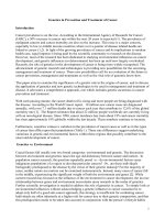
Electrosurgery is used on a daily basis in the operating room, but it remains
poorly understood by those using it. In addition, the physics of electrosur-
gery are far more complicated than those of laser. Common belief notwith-
standing, electrosurgery has an enormous capacity for patient injury if
used incorrectly, even though technology has markedly reduced the likeli-
hood of patient or surgeon injuries. This article is intended to educate the
clinician regarding the basis of electrosurgery and provide an explanation
on how injuries may occur as well as how they may be prevented.
Prevention, Diagnosis, andTreatment of Gynecologic Surgical Site Infections 379
Gweneth B. Lazenby and David E. Soper
Surgical site infections (SSIs) have a significant effect on patient care and
medical costs. This article outlines the risks that lead to SSIs and the pre-
ventive measures, including antimicrobial prophylaxis, which decrease the
incidence of infection. This article also reviews the diagnosis and treatment
of gynecologic SSIs.
Avoiding Major Vessel Injury During Laparoscopic Instrument Insertion 387
Stephanie D. Pickett, Katherine J. Rodewald, Megan R. Billow,
Nichole M. Giannios, and William W. Hurd
Major vessel injuries during laparoscopy most commonly occur during in-
sertion of Veress needle and port trocars through the abdominal wall. This
article reviews methods for avoiding major vessel injury while gaining lap-
aroscopic access, including anatomic relationships of abdominal wall
landmarks to the major retroperitoneal vessels. Methods for periumbilical
placement of the Veress needle and primary trocar are reviewed in terms
of direction and angle of insertion, and alternative methods and locations
are discussed. Methods for secondary port placement are reviewed in
terms of direction, depth, and speed of placement.
Complications of Hysteroscopic and Uterine Resectoscopic Surgery 399
Malcolm G. Munro
Adverse events associated with hysteroscopic procedures are in general
rare, but, with increasing operative complexity, it is now apparent that
Prevention and Management of Complications from Gynecologic Surgery
they are experienced more often. A spectrum of complications exist rang-
ing from those that relate to generic components of procedures such as
patient positioning and anesthesia and analgesia, to a number that are
specific to intraluminal endoscopic surgery (perforation and injuries to sur-
rounding structures and blood vessels). The response of premenopausal
women to excessive absorption of nonionic fluids deserves special atten-
tion. There is also an increasing awareness of uncommon but problematic
sequelae related to the use of monopolar uterine resectoscopes that in-
volve thermal injury to the vulva and vagina. The uterus that has previously
undergone hysteroscopic surgery can behave in unusual ways, at least in
premenopausal women who experience menstruation or who become
pregnant. Better understanding of the mechanisms involved in these ad-
verse events, as well as the use or development of several devices, have
collectively provided the opportunity to perform hysteroscopic and resec-
toscopic surgery in a manner that minimizes risk to the patient.
Gynecologic Surgery and the Management of Hemorrhage 427
William H. Parker and Willis H. Wagner
Surgical blood loss of more than 1000 mL or blood loss that requires
a blood transfusion usually defines intraoperative hemorrhage. Intraoper-
ative hemorrhage has been reported in 1% to 2% of hysterectomy studies.
Preoperative evaluation of the patient can aid surgical planning to help pre-
vent intraoperative hemorrhage or prepare for the management of hemor-
rhage, should it occur. To this effect, the medical and medication history
and use of alternative medication must be gathered. This article discusses
the methods of preoperative management of anemia, including use of iron,
recombinant erythropoietin, and gonadotropin-releasing hormone ago-
nists. The authors have also reviewed the methods of intraoperative and
postoperative management of bleeding.
Understanding Errors During Laparoscopic Surgery 437
William H. Parker
Complications may occur during laparoscopic surgery, even with a skilled
surgeon and under ideal circumstances; human error is inevitable. Video-
taped procedures from malpractice cases are evaluated to ascertain po-
tential contributing cognitive factors, systems errors, equipment issues,
and surgeon training. Situation awareness and principles derived from avi-
ation crew resource management may be adapted to help avoid systems
error. The current process of surgical training may need to be reconsidered.
Postoperative Neuropathy in Gynecologic Surgery 451
Amber D. Bradshaw and Arnold P. Advincula
The development of a postoperative neuropathy is a rare complication that
can be devastating to the patient. Most cases of postoperative neuropathy
are caused by improper patient positioning and the incorrect placement of
surgical retractors. This article presents the nerves that are at greatest risk
of injury during gynecologic surgery through a series of vignettes. Sugges-
tions for protection of each nerve are provided.
Contents
viii
Hollow Viscus Injury During Surgery 461
Howard T. Sharp and Carolyn Swenson
Reproductive tract surgery carries a risk of injury to the bladder, ureter,
and gastrointestinal (GI) tract. This is due to several factors including close
surgical proximity of these organs, disease processes that can distort
anatomy, delayed mechanical and energy effects, and the inability to di-
rectly visualize organ surfaces. The purpose of this article is to review strat-
egies to prevent, recognize, and repair injury to the GI and urinary tract
during gynecologic surgery.
Index 469
Contents
ix
Foreword
William F. Rayburn, MD, MBA
Consulting Editor
A patient’s operative care should be planned with attention to detail and awareness of
potential complications. This issue, guest edited by Dr Howard Sharp, pertains to the
prevention and management of complications from gynecologic surgery. Major objec-
tives are to restore the patient’s physiologic and psychologic health. The operating
room presents the possibility for immediate or delayed errors. Adverse surgical events
are relatively infrequent compared with other types of medical errors, although these
problems often receive increased attention.
This distinguished group of authors comes from academic health centers. Graduate
medical education requires full supervision and assistance by qualified and experi-
enced gynecologists. It is up to the clinical judgment of the supervising surgeon to
allow increasing operative responsibilities for trainees based on their experience, skill,
and level of training. Expanding training by using surgical simulators and virtual training
techniques helps better prepare trainees before entering the operating suite.
The American College of Obstetricians and Gynecologists’ Committee Opinion
Number 328 states that ‘‘ensuring patient safety in the operating room begins before
she enters the operative suite and includes attention to all applicable types of prevent-
able medical errors (including, for example, medication errors) but surgical errors are
unique to this environment.’’ A single error may lead to a grave patient injury even
with the most vigilant supervision. Communication issues, unique terminology, and
special instruments must be understood and shared by all members of the team.
There is a complication rate for every operation. Patients need to understand the
risks and benefits of the procedure, as well as any alternatives, before a gynecologist
initiates any therapy. Informed consent is a discussion, not simply a form. This issue
describes the management of certain complications of gynecologic surgery, which
include electrosurgical energy-related injury, excess hemorrhage, major vessel injury
and venous thromboembolism, and urinary tract and bowel injuries. In the elderly
and obese patients, respiratory insufficiency is an especially common postoperative
problem.
Obstet Gynecol Clin N Am 37 (2010) xi–xii
doi:10.1016/j.ogc.2010.06.002 obgyn.theclinics.com
0889-8545/10/$ – see front matter ª 2010 Elsevier Inc. All rights reserved.
Prevention and Management of Complications from Gynecologic Surgery
Obesity is becoming more prevalent in our surgical patients and represents a much
higher risk for surgical complications. The occurrence of comorbidities, including dia-
betes, hypertension, coronary artery disease, sleep apnea, obesity hypoventilation
syndrome, and osteoarthritis of the knees and hips, are more frequent. These under-
lying alterations in physiology result in increased surgical risks of cardiac failure, deep
venous and pulmonary emboli, aspiration, wound infection and dehiscence, postop-
erative neuropathy, and misdiagnosed intra-abdominal catastrophe.
It is our desire that this issue inspires attention to a vast array of operative compli-
cations. On behalf of Dr Sharp and his excellent team of knowledgeable contributors, I
hope that the practical information provided herein will aid in the implementation of
evidence-based and well-planned approaches to preventing and managing complica-
tions from gynecologic surgery.
William F. Rayburn, MD, MBA
Department of Obstetrics and Gynecology
University of New Mexico School of Medicine
MSC10 5580, 1 University of New Mexico, Albuquerque
NM 87131-0001, USA
E-mail address:
Foreword
xii
Preface
Surgical Complications
Howard T. Sharp, MD
Guest Editor
It is more enjoyable to read about complications than to manage them. Surgical
complications are challenging for several reasons. It is difficult to watch patients and
their families suffer. Although some complications are minor setbacks that resolve
over time, some lead to longstanding disability. As surgeons, we sometimes doubt
ourselves in the wake of a complication and lose confidence in our abilities. In some
cases, surgeons avoid surgery or practice heightened defensive surgery, rendering
them surgically dysfunctional. We should ask ourselves, ‘‘Is there something I should
have done differently?’’ ‘‘Could this have been avoided?’’ and ‘‘Should I have recog-
nized something earlier?’’ These are questions I ask each week at our institution’s
morbidity and mortality conference.
One of my favorite surgical mentors, the great, late Gary Johnson, MD, would
lament, ‘‘If you don’t want surgical complications, don’t do surgery.’’ He had figured
out that complications happen. I do not know that he was any more comfortable
with complications than I, but he recognized an important truth: there is a complication
rate for each surgery performed. Are there ways to reduce complication rates? I think
so. Can all complications be eliminated? I think not.
It has always sounded a bit ridiculous to me when someone says, ‘‘He or she has
the hands of a surgeon,’’ as if the hands have so much to do with being a good
surgeon. Having a steady hand and knowing the patient and how to perform surgery
are given basic prerequisites for taking a patient to the operating room. But there is
much more to being a good surgeon. Surgeons must know anatomy and anatomic
variation, be familiar with surgical instrumentation and its technology, have situational
awareness, and be ever vigilant to recognize risks for complications preoperatively, in-
traoperatively, and postoperatively. Some have said it is good to have a little healthy
paranoia. The reason for vigilance is the recurrent theme of early recognition and
management of complications associated with better outcomes. If there were anything
Obstet Gynecol Clin N Am 37 (2010) xiii–xiv
doi:10.1016/j.ogc.2010.05.005 obgyn.theclinics.com
0889-8545/10/$ – see front matter ª 2010 Elsevier Inc. All rights reserved.
Prevention and Management of Complications from Gynecologic Surgery
to stress in the volume, it is that avoiding complications is much more than just having
‘‘good hands.’’ It is my sincere hope that the words of these fine authors will allow the
readers to avoid and manage complications to the best of their ability.
Howard T. Sharp, MD
Department of Obstetrics and Gynecology
University of Utah Health Sciences Center
Room 2B-200, 1900 East, 30 North
Salt Lake City, UT 84132, USA
E-mail address:
Preface
xiv
Preventing
Electrosurgical
Energy–Related
Injuries
Gary H. Lipscomb, MD
*
, Vanessa M. Givens,
MD
In 1928, Cushing
1
reported a series of 500 neurosurgical procedures on the brain in
which bleeding was controlled by an electrosurgical unit designed by W.T. Bovie.
Since that time, the ‘‘Bovie’’ has become an instrument familiar to every gynecologic
surgeon. Most gynecologic surgeons would consider it a much simpler and safer
instrument than the carbon-dioxide or KTP laser or the argon beam coagulator. This
belief is reinforced by the fact that even weekend introductory laser courses present
a thorough review of laser physics, whereas lectures on electrosurgery are uncommon
even in advanced operative gynecology courses. Common belief notwithstanding,
electrosurgery has an enormous capacity for patient injury if used incorrectly. In addi-
tion, the physics of electrosurgery are far more complicated than those of laser. This
article reviews the principles of electrosurgery and the mechanisms of electrosurgical
injury and discusses the methods of prevention of these injuries.
ELECTROPHYSICS
Although a detailed description of electrophysics is beyond the scope of this article, it
is necessary to review some of the basic principles of electrosurgery to understand
why patient injuries occur. The most fundamental principles of electrosurgery are
that electricity always seeks the ground and the path of least resistance. These 2 prin-
ciples are straightforward and even intuitive. However, most of the other principles of
electrosurgery are not so easily understood. Because most physicians find electro-
physics confusing, it is often easier to relate many of the terms to those of hydraulics,
which are more familiar. Just as a certain amount of water flows through a garden
hose, electric energy consists of a flow of negatively charged particles called elec-
trons. This flow of electrons is referred to as current. Electric current is described by
Section of Obstetrics and Gynecology, Department of Family Medicine, University of Tennessee
Health Science Center, 1301 Primacy Parkway, Memphis, TN 38119, USA
* Corresponding author.
E-mail address:
KEYWORDS
Electrosurgery
Electrode
Cut current
Coagulation current
Obstet Gynecol Clin N Am 37 (2010) 369–377
doi:10.1016/j.ogc.2010.05.007 obgyn.theclinics.com
0889-8545/10/$ – see front matte r ª 2010 Elsevier Inc. All rights reserved.
several interrelated terms.
2
First, current is measured by the number of electrons flow-
ing per second. A flow of 6.24 Â 10
18
electrons (1 coulomb [C]) per second is referred
to as 1 A. This is analogous to a stream of water in which the flow is measured in
gallons per minute. Volt is the unit of force that drives the electron flow against resis-
tance, and 1 V drives 1 A of current through a specified resistance. The volt is similar to
water in a hose under a force of so many pounds per square inch. As with water in
a hose, the higher the water pressure the greater the potential for leaks to occur. Simi-
larly, in the case of electricity, the higher the voltage the greater the possibility of
unwanted stray current. The difficulty that a substance presents to the flow of current
is known as resistance and is sometimes referred to as impedance (I). Resistance is
measured in ohm. The power of current, measured in watts, is the amount of work
produced by the electron flow. Again using the water analogy, power is equivalent
to the work in horsepower produced by a stream of water as it turns a waterwheel.
Power can also be related to the heat output and is often measured in British thermal
unit. Table 1 shows the relationship between these terms.
All variables in electrosurgery are closely interrelated such that a change in one vari-
able leads to changes in the others. Using the analogy of water flowing through a pipe,
it is probably intuitive that if the resistance to flow is increased by decreasing the diam-
eter of the pipe, the pressure forcing the water through the pipe must be increased to
maintain the previous flow rate. Similar events occur with electrosurgery. If the tissue
resistance increases, voltage must also be increased to maintain a constant power.
This interrelationship is known as Ohm’s law, which states that the current in an elec-
tric circuit is directly proportional to the voltage and inversely proportional to the
resistance.
An electric current consists of either a direct or an alternating current. Direct current
is the same current produced by batteries, whereas alternating current is the same
current that is used at home. Fig. 1 illustrates the pattern generated on an oscilloscope
by the 2 different types of currents. Direct current produces a flow of electrons from
one electric pole to another of opposite charge. The flow of current is unidirectional
and continuous. One pole is always negatively charged, and the other is always posi-
tively charged. Direct current is not normally used in electrosurgery.
Unlike direct current in which the poles are always the same charge, in alternating
current the poles reverse polarity periodically. As a result, alternating current alter-
nates the direction of electron flow, first flowing in one direction then reversing flow.
The rate at which the polarity reverses is described in cycles per second and is
referred to as the frequency of the cycle. One cycle per second is 1 Hz. Electric current
used at home is supplied as alternating current at 60 Hz. Voltage of alternating current
is normally measured either from zero baseline to maximum (peak voltage) or from the
maximum in one direction to the maximum in the other (peak-to-peak voltage).
Average or mean voltage when describing alternating current is meaningless because
the positive voltage in one cycle is negated by the identical negative voltage in the
Table 1
Equivalent terms for electricity and hydraulics
Term Unit Hydraulic Equivalent
Current Ampere Gallons per minute
Voltage Volt Pounds per square inch
Impedance Ohm Resistance
Power Watt Horsepower
Lipscomb & Givens
370
same cycle. Thus the average voltage of the current would be zero. To avoid this
problem, the average peak voltage is described using a standard statistical measure
that describes the magnitude using the square root of the mean of the squares of the
values or the root-mean-square (RMS) value. The RMS of household current is 120 V.
Fig. 2 illustrates these terms as illustrated with household current.
EFFECTS
Why do patients do not have muscle contraction or pain when undergoing electrosur-
gical procedures? Common answers are that the patient is grounded or under anes-
thesia. A patient undergoing a loop electrosurgical excision procedure is not under
anesthesia but does not have muscle contraction. Few people would want to ground
themselves by pouring water on the floor and then stick their finger in a light socket.
Why then patients do not experience nerve and muscle excitation?
Normally, when a direct electric current is applied to a tissue, the positively and nega-
tively charged particles in the cells migrate to the oppositely charged poles and the cell
membranes undergo depolarization resulting in muscle contraction and nerve stimula-
tion. This is known as the Faraday effect. With alternating current, the electric poles
reverse with each cycle. If the frequency becomes high enough, there is insufficient
time between cycles for the charged ions to migrate before the poles reverse. At this
point, nerve and muscle depolarization does not occur. This effect occurs at
+
-
0
+
-
0
C.D .
C.A
.
Fig. 1. Direct and alternating current. AC, alternating current; DC, direct current.
Peak to Peak
Voltage
(340 V)
Peak Voltage
(170 V )
RMS Voltage
(120 V)
+
-
60 Hz
0
Fig. 2. Household current (voltage, cycle, and RMS).
Electrosurgical Energy–Related Injuries
371
approximately 100,000 Hz or 100 kHz and is demonstrated in Fig. 3. Most electrosur-
gical units actually operate at 5 to 10 times this frequency. These frequencies are in
the range of amplitude-modulated radiofrequency, and thus the term radiofrequency
current is used to describe the current of electrosurgical units used in medicine.
Often, electrosurgical instruments are referred to as cautery, and the term electro-
cautery is frequently used interchangeably with electrocoagulation to indicate the
coagulation of tissue with electric current. The term cautery is derived from the Greek
‘‘kauterion’’ or hot iron. A cautery transfers heat from a source to the tissue, and there-
fore electrocautery is the transfer of heat from an electrically heated source to tissue.
In electrocoagulation, the heat developed within the tissue is a result of the resistance
to the passage of electron, and any heating of the electrode is secondary to this. Thus,
the terms cautery and electrocautery are technically incorrect.
MONOPOLAR AND BIPOLAR CURRENTS
Electrosurgery can be divided into monopolar or bipolar depending on the number of
electric poles at the site of application. In reality, all electric devices require 2 poles to
complete an electric current. With unipolar current, the Bovie tip is one pole, whereas
the second pole is the grounding pad. With bipolar current, both poles are part of the
tip of the instrument. The main difference between the 2 types of current is the
distance between the poles. Because the human body is a relatively poor conductor
of electric current, a relatively high power output is needed to overcome the long
distance between the poles in unipolar electrosurgery. With bipolar instruments, the
electrodes are only millimeters apart. Because a high-power current would destroy
the instrument, the power output of bipolar instruments is one-third to one-tenth
that of unipolar systems. The relatively low power of bipolar systems is insufficient
to generate the current densities that are needed to cut tissue, and thus these systems
can only desiccate the tissue. Because of the constant inflow of electrons, a nonmodu-
lated cutting waveform produces a more uniform desiccation than a modulated coag-
ulation current. Coagulation current tends to produce a rapid superficial desiccation
that impedes further electron flow into the center. For this reason, bipolar electrosur-
gical generators designed for tubal sterilization produce only cut current because the
use of coagulation current has been associated with higher failure rates.
CUT AND COAGULATION CURRENTS
Electrosurgical generators produce 2 primary types of alternating current, which have,
through common usage, been designated cut and coag or coagulation currents. But,
dlohesuoH
tnerruC
evreN
noitalumitS
yregrusortcelE
VToidaRMFoida
R
MA
zH05
088-45zHM801-88zHk0551-055zHk0002-005zHk001
zHM
Fig. 3. Frequency of spectrum. AM radio, amplitude-modulated radiofrequency; FM radio,
frequency-modulated radiofrequency.
Lipscomb & Givens
372
these labels are misleading because they do not necessarily produce the tissue
effects that are associated with the terms cut and coagulation. In fact, cut current
can coagulate and coag current can cut, but cut current is often the most appropriate
current to use for tissue coagulation. Cut current is more accurately designated as
nonmodulated or undamped current, and coag current is designated as modulated
or damped current. Nonmodulated cut current is characterized by a continuous unin-
terrupted flow of electrons. Modulated coag or damped current consists of a burst of
alternating current interrupted by intervals of no current flow. Fully modulated current
has no current flow for more than 95% of each cycle. Fig. 4 illustrates the 2 types of
currents. At identical power levels, there is less current flow per time interval with
modulated current than with nonmodulated current. Because power (W) 5 volts (V) Â
current flow (I), the peak-to-peak voltage of modulated current must be greater to
produce the same power of nonmodulated current. More simply, for the same
wattage, coag current has a much higher voltage than cut current. As previously
noted, higher voltages are more likely to produce unwanted effects and injuries than
lower voltages. In more simple terms, for the same power levels, cut current is the
safer modality.
ELECTROSURGICAL CUTTING
Electrosurgical cutting occurs when the intracellular temperature increases high and
fast enough to cause the explosive vaporization of water. Electrosurgical cutting
occurs only under extremely high current densities that exist when the current is
confined to arcs traveling between the electrode and tissue (Fig. 5). Cutting is facili-
tated by conditions that encourage the formation of these arcs. Arcs are further
enhanced by the steam envelope formed around the electrode by the vaporization
of cellular water. A continuous current (nonmodulated or cut current) is necessary to
maintain this vapor barrier. Efficient cutting requires the electrode to be moved slowly
but continuously through the tissue. Moving too quickly collapses the steam barrier
and places the electrode in contact with tissue. Because the cross-sectional area of
the electrode is greater than that of the arc, the current density decreases than that
needed for cutting, and the electrode stalls until the steam barrier is regained. There-
fore, if tissue cutting is desired, the electrode should be activated before touching the
tissue and moved slowly to avoid dragging.
Pure Cut
Pure Coag
voltage
On 100% Cycle
On 5% Off 95% Cycle
Fig. 4. Cut versus coag current.
Electrosurgical Energy–Related Injuries
373
SPRAY COAGULATION (FULGURATION)
Spray coagulation or fulguration is different from electrosurgical cutting. In fulguration,
high-voltage, interrupted current (modulated or coag current) is required. The higher
voltages of modulated current compared with those of nonmodulated current allow
arcs to form to the tissue in the absence of a vapor barrier (see Fig. 5). Because the
coagulation waveform is highly interrupted, any steam barrier formed collapses before
the next cycle. The result is that the arcs strike a wider area of tissue in a random
fashion. Much like lightning, it is never said to strike twice in the same spot. With
the coagulation waveform, there is less-rapid heating of tissue because the pause
allows heat to be dissipated to other cells. The end result is more cell heating and
dehydration, with more charring than that occurring with electrosurgical cutting.
However, the effect is superficial.
BLENDED CURRENT
Blended current is not, as is frequently misbelieved, a blend of cut and coag currents,
but it actually refers to a blending of effects. Use of blended current helps to cut tissue
while obtaining some degree of coagulation but avoiding the thicker eschar associ-
ated with cutting in the full coag mode. Blended current is obtained by modulating
the normal cut current so that the cycle is off for a percentage of time less than that
obtained in the full coag mode. The coag setting on the generator is irrelevant. There-
fore, if a setting of blend 1 is selected with a cut setting of 40 W, the effect will be the
same whether the coag setting is 0 or 100 W because only the cut current is modu-
lated. Typically, blend 1 setting is approximately 50% on and 50% off. Blend 3 has
the current cycle off for 75% of the time.
DESICCATION
In both electrosurgical cutting and fulguration, the electrode is not in actual contact
with the tissue. When the electrode is placed in contact with the tissue, the larger
surface area of the tissue results in a relatively slower heating of intracellular water.
Explosive vaporization does not occur, but cellular water is evaporated until the tissue
is dry (desiccated) (see Fig. 5). The higher voltage of the coagulation waveform is more
penetrating and inflicts more damage than cutting current. The current is also more
prone to spark to unwanted areas than a cutting current. The use of coag current in
bipolar electrocoagulation for tubal sterilization has been shown to result in a higher
failure rate.
3
The burst of high-voltage current produces an eschar of carbon at the
surface of the tube. The eschar inhibits penetration of the current into the interior of
Electrosurgical
Fulguration
Electrosurgical
Cutting
Electrosurgical
Desiccation
Fig. 5. Cutting, fulguration, and desiccation.
Lipscomb & Givens
374
the tube and may result in inadequate coagulation, leaving a viable tubal lumen. The
slower crock-pot desiccation produced with cutting current produces a more
complete coagulation.
Coaptive coagulation is another form of desiccation that involves clamping
a bleeding vessel with a conductive clamp and applying current to the clamp to
produce a collagen weld of the vessel. Because of previously mentioned factors,
cutting current is usually the most appropriate current for this purpose, which is oppo-
site to the belief of most surgeons. As noted later in this article, use of coag current in
this setting is much more likely to lead to a surgeon being zapped or burned.
As with lasers, the effect of electric current on tissue depends on the amount of
power applied per square centimeter of surface area multiplied by time. In monopolar
mode, the active electrode is usually small, whereas the ground electrode is relatively
larger. The same amount of current flows out of the ground pad as enters from the
active electrode. However, because the current is dispersed over a wide area, no
tissue damage occurs. Severe burns can occur if the pad becomes detached except
for a small area. The tissue effect at the pad then approaches that encountered at the
active electrode. Similarly, the use of a needle-tip electrode results in high current
densities that cuts the tissue with minimum lateral thermal damage, whereas
a broader-blade electrode produces more thermal tissue damage.
INJURIES WITH ELECTROSURGERY
Unintended burns may occur in several ways during electrosurgery.
4
Burns may occur
at the active electrode as a result of direct coupling, away from the active electrode as
a result of capacitance coupling, or from alternate path burn.
Perhaps the most common type of electrosurgical injury results from direct applica-
tion of current to tissue away from the active electrode itself (direct coupling). The
most easily understood example of this type of injury is when another metal object
such as a probe is touched by the active electrode. The current is conducted through
the probe resulting in injury to the tissue where the probe touches. Injury may also
occur if a metal retractor is touched by a hemostat that is being energized to coagulate
a bleeding patient. The contact may go unnoticed and unrecognized until the following
day when a full-thickness burn is noted on the skin 1 or 2 in away from the incision,
seeming far away from the region where electrosurgery was done. This burnt site is
in reality an area where the retractor was resting on the skin. Another example of
this type of injury occurs when a defect in insulation allows the current to flow from
the defect into the adjacent tissue.
Direct coupling is best avoided by situational awareness of where the active elec-
trode is at all times when the electrode is being energized. Particular care should be
exercised anytime the active electrode is energized near another metal object. Insula-
tion failure is more difficult to avoid. Frequent inspection of reusable insulated instru-
ments may detect insulation flaws before injury occurs. Use of disposable instruments
reduces the chance of repetitive use resulting in damage to insulation that may then
result in patient injuries. There are also conductive sheaths available that when placed
over the instrument monitor for stray current and shut down the generator if such
current is detected.
Alternate Path Burns
The original Bovie electrosurgical unit was a grounded instrument. As such, current
eventually flows into an electron sink or ground, which originally was the earth. As
previously noted, electricity always seeks the path of least resistance to ground. In
Electrosurgical Energy–Related Injuries
375
grounded systems, if there is impedance to the flow of electrons to the ground via the
intended path, other conductive objects may become the path of least resistance. The
other objects could be the metal table in an operating room or the electrocardiogram
(ECG) leads on the patient. Because of the small size of the ECG leads, deep burns
may occur if current is diverted through them.
The likelihood of alternate site burns was considerably reduced by the introduction
of isolated electrosurgical generators in 1972. These generators are no longer con-
nected to a true ground. If a reduction in current flow back to the generator is detected,
the generator shuts down. This mode of action markedly reduces the probability of
alternate site injuries. Another consequence of this technology is that the ground
pads themselves have been eliminated. The correct term for the pads that are avail-
able today is patient return electrode.
Patient Electrode Burn
In monopolar electrosurgery, the current travels through the patient between 2 active
electrodes. Normally, no effect is seen at the return electrode because of its larger
surface area. If for some reason the return electrode becomes detached or has
been improperly placed, patient injury may occur. Because the return electrode is typi-
cally out of direct sight, a large, deep, full-thickness burn may occur without the sur-
geon’s knowledge.
The introduction of return electrode monitoring (REM) technology has essentially
eliminated this type of patient injury. In REM, the return electrode is divided into 2 elec-
trodes that are electrically connected to each other by the patient’s skin. A low-
intensity current is constantly passed between the 2 electrodes. If this current is not
detected because the electrode has become detached or if the surface temperature
increases by more than 2
C, the generator deactivates.
Capacitance Coupling
When unidirectional electric current travels through a conductor, an electromagnetic
field is generated around the conductor. This field can generate a secondary current
in nearby conductors, such as a metal trocar. The amount of current generated is
determined by multiple factors. Capacitance coupling is increased with increasing
voltage. Because coag current is associated with higher voltage it is more likely to
result in a capacitance effect. Open circuit activation (current activation without
touching tissue) also markedly increases voltage and thus the risk of capacitance.
The use of cutting current and limiting the open circuit activation decreases the risk.
Capacitive coupling can produce sufficient current to cause an injury under several
conditions. The most common is when a metal cannula is used with a plastic tissue
anchor. Current can be induced on the metal cannula, but return of current via the
abdominal wall is prevented by the plastic anchor. Up to 70% of the originally applied
current may be inducted in some circumstances. If this current is not conducted away
by the abdominal wall, arcing from the trocar to adjacent tissue may occur. The area
where arcing occurs is frequently out of the visual field of the surgeon and may result in
significant injuries. Thus the old adage, ‘‘use all metal or all plastic trocars.’’
As previously noted, the use of conductive sheaths can conduct stray current from
the surgical site as well as monitor the total amount of current present before deliv-
ering it to the return electrode.
Surgeon Burns
Surgeons may suffer burns from the use of electrosurgical units.
5
Burns may occur
from faulty insulation or contact with the active electrode. The cause of these injuries
Lipscomb & Givens
376
is readily apparent to most clinicians. An injury that is poorly understood by most
surgeons is the burn occurring when zapping a hemostat to coagulate a bleeding
vessel. This type of coagulation is known as coaptive coagulation. It is commonly
believed that coaptive coagulation results from a preexisting hole in the surgical glove;
however, surgical gloves offer minimal insulation to electrosurgical current. In reality,
the current results in a breakdown of the glove material, that is, it blows a hole in the
glove.
There are 4 conditions that increase the likelihood of hemostat burns:
1. The use of coag current with its associated higher voltage is much more likely to
result in surgeon burns than the more appropriate cut current.
2. In anticipation of a possible burn, surgeons often instinctively hold the hemostat
gingerly resulting in a small surface area between the hemostat and the hand.
Similar to the way by which the return electrode’s large surface area does not result
in a patient burn, a broad grip of the hemostat reduces the likelihood of surgeon
burns.
3. The use of open activation circuit producing arcing to the hemostat results in the
highest voltage as the generator tries to complete the circuit. Touching the hemo-
stat before activation produces much-lower voltage and less potential for glove
failure.
4. Because modern electrosurgical units are not grounded, the surgeon has to
become part of the circuit to produce a hemostat burn. Contact with the patient
or metal retractors with the hand not grasping the hemostat allows the surgeon
to be part of the circuit. Lifting the hand off the patient or releasing the retractors
isolates the surgeon and prevents burns from occurring.
SUMMARY
Electrosurgery is used on a daily basis in the operating room, but it remains poorly
understood by those using it. Although technology has markedly reduced the likeli-
hood of patient or surgeon injuries, the potential for serious injuries still exists. This
article is intended to educate the clinician regarding the basis of electrosurgery and
provide an explanation on how injuries may occur as well as how they may be
prevented.
REFERENCES
1. Cushing H. Electrosurgery as an aid to the removal of intracranial tumors. Surg
Gynecol Obstet 1928;47:751–4.
2. Hulka JF, Reich H. Power: electricity and laser. In: Textbook of laparoscopy. Phila-
delphia: WB Saunders; 1994. p. 23–46.
3. Engel T, Harris FW. The electric dynamics of laparoscopic sterilization. J Reprod
Med 1975;15:33–42.
4. Luciano AA, Soderstrom RM, Martin DM. Essential principles of electrosurgery in
operative laparoscopy. J Am Assoc Gynecol Laparosc 1994;1:189–95.
5. Odel RC. Biophysics of electrical energy. In: Soderstrom RM, editor. Operative
laparoscopy: the masters’ technique. New York: Raven Press; 1993. p. 35–44.
Electrosurgical Energy–Related Injuries
377
Prevention,
Diagnosis, and
Treatment of
Gynecologic Surgical
Site Infections
Gweneth B. Lazenby, MD, David E. Soper, MD
*
Surgical site infections (SSIs) are the most common nosocomial infections encoun-
tered during inpatient hospitalization. Approximately two-thirds of these infections
involve superficial incisions, and the remaining involve the deeper tissues and organ
spaces. SSIs have a significant effect on health care costs by prolonging hospitaliza-
tion, requiring additional medications, and potentially additional procedures.
1–3
This
article reviews the pathophysiology, risk factors, prevention strategies, diagnosis,
and treatment of postoperative gynecologic surgical infections.
PATHOPHYSIOLOGY AND MICROBIOLOGY
SSIs are initiated at the time of surgery by endogenous flora of the skin or vagina
contaminating the wound. A foreign body, such as suture, decreases the number of
organisms necessary for the development of SSI. Most endogenous skin flora are
composed of aerobic gram-positive cocci.
4
The most frequent organisms isolated
from SSIs of abdominal incisions are Staphylococcus aureus, coagulase-negative
staphylococci, Enterococcus spp, and Escherichia coli. During gynecologic proce-
dures, potential pathogenic microorganisms may come from the skin or ascend
from the vagina and endocervix to the operative sites, which include abdominal inci-
sion, upper genital tract and/or vaginal cuff. Gynecologic SSIs are more likely to be
infected with gram-negative bacilli, enterococci, group B streptococci, and anaerobes
as a result of incisions involving the vagina, and perineum.
5,6
Postoperative pelvic
abscesses are commonly associated with anaerobes.
6,7
Bacterial vaginosis alters
Department of Obstetrics and Gynecology, Medical University of South Carolina, 96 Jonathon
Lucas Street, Suite 634 MSC 619, Charleston, SC 29425, USA
* Corresponding author.
E-mail address:
KEYWORDS
Surgical site infections
Wound infections
Antibiotic prophylaxis
Obstet Gynecol Clin N Am 37 (2010) 379–386
doi:10.1016/j.ogc.2010.05.001 obgyn.theclinics.com
0889-8545/10/$ – see front matte r ª 2010 Elsevier Inc. All rights reserved.
the vaginal flora to increase the concentration of anaerobes by 1000- to 10,000-fold.
This increase in anaerobes is an important risk factor in the development of postoper-
ative pelvic infection, especially vaginal cuff cellulitus.
8,9
In recent years, methicillin-
resistant S aureus (MRSA) has played a larger role in SSIs.
1
RISK FACTORS
Risk factors for SSIs include diabetes, tobacco abuse, systemic steroid use, surgical site
irradiation, poor nutrition, obesity, prolonged perioperative stay, and transfusion of blood
products.
1,7,10
Preoperative vaginitis due to bacterial vaginosis or Trichomonas vaginalis
is associated with increased risk of posthysterectomy cuff cellulitis.
2,8
Women should be
screened for vaginitis and treated before surgery to decrease this risk.
9
Cervical infection
with Chlamydia trachomatis, Neisseria gonorrhoeae, and mycoplasmas can lead to
ascending infection during transcervical procedures. Preoperative screening for cervi-
citis is recommended in women atrisk for sexuallytransmitted infections. Surgical factors
associated with SSIs include prolonged surgery duration, excessive blood loss, hypo-
thermia, hair removal by shaving, and the use of surgical drains.
1,10–12
Patients under-
going abdominal hysterectomy are more likely to experience febrile morbidity than
those who undergo vaginal hysterectomy.
12
Nasal carriage of Saureusand MRSA has been associated with an increased risk of
SSIs after certain operations; specifically, cardiothoracic, neurosurgical, and orthopedic
surgeries.
10
In these cases, nasal application of mupirocin ointment before surgery has
been shown to decrease the microbial burden.
4,13
There are no data regarding the effect
of Saureusand MRSA decolonization before gynecologic surgeries.
PREVENTION
Surgical practices that decrease the rates of infection include use of antiseptic skin prep-
aration, antimicrobial prophylaxis (AMP), thermoregulation, and following a sterile tech-
nique.
1
Skin preparation with chlorhexidine-alcohol is preferred to povidone-iodine for
preventing SSIs.
14
The goals of AMP are to achieve inhibitory concentrations at the inci-
sion site and to maintain adequate levels of antimicrobial agents for the duration of
surgery. Antimicrobial agents should be administered intravenously no more than 1
hour before making the skin incision.
2,11,12,15,16
If the duration of the procedure exceeds
the expected duration of adequate tissue levels or 2 half-lives of the prophylactic antibi-
otic, an additional dose of the antibiotic should be administered.
1
For cefazolin, the most
commonly used prophylactic antibiotic, a repeat dose should be given if the duration of
surgery exceeds 3 hours.
2
An additional dose of the antibiotic should be administered in
case the estimated blood loss is more than 1500 mL.
3
For patients weighing more than 80
kg, the dose of cefazolin should be doubled to 2 g. With current AMP practices, the rate of
postoperative infections has decreased by approximately 50%.
15,17,18
AMP is recommended for all types of hysterectomies and induced abortion.
19–21
For
hysterectomy, cefazolin is the most commonly used AMP agent. Preoperative admin-
istration of doxycycline is recommended for women who are undergoing surgically
induced abortion.
20,22,23
Gynecologic surgeries for which AMP is not routinely recom-
mended include diagnostic or operative hysteroscopy, endometrial ablation,
24
abdominal myomectomy, and laparoscopy without hysterectomy.
25
AMP
Cephalosporins are the most widely used AMP agents. This class of antibiotics is
effective against gram-positive and gram-negative microorganisms. Secondary to
Lazenby & Soper
380
coverage of the more common microorganisms associated with gynecologic SSIs,
cefazolin is the first choice for most clean-contaminated procedures.
11,12,15
Cephalo-
sporins are not active against Enterococci spp.
If immediate hypersensitivity reaction to penicillin or cephalosporins is reported in
patients, use of alternative broad-spectrum AMP agents is recommended. The Amer-
ican College of Obstetrics and Gynecology recommends a combination of non–b-lac-
tam antibiotics. These combinations include clindamycin and gentamicin, clindamycin
and ciprofloxacin, clindamycin and aztreonam, metronidazole and gentamicin, or
metronidazole and ciprofloxacin.
26
In patients with known history of MRSA infection
or colonization, vancomycin, in addition to the preferred agent, is the AMP agent of
choice. Vancomycin requires an infusion time of 1 hour (Table 1).
1,16
Appropriate use of a single dose of AMP agent minimizes the potential for emerging
microbial resistance.
5,16
Another rare concern after AMP is the development of gastro-
intestinal overgrowth of Clostridium difficile, which can lead to diarrhea, pseudomem-
branous enterocolitis, and potentially fatal toxic megacolon.
2,10,27
Routine
administration of prophylactic antibiotics for the purpose of preventing endocarditis
is no longer recommended for gynecologic surgeries.
28
TYPES AND LOCATIONS OF SSIs
Incisional cellulitis presents with erythema, warmth from the incision, swelling, and/or
localized pain. It is not associated with a fluid collection and does not require drainage.
The most common organisms associated include S aureus, coagulase-negative
staphylococci, and streptococci. Incisional cellulitis without abscess frequently
responds to oral antimicrobial therapy alone.
15
Vaginal cuff cellulitis after hysterec-
tomy is characterized by induration, erythema, and edema of the cuff.
7,17
In the
absence of a cuff abscess, cuff cellulitis can also be treated with oral therapy (Table 2).
SSIs are categorized as superficial incisional, deep incisional, and involving organ/
space and have been defined by the CDC NNIS system.
29
A superficial incisional SSI
or wound infection occurs within 30 days of surgery and involves only the skin or
subcutaneous tissue. At least one of the following findings must be present: purulent
drainage; culture isolation of an organism from the incision; or symptoms of pain,
tenderness, erythema, edema, or warmth from the incision. Deep incisional SSI occurs
within 30 days of the surgery and involves the deep soft tissues, such as fascia and
Table 1
Recommended AMP for gynecologic surgery
Procedure
Preferred
Antibiotic Dose
Alternative Antibiotic
and Intravenous Dose
Hysterectomy Cefazolin 1–2 g Cefotetan, 1 g
Clindamycin, 600 mg; or
metronidazole, 500 mg; with
gentamicin, 1.5 mg/kg
Clindamycin, 600 mg; or
metronidazole, 500 mg; with
ciprofloxacin, 400 mg
Clindamycin, 600 mg; or
metronidazole, 500 mg; with
aztreonam, 1 g
Induced Abortion Doxycycline 100 mg Metronidazole, 500 mg
Data from Refs.
7,10,26
Gynecologic Surgical Site Infections
381
muscle layers. One of the following findings is required for diagnosis: purulent
drainage; a spontaneous dehiscence of a deep incision; the incision is opened due
to signs of fever (temperature, >38
C), localized pain, or tenderness; or an abscess
or other evidence of deep infection is found. An organ/space SSI (includes surgical
site cellulitis, eg, vaginal cuff cellulitis and pelvic abscess, including vaginal cuff
abscess or tubo-ovarian abscess [TOA]) occurs within 30 days of surgery, and the
infection involves any part of the anatomy other than the incision that was manipulated
during surgery. At least one of the following is required: purulent drainage from a drain
placed within the organ/space, culture isolation of an organism from the organ/space,
an abscess or other infections located within the organ/space, or diagnosis is made by
the surgeon.
1
The most serious form of SSI is necrotizing fasciitis. This infection usually presents
with pain disproportionate to physical examination, a thin dishwater drainage, and
possible skin bullae. Necrotizing fasciitis is often caused by a polymicrobial infection
and can lead to the rapid destruction and necrosis of the surrounding tissue, ultimately
resulting in sepsis and end-organ damage. This life-threatening infection requires
immediate wide local debridement of affected tissue after the initiation of broad-
spectrum parenteral antibiotics.
15
DIAGNOSIS
The most common complication after hysterectomy is pelvic infection.
17
Patients with
SSIs often present with pain and tenderness at the operative site and fever. Postop-
erative fever after gynecologic surgery is not uncommon in the first 24 hours. Patients
with temperature greater than 38.4
C (101
F) in the first 24 hours or greater than 38
C
(100.4
F) on 2 occasions at least 4 hours apart excluding the first 24 postoperative
hours should be evaluated for infection. On examination, skin erythema, subcuta-
neous induration, and/or spontaneous drainage of serous or purulent fluid are noted.
Pelvic examination may reveal extraordinary vaginal cuff, paravaginal, or pelvic organ
tenderness. In case of vaginal cuff abscess, a mass may be palpated at the apex of the
vagina. Laboratory tests to evaluate wound infection should include a complete blood
count. Leukocytosis of more than 13,000 cells/mm
3
with or without bandemia and
Table 2
Recommended oral antibiotic therapies for outpatient management of wound cellulitis
Skin and Soft Tissue Infections Suggested Antimicrobial Therapies
Skin Cellulitis, Low Suspicion of MRSA Cephalexin, 500 mg po qid
Dicloxacillin, 500 mg po qid
Ciprofloxacin, 500 mg po bid
Skin Cellulitis, Concern for MRSA Trimethoprim-sulfamethoxazole, DS po bid
Doxycycline, 100 mg po bid
Clindamycin, 300 mg po tid
Vaginal Cuff Cellulitis Amoxicillin/clavulanate, 875/125 mg po bid
Ciprofloxacin, 500 mg po bid; with
metronidazole, 500 mg po bid
Trimethoprim-sulfamethoxazole, DS po bid with
metronidazole, 500 mg po bid
Abbreviation: DS, double strength.
Data from Stevens DL, Bisno AL, Chambers HF, et al. Practice guidelines for the diagnosis and
management of skin and soft-tissue infections. Clin Infect Dis 2005;41(10):1373–406.
Lazenby & Soper
382
increased percentage of polymorphonuclear neutrophils may support the diagnosis of
infection. Gram stain and culture from the incision or abscess drainage can be invalu-
able in directing antimicrobial therapy.
15
Bacteremia is rare
7,30
; therefore, blood
cultures need not be routinely obtained in the presence of a postoperative febrile
morbidity workup unless the patient appears septic.
30
IMAGING
When organ/space SSIs are suspected, radiologic evaluation with computed tomog-
raphy (CT) scan, magnetic resonance imaging (MRI), or ultrasonography can be used
to localize the area of infection.
15
Ultrasonography is the least-expensive method for
identifying a TOA and is well tolerated by patients. The sensitivity and specificity of
ultrasonography in the identification of postoperative intra-abdominal abscess is
81% and 91%, respectively. The classic appearance of a TOA on ultrasonography
is a homogenous, cystic, thin-walled contiguous mass.
31
CT findings characteristic
of a TOA include multiloculated, thick, uniform, enhancing abscess wall with fluid
densities.
32
The appearance of TOA on MRI is similar to CT, demonstrating thick-
walled masses with multiple internal septa, shading, and gas collection. The sensitivity
and specificity of MRI for the diagnosis of TOA are 95% and 89%, respectively.
33,34
The appearance of a postoperative pelvic abscess is similar to that of a TOA, whether
or not the adnexa are involved.
TREATMENT
Not all patients with superficial incisional infections require hospitalization. Patients
with a mild wound cellulitis without evidence of a wound abscess or necrotizing fas-
ciitis can be treated as outpatients with oral therapy. Most deep incisional SSIs will
require hospitalization and parenteral therapy. The physician should use clinical judg-
ment to determine if a patient can be managed as an outpatient when initiating therapy
for the wound infection. Admission should be considered in case of fever with temper-
ature greater than 101F, evidence of peritonitis, intra-abdominal or pelvic abscess,
inability to tolerate oral antibiotics, hypotension, or other physical or laboratory indica-
tors of sepsis. In patients requiring hospital admission, parenteral therapy should be
initiated (Table 3).
Antimicrobial therapy should be directed at the common microbial pathogens asso-
ciated with postoperative gynecologic infection. For incisional cellulitis, antibiotic
therapy should cover gram-positive cocci. In case of localized wound infection, inci-
sion and drainage is indicated. In regions where MRSA is prevalent or a concern, anti-
biotic selection should reflect this. Vaginal cuff cellulitis therapy should be more broad
spectrum, covering gram-positive cocci, anaerobes, and gram-negative enterics (see
Table 2 and Table 3).
Patients requiring admission should receive parenteral antibiotics. In case of deep
incisional or organ/space infections, broad-spectrum antibiotic therapy should be
initiated (see Table 3). Parenteral antibiotics should be continued until the patient is
afebrile and clinically well for at least 24 to 48 hours. If patients do not demonstrate
systemic improvement and if there is no resolution of fever within 48 hours, it is rec-
ommended to consider repeating the imaging to determine if an abscess is present
and/or changing antimicrobials. Drug fever should be considered in well-appearing,
stable patients with persistent fever with or without eosinophilia.
15
Septic pelvic
thrombophlebitis occurs rarely. It is a diagnosis of exclusion and should be considered
in postoperative febrile patients who are not responding to broad-spectrum
Gynecologic Surgical Site Infections
383
antibiotics, in the absence of an abscess or hematoma. Treatment may include contin-
uation of antibiotics and the addition of the intravenous heparin.
7
SURGICAL MANAGEMENT
Superficial incisional abscesses should be opened wide and allowed to drain. The
fascia should be probed to rule out dehiscence. Necrotic tissue within the incision
should be debrided. After debridement, wound healing may be facilitated with
packing, wound vacuum, or secondary closure after adequate regranulation. In the
presence of deep incisional and organ/space infections, debridement and drainage
are occasionally required. Vaginal cuff abscesses can be accessed by opening the
vaginal cuff and probing bluntly to break apart adhesions and allow pus and hema-
tomas to drain. Pelvic and abdominal abscesses may be accessed either surgically
or radiologically with CT assistance or ultrasound-guided needle or catheter.
15
REFERENCES
1. Mangram AJ, Horan TC, Pearson ML, et al. Guideline for prevention of surgical
site infection, 1999. Hospital infection control practices advisory committee.
Infect Control Hosp Epidemiol 1999;20(4):250–78 [quiz: 79–80].
2. Hemsell DL. Prophylactic antibiotics in gynecologic and obstetric surgery. Rev
Infect Dis 1991;13(Suppl 10):S821–41.
3. DiLuigi AJ, Peipert JF, Weitzen S, et al. Prophylactic antibiotic administration prior
to hysterectomy: a quality improvement initiative. J Reprod Med 2004;49(12):
949–54.
Table 3
Recommended parenteral antibiotic therapies for wound and pelvic infections
Skin and Soft Tissue Infections Suggested Antimicrobial Therapies
Superficial SSI (Wound Infection) Cefazolin, 1–2 g IV q 6h
Ceftriaxone, 1–2 g IV q 24h
Cefoxitin, 2 g IV q 6h
Ampicillin/sulbactam, 3 g IV q 6h
Piperacillin/tazobactam, 3.375 g IV q 6h
Deep/Organ SSI (Cuff Cellulitis, Vaginal Cuff
Abscess, TOA, and/or Pelvic Abscess)
Clindamycin, 900 mg IV q 8h; and
gentamicin, 5 mg/kg IV q 24h or
1.5–2 mg/kg IV q 8h
Ceftriaxone, 2 g IV q 24h; and clindamycin,
900 mg IV q 8h
Ampicillin, 2 g IV q 4h; and gentamicin,
5 mg/kg IV q 24h or 1.5–2 mg/kg IV q 8h;
and metronidazole, 500 mg IV q 8h
or clindamycin, 900 mg IV q 8h
Ciprofloxacin, 400 mg IV q 12h; and
metronidazole, 500 mg IV q 8h
Piperacillin/tazobactam, 3.375 g IV q 6h
Doripenem, 500 mg IV q 8h
In cases of MRSA infection, add vancomycin,
20 mg/kg IV q 12h
Abbreviation: IV, intravenous.
Data from Larsen JW, Hager WD, Livengood CH, et al. Guidelines for the diagnosis, treatment
and prevention of postoperative infections. Infect Dis Obstet Gynecol 2003;11(1):65–70.
Lazenby & Soper
384
4. Wenzel RP. Minimizing surgical-site infections. N Engl J Med 2010;362(1):75–7.
5. Duff P, Park RC. Antibiotic prophylaxis in vaginal hysterectomy: a review. Obstet
Gynecol 1980;55(Suppl 5):193S–202.
6. Soper DE. Bacterial vaginosis and postoperative infections. Am J Obstet Gynecol
1993;169(2 Pt 2):467–9.
7. Hager WD. Postoperative infections: prevention and management. 9th edition.
Philadelphia: Lippincott Williams and Wilkins; 2003.
8. Larsson PG, Carlsson B. Does pre- and postoperative metronidazole treatment
lower vaginal cuff infection rate after abdominal hysterectomy among women
with bacterial vaginosis? Infect Dis Obstet Gynecol 2002;10(3):133–40.
9. Soper DE, Bump RC, Hurt WG. Bacterial vaginosis and trichomoniasis vaginitis
are risk factors for cuff cellulitis after abdominal hysterectomy. Am J Obstet
Gynecol 1990;163(3):1016–21 [discussion: 21–3].
10. Kernodle DS, Kaiser A. Surgical and trauma-related infections. 5th edition. Phila-
dephia: Churchill Livingstone; 2000.
11. Tanos V, Rojansky N. Prophylactic antibiotics in abdominal hysterectomy. J Am
Coll Surg 1994;179(5):593–600.
12. Peipert JF, Weitzen S, Cruickshank C, et al. Risk factors for febrile morbidity after
hysterectomy. Obstet Gynecol 2004;103(1):86–91.
13. Bode LG, Kluytmans JA, Wertheim HF, et al. Preventing surgical-site infections in
nasal carriers of Staphylococcus aureus. N Engl J Med 2010;362(1):9–17.
14. Darouiche RO, Wall Jr. MJ, Itani KM, et al. Chlorhexidine-alcohol versus
povidone-iodine for surgical-site antisepsis. N Engl J Med 2010;362(1):18–26.
15. Larsen JW, Hager WD, Livengood CH, et al. Guidelines for the diagnosis, treat-
ment and prevention of postoperative infections. Infect Dis Obstet Gynecol
2003;11(1):65–70.
16. Bratzler DW, Houck PM. Antimicrobial prophylaxis for surgery: an advisory state-
ment from the national surgical infection prevention project. Clin Infect Dis 2004;
38(12):1706–15.
17. Hemsell DL. Gynecologic postoperative infections. New York: Raven Press; 1994.
18. Mittendorf R, Aronson MP, Berry RE, et al. Avoiding serious infections associated
with abdominal hysterectomy: a meta-analysis of antibiotic prophylaxis. Am J Ob-
stet Gynecol 1993;169(5):1119–24.
19. McCausland VM, Fields GA, McCausland AM, et al. Tuboovarian abscesses after
operative hysteroscopy. J Reprod Med 1993;38(3):198–200.
20. Moller BR, Allen J, Toft B, et al. Pelvic inflammatory disease after hysterosalpin-
gography associated with Chlamydia trachomatis and Mycoplasma hominis.
Br J Obstet Gynaecol 1984;91(12):1181–7.
21. Goldstein S. Sonohysterography. New York: Churchill Livingstone; 1995.
22. Sawaya GF, Grady D, Kerlikowske K, et al. Antibiotics at the time of induced abor-
tion: the case for universal prophylaxis based on a meta-analysis. Obstet
Gynecol 1996;87(5 Pt 2):884–90.
23. Pittaway DE, Winfield AC, Maxson W, et al. Prevention of acute pelvic inflamma-
tory disease after hysterosalpingography: efficacy of doxycycline prophylaxis.
Am J Obstet Gynecol 1983;147(6):623–6.
24. Bhattacharya S, Parkin DE, Reid TM, et al. A prospective randomised study of
the effects of prophylactic antibiotics on the incidence of bacteraemia
following hysteroscopic surgery. Eur J Obstet Gynecol Reprod Biol 1995;
63(1):37–40.
25. Kocak I, Ustun C, Emre B, et al. Antibiotics prophylaxis in laparoscopy. Ceska
Gynekol 2005;70(4):269–72.
Gynecologic Surgical Site Infections
385
26. ACOG Committee on Practice Bulletins–Gynecology. ACOG practice bulletin no.
104: antibiotic prophylaxis for gynecologic procedures. Obstet Gynecol 2009;
113(5):1180–9.
27. Garey KW, Jiang ZD, Yadav Y, et al. Peripartum Clostridium difficile infection:
case series and review of the literature. Am J Obstet Gynecol 2008;199(4):332–7.
28. Wilson W, Taubert KA, Gewitz M, et al. Prevention of infective endocarditis: guide-
lines from the American Heart Association: a guideline from the American Heart
Association Rheumatic fever, Endocarditis, and Kawasaki Disease Committee,
Council on Cardiovascular Disease in the Young, and the Council on Clinical
Cardiology, Council on Cardiovascular Surgery and Anesthesia, and the Quality
of Care and Outcomes Research Interdisciplinary Working Group. Circulation
2007;116(15):1736–54.
29. Horan TC, Gaynes RP, Martone WJ, et al. CDC definitions of nosocomial surgical
site infections: a modification of CDC definitions of surgical wound infections.
Infect Control Hosp Epidemiol 1992;13(10):606–8.
30. de la Torre SH, Mandel L, Goff BA. Evaluation of postoperative fever: usefulness
and cost-effectiveness of routine workup. Am J Obstet Gynecol 2003;188(6):
1642–7.
31. Moir C, Robins RE. Role of ultrasonography, Gallium scanning, and computed
tomography in the diagnosis of intraabdominal abscess. Am J Surg 1982;
143(5):582–5.
32. Hiller N, Sella T, Lev-Sagi A, et al. Computed tomographic features of tuboovarian
abscess. J Reprod Med 2005;50(3):203–8.
33. Tukeva TA, Aronen HJ, Karjalainen PT, et al. MR imaging in pelvic inflammatory
disease: comparison with laparoscopy and US. Radiology 1999;210(1):209–16.
34. Ha HK, Lim GY, Cha ES, et al. MR imaging of tubo-ovarian abscess. Acta Radiol
1995;36(5):510–4.
Lazenby & Soper
386
Avoiding Major
Vessel Injury During
Laparoscopic
Instrument Insertion
Stephanie D. Pickett, MD
a
, Katherine J. Rodewald, MD
a
,
Megan R. Billow,
DO
a
, Nichole M. Giannios, DO
a
,
William W. Hurd,
MD, MSc
b,c,
*
Laparoscopy is one of the most common surgical approaches performed in the United
States today. This surgical approach has gained popularity compared with traditional
laparotomy due to increased safety, better outcomes, and shorter recovery periods.
Each year gynecologists and general surgeons perform an estimated 2 million laparo-
scopic procedures, including cholecystectomies, tubal ligations, appendectomies,
hysterectomies, urogynecologic repairs, and cancer staging, to name a few.
1
Advances in laparoscopic technology and the development of robotic surgery are
likely to further increase the number of cases performed laparoscopically. Fortunately,
major complications related to laparoscopy are uncommon, occurring in less than 2%
of procedures.
2
One of the most serious laparoscopic complications is injury to major vessels, which
reportedly occurs in approximately 0.04% of cases.
3
Vessel injury occurs most
commonly while gaining intra-abdominal access during insertion of the Veress needle
and port trocars through the abdominal wall.
2,4
Although vessel injury occurrence is
low, mortality is high. Injury to one or more major vessels can quickly result in fatal
exsanguinations, with a majority of these deaths occurring within the first 24 hours
The authors have nothing to disclose.
a
Department of Obstetrics and Gynecology, University Hospitals Case Medical Center, 11100
Euclid Avenue MAC 5034, Cleveland, OH 44106, USA
b
Division of Reproductive Endocrinology and Infertility, Department of Obstetrics and
Gynecology, University Hospitals Case Medical Center, 11100 Euclid Avenue MAC 5034,
Cleveland, OH 44106, USA
c
Department of Reproductive Biology, Case Western Reserve University School of Medicine,
11100 Euclid Avenue MAC 5034, Cleveland, OH 44106, USA
* Corresponding author.
E-mail address:
KEYWORDS
Laparoscopy
Intraoperative complications
Blood vessel injuries
Obstet Gynecol Clin N Am 37 (2010) 387–397
doi:10.1016/j.ogc.2010.05.002 obgyn.theclinics.com
0889-8545/10/$ – see front matte r ª 2010 Elsevier Inc. All rights reserved.