Báo cáo khoa học: Organizational constraints on Ste12 cis-elements for a pheromone response in Saccharomyces cerevisiae docx
Bạn đang xem bản rút gọn của tài liệu. Xem và tải ngay bản đầy đủ của tài liệu tại đây (694.57 KB, 14 trang )
Organizational constraints on Ste12 cis-elements for a
pheromone response in Saccharomyces cerevisiae
Ting-Cheng Su
1,2
, Elena Tamarkina
1
and Ivan Sadowski
1
1 Department of Biochemistry and Molecular Biology, Molecular Epigenetics, LSI, University of British Columbia, Vancouver, Canada
2 Graduate Program in Genetics, University of British Columbia, Vancouver, Canada
Introduction
Ste12 protein of the budding yeast Saccharomyces
cerevisiae has attracted considerable interest as a
model eukaryotic transcription factor because, much
like metazoan factors with a similar function, it regu-
lates multiple distinct classes of genes in response to
combinations of signal transduction pathways. In hap-
loid yeast, Ste12 activates genes required for mating
between MATa and MATa cells to form diploids, in
response to peptide pheromones produced by the
opposite mating type [1]. Ste12 also activates genes
necessary for filamentous growth in response to nutri-
ent limitation in a process known as invasive or
pseudohyphal growth. In both cases, Ste12 activity is
regulated by two inhibitor proteins, Dig1 and Dig2
[2], whose functions are considered to be antagonized
by a prototypical mitogen-activated protein kinase
(MAPK) signaling cascade [3–5]. Genes induced by
pheromones include those that encode many of the
Keywords
gene regulation; pheromone response; PRE,
Ste12; yeast
Correspondence
I. Sadowski, Department of Biochemistry
and Molecular Biology, University of British
Columbia, 2350 Health Sciences Mall,
Vancouver, BC, V6T 1Z3, Canada
Fax: +1 604 822 9311
Tel: +1 604 822 4524
E-mail:
(Received 1 April 2010, revised 30 May
2010, accepted 3 June 2010)
doi:10.1111/j.1742-4658.2010.07728.x
Ste12 of Saccharomyces cerevisiae binds to pheromone-response cis-elements
(PREs) to regulate several classes of genes. Genes induced by pheromones
require multimerization of Ste12 for binding of at least two PREs on respon-
sive promoters. We have systematically examined nucleotides of the consen-
sus PRE for binding of wild-type Ste12 to DNA in vitro, as well as the
organizational requirements of PREs to produce a pheromone response
in vivo. Ste12 binds as a monomer to a single PRE in vitro, and two PREs
upstream of a minimal core promoter cause induction that is proportional to
their relative affinity for Ste12 in vitro. Although consensus PREs are
arranged in a variety of configurations in the promoters of responsive genes,
we find that there are severe constraints with respect to how they can be posi-
tioned in an artificial promoter to cause induction. Two closely-spaced PREs
can induce transcription in a directly-repeated or tail-to-tail orientation,
although PREs separated by at least 40 nucleotides are capable of inducing
transcription when oriented in a head-to-head or tail-to-tail configuration.
We characterize several examples of promoters that bear multiple consensus
PREs or a single PRE in combination with a PRE-like sequence that match
these requirements. A significant number of responsive genes appear to pos-
sess only a single PRE, or PREs in configurations that would not be expected
to enable induction, and we suggest that, for many pheromone-responsive
genes, Ste12 must activate transcription by binding to cryptic or sub-optimal
sites on DNA, or may require interaction with additional uncharacterized
DNA bound factors.
Abbreviations
EMSA, electrophoretic mobility shift assay; FRE, filamentous response element; MAPK, mitogen-activated protein kinase; PRE, pheromone
response element; RCS, relative competition strength; TCS, Tec1 binding site.
FEBS Journal 277 (2010) 3235–3248 ª 2010 The Authors Journal compilation ª 2010 FEBS 3235
mating pheromone response pathway components,
proteins that cause G
1
cell cycle arrest along with the
morphological alterations necessary for mating, and
gene products that eventually contribute to down-
regulation of the pheromone response, allowing re-
entry into the cell cycle following mating [6,7].
Nutrient limitation induces filamentous growth
through up-regulation of genes that alter cell cycle
progression, budding pattern, formation of an elon-
gated cellular morphology, increased agar invasiveness
and enhanced cellular adhesion [8,9]. The regulation
of this response involves Ste12 in combination with a
host of additional DNA bound factors, including
Tec1, Phd1, Flo8 and Sok2 [10], through signals
transmitted by the pheromone response MAPK, Ras-
cAMP-protein kinase A and Snf1⁄ AMP-activated pro-
tein kinase pathways [11,12].
The capacity of Ste12 to activate these multiple dis-
tinct classes of genes in response to pheromone and
nutrient signals is considered to involve the binding to
DNA at pheromone-response elements (PREs), with
the consensus 5¢-ATGAAACA-3¢ [13] in combination
with additional factors bound to adjacent sites [7,14–
16]. For example, the function of Ste12 with respect to
the activation of genes involved in filamentous growth
requires interaction with another transcription factor,
Tec1 [17]. Some filamentous response genes have a
PRE adjacent to a Tec1 binding site (TCS) element,
and this combination of cis-elements is designated a fil-
amentous response element (FRE) [15]. Ste12 and
Tec1 bind cooperatively to FREs from the TEC1,
FLO11 and TY1 promoters in vitro [15]. Several differ-
ent classes of genes can also be distinguished amongst
the pheromone-responsive genes. MATa and MATa-
specific pheromone-inducible genes, including those
encoding the peptide-mating pheromones and their
receptors, appear to be regulated by Ste12 bound to
DNA in combination with Mcm1 and a1 protein,
respectively [14,16]. By contrast, pheromone-responsive
genes common to both MATa and MATa haploids are
considered to require multimerization of Ste12 for
binding to multiple adjacent PREs. Additionally, genes
that become activated later during the phero-
mone response, such as KAR3 and PRM2 involved in
karyogamy, may be regulated by Ste12 in combination
with Kar4, whose expression is itself induced by a
pheromone [18].
Despite having served as an important model for
eukaryotic signal-responsive transcription factors for
several decades, there is presently little mechanistic or
structural information available regarding how Ste12
forms multimers and interacts with additional factors
for the regulation of these different classes of genes.
Global localization of Ste12 indicates that there are
more than 800 target genes in untreated cells
[7,19,20], presumably representing those involved in
both pheromone and filamentous responses. It is gen-
erally accepted that Ste12 activates genes for the fila-
mentous response when bound cooperatively to DNA
at PREs closely positioned to a binding site for Tec1
[15,21]. However, an examination of the arrangement
of Ste12 and Tec1 binding sites in promoters of this
class reveals a variety of spacing and orientations
between PREs and TCS elements, and the FRE-like
orientation as characterized from the TY1 and TEC1
promoters is quite rare. An implication of this obser-
vation is that cooperative interaction between Ste12
and Tec1 must be accommodated by a variety of ori-
entations between their sites. Similarly, haploid-
specific pheromone-responsive genes, common to both
MATa and MATa haploid cells, are presumed to be
solely activated by Ste12 multimers bound to adjacent
PREs [2]. Global expression analysis indicates that more
than 200 genes become induced within 30 min of
treatment with mating pheromone [6,7]. Examination of
the promoters of a group of the most strongly induced
pheromone-responsive genes does not reveal a simple
correlation between either the number or arrangement
of predicted consensus pheromone response elements
(PREs) and the relative level of inducibility (Fig. 1), and
there are also a significant number of pheromone-
induced genes that appear to completely lack PREs (not
shown in Fig. 1) [6,7]. It might be concluded that there
are few restrictions on the arrangement of multiple
PREs to enable cooperative interaction for DNA bind-
ing of Ste12 for activation of pheromone response.
Most analyses of Ste12- and pheromone-responsive
transcription have been performed in the context of the
FUS1 promoter, which contains four PREs within
a 100 nucleotide upstream sequence (Fig. 1), and whose
expression is strongly induced in both MATa and
MATa haploid cells in response to a- and a-factor,
respectively [13,22]. Within the FUS1 promoter,
a single PRE was found to confer some responsiveness
to pheromone, although a minimum of two were shown
to be necessary for a significant response. Deletion of
all four PREs eliminated the response to pheromone,
and the response could be restored by insertion
of oligonucleotides bearing the PRE consensus [13]. The
contribution of spatial and orientation differences
between multiple PREs to produce pheromone response
was not examined in this previous study and,
in any case, the experiments were performed using
high copy reporter genes, making it difficult to com-
pare requirements for the expression of chromosomal
genes.
Organization of PREs for a pheromone response T C. Su et al.
3236 FEBS Journal 277 (2010) 3235–3248 ª 2010 The Authors Journal compilation ª 2010 FEBS
Given the apparently relaxed organizational require-
ments for PREs on pheromone-responsive genes, we
expected that it should be relatively trivial to produce
artificial pheromone-responsive promoters. Instead, in
the present study, we find that there are rather strin-
gent constraints on how two consensus PREs can be
positioned within a minimal artificial promoter
to enable a response to pheromone. Wild-type Ste12
binds to a single PRE as a monomer in vitro, and a
minimum of two PREs positioned in specific orienta-
tions are necessary to cause induction in vivo.Wefind
that there is a direct linear relationship between the
response to pheromone and the combined strength of
the two PREs positioned in an optimal orientation.
Many natural pheromone-responsive promoters do not
possess PREs in optimal orientations [7] and, for these
genes, we propose that Ste12 must activate transcrip-
tion when bound to cryptic or sub-optimal sites, or in
cooperation with additional uncharacterized transcrip-
tion factors.
Results
Recombinant wild-type Ste12 binds as a
monomer to a single PRE in vitro
Several previous studies have examined the binding of
recombinant maltose-binding domain-Ste12 fusions or
Ste12 DNA-binding domain fragments to an FRE
[15], or the FUS1 promoter in vitro [23]. We have
expressed 6-His-Ste12 in insect cells using baculovirus,
and found that the protein is capable of forming com-
plexes in vitro with an oligonucleotide (S26D) contain-
ing two directly-repeated PREs from the FUS1
promoter, previously shown to be capable of confer-
ring pheromone-responsiveness in vivo (Fig. 2A, lane
2). Antibodies recognizing various Ste12 regions inhi-
bit the formation of the complex (Fig. 2A, lanes 8–10)
but not control antibodies (Fig. 2A, lane 11). Addi-
tionally, competition with unlabeled wild-type S26D
oligo inhibits complex formation (Fig. 2A, lane 3) but
not competition with an oligonucleotide bearing a cis-
element for an unrelated transcription factor (Fig. 2A,
lane 6), demonstrating that recombinant wild-type
Ste12 protein produced in insect cells forms a
sequence-specific interaction with a PRE-containing
oligonucleotide in vitro. The complex that we observed
in an electrophoretic mobility shift assay (EMSA)
likely represents the binding of Ste12 to a single PRE
on the oligo because competition with an unlabeled
competitor bearing a mutation of only one of the
PREs does not prevent its formation (Fig. 2A, lane 4),
although a competitor bearing mutations of both
PREs does not compete for binding of Ste12 (Fig. 2A,
lane 5). Furthermore, oligonucleotide probes contain-
ing only a single PRE produce a complex with identi-
cal mobility to that produced by oligos with two
PREs (not shown; Figs 3 and 4). Recombinant full-
length Ste12 appears to have an autoinhibitory effect
because the addition of greater concentrations of pro-
tein causes the loss of DNA binding activity altogether
(not shown), rather than producing multiple com-
plexes. This effect appears to require the C-terminus
because a truncated derivative lacking the C-terminal
73 amino acids is able to form multiple complexes on
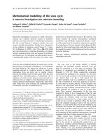
Fig. 1. Organization of a selection of strongly inducible pheromone-
responsive promoters. Schematic representation of the organization
of consensus PREs within nine of the 35 most strongly induced
pheromone response genes (excluding pseudogenes and genes
without obvious PREs), as identified by global expression analysis
(30 min of a-factor treatment) [6,7]. Numbers between any two
PREs indicate the spacing in nucleotides, whereas the number
furthest to the right indicates the distance to the translation start
site. The promoters are arranged in the relative order of inducibility
(top to bottom). STE12 is within the top 100 pheromone-inducible
genes, and was included here because we have examined this
promoter in some detail.
T C. Su et al. Organization of PREs for a pheromone response
FEBS Journal 277 (2010) 3235–3248 ª 2010 The Authors Journal compilation ª 2010 FEBS 3237
this same probe (not shown). By contrast, recombinant
Ste12 and Tec1, both produced in insect cells, are
capable of binding individually to an FRE-containing
oligonucleotide in vitro (Fig. 2C, lanes 1 and 2), and
form a higher-order complex when added together in
binding reactions (Fig. 2C, lane 3). This indicates that
recombinant Ste12, although capable of forming terni-
ary complexes with Tec1 in vitro, is excluded from
forming multimerized complexes with two closely-
spaced PREs in vitro, which indicates that the binding
of wild-type Ste12 to multiple PREs in vivo may
require additional factors or post-translational modifi-
cations. We are currently investigating the significance
of this feature with respect to the pheromone response,
and we discuss the implications of these observations
below.
To determine the stoichiometry of Ste12 bound to a
single PRE in vitro, we expressed a series of C-terminal
truncations for use in the analysis of hetero-complex
formation. Wild-type Ste12 protein produced in insect
cells (Fig. 3A, lane 1) or truncated versions of Ste12
containing residues 1–476 (Fig. 3A, lane 2), 1–350
(Fig. 3A, lane 3) or 1–215 (Fig. 3A, lane 4), produced
by in vitro transcription and translation, each were
capable of forming complexes with a single PRE-con-
taining oligo in EMSA. We then mixed the full-length
protein together with the truncated forms in vitro prior
to adding the labeled oligonucleotide probe and per-
forming EMSA. In these experiments, none of the
truncated species caused the production of an inter-
mediate complex in combination with wild-type Ste12
(Fig. 3A, lanes 5–7), which would be expected if there
were multiple protein molecules bound to a single
PRE. Because it is possible that co-translation of Ste12
may be necessary for hetero-complex formation, as is
the case with proteins such as GCN4 and GAL4
[24,25], we also performed this experiment using co-
translation of the truncated Ste12 derivatives (Fig. 3B).
We found that when the 1–476 and 1–350 or 1–350
and 1–215 derivatives are produced by co-translation
(Fig. 3B, lanes 4 and 8, respectively), we also do not
observe intermediate-sized complexes that would indi-
cate formation of hetero-multimers. From these
results, we argue that Ste12 protein likely binds to a
single PRE as a monomer.
Sequence requirement of the PRE for binding
Ste12 in vitro
The sequence requirements for binding of Ste12 to
DNA have largely been inferred from a comparative
AB
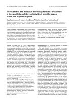
Fig. 3. Ste12 binds to a PRE as a monomer. (A) EMSA reactions
were performed with a labeled oligo containing a single PRE
(IS1430 ⁄ 1431) and full-length Ste12 (lane 1), Ste12 1–476 (lane 2),
Ste12 1–350 (lane 3) and Ste12 1–215 (lane 4). Full-length Ste12
was mixed with 1–476 (lane 5), 1–350 (lane 6) or 1–215 (lane 7)
prior to adding the labeled oligo and performing the binding reac-
tion. (B) Reactions were performed with in vitro translated Ste12
1–476 (lanes 1, 3 and 4), 1–350 (lanes 2, 3, 4, 5, 7 and 8) or 1–215
(lanes 6–8). The Ste12 derivatives were synthesized separately
in vitro and then mixed prior to EMSA (lanes 3 and 7) or were
co-translated (lanes 4 and 8).
AC
B
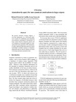
Fig. 2. Recombinant Ste12 produced in insect cells binds to a sin-
gle PRE in vitro. (A) EMSA reactions were performed with extracts
of Sf21 insect cells producing recombinant Ste12 protein (lanes
2–11) or uninfected cells (lane 1) using an oligonucleotide probe
containing two directly-repeated PREs (sites II and III from the
FUS1 promoter, S26D). Unlabeled oligonucleotide competitor oligos
were added at ten-fold molar excess (lanes 3–5), as indicated in
(B). The binding reaction in lane 6 contained a ten-fold molar excess
of an RBEIII oligonucleotide [37]. Antibodies against Ste12 (lanes
8–10) or preimmune serum (lane 11) were added to the binding
reactions. (C) Full-length recombinant Ste12 and Tec1 form a com-
plex on an FRE in vitro. EMSA reactions using a labeled FRE probe
(CN140 ⁄ 141) derived from the TY1 LTR were performed with
Ste12 (lane 1), Tec1-flag (lane 2) or both Ste12 and Tec-1 flag
(lanes 3 and 4). Anti-flag sera were added to the binding reaction in
lane 4.
Organization of PREs for a pheromone response T C. Su et al.
3238 FEBS Journal 277 (2010) 3235–3248 ª 2010 The Authors Journal compilation ª 2010 FEBS
analysis of pheromone-responsive promoters and geno-
mic localization of Ste12 protein in vivo [7,10,19]. To
characterize residues of the PRE that are necessary for
affinity of Ste12 in vitro, we performed a systematic
analysis using competitions with mutant oligonucleo-
tides in EMSA (Fig. 4A). Within the eight nucleotide
consensus (ATGAAACA), we found that mutation of
each of the residues impairs the ability to compete for
binding to the wild-type oligo (Fig. 4A, PRE mutants).
In particular, mutations of residues A5 and A6 of the
central AAA trinucleotide to G significantly impair
competition (Fig. 4A, lines 5 and 6), as does substitu-
tion of G3 with a pyrimidine (C or T) (Fig. 4A, line 2;
Table 1). We also compared the relative affinities of
the four PREs within the FUS1 promoter (Fig. 4B,
designated I, II, III and IV, 5¢–3¢, top). Amongst these,
site II is identical to the eight nucleotide consensus,
sites III and IV have substitutions of the outer 3¢ and
5¢ nucleotides, respectively, and site I has a substitu-
tion of A5 within the AAA trinucleotide. Using com-
petition experiments, we were able to rank the relative
strengths of PREs within the FUS1 promoter as sites
II, IV, III and I (Fig. 4B, strongest to weakest;
Table 1).
Because higher concentrations of recombinant Ste12
produce an autoinhibitory effect, we were unable to
determine affinity constants using EMSA with this
reagent. However, for each mutant oligonucleotide, we
calculated a relative competition strength (RCS) value,
which represents the ratio of competitor oligonucleo-
tide required to compete for 50% binding of total
Ste12 relative to the consensus oligonucleotide within
the same experiment (Fig. S1 and Table 1). From the
RCS values, we predict the relative contribution of
each nucleotide within the consensus PRE for binding
of wild-type Ste12 in vitro, as shown in Fig. 4C.
Relative affinity of Ste12 for PREs in vitro
correlates directly with the pheromone response
in vivo
To determine by how much the relative affinity of
Ste12 for PREs in vitro contributes to the pheromone
response in vivo, we inserted oligonucleotides bearing
the consensus or mutant PREs into a reporter with a
minimal GAL1 core promoter upstream of LacZ,
which were integrated in single copy at a lys2 disrup-
tion. We found that none of the PREs inserted individ-
ually upstream of the GAL1 core element were capable
of inducing a response to pheromone, even with the
strongest of the PREs from the FUS1 promoter (not
shown). By contrast, reporters with an insertion of two
identical directly-repeated PREs, in either orientation
relative to the transcriptional start site (not shown),
and arranged in the same context as FUS1 PREs II
and III (Fig. 4B), all produced a response to phero-
mone and, interestingly, the level of inducibility corre-
lated with the RCS values for the PREs as determined
in vitro (Fig. 5A). Accordingly, a duplicated PRE with
a substitution of residue A5 of the central AAA
A
B
C
Fig. 4. Nucleotides required for binding of full-length Ste12 to the
consensus PRE in vitro. (A) EMSA reactions were performed with
recombinant wild-type Ste12 and a labeled oligonucleotide bearing
a single consensus PRE (RS010 ⁄ 011). Binding reactions contained
no competitor (lane 1), or a 0.625- (lanes 2 and 7), 1.25- (lanes 3
and 8), 2.5- (lanes 4 and 9), 5- (lanes 5 and 10) or 10- (lanes 6 and
11) fold molar excess of unlabeled consensus oligo (lanes 2–6) or
the indicated mutant oligos (lanes 7–11). Mutant oligos (lines 1–7)
contained a single nucleotide substitution from the consensus PRE
(Table S1). (B) The sequence of the FUS1 promoter indicating the
position of four PREs (designated sites I, II, III and IV, 5¢–3¢). EMSA
reactions were performed as in (A) but using a labeled oligonucleo-
tide bearing PRE IV (IS1428 ⁄ 1429), and with the unlabeled compet-
itors as indicated. (C) The RCS was calculated for each mutant
oligo (Table 1). The effect that mutation of each nucleotide of the
consensus PRE has on the binding of Ste12 in vitro is indicated
proportional to the font size for each residue.
T C. Su et al. Organization of PREs for a pheromone response
FEBS Journal 277 (2010) 3235–3248 ª 2010 The Authors Journal compilation ª 2010 FEBS 3239
trinucleotide to G, which seriously inhibits binding of
Ste12 in vitro, produces a small but detectable level of
inducibility (Fig. 5A, line 4), whereas the duplicated
consensus PRE causes a level of pheromone response
comparable to the full FUS1 promoter (Fig. 5A, lines
1 and 5).
Because the inducibility of reporters bearing
two directly-repeated PREs appeared to be approxi-
mately proportional to the relative affinity for Ste12
in vitro, we were interested in determining the extent
that mutations of one PRE would have in combination
with a strong consensus element. To address this, we
introduced mutations of the central AAA trinucleotide
into the 3¢ PRE of the artificial reporter constructs.
Mutation of the central A5 residue of the trinucleotide,
causes an approximately three-fold reduction in phero-
mone inducibility in combination with a consensus
PRE (compare Fig. 5B, line 1, with Fig. 5A, line 1).
Mutation of two of the central A residues compro-
mises the response by approximately ten-fold (Fig. 5B,
line 2), and a PRE bearing substitution of all three A
residues completely prevents the response to phero-
mone (lines 3–5). The latter mutation also completely
prevents binding of Ste12 in vitro (not shown) and, in
effect, the reporters indicated in lines 4 and 5 of
Fig. 5B possess only a single functional PRE. We also
examined the effect that mutations in both directly-
repeated PREs have on pheromone response, and
observed that inducibility was reduced significantly
when both elements have mutations that limit binding
of Ste12 in vitro. For example, directly-repeated PREs
with substitutions of residues A1 and A8, respectively,
comprising mutations that have a relatively minor
effect on binding Ste12 in vitro, cause an approxi-
mately four-fold defect in inducibility relative to two
consensus PREs (Fig. 5C, line 5). Combinations of
PREs that have more serious defects in binding Ste12
produce proportionally less response (Fig. 5C, lines 6
and 7), although even two quite weak directly-repeated
PREs retain a detectable level of inducibility (Fig. 5C,
line 8). These results demonstrate that a significant
Table 1. RCS of mutant PREs for binding of wild-type Ste12 to a
PRE consensus (ATGAAACA) in vitro.
FUS1 PRE
a
Sequence RCS
b
II ATGAAACA 1.00
IV tTGAAACA 0.27
c
AaGAAACA 0.14
ATaAAACA 0.81
ATcAAACA 0.03
c
ATtAAACA 0.01
c
ATGcAACA 0.69
ATGgAACA 0.20
c
I ATGAgACA 0.02
c
ATGAAgCA 0.05
c
ATGAAAgA 0.30
III ATGAAACg 0.26
a
PREs represented in the FUS1 promoter (Fig. 4B).
b
RCS for each
oligo was calculated from the concentration of unlabeled competi-
tor oligonucleotide required to compete 50% of total Ste12 protein
bound to the consensus PRE, relative to competition in the same
experiment with a wild-type PRE (Fig. S1).
c
Concentrations of oligo
required for 50% competition was calculated by extrapolation.
A
B
C
Fig. 5. The pheromone response conferred by two directly-
repeated PREs in vivo is proportional to their relative affinity for
Ste12 in vitro. (A) Strains bearing single-copy integrations of a mini-
mal GAL1-LacZ reporter bearing two copies of the indicated PRE
(lines 1–4) were left untreated (red bars) or treated with a-factor for
60 min (blue bars) prior to harvesting the cells and assaying b-galac-
tosidase activity. The shading of the boxes containing the PRE
sequence indicates the relative competition strength for Ste12
in vitro, with the stronger PREs being shaded darker and the
weaker PREs shaded lighter. Line 5 shows results from a strain
bearing the full FUS1-LacZ promoter. (B) Reporter genes bearing a
consensus PRE and PREs containing substitutions of the central
AAA trinucleotide were assayed as in (A). (C) Combinations of
consensus PREs and PREs bearing the indicated mutations were
assayed in the same context as described above.
Organization of PREs for a pheromone response T C. Su et al.
3240 FEBS Journal 277 (2010) 3235–3248 ª 2010 The Authors Journal compilation ª 2010 FEBS
response to pheromone can be conferred by a single
strong consensus PRE in combination with much
weaker adjacent PREs, with a level of inducibility
proportional to the relative strength of the second
PRE. Additionally, duplicated PREs with substitutions
that inhibit Ste12 binding are capable of inducing a
response to pheromone, but at significantly lower
levels. Interestingly, when we examined the effect of
the combined RCS of two directly-repeated PREs on
the response to pheromone, we observed a direct and
simple linear relationship between the product of the
RCS values and pheromone responsiveness (Fig. 6).
This analysis indicates that, in the context of the mini-
mal GAL1 promoter, the limiting factor for transcrip-
tional activation in pheromone-treated cells appears to
be binding of Ste12 multimers to DNA.
Organizational constraints on multiple PREs for a
pheromone response
When examining the promoters of some of the most
strongly induced pheromone response genes (Fig. 1),
we noted that PREs are arranged in a variety of con-
figurations. Most promoters have PREs in a directly-
repeated orientation, although there are many
instances of PREs arranged in a tail-to-tail configura-
tion (PRM6, FUS1, AGA1 and STE12). Also, there is
considerable variability in spacing between multiple
PREs (Fig. 1). To examine the significance that these
differences in configuration have for pheromone
response, we compared the responses of a GAL1 mini-
mal promoter bearing two consensus PREs positioned
at different orientations with respect to each other
(Fig. 7). In the FUS1 promoter, two PREs (sites II
and III) are positioned in a directly-repeated orienta-
tion separated by three nucleotides (Fig. 7A, line 2)
(i.e. the same context as the experiments described
above). We found that inverting one of the PREs
such that they are positioned in a head-to-head orien-
tation completely prevented the response to phero-
mone (Fig. 7A, line 3). By contrast, two consensus
PREs from the STE12 promoter positioned in a tail-
to-tail configuration, separated by a single nucleotide,
caused considerably greater induction compared to the
directly-repeated PREs from FUS1 (Fig. 7A, line 1).
This indicates that there are severe organizational con-
straints for closely-positioned PREs that must limit
Fig. 6. The combined relative strength of two directly-repeated
PREs produces a proportionally linear response to pheromone.
A combined relative PRE strength for each of the reporter genes
described in Fig. 5 was calculated as log(RCS
PRE1
· RCS
PRE2
) and
plotted against the respective pheromone responsiveness for each
reporter (b-galactosidase activity (· 10
)3
).
A
B
Fig. 7. Organizational constraints on closely-spaced PREs for
pheromone response in vivo. (A) Pheromone responsiveness of
minimal promoters containing PREs II and III from the STE12 pro-
moter in a tail-to-tail orientation (line 1), directly-repeated consensus
PREs from the FUS1 promoter (PRE II, line 2) or with the second
consensus PRE inverted into a head-to-head orientation (line 3). (B)
The consensus PREs from the FUS1 promoter were moved apart
to produce an intervening spacing of ten (lines 7–9), 20 (lines 4–6)
or 40 (lines 1–3) nucleotides, with the orientation of the PREs as
indicated.
T C. Su et al. Organization of PREs for a pheromone response
FEBS Journal 277 (2010) 3235–3248 ª 2010 The Authors Journal compilation ª 2010 FEBS 3241
binding and activation by Ste12. We then examined
how the spacing between two directly-repeated consen-
sus PREs affects the observed response, and found
that they could not be moved apart without seriously
compromising induction (Fig. 7B). Separation of PREs
by even one nucleotide completely prevented induc-
tion, as did separation by three, five, seven (not
shown), 10 or 20 nucleotides (Fig. 7B, lines 4–9).
Curiously, however, two PREs spaced 40 nucleotides
apart in either a head-to-head or tail-to-tail orientation
produced a significant level of pheromone response
(Fig. 7B, lines 2 and 3, respectively). Taken together,
these results indicate that there must be structural con-
straints on Ste12 that allow binding to closely-spaced
PREs in several different configurations. Additionally,
the fact that head-to-head and tail-to-tail PREs sepa-
rated by 40 nucleotides allow induction implies that a
sufficient length of intervening DNA is required to
bend or twist into a conformation enabling an inter-
action between Ste12 proteins bound to these PREs.
We discuss the possible implications of these results
further below.
PREs from the STE12 promoter demonstrate
organizational constraints
To examine whether the organizational constraints that
we observe on artificially produced arrangements of
PREs are representative of pheromone-responsive pro-
moters in vivo, we examined the contribution of PREs
within the STE12 promoter, which contains four PREs:
three in the forward orientation and one in the reverse
orientation (Fig. 1, bottom). We found that a sub-frag-
ment bearing only the three 5¢ elements (sites I, II and
III) caused an elevated level of basal expression, which
is dependent upon STE12 (Fig. 8, basal expression,
compare lines 1 and 2) and, furthermore, that a sub-
fragment bearing only the inverted PREs II and III
could account for almost all pheromone inducibility of
the STE12 promoter (Fig. 8, pheromone induction, line
1, compare lines 1 and 4). Similarly, mutation of site I
had only a small negative effect on the response
(Fig. 8, line 3), whereas mutation of either sites II or
III completely prevented induction (Fig. 8, lines 5 and
6). These observations indicate that, although PREs
may be scattered throughout the promoters of phero-
mone-responsive genes, in some cases, the majority of
pheromone response may involve only two properly
spaced and oriented binding sites for Ste12.
Pheromone response of promoters with a single
consensus PRE
Considering the results reported above, we questioned
how it is possible that a number of genes amongst
those that are strongly induced by pheromone have
only a single consensus PRE (Fig. 1) [7]. CIK1, for
example, is one of the most strongly induced genes in
pheromone-treated cells, and apparently has only a
single consensus PRE. We examined the CIK1 pro-
moter to determine whether there were potential
weaker binding sites for Ste12 falling within the con-
straints that we observed on the artificial promoters
described above. Accordingly, we noted that the CIK1
PRE is positioned only three nucleotides downstream
of a PRE-like sequence with substitution at residues
T1 and A6 of the consensus (Figs 4C and 9A, top). A
portion of the CIK1 promoter bearing these elements
inserted upstream of a minimal promoter was found to
be strongly induced by pheromone, although deletion
Fig. 8. Orientation and spacing of PREs
contributing to response of the STE12
promoter. The sequence of the STE12
promoter region containing the three most
distal PREs (designated I, II, and III, 5¢–3¢)is
indicated. An oligonucleotide representing
this sequence, or bearing mutations or
deletions as indicated, was inserted
upstream of the minimal GAL1 core
promoter-LacZ reporter gene. The
expression of the reporter was measured in
untreated cells (basal expression, left) or
cells treated with a-factor for 60 min
(pheromone induction).
Organization of PREs for a pheromone response T C. Su et al.
3242 FEBS Journal 277 (2010) 3235–3248 ª 2010 The Authors Journal compilation ª 2010 FEBS
of the PRE-like sequence completely prevented the
response (Fig. 9A), indicating that this element does
contribute to induction by Ste12 multimers in vivo.
Similarly, on the PRM3 promoter, we observed the
PRE-like sequence 5¢-ATAAAACA-3¢ 36 nucleotides
upstream of the consensus PRE, positioned in a head-
to-head orientation (Fig. 9B). In vitro, we found that
an oligonucleotide bearing this sequence competes for
binding to Ste12 only slightly less efficiently than does
a consensus PRE (Table 1). A region including these
elements inserted upstream of the GAL1 core pro-
moter was responsive to pheromone (Fig. 9B, line 1),
although the response was reduced considerably when
the PRE-like sequence was deleted (Fig. 9B, line 2).
These results indicate that this PRE-like sequence can
produce a pheromone response by Ste12 multimers ori-
ented in a head-to-head conformation approximately
40 nucleotides away from a consensus PRE, and we
had demonstrated this effect with the artificial promo-
ters. Taken together, these results indicate that, for
some pheromone-responsive genes, Ste12 must activate
transcription from sub-optimal binding sites, in combi-
nation with a single consensus PRE whose arrange-
ment falls within specific organizational constrains.
A
B
Fig. 9. A single consensus PRE can confer pheromone responsive-
ness in conjunction with PRE-like sequences. (A) Sequence of the
CIK1 promoter region, indicting the consensus PRE and a PRE-like
sequence. An oligonucleotide representing this sequence, or bear-
ing a deletion of the PRE-sequence, was inserted upstream of the
minimal GAL1 core promoter-LacZ reporter, and expression was
measured in untreated and pheromone-treated cells. (B) Sequence
of the PRM3 promoter indicating the location of a consensus PRE
and PRE-like sequence. The pheromone responsiveness of the min-
imal promoter bearing oligonucleotides representing the wild-type
or mutant promoter sequences was measured in untreated and
pheromone-treated cells.
A
B
C
D
Fig. 10. Structural constraints on Ste12 for binding closely-posi-
tioned PREs. Schematic representation of a possible mechanism
for the recognition of closely-spaced PREs in different conforma-
tions by Ste12 multimers. Interaction with directly-repeated PREs,
positioned three nucleotides apart (A) or in a tail-to-tail orientation
(B) may involve an interaction with C-terminal sequences separated
from the N-terminal DNA binding domain by a flexible linker region.
Some closely-spaced configurations appear to be excluded from
binding Ste12 multimers, as in a closely-spaced head-to-head orien-
tation (C). Head-to-head and tail-to-tail orientations may be accom-
modated providing that the sites are separated sufficiently to allow
bending or twisting of the intervening DNA to enable binding of
Ste12 multimers (D).
T C. Su et al. Organization of PREs for a pheromone response
FEBS Journal 277 (2010) 3235–3248 ª 2010 The Authors Journal compilation ª 2010 FEBS 3243
We note, however, that we have only examined sub-
fragments for both of these promoters, and there are
likely to be additional factors that contribute to
response. In this vein, it is important to note that both
were shown to be Kar4-dependent [18].
Discussion
The pheromone response pathway of Saccharomyces
has provided an important model for understanding
how genes are regulated in response to signals trans-
mitted through MAP kinase cascades. However,
despite almost 20 years of intensive research, there
remain many unanswered questions regarding the func-
tion of Ste12, including the molecular mechanisms that
control its activity by upstream MAPKs, how it causes
transcriptional activation, and the nature of its interac-
tion with PREs on DNA. To begin addressing the lat-
ter issue, we have performed a systematic analysis of
Ste12 binding to the PRE in vitro, and studied the rela-
tionship between binding affinity and spatial orienta-
tion between two PREs for pheromone responsiveness
in vivo. Ste12 likely binds to a single PRE in vitro as a
monomer, and therefore the protein must require mul-
timerization in vivo to bind DNA and activate the
haploid-specific pheromone response because a mini-
mum of two PREs are required.
Surprisingly, based on analysis of artificial promot-
ers containing two PREs, there appear to be serious
constraints with respect to how these can be positioned
relative to one another to enable pheromone response
of an artificial promoter. Two directly-repeated PREs
cause activation only when located within three nucle-
otides of each other. By contrast, PREs inverted in a
tail-to-tail conformation separated by a single nucleo-
tide produce a very strong response. Additionally,
PREs oriented in head-to-head or tail-to-tail configura-
tions are only able to cause a pheromone response
when separated by approximately 40 nucleotides.
Taken together, these observations indicate that Ste12
must have structural features that can accommodate
multimerization for binding of closely-spaced sites
oriented in several different conformations (Fig. 10),
such that binding to closely-positioned PREs in either
a directly-repeated (Fig. 10A) or tail-to-tail conforma-
tion (Fig. 10B) may form multimers through interac-
tion between surfaces on the Ste12 protein that are
separated from the DNA-binding domain by a flexible
linker in order to accommodate different orientations.
Because PREs oriented in a head-to-head manner do
not produce a response, the flexibility of Ste12 may
not be able to accommodate this particular orienta-
tion, or perhaps the N-terminal DNA binding domain
is sterically precluded from such an interaction
(Fig. 10C). PREs oriented in either a head-to-head or
tail-to-tail conformation are capable of inducing a
pheromone response if positioned 40 nucleotides apart
(i.e. approximately four helical turns of DNA), sug-
gesting that Ste12 is capable of forming multimers that
can bind these configurations, provided that the inter-
vening DNA is able to bend or twist into a conforma-
tion that can accommodate the interaction (Fig. 10D).
An additional possibility is that Ste12 multimerization
in vivo, enabling accommodation of various PRE
arrangements, may require additional nuclear factors.
Accordingly, Ste12 was shown to associate on phero-
mone response promoters in vivo with both inhibitor
proteins Dig1 and Dig2 [2], and so it is possible these
proteins facilitate the binding of Ste12 to PREs
arranged in various configurations. However, we con-
sider this to be unlikely considering that the activation
of Ste12-dependent genes appears to be constitutive in
dig1 dig2 null strain backgrounds [3–5], presumably
including genes requiring a variety of PRE orientations
for a pheromone response.
Curiously, recombinant wild-type Ste12 produced in
insect cells is incapable of forming multimers on oligos
containing two PREs in vitro, despite the fact that the
same arrangement of PREs confers a strong response
to pheromone in vivo. Furthermore, full-length Ste12
appears to have an autoinhibitory function because
high concentrations of protein completely prevent
binding to DNA. Because the deletion of the C-terminus
prevents these effects (not shown), we suggest that
multimerization of Ste12 in vivo must be regulated
through a mechanism involving the C-terminus. Ste12
produced in insect cells becomes phosphorylated on
most of the same residues that we have observed in
yeast [26,27], and we find that mild treatment with
phosphatase in vitro produces slower migrating com-
plexes with oligos containing two PREs (T C. Su and
I. Sadowski, unpublished results), suggesting that
phosphorylation may regulate the ability to bind multi-
ple adjacent PREs. By contrast, recombinant wild-type
Ste12 does produce terniary complexes with Tec1 on
an FRE-containing oligo in vitro (Fig. 2C). These
results suggest that activation of haploid-specific pher-
omone-responsive genes, but not Ste12 ⁄ Tec1-respon-
sive genes, may require additional regulation in vivo
involving dephosphorylation. The results obtained in
the present study also raise the important question of
why two PREs are required for pheromone response if
wild-type Ste12 is able to bind to a single PRE in vitro.
This indicates that either the activation domain of
Ste12 is incapable of activating transcription when
bound to a single site, or that binding to a single site
Organization of PREs for a pheromone response T C. Su et al.
3244 FEBS Journal 277 (2010) 3235–3248 ª 2010 The Authors Journal compilation ª 2010 FEBS
in vivo is limited by additional factors. Consistent with
the latter possibility, it was shown that Ste12 does not
interact with filamentous response promoters (contain-
ing a single PRE) in the absence of Tec1 [2], indicating
that Ste12 is prevented from binding a single PRE
in vivo on its own. This effect is likely mediated by the
inhibitor proteins Dig1 and ⁄ or Dig2 [3,28] and, consis-
tent with this, we found that binding of wild-type
Ste12 to a single PRE in vitro is inhibited by the addi-
tion of recombinant Dig1 and Dig2 (T C. Su and
I. Sadowski, unpublished results).
We have systematically examined nucleotides within
the PRE by mutagenesis, and have compared the rela-
tive affinity of natural sites within the FUS1 promoter
for binding of wild-type Ste12 in vitro. Using an artifi-
cial reporter bearing two PREs arranged in a directly-
repeated orientation, we find that there is a significant
and simple linear relationship between the combined
relative strength of the two PREs in vitro and the level
of pheromone responsiveness in vivo (Fig. 6). This sug-
gests that, in pheromone-treated cells, using a concen-
tration of pheromone where presumably Ste12 is free
of inhibition by the regulatory proteins Dig1 and Dig2
[2], the association of Ste12 protein with cis-elements
on DNA is probably the limiting interaction for induc-
tion, at least in the context of our artificial promoters.
However, we envisage that many, if not most, natural
promoters controlled by Ste12 will also be subject to
the additional effects of nucleosome positioning, which
likely would significantly alter the effects produced by
combinations of PREs with different affinities for
Ste12 protein, as previously shown for transcriptional
activation by Pho4 [29,30].
Upon cursory examination of the most strongly
induced pheromone-responsive promoters in vivo,it
could not be predicted that there should be such severe
constraints on the organization of PREs for induction
by pheromone (Fig. 1). Most of these promoters
appear to have PREs arranged without any particular
defined conformation, some promoters appear to only
have a single PRE, and other pheromone-responsive
promoters have none (not shown in Fig. 1). On the
basis of the results obtained in the present study, we
expect that many pheromone-responsive genes must
rely on nonconsensus weaker binding sites for Ste12,
which are positioned adjacent to consensus PREs in a
conformation that can accommodate the binding of
Ste12 multimers. We have detailed such instances on
sub-fragments of the CIK1 and PRM3 promoters
(Fig. 9). Both of these genes are also regulated by
Kar4 [18], and it will be interesting to determine how
these factors interact within the context of their full
promoters to promote induction during pheromone
response. On several promoters, including FUS1 and
STE12, we find that only two PREs oriented in an
optimal configuration can account for the majority of
pheromone response, and this suggests that many
genes strongly induced by pheromone may only
require two properly oriented PREs. Many phero-
mone-responsive promoters bear consensus PREs posi-
tioned some distance apart, and the results obtained in
the present study indicate that two consensus elements
oriented in a head-to-head or tail-to-tail orientation at
least 40 nucleotides apart can confer a significant
response. Such configurations are observed on many
natural pheromone-responsive promoters, including
FUS3 and PRM6 (Fig. 1). Furthermore, PRE I of the
FUS1 promoter is oriented in a tail-to-tail conforma-
tion with respect to the three more proximal sites (II,
III, and IV) and, consequently, this may allow activa-
tion by Ste12 multimers from any combination of
these proximal three sites. Several other promoters,
with either a single consensus PRE or with two PREs
in orientations that should occlude a pheromone
response based on our data, have potential weaker
Ste12 binding sites positioned in a tail-to-tail orienta-
tion. We find such examples within the FIG1 and
PRM4 promoters (Fig. 1).
There also genes that are strongly induced by phero-
mone but appear to lack a consensus PRE, including
many of the PRMs, ASG7, FIG2, FIG3, ECM18 and
MCH2 (not shown). In these cases, Ste12 must activate
from multiple nonconsensus binding sites or through
cooperative interaction on weaker elements with addi-
tional DNA binding proteins, such as Mcm1 [16,31]
and Kar4 [18], and perhaps with previously unrecog-
nized additional factors. Consistent with this possibil-
ity, it was recently shown that there is a strong
correlation between the association of Ste12 on phero-
mone-responsive promoters with potential binding sites
for Flo8, suggesting that pheromone response for
many genes may involve an association between these
factors [7]. Accordingly, it is interesting that the func-
tion of Ste12 with respect to activating transcription in
response to the pheromone-response MAPK pathway
is remarkably similar to TFII-I, which is a protein in
mammalian cells that performs this function in
response to MAPK signaling downstream of RAS
through cooperative interactions on upstream elements
with a number of factors, including serum response
factor, PHOX1, nuclear factor-jB and upstream stimu-
latory factor [32,33].
The results reported in the present study demonstrate
that many aspects of Ste12 regulation at the molecular
level are still not well understood. This protein appears
to bind as a monomer to a single PRE in vitro,
T C. Su et al. Organization of PREs for a pheromone response
FEBS Journal 277 (2010) 3235–3248 ª 2010 The Authors Journal compilation ª 2010 FEBS 3245
although at least two properly configured PREs are
necessary for a pheromone response in vivo. It will be
important to elucidate the structural features of Ste12
that impose these restrictions, as well as the mecha-
nisms controlling the interaction of this factor with
multiple PREs in vivo to mediate pheromone response.
Materials and methods
Oligoucleotides, plasmids and yeast strains
Sequences of oligonucleotides for construction of minimal
promoter reporters are detailed in Table S2. Oligonucleo-
tides for construction of reporter genes were annealed and
cloned into the XhoI ⁄ XbaI sites of pIS341, which is a lys2
disintegrator vector [34], bearing the GAL1 core promoter
region upstream of LacZ and the ADH1 terminator. All
experiments were performed in a W303-1A strain back-
ground (MATa ade2 leu2 trp1 ura3 can1). Reporter gene
plasmids were linearized by digestion with NruI prior to
transformation into yeast using the LiAc technique [35].
URA
+
transformants were allowed to grow nonselectively
on yeast extract peptone dextrose for 3 days to allow rear-
rangement of the disintegrator, prior to streaking for single
colonies on 5-fluoroorotic acid. Strains bearing reporter
gene integrants at the lys2 disruption were identified by rep-
lica plating, and single copy integration was verified by
analysis of chromosomal DNA using PCR [34]. The phero-
mone responsiveness of strains bearing the reporter genes
was assayed in cultures grown in yeast extract peptone dex-
trose until A
600
of 0.6 was reached. Pheromone was added
at a concentration of 2 lgÆ mL
)1
. The cells were collected
and b-galactosidase activity was assayed as described previ-
ously [36]; the results represent an average of three indepen-
dent experiments.
Recombinant proteins and EMSA
Full-length Ste12 protein was expressed as an N-terminal
6-His fusion in insect cells using baculovirus in the Sf21
insect cell line [26]. Tec1 was expressed with a 6-His-N-ter-
minal and C-terminal flag epitope tag using the Bac-to-Bac
system (Invitrogen, Carlsbad, CA, USA). Antibodies A3,
B3 and F3 were raised against Escherichia coli TrpE fused
to Ste12 residues (265–688), (314–688) and (1–215), respec-
tively. Sf21 cells infected with Ste12 and Tec1 virus were
collected and washed in ice-cold lysis buffer (20 mm Tris,
pH 8.0, 40 mm NaCl, 1 mm dithiothreitol, 5% glycerol,
2.5 mm MgCl
2
,1mm Na
3
VO
4
,5mm EGTA, 50 mm NaF
and 20 mm b-glycerol phosphate). The cells were lysed by
forcing through a 27-gauge needle ten times, and then soni-
cated for 10 s. A clarified supernatant was obtained by cen-
trifugation at 10 000 g for 10 min. Ste12 proteins were
produced by in vitro transcription and translation using the
TNT T7 Quick Coupled Transcription⁄ Translation System
(Promega, Madison, WI, USA). Briefly, plasmid pSC4,
which contains a full-length genomic clone of STE12, was
used as template for amplification with oligonucleotide
oIS1144, in combination with oVT2, oET30 and oIS1146
(Table S3), to produce fragments with a 5¢ T7 RNA poly-
merase promoter and encoding Ste12 (1–215), Ste12 (1–350)
and Ste12 (1–479), respectively. The Ste12 derivatives were
produced individually or by co-translation in 50 l L reac-
tions containing 1 lL of T7 RNA polymerase and 40 lLof
rabbit reticulocyte lysate. The reactions were carried out at
30 °C for 90 min and then assayed immediately for DNA
binding activity.
Oligonucleotides used for EMSA are detailed in
Table S1, and were annealed and labeled using Klenow
(New England Biolabs, Beverly, MA, USA) with
[
32
P]adATP and [
32
P]adTTP, as described previously [37].
The 5¢ overhangs of unlabeled competitor oligonucleotides
were filled in using Klenow and an unlabeled dNTP mix-
ture. EMSA reactions contained 1 lL of labeled oligonu-
cleotide probe (2 pmol), 2 l g of poly(dI-dC) (Sigma,
St Louis, MO, USA), 2.5 m m MgCl
2
, 1% glycerol, 20 mm
Tris (pH 8.0), 40 mm NaCl and 1 l L of Sf21 extract or
in vitro translation reaction in a total volume of 20 lL.
Labeled oligonucleotide probes were added to the binding
reactions after a 30 min pre-incubation on ice with unla-
beled competitor oligos or specific antibodies. Binding reac-
tions were performed at room temperature for 30 min and
the reactions were resolved on nondenaturing polyacryl-
amide gels containing 0.5 · TBE (89 mm Tris, 89 mm Boric
acid, 2 mm EDTA, pH 8.0) buffer and 1% glycerol at
200 V for 3 h. Signals produced in the EMSA reactions
were quantitated using imagequant software (GE Health-
care, Milwaukee, WI, USA).
Acknowledgements
We thank LeAnn Howe, Mike Kobor, Viven Measday,
Sheetal Raithatha and Kris Barretto for their helpful
comments. This research was supported by funds from
the Canadian Cancer Society Research Institute (grant
018436).
References
1 Fields S, Chaleff DT & Sprague GF Jr (1988) Yeast
STE7, STE11, and STE12 genes are required for expres-
sion of cell-type-specific genes. Mol Cell Biol 8, 551–556.
2 Chou S, Lane S & Liu H (2006) Regulation of mating
and filamentation genes by two distinct Ste12 complexes
in Saccharomyces cerevisiae. Mol Cell Biol 26, 4794–
4805.
3 Tedford K, Kim S, Sa D, Stevens K & Tyers M (1997)
Regulation of the mating pheromone and invasive
Organization of PREs for a pheromone response T C. Su et al.
3246 FEBS Journal 277 (2010) 3235–3248 ª 2010 The Authors Journal compilation ª 2010 FEBS
growth responses in yeast by two MAP kinase sub-
strates. Curr Biol 7, 228–238.
4 Bardwell L, Cook JG, Zhu-Shimoni JX, Voora D &
Thorner J (1998) Differential regulation of transcrip-
tion: repression by unactivated mitogen-activated pro-
tein kinase Kss1 requires the Dig1 and Dig2 proteins.
Proc Natl Acad Sci USA 95, 15400–15405.
5 Cook JG, Bardwell L, Kron SJ & Thorner J (1996)
Two novel targets of the MAP kinase Kss1 are negative
regulators of invasive growth in the yeast Saccharomy-
ces cerevisiae. Genes Dev 10, 2831–2848.
6 Roberts CJ, Nelson B, Marton MJ, Stoughton R,
Meyer MR, Bennett HA, He YD, Dai H, Walker WL,
Hughes TR et al. (2000) Signaling and circuitry of mul-
tiple MAPK pathways revealed by a matrix of global
gene expression profiles. Science 287, 873–880.
7 Zheng W, Zhao H, Mancera E, Steinmetz LM & Snyder
M (2010) Genetic analysis of variation in transcription
factor binding in yeast. Nature 464, 1187–1191.
8 Wittenberg C & La Valle R (2003) Cell-cycle-regulatory
elements and the control of cell differentiation in the
budding yeast. Bioessays 25, 856–867.
9 Gagiano M, Bauer FF & Pretorius IS (2002) The sens-
ing of nutritional status and the relationship to filamen-
tous growth in Saccharomyces cerevisiae. FEMS Yeast
Res 2, 433–470.
10 Borneman AR, Leigh-Bell JA, Yu H, Bertone P,
Gerstein M & Snyder M (2006) Target hub proteins
serve as master regulators of development in yeast.
Genes Dev 20, 435–448.
11 Zaman S, Lippman SI, Zhao X & Broach JR (2008)
How Saccharomyces responds to nutrients. Annu Rev
Genet 42, 27–81.
12 Orlova M, Ozcetin H, Barrett L & Kuchin S (2009)
Roles of the Snf1-activating kinases during nitrogen
limitation and pseudohyphal differentiation in
Saccharomyces cerevisiae. Eukaryot Cell 9, 208–214.
13 Hagen DC, McCaffrey G & Sprague GF Jr (1991)
Pheromone response elements are necessary and suffi-
cient for basal and pheromone-induced transcription of
the FUS1 gene of Saccharomyces cerevisiae. Mol Cell
Biol 11, 2952–2961.
14 Sengupta P & Cochran BH (1991) MAT alpha 1 can
mediate gene activation by a-mating factor. Genes Dev
5, 1924–1934.
15 Madhani HD & Fink GR (1997) Combinatorial control
required for the specificity of yeast MAPK signaling.
Science 275, 1314–1317.
16 Yuan YO, Stroke IL & Fields S (1993) Coupling of cell
identity to signal response in yeast: interaction between
the alpha 1 and STE12 proteins. Genes Dev 7, 1584–
1597.
17 Gavrias V, Andrianopoulos A, Gimeno CJ &
Timberlake WE (1996) Saccharomyces cerevisiae TEC1
is required for pseudohyphal growth. Mol Microbiol 19,
1255–1263.
18 Lahav R, Gammie A, Tavazoie S & Rose MD (2007)
Role of transcription factor Kar4 in regulating
downstream events in the Saccharomyces cerevisiae
pheromone response pathway. Mol Cell Biol 27,
818–829.
19 Zeitlinger J, Simon I, Harbison CT, Hannett NM,
Volkert TL, Fink GR & Young RA (2003) Program-
specific distribution of a transcription factor dependent
on partner transcription factor and MAPK signaling.
Cell 113, 395–404.
20 Lefrancois P, Euskirchen GM, Auerbach RK, Rozow-
sky J, Gibson T, Yellman CM, Gerstein M & Snyder
M (2009) Efficient yeast ChIP-Seq using multiplex
short-read DNA sequencing. BMC Genomics 10, 37.
21 Kohler T, Wesche S, Taheri N, Braus GH & Mosch
HU (2002) Dual role of the Saccharomyces cerevisiae
TEA ⁄ ATTS family transcription factor Tec1p in regula-
tion of gene expression and cellular development.
Eukaryot Cell 1, 673–686.
22 Fields S & Herskowitz I (1985) The yeast STE12 prod-
uct is required for expression of two sets of cell-type
specific genes. Cell 42, 923–930.
23 Yuan YL & Fields S (1991) Properties of the DNA-
binding domain of the Saccharomyces cerevisiae STE12
protein. Mol Cell Biol 11, 5910–5918.
24 Carey M, Kakidani H, Leatherwood J, Mostashari F &
Ptashne M (1989) An amino-terminal fragment of
GAL4 binds DNA as a dimer. J Mol Biol 209, 423–432.
25 Hope IA & Struhl K (1987) GCN4, a eukaryotic tran-
scriptional activator protein, binds as a dimer to target
DNA. EMBO J 6, 2781–2784.
26 Nelson C, Goto S, Lund K, Hung W & Sadowski I
(2003) Srb10 ⁄ Cdk8 regulates yeast filamentous growth
by phosphorylating the transcription factor Ste12.
Nature 421, 187–190.
27 Hung W, Olson KA, Breitkreutz A & Sadowski I
(1997) Characterization of the basal and pheromone-
stimulated phosphorylation states of Ste12p. Eur J
Biochem 245, 241–251.
28 Olson KA, Nelson C, Tai G, Hung W, Yong C, Astell
C & Sadowski I (2000) Two regulators of Ste12p inhibit
pheromone-responsive transcription by separate mecha-
nisms. Mol Cell Biol 20, 4199–4209.
29 Kim HD & O’Shea EK (2008) A quantitative model of
transcription factor-activated gene expression. Nat
Struct Mol Biol 15, 1192–1198.
30 Lam FH, Steger DJ & O’Shea EK (2008) Chromatin
decouples promoter threshold from dynamic range.
Nature 453, 246–250.
31 Hwang-Shum JJ, Hagen DC, Jarvis EE, Westby CA &
Sprague GF Jr (1991) Relative contributions of MCM1
and STE12 to transcriptional activation of a- and
T C. Su et al. Organization of PREs for a pheromone response
FEBS Journal 277 (2010) 3235–3248 ª 2010 The Authors Journal compilation ª 2010 FEBS 3247
alpha-specific genes from Saccharomyces cerevisiae. Mol
Gen Genet 227, 197–204.
32 Chen J, Malcolm T, Estable MC, Roeder RG &
Sadowski I (2005) TFII-I regulates induction of
chromosomally integrated human immunodeficiency
virus type 1 long terminal repeat in cooperation with
USF. J Virol 79, 4396–4406.
33 Kim DW, Cheriyath V, Roy AL & Cochran BH (1998)
TFII-I enhances activation of the c-fos promoter
through interactions with upstream elements. Mol Cell
Biol 18, 3310–3320.
34 Sadowski I, Su TC & Parent J (2007) Disintegrator
vectors for single-copy yeast chromosomal integration.
Yeast 24, 447–455.
35 Gietz RD & Schiestl RH (2007) High-efficiency yeast
transformation using the LiAc ⁄ SS carrier DNA ⁄ PEG
method. Nat Protoc 2, 31–34.
36 Amberg DC, Burke DJ & Strathern JN (2005) Assay of
b-galactosidase in yeast: assay of crude extracts. In
Methods in Yeast Genetics. Cold Spring Harbor Labora-
tory Press, Cold Spring Harbor, NY.
37 Chen J, Malcolm T, Estable M, Roeder R & Sadowski
I (2005) TFII-I regulates induction of chromosomally
integrated human immunodeficiency virus type 1 long
terminal repeat in cooperation with USF. J Virol 79,
4396–4406.
Supporting information
The following supplementary material is available:
Fig. S1. Graphical representation of competition
experiments for Ste12 DNA binding in EMSA.
Table S1. Annealed double-stranded oligonucleotides
for use as probes and competitors in EMSA reactions.
Table S2. Annealed oligonucleotides used for construc-
tion of reporter gene plasmids and yeast strains.
Table S3. Oligonucletides for production of templates
for in vitro transcription and translation reactions.
This supplementary material can be found in the
online version of this article.
Please note: As a service to our authors and readers,
this journal provides supporting information supplied
by the authors. Such materials are peer-reviewed and
may be re-organized for online delivery, but are not
copy-edited or typeset. Technical support issues arising
from supporting information (other than missing files)
should be addressed to the authors.
Organization of PREs for a pheromone response T C. Su et al.
3248 FEBS Journal 277 (2010) 3235–3248 ª 2010 The Authors Journal compilation ª 2010 FEBS