Báo cáo khoa học: The N-terminal hybrid binding domain of RNase HI from Thermotoga maritima is important for substrate binding and Mg2+-dependent activity pot
Bạn đang xem bản rút gọn của tài liệu. Xem và tải ngay bản đầy đủ của tài liệu tại đây (1.33 MB, 16 trang )
The N-terminal hybrid binding domain of RNase HI from
Thermotoga maritima is important for substrate binding
and Mg
2+
-dependent activity
Nujarin Jongruja
1
, Dong-Ju You
1
, Eiko Kanaya
1
, Yuichi Koga
1
, Kazufumi Takano
1,2
and
Shigenori Kanaya
1
1 Department of Material and Life Science, Graduate School of Engineering, Osaka University, Japan
2 CRESTO, JST, Osaka, Japan
Introduction
Ribonuclease H (RNase H; EC 3.1.26.4) is an enzyme
that specifically cleaves RNA of RNA⁄ DNA hybrids
[1]. It requires divalent metal ions, such as Mg
2+
and
Mn
2+
, for activity. RNase H is widely present in bac-
teria, archaea and eukaryotes. These RNase H are
involved in DNA replication, repair and transcription
Keywords
cleavage site specificity; hybrid binding
domain; metal preference; RNase H;
substrate binding affinity;
Thermotoga maritima
Correspondence
S. Kanaya, Department of Material and Life
Science, Graduate School of Engineering,
Osaka University, 2-1, Yamadaoka, Suita,
Osaka 565-0871, Japan
Fax: +81 6 6879 7938
Tel.: +81 6 6879 7938
E-mail:
(Received 18 June 2010, revised 6 August
2010, accepted 27 August 2010)
doi:10.1111/j.1742-4658.2010.07834.x
Thermotoga maritima ribonuclease H (RNase H) I (Tma-RNase HI) con-
tains a hybrid binding domain (HBD) at the N-terminal region. To analyze
the role of this HBD, Tma-RNase HI, Tma-W22A with the single mutation
at the HBD, the C-terminal RNase H domain (Tma-CD) and the N-termi-
nal domain containing the HBD (Tma-ND) were overproduced in Escheri-
chia coli, purified and biochemically characterized. Tma-RNase HI prefers
Mg
2+
to Mn
2+
for activity, and specifically loses most of the Mg
2+
-depen-
dent activity on removal of the HBD and 87% of it by the mutation at the
HBD. Tma-CD lost the ability to suppress the RNase H deficiency of an
E. coli rnhA mutant, indicating that the HBD is responsible for in vivo
RNase H activity. The cleavage-site specificities of Tma-RNase HI are not
significantly changed on removal of the HBD, regardless of the metal
cofactor. Binding analyses of the proteins to the substrate using surface
plasmon resonance indicate that the binding affinity of Tma-RNase HI is
greatly reduced on removal of the HBD or the mutation. These results
indicate that there is a correlation between Mg
2+
-dependent activity and
substrate binding affinity. Tma-CD was as stable as Tma-RNase HI,
indicating that the HBD is not important for stability. The HBD of
Tma-RNase HI is important not only for substrate binding, but also for
Mg
2+
-dependent activity, probably because the HBD affects the interaction
between the substrate and enzyme at the active site, such that the scissile
phosphate group of the substrate and the Mg
2+
ion are arranged ideally.
Abbreviations
Bha-RNase HI, Bacillus halodurans RNase HI; Bst-RNase HIII, Bacillus stearothermophilus RNase HIII; Bsu-RNase HII, Bacillus subtilis RNase
HII; D13-R4-D12 ⁄ D29, 29 bp DNA
13
-RNA
4
-DNA
12
⁄ DNA duplex; D15-R1-D13 ⁄ D29, 29 bp DNA
15
-RNA
1
-DNA
13
⁄ DNA duplex; Eco-RNase HI,
Escherichia coli RNase HI; Eco-RNase HII, Escherichia coli RNase HII; GdnHCl, guanidine hydrochloride; HBD, hybrid binding domain; HIV-1
RNase H, RNase H domain of HIV-1 reverse transcriptase; Hsa-RNase H1, human RNase H1; IPTG, isopropyl thio-b-
D-galactoside; MMLV
RNase H, Moloney murine leukemia virus reverse transcriptase; R12 ⁄ D12, 12 bp RNA ⁄ DNA hybrid; R29 ⁄ D29, 29 bp RNA ⁄ DNA hybrid;
R9-D9 ⁄ D18, 18 bp RNA
9
-DNA
9
⁄ DNA duplex; RNase H, ribonuclease H; Sce-RNase H1, Saccharomyces cerevisiae RNase H1; Sto-RNase HI,
Sulfolobus tokodaii RNase HI; Tma-CD, C-terminal catalytic domain (residues 64–223) of Tma-RNase HI; Tma-ND, N-terminal domain
(residues 1–63) of RNase HI from Thermotoga maritima containing HBD; Tk-RNase HII, Thermococcus kodakaraensis RNase HII.
4474 FEBS Journal 277 (2010) 4474–4489 ª 2010 The Authors Journal compilation ª 2010 FEBS
[2–6]. In antisense therapy, RNase H is involved in the
recognition and cleavage of a disease-causative mRNA
[7]. Mutations in human RNase H2, which do not
necessarily significantly affect the activity [8], cause
severe neurological disorder termed Aicardi–Goutieres
syndrome [9]. RNase H is also present in retroviruses
as a C-terminal domain of reverse transcriptase. Retro-
viral RNases H are required for the conversion of sin-
gle-stranded genomic RNA into double-stranded
DNA, which is an initial step of viral proliferation,
and are therefore regarded as one of the targets for
AIDS therapy [10].
RNases H are classified into two major families
(type 1 and type 2 RNases H) based on the difference
in their amino acid sequences [11]. Four acidic active-
site residues are fully conserved in these RNases H,
except that from compost metagenome [12], and their
geometrical configurations are well conserved [13].
According to the crystal structures of the C-terminal
catalytic domains of Bacillus halodurans RNase HI
(Bha-RNase HI) [14] and human RNase H1 [15] in
complex with the RNA ⁄ DNA substrate, type 1 RNase
H binds to the minor groove of the substrate, such
that one depression containing the active site interacts
with the RNA backbone and the other depression con-
taining the phosphate-binding pocket interacts with
the DNA backbone. These two depressions are sepa-
rated by a ridge, which is composed of three highly
conserved Asn ⁄ Gln residues. Because two metal ions
are coordinated by the four acidic active site residues,
the scissile phosphate group of the substrate and water
molecules, a two-metal ion catalysis mechanism has
been proposed for RNase H [14,16,17]. According to
this mechanism, one metal ion is required for sub-
strate-assisted nucleophile formation and product
release, whereas the other is required to destabilize the
enzyme–substrate complex and thereby promote the
phosphoryl transfer reaction.
Thermotoga maritima is a strictly anaerobic, extre-
mely thermophilic eubacterium, isolated from various
geothermally heated locales on the sea floor, and
grows in the temperature range 55–90 °C with an opti-
mum at 80 °C [18]. Its genome sequence has been
determined previously [19]. The genome contains single
rnhA and single rnhB genes, encoding type 1 (Tma-
RNase HI; accession no. AAD36370) and type 2
(Tma-RNase HII; accession no. AAD35996) RNases
H, respectively. Tma-RNase HI is composed of 223
amino acid residues and contains a hybrid binding
domain (HBD) at the N-terminal region. Without this
domain, Tma-RNase HI shows relatively low (£ 20%)
amino acid sequence identities to any one of the repre-
sentative members of type 1 RNases H, which have
been biochemically characterized. Therefore, it would
be informative to characterize Tma-RNase HI and
compare its biochemical properties with those of other
type 1 RNases H.
HBD, previously termed as double-stranded RNA
and hybrid binding domain [20], consists of approxi-
mately 40 amino acid residues and is commonly
present at the N-terminal regions of eukaryotic type 1
RNases H (RNase H1) [21]. According to the crystal
structure of the HBD of human RNase H1 in complex
with the RNA ⁄ DNA substrate, HBD consists of a
three-stranded anti-parallel b-sheet (b1–b3) and two
helices (aA and aB) [22]. It binds to the minor groove
of the substrate, such that a loop between aA and b3
interacts with the RNA backbone and a positively-
charged depression interacts with the DNA backbone.
The importance of HBD with respect to substrate
binding has been reported for yeast [20,23], mouse [24]
and human [22,25,26] RNases H1. The requirement of
HBD for processivity [24] and positional preference
[25,26] has also been reported for mouse and human
RNases H1, respectively.
HBD is also present in several bacterial type 1
RNases H, including Tma-RNase HI, Bha-RNase HI
and RBD-RNase HI from Shewanella sp. SIB1 [13].
However, it remains to be determined whether these
HBDs have a role similar to those of eukaryotic
RNases H1, although the isolated HBD from SIB1
RBD-RNases HI, which is renamed as SIB1 HBD-
RNase HI in the present study, has been reported to
bind to the RNA ⁄ DNA substrate [27]. Attempts to
overproduce the SIB1 HBD-RNase HI derivative lack-
ing the HBD have so far been unsuccessful, probably
as a result of the instability of the protein (T. Tadok-
oro, unpublished data). In the present study, we over-
produced, purified and biochemically characterized
Tma-RNase HI and its derivatives lacking the HBD or
RNase H domain. On the basis of the results obtained,
we discuss the role of the HBD from Tma-RNase HI.
Results
Protein preparations
The amino acid sequence of Tma-RNase HI is com-
pared with those of the representative members of type
1 RNases H, Bha-RNase HI, HBD-RNase HI from
Shewanella sp. SIB1, Saccharomyces cerevisiae RNase
H1 (Sce-RNase H1), human RNase H1 (Hsa-RNase
H1), E. coli RNase HI (Eco-RNase HI) and the
RNase H domain of HIV-1 reverse transcriptase (HIV-
1 RNase H) in Fig. 1. The HBD of Tma-RNase HI
shows relatively high amino acid sequence identities of
N. Jongruja et al. Role of HBD from T. maritima RNase HI
FEBS Journal 277 (2010) 4474–4489 ª 2010 The Authors Journal compilation ª 2010 FEBS 4475
43%, 42%, 33% and 32% with respect to those of
Sce-RNase H1, Bha-RNase HI, SIB1 HBD-RNase HI
and Hsa-RNase H1, whereas the RNase H domain of
Tma-RNase HI shows relatively low amino acid
sequence identities of 20% to Hsa-RNase H1, 19% to
Eco-RNase HI, 18% to SIB1 HBD-RNase HI, Sce-
RNase H1 and Bha-RNase HI, and 17% to HIV-1
RNase H. Nevertheless, all active-site residues (four
acidic and one histidine residues) are fully conserved in
Tma-RNase HI as Asp71, Glu111, Asp135, His179
and Asp189.
To analyze the role of the HBD of Tma-RNase HI,
the Tma-RNase HI derivatives lacking either the HBD
(Tma-CD, residues 64–223) or the RNase H domain
(Tma-ND, residues 1–63) were constructed. Tma-ND
contains the entire HBD of Tma-RNase HI (Fig. 1).
Tma-RNase HI, Tma-CD and Tma-ND were over-
produced in the rnhA deficient strain E. coli
MIC3001(DE3) to avoid a contamination of host-
derived RNase HI. Upon overproduction, these pro-
teins accumulated in E. coli cells in a soluble form and
were purified to give a single band on SDS ⁄ PAGE
(Fig. 2). The amount of the protein purified from 1L
culture was approximately 35 mg for Tma-RNase HI,
15 mg for Tma-CD and 30 mg for Tma-ND. The
molecular masses of these proteins were estimated to be
28 kDa for Tma-RNase HI, 16 kDa for Tma-CD and
8 kDa for Tma-ND by gel filtration column chromato-
graphy. These values are comparable to those calculated
from the amino acid sequences (25 967 for Tma-RNase
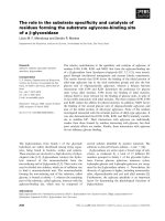
Fig. 1. Alignment of the amino acid sequences. The amino acid sequence of Tma-RNase HI (Tma) is compared with those of Bha-RNase HI
(Bha), SIB1 HBD-RNase HI (SIB1HBD), Sce-RNase H1 (Sce), Hsa-RNase H1 (Hsa), Eco-RNase HI (Eco) and HIV-1 RNase H (HIV1). The
accession numbers are AAD36370 for Tma-RNase HI, BAF73617 for SIB1 HBD-RNase HI, DAA10134 for Sce-RNase H1, EAX01061 for Hsa-
RNase H1, P0A7Y4 for Eco-RNase HI and ABU62661 for HIV-1 RNase H. The ranges of the secondary structures of Hsa-RNase H1 are
shown above the sequence, based on the crystal structures of its HBD (Protein Data Bank code: 3BSU) and RNase H domain (Protein Data
Bank code: 2KQ9), which were independently determined in complex with the substrate. The range of HBD is also shown. The amino acid
residues, which are conserved in at least three (for HBD) or four (for RNase H domain) different proteins, are highlighted in black. The five
active-site residues are denoted by filled circles above the sequences. The amino acid residues that contact the substrate in the co-crystal
structure of the HBD of Hsa-RNase H1 with the substrate are also denoted by open circles above the sequence. The amino acid residue that
is mutated in the present study is indicated by an arrow. Gaps are denoted by dashes. The numbers represent the positions of the amino
acid residues relative to the initiator methionine for each protein.
Role of HBD from T. maritima RNase HI N. Jongruja et al.
4476 FEBS Journal 277 (2010) 4474–4489 ª 2010 The Authors Journal compilation ª 2010 FEBS
HI, 18 860 for Tma-CD and 7107 for Tma-ND), sug-
gesting that all proteins exist as a monomer in solution.
CD spectra
The far- and near-UV CD spectra of Tma-RNase HI,
Tma-CD and Tma-ND were measured at 20 °C and
pH 9.0, and comparisons are shown in Fig. 3. The far-
and near-UV CD spectra of Tma-CD are similar to
those of Tma-RNase HI, suggesting that removal
of the HBD does not significantly affect the structure
of the RNase H domain of Tma-RNase HI. The
far- and near-UV CD spectra of Tma-ND were sig-
nificantly different from those of Tma-RNase HI,
probably because the secondary structure contents and
environment of the aromatic residues are different in
these proteins. According to the crystal structures of
the HBD [22] and RNase H domain [15] of Hsa-
RNase HI, the b-strand contents are 37% for HBD
and 21% for RNase H domain, whereas the a-helix
contents of these domains are similar to each other
(39% for HBD and 38% for RNase H domain).
Enzymatic activity
The dependencies of the Tma-RNase HI and Tma-
CD activities on pH, salt and metal ion were
analyzed at 30 °C by changing one of the conditions
used for assay [10 mm Tris ⁄ HCl, 1 mm MgCl
2
,
50 mm KCl (pH 9.0) for Tma-RNase HI, and 10 mm
Tris ⁄ HCl, 1 mm MnCl
2
,10mm KCl (pH 9.0) for
Tma-CD]. The M13 DNA ⁄ RNA hybrid was used as
a substrate. The enzymatic activities of these proteins
were determined at the temperature (30 °C), which
could be much lower than the optimum one because
the substrate used for assay is not fully stable at
‡ 60 °C. When the enzymatic activity was determined
over the range pH 5–12, both proteins exhibited the
highest activities at around pH 9.0 (data not shown).
They exhibited approximately 50% of the maximal
activities at pH 7.0 and 11.0. When the enzymatic
activity was determined in the presence of various
concentrations of NaCl or KCl, Tma-RNase HI
exhibited the highest activity in the presence of
50 mm KCl, whereas Tma-CD exhibited it in the
presence of 10 mm KCl (Fig. 4). Their enzymatic
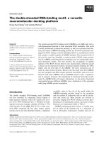
Fig. 2. SDS ⁄ PAGE of Tma-RNase HI and its derivatives. The
purified proteins of Tma-RNase HI (lane 1), Tma-CD (lane 2) and
Tma-ND (lane 3) were subjected to electrophoresis on a 15% poly-
acrylamide gel in the presence of SDS. After electrophoresis, the
gel was stained with Coomassie Brilliant Blue. Lane M, a low-
molecular-weight marker kit (GE Healthcare, Tokyo, Japan).
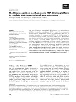
Fig. 3. CD spectra of Tma-RNase HI and its derivatives. Far-UV (A)
and near-UV (B) CD spectra of Tma-RNase HI (thick solid dark line),
Tma-W22A (thin solid dark line), Tma-CD (dashed dark line) and
Tma-ND (thick solid gray line) are shown. These spectra were mea-
sured at pH 9.0 and 20 °C, as described in the Experimental proce-
dures.
N. Jongruja et al. Role of HBD from T. maritima RNase HI
FEBS Journal 277 (2010) 4474–4489 ª 2010 The Authors Journal compilation ª 2010 FEBS 4477
activities decreased to a large extent at higher
(‡ 0.2 m) salt concentrations. When the enzymatic
activity was determined in the presence of various
concentrations of MgCl
2
, MnCl
2
, NiCl
2
, ZnCl
2
,
CoCl
2
or CaCl
2
, both Tma-RNase HI and Tma-CD
exhibited the highest activities in the presence of
1mm MgCl
2
and 0.1–5 mm MnCl
2
(Fig. 5). Both
proteins exhibited little activity (less than 0.01% of
the maximal activity) in the presence of NiCl
2
, ZnCl
2
,
CoCl
2
or CaCl
2
. The maximal Mg
2+
- and Mn
2+
-
dependent activities of these proteins are summarized
in Table 1. Tma-RNase HI prefers Mg
2+
to Mn
2+
because its maximal Mg
2+
-dependent activity is
higher than its maximal Mn
2+
-dependent activity by
16-fold. By contrast, Tma-CD prefers Mn
2+
to Mg
2+
because its maximal Mn
2+
-dependent activity is
higher than its maximal Mg
2+
-dependent activity by
69-fold. Interestingly, the maximal Mn
2+
-dependent
activity of Tma-CD is comparable to that of Tma-
RNase HI. These results indicate that removal of the
HBD severely reduces the Mg
2+
-dependent activity of
Tma-RNase HI without significantly affecting its
Mn
2+
-dependent activity.
The kinetic parameters of Tma-CD were determined
at 30 °C in the presence of 1 mm MgCl
2
or MnCl
2
and
compared with those of Tma-RNase HI (Table 1). The
V
max
values of Tma-CD determined in the presence of
1mm MgCl
2
and MnCl
2
were 410-fold lower and
1.6-fold higher than those of Tma-RNase HI. The K
m
values of Tma-CD determined in the presence of 1 mm
MgCl
2
and MnCl
2
were 5.1- and 6.8-fold higher than
those of Tma-RNase HI. These results indicate that
the substrate binding affinity of Tma-RNase HI is
reduced by five- to seven-fold on removal of the HBD,
regardless of the metal cofactor, and the large reduc-
tion in Mg
2+
-dependent activity on removal of the
HBD is not a result of the marked decrease in
substrate binding affinity.
Fig. 4. Salt dependencies of Tma-RNase HI and Tma-CD. The enzy-
matic activities of Tma-RNase HI (A) and Tma-CD (B) were deter-
mined at 30 °Cin10m
M Tris ⁄ HCl (pH 9.0) containing 1 mM MgCl
2
(Tma-RNase HI) or 1 mM MnCl
2
(Tma-CD), 1 mM b-mercaptoe-
thanol, 50 lgÆmL
)1
BSA, and various concentrations of NaCl (open
circle) or KCl (closed circle), using M13 DNA ⁄ RNA hybrid as a sub-
strate. Experiments were carried out at least twice and the average
values are shown together with the errors.
Fig. 5. Metal ion dependencies of Tma-RNase HI and Tma-CD.
The enzymatic activities of Tma-RNase HI (A) and Tma-CD (B)
were determined at 30 °Cin10m
M Tris ⁄ HCl (pH 9.0) containing
50 m
M KCl (Tma-RNase HI) or 10 mM KCl (Tma-CD), 1 mM
b-mercaptoethanol, 50 lgÆmL
)1
BSA, and various concentrations of
MgCl
2
(open circle) or MnCl
2
(closed circle), using M13 DNA ⁄ RNA
hybrid as a substrate. Experiments were carried out at least twice
and the average values are shown together with the errors.
Role of HBD from T. maritima RNase HI N. Jongruja et al.
4478 FEBS Journal 277 (2010) 4474–4489 ª 2010 The Authors Journal compilation ª 2010 FEBS
Complementation assay
E. coli MIC3001 shows an RNase H-dependent
temperature-sensitive growth phenotype [28]. E. coli
MIC3001(DE3) also displays this phenotype. To exam-
ine whether the genes encoding Tma-RNase HI and
Tma-CD complement the temperature-sensitive growth
phenotype of MIC3001(DE3), E. coli MIC3001(DE3)
transformants for overproduction of these proteins
were grown in the absence of isopropyl thio-b-d-galac-
toside (IPTG) at permissive (30 °C) and nonpermissive
(42 °C) temperatures. The results showed that the
Tma-RNase HI gene complements the temperature-
sensitive growth phenotype of MIC3001(DE3),
whereas the Tma-CD gene does not (data not shown).
These results suggest that HBD is required for in vivo
function of Tma-RNase HI. It is unlikely that Tma-
CD is not produced or produced in a nonfunctional
form in E. coli cells in the absence of IPTG because
the protein is overproduced in a soluble and functional
form upon overproduction, as noted above.
Cleavage-site specificity
The cleavage-site specificities of Tma-RNase HI and
Tma-CD were analyzed by using 12 bp RNA ⁄ DNA
hybrid (R12 ⁄ D12), 29 bp DNA
13
-RNA
4
-DNA
12
⁄
DNA duplex (D13-R4-D12 ⁄ D29), 29 bp DNA
15
-
RNA
1
-DNA
13
⁄ DNA duplex (D15-R1-D13 ⁄ D29) and
18 bp RNA
9
-DNA
9
⁄ DNA duplex (R9-D9 ⁄ D18). For
comparative purposes, these substrates were cleaved
by Eco-RNase HI, Sulfolobus tokodaii RNase HI
(Sto-RNase HI) and Thermococcus kodakaraensis
RNase HII (Tk-RNase HII) as well. D13-R4-D12
and D15-R1-D13 are the chimeric oligonucleotides,
in which four and single ribonucleotides are flanked
by 12–15 bp of DNA at both sides. R9-D9 ⁄ D18 is a
Okazaki fragment-like substrate, in which the 18 base
chimeric oligonucleotide (RNA
9
-DNA
9
) is hybridized
to the 18 base complementary DNA.
Cleavage of the R12 ⁄ D12 substrate with various
RNase H enzymes is summarized in Fig. 6A,B. Tma-
RNase HI, Eco-RNase HI, Sto-RNase HI and Tk-
RNase HII cleaved this substrate at multiple sites,
although with different site specificities. Tma-RNase
HI cleaved this substrate slightly more efficiently in the
presence of Mg
2+
than in the presence of Mn
2+
. Tma-
CD cleaved this substrate with much less and compa-
rable efficiencies compared to those of Tma-RNase HI
in the presence of Mg
2+
and Mn
2+
, respectively.
These results are consistent with those obtained by
using M13 DNA ⁄ RNA as a substrate. The cleavage
sites of the R12 ⁄ D12 substrate with Tma-CD are simi-
lar to those with Tma-RNase HI, regardless of the
metal cofactor, although their preferable cleavage sites
are slightly different with each other. The cleavage
sites of this substrate and their susceptibilities to cleav-
age with Eco-RNase HI, Sto-RNase HI, and Tk-
RNase HII are essentially the same as those reported
previously [27–30].
Cleavage of the D13-R4-D12 ⁄ D29 substrate with
various RNase H enzymes is summarized in Fig. 6C,D.
Tma-RNase HI, Eco-RNase HI, Sto-RNase HI and
Tk-RNase HII cleaved this substrate most preferably
at a16-a17, a15-a16, a14-a15 and a16-a17, respectively.
The cleavage sites of this substrate with Eco-RNase
HI and Tk-RNase HII are the same as those reported
previously [30]. The a16-a17 site has been reported to
be exclusively cleaved only by type 2 RNases H, except
for bacterial RNases HIII [31,32]. Therefore, Tma-
RNase HI is the first type 1 RNase H enzyme that
exclusively cleaves this substrate at this site. Tma-CD
also cleaved this substrate at a16-a17 with a similar
efficiency to that of Tma-RNase HI. However, these
enzymes cleaved this substrate only in the presence of
Mn
2+
.
Table 1. Specific activities and kinetic parameters of Tma-RNase HI and its derivatives. Hydrolysis of the M13 DNA ⁄ RNA hybrid by the
enzyme was carried out at 30 °C under the conditions described in the Experimental procedures. ND, not determined.
Protein Metal Salt
Specific activity
(UÆmg
)1
)
Relative
activity
a
(%) K
m
(lM) V
max
(UÆmg
)1
)
Tma-RNase HI 1 m
M MgCl
2
50 mM KCl 3.6 ± 0.52 100 0.39 ± 0.064 7.3 ± 1.4
1m
M MnCl
2
10 mM KCl 0.23 ± 0.026 6.4 0.25 ± 0.042 0.62 ± 0.071
Tma-W22A 1 m
M MgCl
2
50 mM KCl 0.48 ± 0.057 13 ND ND
1m
M MnCl
2
10 mM KCl 0.35 ± 0.039 9.7 ND ND
Tma-CD 1 m
M MgCl
2
50 mM KCl 0.0048 ± 0.00068 0.13 2.0 ± 0.24 0.018 ± 0.0042
1m
M MnCl
2
10 mM KCl 0.33 ± 0.0081 9.2 1.7 ± 0.33 1.0 ± 0.14
Eco-RNase HI 10 m
M MgCl
2
50 mM NaCl 8.3 ± 0.22 231 ND ND
a
The specific activities of the proteins relative to that of Tma-RNase HI determined in the presence of 1 mM MgCl
2
and 50 mM KCl.
N. Jongruja et al. Role of HBD from T. maritima RNase HI
FEBS Journal 277 (2010) 4474–4489 ª 2010 The Authors Journal compilation ª 2010 FEBS 4479
Role of HBD from T. maritima RNase HI N. Jongruja et al.
4480 FEBS Journal 277 (2010) 4474–4489 ª 2010 The Authors Journal compilation ª 2010 FEBS
The D15-R1-D13 ⁄ D29 substrate was used to con-
firm that Tma-RNase HI and Tma-CD do not cleave
the DNA-RNA-DNA ⁄ DNA substrate containing a
single ribonucleotide. This substrate is not cleaved by
type 1 RNases H but is cleaved by type 2 RNases H,
except for bacterial RNases HIII, at the DNA-RNA
junction [21,27,32]. As expected, this substrate was not
cleaved with Tma-RNase HI, Sto-RNase HI and Eco-
RNase HI, although it was cleaved with Tk-RNase
HII at the DNA-RNA junction (data not shown).
These results exclude the possibility that the cleavage
of the D13-R4-D12 ⁄ D29 substrate with Tma-RNase
HI at a16-a17 is caused by the contamination of a type
2 RNase H enzyme.
Cleavage of the R9-D9 ⁄ D18 substrate with various
RNase H enzymes is summarized in Fig. 6E,F. Tma-
RNase HI and Tma-CD cleaved this substrate most
preferably at g7-c8 and c8-c9, and much less preferably
at u6-g7 and c9-T10 in the presence of Mn
2+
. They
cleaved this substrate with similar site specificities in
the presence of Mg
2+
. However, their abilities to
cleave this substrate are greatly reduced in the presence
of Mg
2+
by more than 100-fold. Eco-RNase HI and
Sto-RNase HI cleaved this substrate at all sites
between a5 and c9 and between a5 and T10, respec-
tively, as reported previously [33]. However, both
enzymes showed a preference for the sites far from the
RNA-DNA junction (a5-u6, u6-g7 and g7-c8 for Eco-
RNase HI, and u5-a6 and u6-g7 for Sto-RNase HI).
Tk-RNase HII cleaved this substrate almost exclusively
at c8-c9. Eco-RNase HI and Tk-RNase HII cleaved
the RNA-DNA junction (c9-T10) as well, although
with very poor efficiency.
It has been demonstrated for mouse RNase H1 that
the HBD is required for processivity of the enzyme
[24]. Tma-RNase HI did not show the processivity
for cleavage of the R12 ⁄ D12 substrate (Fig. 6). How-
ever, this result does not necessarily indicate that
Tma-RNase HI shows no processivity because mouse
RNase H1 shows the processivity only for long
RNA ⁄ DNA substrates. Therefore, it would be infor-
mative to examine whether Tma-RNase HI shows
processivity for long RNA ⁄ DNA substrates and loses
this processivity on removal of the HBD.
Binding to substrate
To examine whether the HBD of Tma-RNase HI is
important for substrate binding, the binding affinities
of Tma-RNase HI, Tma-CD and Tma-ND to the
29 bp RNA ⁄ DNA hybrid (R29 ⁄ D29) were analyzed in
the absence of the metal cofactor using surface plas-
mon resonance. These proteins were injected onto the
sensor chip, on which the R29 ⁄ D29 substrate was
immobilized. The sensorgrams obtained by injecting
1 lm of these proteins are shown in Fig. 7. The disso-
ciation constants, K
D
, of the proteins for binding to
the R29 ⁄ D29 substrate, which were determined by
measuring the equilibrium-binding responses at various
concentrations of the proteins, are summarized in
Table 2. The K
D
value of Tma-ND was higher than
(although comparable to) that of Tma-RNase HI. By
Fig. 7. Binding of Tma-RNase HI and its derivatives to the sub-
strate. Sensorgrams from Biacore X showing binding of 1 l
M of
Tma-RNase HI (thick solid dark line), Tma-W22A (thin solid dark
line), Tma-CD (dashed dark line) and Tma-ND (thick solid gray line)
to the immobilized R29 ⁄ D29 substrate are shown. Injections were
performed at time zero for 60 s.
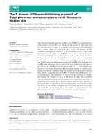
Fig. 6. Cleavage of various oligomeric substrates with various RNases H. (A, C, E) Separation of the hydrolysates by urea gel. The 5¢-end
labeled R12 ⁄ D12 (A), 5¢-end labeled D13-R4-D12 ⁄ D29 (C) and 3¢-end labeled R9-D9 ⁄ D18 (E) were hydrolyzed by the enzyme at 30 °C for
15 min and the hydrolysates were separated on a 20% polyacrylamide gel containing 7
M urea, as described in the Experimental procedures.
The concentration of the substrate was 1.0 l
M. The amount of the enzyme added to the reaction mixture (10 lL) is indicated above each
lane. The metal cofactors used to cleave these substrates with Tma-RNase HI and Tma-CD are also shown above the gel together with their
concentrations. The complete sequence of R12 (A) as well as the partial sequences of D13-R4-D12 (C) and R9-D9 (E) are indicated along the
gel. (B, D, F) Schematic representation of the sites and extents of cleavage by various RNases H. Cleavage sites of R12 ⁄ D12 (B), D13-R4-
D12 ⁄ D29 (D) and R9-D9 ⁄ D18 (F) by the enzyme are shown by arrows. In these panels, only the sequences of the oligonucleotides cleaved
by the enzyme are shown. The differences in the lengths of the arrows reflect relative cleavage intensities at the position indicated. These
lengths do not necessarily reflect the amount of the products accumulated upon complete hydrolysis of the substrate. Deoxyribonucleotides
are indicated by capital letters and ribonucleotides are indicated by lowercase letters.
N. Jongruja et al. Role of HBD from T. maritima RNase HI
FEBS Journal 277 (2010) 4474–4489 ª 2010 The Authors Journal compilation ª 2010 FEBS 4481
contrast, the K
D
value of Tma-CD was considerably
higher than that of Tma-RNase HI by 49-fold. These
results indicate that the HBD of Tma-RNase HI is
important for substrate binding. When binding of
Tma-RNase HI to the R29 ⁄ D29 substrate was
analyzed in the presence of 0.5 m NaCl, only a small
positive signal was observed, even when 4 lm of the
protein was injected, indicating that the binding affin-
ity of Tma-RNase HI to the substrate is severely
decreased at high salt concentration.
Thermal stability
To examine whether removal of the N- or C-terminal
domain affects the stability of Tma-RNase HI, the
thermal stabilities of Tma-RNase HI, Tma-CD and
Tma-ND were determined by monitoring changes of
the CD values at 222 nm. In the presence of 3 m
guanidine hydrochloride (GdnHCl) and 10 mm dith-
iothreitol at pH 9, all proteins unfolded in a single
cooperative fashion in a reversible manner. The ther-
mal denaturation curves of these proteins are com-
pared with one another in Fig. 8. The parameters
characterizing the thermal denaturation of these pro-
teins are summarized in Table 2. A comparison of
these parameters indicates that Tma-CD and Tma-ND
are less stable than Tma-RNase HI by 1.3 and 10.8 °C
in T
m
, respectively. These results suggest that the inter-
actions between the N- and C-terminal domains of
Tma-RNase HI do not significantly contribute to the
stabilization of its C-terminal domain but contribute
to the stabilization of its N-terminal domain. Tma-
RNase HI is thermally denatured in a single coopera-
tive fashion, probably because its N-terminal domain
is denatured immediately after its C-terminal RNase H
domain is denatured. It is noted that the DH
m
and
DS
m
values of Tma-CD are considerably higher than
those of Tma-RNase HI and Tma-ND, which are
comparable to each other. The reason why the DH
m
and DS
m
values of Tma-RNase HI increase on
removal of the N-terminal domain remains to be
clarified.
Analysis for interaction between two domains
To examine whether the HBD of Tma-RNase HI
strongly interacts with the RNase H domain, Tma-ND
was mixed with Tma-CD in a 1 : 1 molar ratio and
applied to gel filtration column chromatography. Both
proteins were eluted from the column as two inde-
pendent peaks (data not shown), indicating that
Tma-ND does not form a stable complex with
Table 2. Dissociation constants and parameters characterizing thermal denaturation of Tma-RNase HI and its derivatives. ND, not
determined.
Protein K
D
a
(lM) T
m
b
(°C) DT
m
b
(°C) DH
m
b
(kJÆmol
)1
) DS
m
b
(kJ.mol
)1
ÆK
)1
)
Tma-RNase HI 0.16 ± 0.013 67.0 ± 0.83 – 115.9 ± 11.1 0.34 ± 0.032
Tma-W22A 3.3 ± 0.54 ND ND ND ND
Tma-CD 7.8 ± 0.47 65.7 ± 4.3 )1.3 205.7 ± 22.3 0.58 ± 0.067
Tma-ND 0.40 ± 0.083 56.2 ± 3.2 )10.8 111.7 ± 7.43 0.33 ± 0.038
a
Dissociation constant of the protein for binding to the R29 ⁄ D29 substrate was determined by measuring equilibrium-binding responses at
various concentrations of the protein using surface plasmon resonance (Biacore) as described in the Experimental procedures.
b
Parameters
characterizing thermal denaturation of the proteins were determined from the thermal denaturation curves shown in Fig. 8. The melting tem-
perature (T
m
) is temperature of the midpoint of the thermal denaturation transition. DT
m
is the difference in T
m
between the intact and trun-
cated proteins and is calculated as: T
m
(truncated) ) T
m
(intact). DH
m
and DS
m
are the enthalpy and entropy changes of unfolding at T
m
calculated by van’t Hoff analysis.
Fig. 8. Thermal denaturation curves. Thermal denaturation curves
of Tma-RNase HI (closed circl), Tma-CD (open square) and Tma-ND
(closed triangle) are shown. These curves were obtained at pH 9.0
in the presence of 3
M GdnHCl and 10 mM dithiothreitol by monitor-
ing the change in the CD value at 222 nm, as described in the
Experimental procedures. The theoretical curves are drawn on
the assumption that the proteins are denatured via a two-state
mechanism.
Role of HBD from T. maritima RNase HI N. Jongruja et al.
4482 FEBS Journal 277 (2010) 4474–4489 ª 2010 The Authors Journal compilation ª 2010 FEBS
Tma-CD. In addition, the Mg
2+
-dependent activity of
Tma-CD was not significantly changed in the presence
of a 10–1000 molar excess of Tma-ND, indicating that
the Mg
2+
-dependent activity of Tma-CD is not
restored in the presence of Tma-ND. It has been pro-
posed for eukaryotic type 1 RNases H that the HBD
and RNase H domain are separated by a flexible linker
and move rather freely [21]. The HBD of Tma-RNase
HI also may not strongly interact with the RNase H
domain.
Biochemical properties of Tma-W22A
According to the crystal structure of the HBD of Hsa-
RNase H1 in complex with the substrate, Tyr29,
Trp43, Phe58, Lys59 and Lys60 interact with the DNA
strand of the substrate [22]. These residues, except for
Lys60, are well conserved in various HBDs, suggesting
that the HBDs of other type 1 RNases H bind to the
substrate in a manner similar to the interaction of the
HBD of Hsa-RNase H1. The mutation of Trp43 to
Ala greatly reduces both the substrate binding affinities
and enzymatic activities of Hsa-RNase H1 [22,26] and
mouse RNase H1 [24]. To examine whether the corre-
sponding tryptophan residue (Trp22) is important for
substrate binding and enzymatic activity of Tma-
RNase HI, the mutant protein, Tma-W22A, was con-
structed, overproduced and purified. The production
level and purification yield of Tma-W22A were compa-
rable to those of Tma-RNase HI. The far- and near-
UV CD spectra of Tma-W22A are similar to those of
Tma-RNase HI (Fig. 3), suggesting that the mutation
at Trp22 does not significantly affect the structure of
Tma-RNase HI.
The pH, salt and metal ion dependencies of Tma-
W22A were similar to those of Tma-RNase HI (data
not shown). Its maximal Mn
2+
-dependent activity was
also similar to that of Tma-RNase HI (Table 1). How-
ever, its maximal Mg
2+
-dependent activity was lower
than that of Tma-RNase HI by 7.5-fold (Table 1),
indicating that the mutation of Trp22 to Ala con-
siderably reduces the Mg
2+
-dependent activity of
Tma-RNase HI without significantly affecting the
Mn
2+
-dependent activity. The binding affinity of
Tma-W22A to the R29 ⁄ D29 substrate was analyzed in
the absence of the metal cofactor using surface plas-
mon resonance and compared with that of Tma-RNase
HI. The K
D
value of Tma-W22A was higher than that
of Tma-RNase HI by 21-fold, suggesting that Trp22 of
Tma-RNase HI is involved in substrate binding. These
results suggest that the HBD of Tma-RNase HI inter-
acts with the substrate in a manner similar to the inter-
action of the HBD of Hsa-RNase H1.
The cleavage site specificities of Tma-W22A were
not analyzed because the cleavage site specificities of
Tma-RNase HI are not significantly changed even
when its N-terminal domain is removed, and therefore
it is highly likely that the cleavage site specificities of
Tma-W22A are similar to those of Tma-RNase HI.
Likewise, the stability of Tma-W22A was not analyzed
because the stability of Tma-W22A is not significantly
changed even when the HBD is completely removed,
and therefore it is highly likely that Tma-W22A is as
stable as Tma-RNase HI.
Discussion
Importance of HBD for substrate binding
In the present study, we showed that the HBD of
Tma-RNase HI is important for substrate binding.
However, on removal of the HBD, the K
m
value of
Tma-RNase HI increases by 5–7-fold, whereas its K
D
value increases by 49-fold. Because the K
m
and K
D
val-
ues are determined in the presence and absence of the
metal cofactor, these results suggest that the difference
in substrate binding affinity between Tma-RNase HI
and Tma-CD determined in the presence of the metal
cofactor is smaller than that determined in its absence.
Presumably, the HBD governs binding of Tma-RNase
HI to the substrate and its substrate binding affinity is
not significantly changed either in the presence or
absence of the metal cofactor. By contrast, the sub-
strate binding affinity of the RNase H domain proba-
bly increases in the presence of the metal cofactor
compared to that in its absence. The cleavage-site spec-
ificity of Tma-RNase HI is not significantly changed
on removal of the HBD, probably because the HBD
of Tma-RNase HI facilitates initial nonsite-specific
interactions with the substrate and promotes the site-
specific interactions between the RNase H domain of
Tma-RNase HI and substrate.
Importance of HBD for Mg
2+
-dependent activity
Removal of the HBD severely reduces the Mg
2+
-
dependent activity of Tma-RNase HI by 750-fold
without significantly affecting the Mn
2+
-dependent
activity. Similarly, single mutation at the HBD
(Trp22 to Ala) reduces the Mg
2+
-dependent activity
of Tma-RNase HI by 7.5-fold without significantly
affecting the Mn
2+
-dependent activity. Removal of
the HBD and single mutation at the HBD reduces
the binding affinity of Tma-RNase HI by 49- and 21-
fold, respectively. Thus, there is a correlation between
the Mg
2+
-dependent activity of Tma-RNase HI and
N. Jongruja et al. Role of HBD from T. maritima RNase HI
FEBS Journal 277 (2010) 4474–4489 ª 2010 The Authors Journal compilation ª 2010 FEBS 4483
the substrate binding affinity of the HBD. High simi-
larity in the conformation of the active site between
the Hsa-RNase H1 derivative lacking the HBD and
Eco-RNase HI [15] suggests that the conformation of
the active site is not significantly changed on removal
of the HBD. Because the HBD is important for sub-
strate binding, the HBD may affect the interaction
between the enzyme and substrate at the active site.
Because not only the active-site residues, but also the
substrate provide ligands for coordination of the
metal ion [14], removal of the HBD or mutation at
the HBD may alter the interaction between the sub-
strate and metal ion, such that the scissile phosphate
group of the substrate and the Mg
2+
ion are
arranged ideally. The effect of this alteration on the
coordination of the metal ion varies for Mg
2+
and
Mn
2+
, probably because Mn
2+
is a transition metal
having coordinates with a different geometry than
Mg
2+
.
Similar results have been reported for Eco-RNase
HI [34], RNase H of Moloney murine leukemia virus
reverse transcriptase (MMLV RNase H) [35] and
Bacillus stearothermophilus RNase HIII (Bst-RNase
HIII) [36]. Eco-RNase HI and Bst-RNase HIII prefer
Mg
2+
to Mn
2+
, whereas MMLV RNase H prefer
Mn
2+
to Mg
2+
, although all of them specifically lose
or greatly reduce Mg
2+
-dependent activity by deletion
of the basic protrusion (for Eco-RNase HI and
MMLV RNase H) or removal of the N-terminal sub-
strate binding domain (for Bst-RNase HIII). The metal
ion preference of Hsa-RNase HI is also changed on
removal of the HBD because Hsa-RNase HI prefers
Mg
2+
to Mn
2+
[27], whereas its derivative without the
HBD prefers Mn
2+
to Mg
2+
[15]. The mutation or
deletion at other regions than those involved in sub-
strate binding also often alter the metal ion preference
of RNase H, so that they show a strong preference to
Mn
2+
. For example, Eco-RNase HI [37] and HIV-1
RNase H [38] specifically loses Mg
2+
-dependent activ-
ity by the mutation of the active-site residue (Glu48 or
Asp134 for Eco-RNase HI and Glu478 for HIV-1
RNase H). Eco-RNase HI specifically loses the Mg
2+
-
dependent activity by deletion of the last helix [39]. In
all cases, the conformation of the metal binding site is
probably slightly changed, so that it becomes unfavor-
able for binding of Mg
2+
but is kept favorable for
binding of Mn
2+
.
The finding that the D13-R4-D12 ⁄ D29 and R9-
D9 ⁄ D18 substrates cannot be effectively cleaved by
Tma-RNase HI in the presence of Mg
2+
suggests
that the RNA ⁄ DNA hybrid region in these substrates
is too short to accommodate both the HBD and the
RNase H domain. According to the crystal structure
of the catalytic domain of human RNase H1 in com-
plex with the substrate [15], the enzyme interacts with
several RNA residues preceding the scissile bond. The
distance between the most preferable cleavage site of
Tma-RNase HI and 5¢-end of the RNA ⁄ DNA hybrid
region is five bases for the R12 ⁄ D12 substrate
(Fig. 6B), which can be effectively cleaved by the
enzyme in the presence of Mg
2+
, and seven or eight
bases for the R9-D9 ⁄ D18 substrate (Fig. 6F), whereas
the distance between this cleavage site and the 3¢-end
of the RNA ⁄ DNA hybrid region is seven bases for
the R12 ⁄ D12 substrate (Fig. 6B) and one or two
bases for the R9-D9 ⁄ D18 substrate (Fig. 6F). These
results suggest that the HBD of Tma-RNase HI binds
to the downstream region of the substrate from the
scissile bond. Tma-RNase HI cannot effectively cleave
the D13-R4-D12 ⁄ D29 and R9-D9 ⁄ D18 substrates in
the presence of Mg
2+
, probably because the HBD
cannot bind to double-stranded DNA, which is
located downstream from the scissile bond of the
substrate.
Importance of HBD for in vivo function
Tma-RNase HI complements the temperature-sensitive
growth phenotype of E. coli MIC3001, indicating that
it functions as a substitute of Eco-RNase HI in vivo.
By contrast, Tma-CD does not function as a substitute
of Eco-RNase HI in vivo. These results suggest that
HBD is required for in vivo RNase H activity of Tma-
RNase HI. Because Tma-RNase HI and Tma-CD
greatly differ in Mg
2+
-dependent activity, the Mg
2+
-
dependent activity of Tma-RNase HI may be responsi-
ble for its RNase H activity in vivo. It has been
reported that E. coli RNase HII (Eco-RNase HII) [40]
and Bacillus subtilis RNase HII (Bsu-RNase HII) [41],
which prefer Mn
2+
to Mg
2+
for activity, complement
the temperature-sensitive growth phenotype of E. coli
MIC3001. Both proteins exhibit the highest Mn
2+
-
dependent activities ( 0.5 UÆmg
)1
) in the presence of
10 mm MnCl
2
[31], which are comparable to that of
Tma-CD (0.33 UÆmg
)1
at 1 mm MnCl
2
) (Table 1).
Eco-RNase HII and Bsu-RNase HII exhibit the high-
est Mg
2+
-dependent activities ( 0.03 UÆmg
)1
) in the
presence of 10 mm MgCl
2
[31], which are considerably
higher than that of Tma-CD (0.0048 UÆmg
)1
)
(Table 1). These results suggest that Mg
2+
-dependent
activities, instead of Mn
2+
-dependent activities, are
responsible for in vivo RNase H activities of
Eco-RNase HII and Bsu-RNase HII as well. Mg
2+
-
dependent activity of Tma-CD may be too low to
complement the temperature-sensitive growth pheno-
type of E. coli MIC3001.
Role of HBD from T. maritima RNase HI N. Jongruja et al.
4484 FEBS Journal 277 (2010) 4474–4489 ª 2010 The Authors Journal compilation ª 2010 FEBS
Salt dependencies of enzymatic activity
Both the Tma-RNase HI and Tma-CD activities are
sensitive to salt. Both activities decrease sharply with
the increase of the concentration of salt beyond the
optimum one (Fig. 4). The surface plasmon resonance
analyses for binding to the R29 ⁄ D29 substrate indicate
that the activities of these proteins greatly decrease at
high salt concentrations because the binding affinities
of these proteins to the substrate greatly decrease.
However, the highest activities of Tma-RNase HI and
Tma-CD are observed at 50 and 10 mm KCl, respec-
tively, indicating that Tma-RNase HI requires higher
concentrations of KCl for maximal activity than does
Tma-CD. It has been reported that Hsa-RNase H1
requires a physiological salt concentration (125 mm
NaCl) for maximal activity to overcome the nonspe-
cific charge–charge interactions between HBD and
nucleic acids [22]. Because the basic residues that con-
tribute to these interactions are well conserved in the
HBD of Tma-RNase HI, Tma-RNase HI probably
requires 50 mm KCl for maximal activity to overcome
these nonspecific interactions.
Experimental procedures
Cells and plasmids
E. coli MIC3001 [F
)
, supE44, supF58, lacY1 or D(lacIZY)6,
trpR55, galK2, galT22, metB1, hsdR14(r
K
)
m
K
+
), rnhA339::
cat, recB270] [28] was kindly donated by M. Itaya (Keio
University, Tsuruoka, Japan). E. coli MIC3001(DE3) was
constructed by lysogenizing E. coli MIC3001 with kDE3
using a kDE3 Lysogenization Kit (Novagen, Madison, WI,
USA). Plasmid pET25b was obtained from Novagen.
Plasmid construction
The pET25b derivatives for overproduction of Tma-RNase
HI, Tma-CD and Tma-ND were constructed by PCR. The
genomic DNA of T. maritima MSB8, which was obtained
from the American Type Culture Collection (Manassa, VA,
USA), was used as a template. The sequences of the PCR
primers are 5¢- TGGGTTTGAGAG
CATATGAAGTTGG
CAAAAAAATACTAC-3¢ for primer 1, 5¢-CG
CATATG
GAGACGATGATCGCCTACGTCGATG-3¢ for primer 2,
5¢-ACCGTT
AAGCTTTCATAAACATCCTCCTTT-3¢ for
primer 3, and 5¢-CG
GAATTCTCATGTGTCCAGTTCTG
GACAGATGCACTC-3¢ for primer 4, where the NdeI
(primers 1 and 2), HindIII (primer 3) and EcoRI (primer 4)
sites are shown underlined. Primer 2 is designed such that
the ATG codon is attached to the 5¢-terminus of the gene
encoding Tma-CD. Primer 4 is designed such that the TGA
codon is attached to the 3¢-terminus of the gene encoding
Tma-ND. Primers 1 and 3, primers 2 and 3, and primers 1
and 4 were used to amplify the genes encoding Tma-RNase
HI, Tma-CD and Tma-ND, respectively. The resultant
DNA fragments were digested with NdeI and HindIII or
EcoRI, and ligated into the NdeI-HindIII or NdeI-EcoRI
sites of pET25b.
The pET25b derivative for overproduction of Tma-
W22A was constructed by site-directed mutagenesis using
PCR, as described previously [42]. The pET25b derivative
for overproduction of Tma-RNase HI was used as a tem-
plate. The mutagenic primers were designed such that the
codon for Trp22 (TGG) is changed to GCG for Ala. The
resultant DNA fragment was digested with NdeI and Hin-
dIII, and ligated into the NdeI-HindIII sites of pET25b.
All DNA oligomers for PCR were synthesized by Hokka-
ido System Science (Sapporo, Japan). PCR was performed
in 25 cycles using a thermal cycler (Gene Amp PCR System
2400; Applied Biosystems, Tokyo, Japan) and KOD DNA
polymerase (Toyobo Co. Ltd, Kyoto, Japan). The DNA
sequences of the genes encoding all proteins described
above were confirmed by ABI Prism 310 DNA sequencer
(Applied Biosystems).
Overproduction and purification
For overproduction of Tma-RNase HI, Tma-CD, Tma-ND
and Tma-W22A, the E. coli MIC3001(DE3) transformants
with the pET25b derivatives were grown at 30 °C. When
A
600
reached a value of approximately 0.5, 1 mm IPTG was
added to the culture medium and cultivation was continued
at 30 °C for an additional 30 min. Then, the temperature
of the growth medium was shifted to 25 °C and cultivation
was continued at 25 °C for an additional 16 h. The subse-
quent purification procedures were carried out at 4 °C.
Cells were harvested by centrifugation at 8000 g for 10 min,
suspended in 10 mm Tris ⁄ HCl (pH 8.0) containing 1 mm
EDTA and 1 mm dithiothreitol (buffer A), disrupted by
sonication lysis, and centrifuged at 30 000 g for 30 min.
The supernatant was collected, dialyzed against buffer A
and loaded onto a HiTrap SP column (GE Healthcare,
Tokyo, Japan) equilibrated with the same buffer. The pro-
tein was eluted from the column with a linear gradient of
NaCl from 0 to 1 m. The fractions containing the protein
were collected, dialyzed against buffer A and loaded onto a
HiTrap Heparin column (GE Healthcare) equilibrated with
the same buffer. The protein was eluted from the column
with a liner gradient of NaCl from 0 to 1 m. The fractions
containing the protein were collected and used for biochem-
ical characterization. Eco-RNase HI [42], Sto-RNase HI
[29] and Tk-RNase HII [32] were overproduced and puri-
fied as described previously.
The purity of the protein was analyzed by SDS ⁄ PAGE
using a 15% polyacrylamide gel [43], followed by staining
with Coomassie Brilliant Blue. The protein concentration
was determined from UV absorption using a cell with an
N. Jongruja et al. Role of HBD from T. maritima RNase HI
FEBS Journal 277 (2010) 4474–4489 ª 2010 The Authors Journal compilation ª 2010 FEBS 4485
optical path length of 1 cm and an A
280
value for 0.1%
(1 mgÆmL
)1
) solution of 1.79 for Tma-RNase HI, 1.75 for
Tma-CD, 1.85 for Tma-ND, 1.58 for Tma-W22A, 2.0 for
Eco-RNase HI, 0.97 for Sto-RNase HI and 0.56 for
Tk-RNase HII. These values were calculated by using
absorption coefficients of 1576 m
)1
Æcm
)1
for Tyr and
5225 m
)1
Æcm
)1
for Trp at 280 nm [44].
Gel filtration chromatography
For estimation of the molecular masses of proteins, the
proteins were applied to a HiLoad 16 ⁄ 60 Superdex 200pg
column (GE Healthcare) equilibrated with 10 mm
Tris ⁄ HCl (pH 8.0) containing 10 mm dithiothreitol and
0.2 m NaCl. Thyroglobulin (670 kDa), bovine gamma
globulin (158 kDa), chicken ovalbumin (44 kDa), horse
myoglobin (17 kDa) and vitamin B
12
(1.35 kDa) were used
as markers.
Enzymatic activity
The RNase H activity was determined by measuring the
amount of the acid-soluble digestion product from the sub-
strate,
3
H-labeled M13 DNA ⁄ RNA hybrid, accumulated
upon incubation at 30 °C for 15 min, as described previ-
ously [45]. The reaction buffers were 10 m m Tris ⁄ HCl
(pH 9.0) containing 1 mm MgCl
2
,50mm KCl, 1 mm b-
mercaptoethanol (b-Me) and 50 lgÆmL
)1
BSA for Tma-
RNase HI and Tma-W22A, and 10 mm Tris ⁄ HCl (pH 9.0)
containing 1 mm MnCl
2
,10mm KCl, 1 mm b-Me and
50 lgÆmL
)1
BSA for Tma-CD. The substrate concentration
(RNA nucleotide phosphate concentration), which was cal-
culated on the assumption that the entire region of single-
stranded M13 DNA is converted to DNA ⁄ RNA hybrid,
was 0.3 lm. One unit was defined as the amount of enzyme
producing 1 lmolÆmin
)1
of acid-soluble material at 30°C.
The specific activity was defined as the enzymatic activity
per milligram of protein.
For determination of the kinetic parameters, the substrate
concentration was varied over the range 0.18–7.7 lm. The
hydrolysis of the M13 DNA ⁄ RNA hybrid by the enzyme fol-
lowed Michaelis–Menten kinetics and the kinetic parameters
were determined from the Lineweaver–Burk plot.
For cleavage of the oligomeric substrates, R12 ⁄ D12,
D13-R4-D12 ⁄ D29, D15-R1-D13 ⁄ D29 and R9-D9 ⁄ D18
were prepared by hybridizing 1 lm of the 5¢-FAM-labeled
12 base RNA (5¢-cggagaugacgg-3¢), 29 base DNA
13
-RNA
4
-
DNA
12
(5¢-AATAGAGAAAAAGaaaaAAGATGGCAA
AG-3¢), 29 base DNA
15
-RNA
1
-DNA
13
(5¢-AATAGAGAA
AAAGAAaAAAGATGGCAAAG-3¢) and 3¢-FAM-labeled
18 base RNA
9
-DNA
9
(5¢-uugcaugccTGCAGGTCG-3¢) with
a 1.5 molar equivalent of the complementary DNA, as
described previously [41] (in these sequences, DNA and
RNA are represented by capital and lowercase letters,
respectively; FAM represents 6-carboxyfluorescein). All
oligonucleotides were synthesized by Hokkaido System
Science. Hydrolysis of the substrate at 30 °C for 15 min
and separation of the products on a 20% polyacrylamide
gel containing 7 m urea were carried out as described previ-
ously [41]. The reaction buffers for Tma-RNase HI and
Tma-CD were the same as those for the hydrolysis of the
M13 DNA ⁄ RNA hybrid. The reaction buffers were 10 mm
Tris-HCl (pH 8.0) containing 10 mm MgCl
2
,50mm NaCl,
1mm b-Me and 50 lgÆmL
)1
BSA for Eco-RNase HI [46],
10 mm Tris-HCl (pH 8.5) containing 5 mm MgCl
2
,10mm
NaCl, 1 mm b-Me and 50 lg ÆmL
)1
BSA for Sto-RNase HI
[29], and 50 mm Tris-HCl (pH 8.0) containing 10 mm
MgCl
2
,50mm NaCl, 1 mm dithiothreitol and 0.01% BSA
for Tk-RNase HII [32]. The products were detected by
Typhoon 9240 Imager (GE Healthcare). They were identi-
fied by comparing their migration on the gel with those of
the oligonucleotides generated by digestion of substrates
with Eco-RNase HI [27], Sto-RNase HI [29,33] and
Tk-RNase HII [30,32].
CD spectra
The CD spectra were measured on a J-725 spectropolarime-
ter (Japan Spectroscopic, Tokyo, Japan) at 20 °C. The pro-
teins were dissolved in 5 mm Tris ⁄ HCl (pH 9.0). For
measurement of the far-UV CD spectra (200–260 mm), the
protein concentration was approximately 0.1 mgÆmL
)1
and
a cell with an optical path length of 2 mm was used. For
measurement of the near-UV CD spectra (250–320 mm),
the protein concentration was approximately 1.0 mgÆmL
)1
and a cell with an optical path length of 10 mm was used.
The mean residue ellipticity, h, which has the units of degÆc-
m
)2
Ædmol
)1
, was calculated by using an average amino acid
molecular weight of 110.
Binding analysis to substrate
Binding of proteins to the substrate was analyzed using
the Biacore X instrument (Biacore, Uppsala, Sweden).
R29 ⁄ D29 with the same sequence as that of D13-R4-
D12 ⁄ D29 or D15-R1-D13 ⁄ D29 was prepared so that the
RNA strand was biotinylated at the 5¢-end. The biotinylat-
ed oligonucleotide was synthesized by Hokkaido System
Science. The substrate was immobilized on the SA sensor
chip (Biacore), on which streptavidin is covalently linked,
by injecting 10 lL of NaCl ⁄ Tris buffer (10 mm Tris ⁄ HCl,
1mm EDTA, 50 mm NaCl, 1 mm b-Me, 0.005% Tween
P20, pH 9.0) containing 100 nm of biotinylated R29 ⁄ D29,
as described previously [47]. The proteins were dissolved
in NaCl ⁄ Tris buffer and injected at 25 °C at a flow rate
of 10 lLÆmin
)1
onto the sensor chip surface, on which
R29 ⁄ D29 has been immobilized. Binding surface was regen-
erated by washing with 2 m NaCl.
To determine the dissociation constant, K
D
, the concen-
tration of the protein injected onto the sensor chip was
Role of HBD from T. maritima RNase HI N. Jongruja et al.
4486 FEBS Journal 277 (2010) 4474–4489 ª 2010 The Authors Journal compilation ª 2010 FEBS
varied in the range 0.05–1.5 lm for Tma-RNase HI, 10–
50 lm for Tma-CD, 1.0–10 lm for Tma-ND and 0.5–50 lm
for Tma-W22A. From the plot of the equilibrium binding
responses as a function of the concentrations of the pro-
teins, the K
D
value was determined using the steady-state
affinity software available in biaevaluation (Biacore).
Thermal denaturation
Thermal denaturation curves of the proteins were obtained
by monitoring the change in CD values at 222 nm as the
temperature was increased. The proteins were dissolved in
5mm N-cyclohexyl-3¢-aminopropanesulfonic acid-NaOH
(pH 9.0) containing 3 m GdnHCl and 10 mm dithiothreitol.
The protein concentration and optical path length were
0.1 mgÆmL
)1
and 2 mm, respectively. The temperature of
the protein solution was linearly increased by approximately
1.0 °CÆmin
)1
. The thermal denaturation of these proteins
was fully reversible under this condition. The temperature of
the midpoint of the transition, T
m
, was calculated from
curve fitting of the resultant CD values versus temperature
data on the basis of a least squares analysis. The enthalpy
(DH
m
) and entropy (DS
m
) changes for thermal denaturation
at T
m
were calculated by van’t Hoff analysis.
Acknowledgements
This work was supported in part by a Grant
(21380065) from the Ministry of Education, Culture,
Sports, Science, and Technology of Japan, and by an
Industrial Technology Research Grant Program from
the New Energy and Industrial Technology Develop-
ment Organization (NEDO) of Japan.
References
1 Crouch RJ & Dirksen ML (1982) Ribonuclease H. In
Nuclease (Linn SM & Roberts RJ eds), pp. 211–241.
Cold Spring Harbor Laboratory, Cold Spring Harbor,
New York.
2 Kogoma T & Foster PL (1998) Physiological
functions of E. coli RNase HI. In Ribonuclease H
(Crouch RJ & Toulme JJ eds), pp. 39–66. INSERM,
Paris.
3 Qiu J, Qian Y, Frank P, Wintersberger U & Shen B
(1999) Saccharomyces cerevisiae RNase H(35) functions
in RNA primer removal during lagging-strand DNA
synthesis, most efficiently in cooperation with Rad27
nuclease. Mol Cell Biol 19, 8361–8371.
4 Arudchandran A, Cerritelli SM, Narimatsu SK, Itaya
M, Shin DY, Shimada Y & Crouch RJ (2000) The
absence of ribonuclease H1 or H2 alters the sensitivity
of Saccharomyces cerevisiae to hydroxyurea, caffeine
and ethyl methanesulphonate: implications for roles of
RNases H in DNA replication and repair. Genes Cells
5, 789–802.
5 Rydberg B & Game J (2002) Excision of misincorporat-
ed ribonucleotides in DNA by RNase H (Type 2) and
FEN-1 in cell-free extracts. Proc Natl Acad Sci USA 99,
16654–16659.
6 Cerritelli SM, Frolova EG, Feng C, Grinberg A, Love
PE & Crouch RJ (2003) Failure to produce mitochon-
drial DNA results in embryonic lethality in Rnaseh1
null mice. Mol Cell 11, 807–815.
7 Dean NM & Bennett CF (2003) Antisense oligonucleo-
tide-based therapeutics for cancer. Oncogene 22, 9087–
9096.
8 Chon H, Vassilev A, DePamphilis ML, Zhao Y, Zhang
J, Burgers PM, Crouch RJ & Cerritelli SM (2009) Con-
tributions of the two accessory subunits, RNASEH2B
and RNASEH2C, to the activity and properties of the
human RNase H2 complex. Nucleic Acids Res 37, 96–
110.
9 Crow YJ, Leitch A, Hayward BE, Garner A, Parmar
R, Griffith E, Ali M, Semple C, Aicardi J, Babul-Hirji
R et al. (2006) Mutations in genes encoding ribonucle-
ase H2 subunits cause Aicardi-Goutieres syndrome and
mimic congenital viral brain infection. Nat Genet 38,
910–916.
10 Hughes SH, Arnold E & Hostomsky Z (1998) RNase H
of retroviral reverse transcriptases. In Ribonucleases H
(Crouch RJ & Toulme JJ eds), pp 195–224. Inserm, Paris.
11 Ohtani N, Haruki M, Morikawa M & Kanaya S (1999)
Molecular diversities of RNases H. J Biosci Bioeng 88,
12–19.
12 Kanaya E, Sakabe T, Nguyen NT, Koikeda S,
Koga Y, Takano K & Kanaya S(2010) Cloning of
the RNase H genes from a metagenomic DNA
library: identification of a new type 1 RNase H
without a typical active-site motif. J Appl Microbiol
109, 974–983.
13 Tadokoro T & Kanaya S (2009) Ribonuclease H:
molecular diversities, substrate binding domains, and
catalytic mechanism of the prokaryotic enzymes. FEBS
J 276, 1482–1493.
14 Nowotny M, Gaidamakov SA, Crouch RJ &
Yang W (2005) Crystal structures of RNase H
bound to an RNA ⁄ DNA hybrid: substrate
specificity and metal-dependent catalysis. Cell 121,
1005–1016.
15 Nowotny M, Gaidamakov SA, Ghirlando R, Cerritelli
SM, Crouch RJ & Yang W (2007) Structure of human
RNase H1 complexed with an RNA ⁄ DNA hybrid:
insight into HIV reverse transcription. Mol Cell
28,
264–276.
16 Nowotny M & Yang W. (2006) Stepwise analyses of
metal ions in RNase H catalysis from substrate
destabilization to product release. EMBO J 25, 1924–
1933.
N. Jongruja et al. Role of HBD from T. maritima RNase HI
FEBS Journal 277 (2010) 4474–4489 ª 2010 The Authors Journal compilation ª 2010 FEBS 4487
17 Yang W, Lee JY & Nowotny M (2006) Making and
breaking nucleic acids: two-Mg
2+
-ion catalysis and sub-
strate specificity. Mol Cell 22, 5–13.
18 Huber R, Langworthy TA, Ko
¨
nig H, Thomm M,
Woese CR, Sleytr UB & Stetter KO (1986) Thermotoga
maritima sp. nov. represents a new genus of unique
extremely thermophilic eubacteria growing up to 90°C.
Arch Microbiol 144, 324–333.
19 Kyrpides NC, Ouzounis CA, Iliopoulos I, Vonstein V
& Overbeek R (2000) Analysis of the Thermotoga mari-
tima genome combining a variety of sequence similarity
and genome context tools. Nucleic Acids Res 28, 4573–
4576.
20 Cerritelli SM, Fedoroff OY, Reid BR & Crouch RJ
(1998) A common 40 amino acid motif in eukaryotic
RNase H1 and caulimovirus ORF VI proteins binds to
duplex RNAs. Nucleic Acids Res 26, 1834–1840.
21 Cerritelli SM & Crouch RJ (2009) Ribonuclase H: the
enzymes in eukaryotes. FEBS J 276, 1494–1505.
22 Nowotny M, Cerritelli SM, Ghirlando R, Gaidamakov
SA, Crouch RJ & Yang W (2008) Specific recognition
of RNA ⁄ DNA hybrid and enhancement of human
RNase H1 activity by HBD. EMBO J 27, 1172–1181.
23 Cerritelli SM & Crouch RJ (1995) The non-RNase H
domain of Saccharomyces cerevisiae RNase H1 binds
double-stranded RNA: magnesium modulates the
switch between double-stranded RNA binding and
RNase H activity. RNA 1, 246–259.
24 Gaidamakov SA, Gorshkova II, Schuck P, Steinbach
PJ, Yamada H, Crouch RJ & Cerritelli SM (2005)
Eukaryotic RNases H1 act processively by interactions
through the duplex RNA-binding domain. Nucleic Acids
Res 33, 2166–2175.
25 Wu H, Lima WF & Crooke ST (2001) Investigating the
structure of human RNase H1 by site-directed mutagen-
esis. J Biol Chem 276, 23547–23553.
26 Lima WF, Wu H, Nichols JG, Prakash TP, Ravikumer
V & Crooke ST (2003) Human RNase H1 uses one
tryptophan and two lysines to position the enzyme at
the 3¢-DNA ⁄ 5¢-RNA terminus of the heteroduplex sub-
strate. J Biol Chem 278, 49860–49867.
27 Tadokoro T, Chon H, Koga Y, Takano K & Kanaya S
(2007) Identification of the gene encoding a Type 1
RNase H with an N-terminal double-stranded RNA
binding domain from a psychrotrophic bacterium.
FEBS J 274, 3715–3727.
28 Itaya M & Crouch RJ (1991) A combination of RNase
H(rnh) and recBCD or sbcB mutations in Escherichia
coli K12 adversely affects growth. Mol Gen Genet 227,
424–432.
29 You DJ, Chon H, Koga Y, Takano K & Kanaya S
(2007) Crystal structure of Type 1 ribonuclease H from
hyperthermophilic archaeon Sulfolobus tokodaii
: role of
arginine 118 and C-terminal anchoring. Biochemistry
46, 11494–11503.
30 Haruki M, Hayashi K, Kochi T, Muroya A, Koga Y,
Morikawa M, Imanaka T & Kanaya S (1998) Gene
cloning and characterization of recombinant ribonucle-
ase HII from a hyperthermophilic archaeon. J Bacteriol
180, 6207–6214.
31 Kanaya S (2001) Prokaryotic type 2 RNases H. Meth-
ods Enzymol 341, 377–394.
32 Rohman MS, Koga Y, Takano K, Chon H, Crouch RJ
& Kanaya S (2008) Effect of the disease-causing
mutations identified in human ribonuclease (RNase) H2
on the activities and stabilities of yeast RNase H2 and
archaeal RNase HII. FEBS J 275, 4836–4849.
33 Ohtani N, Yanagawa H, Tomita M & Itaya M (2004)
Cleavage of double-stranded RNA by RNase HI from
a thermoacidophilic archaeon, Sulfolobus tokodaii 7.
Nucleic Acids Res 32, 5809–5819.
34 Keck JL & Marqusee S (1996) The putative substrate
recognition loop of Escherichia coli ribonuclease H
is not essential for activity. J Biol Chem 271, 19883–
19887.
35 Blain SW & Goff SP (1996) Differential effects of
Moloney murine leukemia virus reverse transcriptase
mutations on RNase H activity in Mg
2+
and Mn
2+
.
J Biol Chem 271, 1448–1454.
36 Chon H, Matsumura H, Koga Y, Takano K & Kanaya
S (2006) Crystal structure and structure-based muta-
tional analyses of RNase HIII from Bacillus stearother-
mophilus: a new Type 2 RNase H with TBP-like
substrate-binding domain at the N-terminus. J Mol Biol
356, 165–178.
37 Tsunaka Y, Haruki M, Morikawa M, Oobatake M &
Kanaya S (2003) Dispensability of Glu
48
and Asp
134
for
Mn
2+
-dependent activity of E. coli ribonuclease HI.
Biochemistry 42, 3366–3374.
38 Cirino NM, Cameron CE, Smith JS, Rausch JW, Roth
MJ, Benkovic SJ & Le Grice SF (1995) Divalent cation
modulation of the ribonuclease functions of human
immunodeficiency virus reverse transcriptase. Biochemis-
try 34, 9936–9943.
39 Goedken ER, Raschke TM & Marqusee S (1997)
Importance of the C-terminal helix to the stability and
enzymatic activity of Escherichia coli ribonuclease H.
Biochemistry 36, 7256–7263.
40 Itaya M (1990) Isolation and characterization of a sec-
ond RNase H (RNase HII) of Escherichia coli K-12
encoded by the rnhB gene. Proc Natl Acad Sci USA 87,
8587–8591.
41 Ohtani N, Haruki M, Morikawa M, Crouch RJ, Itaya
M & Kanaya S (1999b) Identification of the genes
encoding Mn
2+
-dependent RNase HII and Mg
2+
-
dependent RNase HIII from Bacillus subtilis: classifica-
tion of RNases H into three families. Biochemistry 38,
605–618.
42 Kanaya S, Oobatake M, Nakamura H & Ikehara M
(1993) pH-dependent thermostabilization of Escherichia
Role of HBD from T. maritima RNase HI N. Jongruja et al.
4488 FEBS Journal 277 (2010) 4474–4489 ª 2010 The Authors Journal compilation ª 2010 FEBS
coli ribonuclease HI by histidine to alanine substitu-
tions. J Biotech 28, 117–136.
43 Laemmli UK (1970) Cleavage of structural proteins
during the assembly of the head of bacteriophage T.
Nature 227, 680–685.
44 Goodwin TW & Morton RA (1946) The spectrophoto-
metric determination of tyrosine and tryptophan in pro-
teins. Biochem J 40, 628–632.
45 Kanaya S, Katsuda C, Kimura S, Nakai T, Kitakuni E,
Nakamura H, Katayanagi K, Morikawa K & Ikehara
M (1991) Stabilization of Escherichia coli ribonuclease
H by introduction of an artificial disulfide bond. J Biol
Chem 266, 6038–6044.
46 Ohtani N, Haruki M, Morikawa M & Kanaya S (2001)
Heat labile ribonuclease HI from a psychrotrophic bac-
terium: gene cloning, characterization, and site-directed
mutagenesis. Protein Eng 14 , 975–982.
47 Haruki M, Noguchi E, Kanaya S & Crouch RJ (1997)
Kinetic and stoichiometric analysis for the binding of
Escherichia coli ribonuclease HI to RNA-DNA hybrids
using surface plasmon resonance. J Biol Chem 272,
22015–22022.
N. Jongruja et al. Role of HBD from T. maritima RNase HI
FEBS Journal 277 (2010) 4474–4489 ª 2010 The Authors Journal compilation ª 2010 FEBS 4489