Báo cáo khoa học: PI3K⁄Akt signalling-mediated protein surface expression sensed by 14-3-3 interacting motif pot
Bạn đang xem bản rút gọn của tài liệu. Xem và tải ngay bản đầy đủ của tài liệu tại đây (947.26 KB, 12 trang )
PI3K
⁄
Akt signalling-mediated protein surface expression
sensed by 14-3-3 interacting motif
Jean-Ju Chung
1,
*, Yukari Okamoto
2
, Brian Coblitz
1,
, Min Li
1
, Yun Qiu
3
and Sojin Shikano
2
1 Department of Neuroscience, Johns Hopkins University, Baltimore, MD, USA
2 Department of Biochemistry and Molecular Genetics, University of Illinois at Chicago, IL, USA
3 Department of Pharmacology and Experimental Therapeutics, University of Maryland, Baltimore, MD, USA
Keywords
14-3-3; Akt; GPR15; PI3K; regulation of
surface expression
Correspondence
S. Shikano, Department of Biochemistry
and Molecular Genetics, University of Illinois
at Chicago, 900 S. Ashland Ave., Chicago,
IL 60607, USA
Fax: +1 312 413 0353
Tel: +1 312 413 2029
E-mail:
Present addresses
*Howard Hughes Medical Institute,
Department of Cardiology, Children’s
Hospital Boston, Harvard Medical School,
Boston, MA, USA
Department of Biological Sciences,
Columbia University, New York, NY, USA
(Received 21 May 2009, revised 2 July
2009, accepted 24 July 2009)
doi:10.1111/j.1742-4658.2009.07241.x
The regulation of protein expression on the cell surface membrane is an
important component of the cellular response to extracellular signalling.
The translation of extracellular signalling into specific protein localization
often involves the post-translational modification of cargo proteins. Using
a genetic screen of random peptides, we have previously identified a group
of C-terminal sequences, represented by RGRSWTY-COOH (termed
‘SWTY’), which are capable of overriding an endoplasmic reticulum locali-
zation signal and directing membrane proteins to the cell surface via spe-
cific binding to 14-3-3 proteins. The identity of the kinase signalling
pathways that drive phosphorylation and 14-3-3 binding of the SWTY
sequence is not known. In this study, we report that the activation of the
phosphoinositide 3-kinase (PI3K) ⁄ protein kinase B (Akt) pathway by the
over-expression of active kinases, stimulation with fetal bovine serum or
growth factors can: (a) phosphorylate the SWTY sequence; (b) recruit
14-3-3 proteins to SWTY; and (c) promote surface expression of the chime-
ric potassium channel fused with the SWTY sequence. The expression of
the dominant negative Akt inhibited the enhancement of surface expression
by fetal bovine serum. In addition, the activation of PI3K significantly
enhanced the 14-3-3 association and cell surface expression of GPR15,
a G protein-coupled receptor which carries an endogenous SWTY-like,
C-terminal, 14-3-3 binding sequence and is known to serve as a HIV
co-receptor. Given the wealth and specificity of both kinase activity and
14-3-3 binding sequences, our results suggest that the C-terminal SWTY-
like motif may serve as a sensor that can selectively induce the cell surface
expression of membrane proteins in response to different extracellular
signals.
Structured digital abstract
l
MINT-7233053: PKA (uniprotkb:P17612) phosphorylates (MI:0217) Kir2.1 (uni-
protkb:
P35561)byprotein kinase assay (MI:0424)
l
MINT-7233066: GPR15 (uniprotkb:Q9BG77) physically interacts (MI:0915) with 14-3-3
protein beta (uniprotkb:
P31946)byanti tag coimmunoprecipitation (MI:0007)
Abbreviations
Akt, protein kinase B; CaMKII, calcium and calmodulin-dependent kinase II; ER, endoplasmic reticulum; HA, hemagglutinin; GPR15,
G protein-coupled receptor 15; GST, glutathione S-transferase; PDGF, platelet-derived growth factor; PI3K, phosphoinositide 3-kinase;
PKA ⁄ C, protein kinase A ⁄ C; SIV, simian immunodeficiency virus.
FEBS Journal 276 (2009) 5547–5558 ª 2009 The Authors Journal compilation ª 2009 FEBS 5547
Introduction
Membrane proteins comprise approximately 30% of
the proteome [1]. For plasma membrane proteins, their
density on the cell surface is often a key determinant
of their overall function in a cell. In addition to tran-
scriptional regulation, the surface expression of mem-
brane proteins is tightly regulated at various
post-transcriptional and post-translational levels in
response to extracellular signals.
Increasing evidence suggests that the phosphoryla-
tion signal plays a critical role in protein targeting to
the plasma membrane. For example, extracellular sig-
nalling that activates phosphoinositide 3-kinase (PI3K)
causes a significant increase in the surface expression
of voltage-gated calcium channels and transient recep-
tor potential channels [2,3]. In the KCNK3 potassium
channel, the deletion of a 14-3-3 binding motif results
in a substantial loss of surface expression, implicating
the influence of the phosphorylation signal on the
cargo protein [4,5]. In this channel, the recognition of
phosphorylated peptide signals by 14-3-3 proteins and
the concurrent steric masking of the adjacent endoplas-
mic reticulum (ER) localization signal allow efficient
forward transport of the channel [6,7]. However, no
experimental evidence is yet available regarding the
signalling pathways responsible for the phosphoryla-
tion of this 14-3-3 binding signal.
Using a novel random peptide display system, we
have previously isolated a series of peptide motifs that
override the ER localization signal and confer surface
expression of reporter membrane protein. Among
them, the C-terminal ‘SWTY’ peptides (represented by
RGRSWTY-COOH) were found to mediate surface
expression by interacting with 14-3-3 [8]. Bioinformat-
ics analysis identified native membrane proteins
that carry SWTY-like sequences, including an HIV
co-receptor GPR15 [8].
14-3-3 proteins have been reported to interact with
more than 300 proteins and regulate a wide variety of
biological pathways [9,10]. Most of the 14-3-3 interac-
tions are mediated by two canonical internal binding
motifs, mode I (RSXpS ⁄ pTXP) and mode II (RX/
XpS ⁄ pTXP) (/ is an aromatic or aliphatic amino acid,
X is any amino acid, pS ⁄ pT represents phosphorylated
Ser or Thr) [11,12]. However, a number of earlier
reports recognized the interaction between 14-3-3 and
the C-termini of membrane proteins. These include
plant plasma membrane H
+
-ATPase and the Iba sub-
unit of the glycoprotein complex Ib-X-V, to which
14-3-3 binding is now known to cause significant
changes in structure and function (see review [13]).
With increasing evidence of specific and functional
interactions of 14-3-3 with protein C-termini, including
SWTY, this characteristic binding is proposed as
mode III [13–17].
14-3-3 binding to the SWTY motif is dependent on
phosphorylation and thus relies on the activity of
kinases that phosphorylate the motif [8]. To under-
stand the physiological role of 14-3-3-mediated cell
surface transport in membrane proteins, it is essential
to identify the signalling pathways that drive the phos-
phorylation of target proteins and the recruitment of
14-3-3. In this study, we demonstrate that SWTY
motifs serve as sensors that translate extracellular sig-
nals into the cell surface localization of membrane pro-
teins through protein kinase B (Akt) activity. Akt, a
downstream kinase of PI3K, directly phosphorylates
the SWTY sequence on fetal bovine serum or growth
factor stimulation, and recruits 14-3-3, which subse-
quently confers the cell surface expression of the
SWTY-carrying reporter potassium channel. In addi-
tion, we show that the surface expression of GPR15, a
G protein-coupled receptor that bears a C-terminal
SWTY-like 14-3-3 binding motif [8], is promoted by
the activation of PI3K.
Results
Serum-induced surface expression of an SWTY
reporter protein
In our earlier study, we observed that the fusion of the
ER localization signal ‘RKR’ [18] to the surface mem-
brane potassium channel Kir2.1 efficiently retained this
channel intracellularly [8,19]. The SWTY motif, when
placed at the extreme C-terminus of this chimeric
channel, overrode the RKR signal and potentiated sur-
face expression; Kir2.1-RKR-SWTY showed a four- to
six-fold higher surface expression than that of wild-
type Kir2.1. Specific interaction of 14-3-3 proteins with
the SWTY motif was found to be necessary and suffi-
cient to confer surface expression in HEK293 cells [8].
With the strict phosphorylation dependence of the
SWTY–14-3-3 interaction in an in vitro binding assay
[8], HEK293 cells should possess active protein
kinase(s) phosphorylating the SWTY sequence.
We first tested whether serum components can regu-
late SWTY-mediated surface expression. HEK293 cells
transfected with chimeric Kir2.1 constructs were
cultured for 24 h in serum-deprived medium, and then
stimulated with 10% fetal bovine serum. Figure 1A
shows that fetal bovine serum treatment caused a two-
fold increase in the surface expression of Kir2.1-RKR-
14-3-3-mediated cell surface expression J J. Chung et al.
5548 FEBS Journal 276 (2009) 5547–5558 ª 2009 The Authors Journal compilation ª 2009 FEBS
SWTY, but no changes for the Kir2.1-RAA-SIS con-
trol, which carries neither the RKR nor SWTY signal.
Furthermore, mutation of Thr at the –2 position
(RKR-SWAY), which abrogates 14-3-3 binding [8],
abolished both the surface expression and sensitivity to
fetal bovine serum treatment. Quantification of the
surface expression indicated that, under fetal bovine
serum treatment, SWTY conferred a four-fold higher
expression than that of the Kir2.1-RAA-SIS control
(Fig. 1B). Immunoblot analyses of protein expression
indicated little change in total Kir2.1 or 14-3-3 pro-
teins among different constructs with or without fetal
bovine serum treatment (Fig. 1C). Examination of
14-3-3 binding revealed both specificity for Kir2.1-
RKR-SWTY and an approximate two-fold increase in
binding on fetal bovine serum treatment (Fig. 1C,
lanes 3 and 4). These results demonstrate that an
SWTY motif confers sensitivity to fetal bovine serum
induction and the fetal bovine serum-induced increase
in surface expression correlates with 14-3-3 binding.
Akt phosphorylation of SWTY signals
We have previously shown that phosphorylation at the
)2 position of an SWTY motif is necessary for 14-3-3
interaction [8]. Various kinases, including protein kina-
se A (PKA), Akt, protein kinase C (PKC), and calcium
and calmodulin-dependent kinase II (CaMK II), have
been implicated in the generation of 14-3-3 binding
sites [20–25], which comprise the internal peptides
defined as canonical mode I and II 14-3-3 binding
motifs (Fig. 2A). However, little is known about the
kinases responsible for the generation of C-terminal
14-3-3 binding sites, recently proposed as new mode III
14-3-3 binding (see review [13]). Figure 2A compares
the SWTY motif with mode I, II and III 14-3-3 binding
10
0
10
1
10
2
10
3
10
4
10
0
10
1
10
2
10
3
10
4
Kir2.1-
RAA-SIS
Kir2.1-
RKR-SWTY
Kir2.1-
RKR-SWAY
Fetal bovine serum
+–
0.5
1.5
RAA-SIS
2.5
0
1.0
2.0
RKR-SWTY
Normalized fluorescence (A.U.)
None
Fetal bovine serum
RKR-SWAY
RKR-SWTY
RAA-SIS
RKR-SWAY
Fetal bovine
serum
–
+++
Input
IP
Kir2.1
––
Kir2.1-
123456
14-3-3
*
*
*
**
**
14-3-3
10
0
10
1
10
2
10
3
10
4
10
0
10
1
10
2
10
3
10
4
10
0
10
1
10
2
10
3
10
4
10
0
10
1
10
2
10
3
10
4
A
B
C
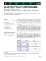
Fig. 1. Serum-induced surface expression of SWTY reporter protein.
(A) Flow cytometric analyses of hemagglutinin (HA)-tagged Kir2.1
channel in HEK293 cells. The shaded areas are staining signals using
anti-HA IgG. Histograms display the cell number (vertical axis) versus
the logarithmic fluorescence intensity (horizontal axis). Mock-trans-
fected cells stained with primary and secondary antibodies served
as background (unshaded areas). The transfected constructs are as
indicated. After starvation, cells were incubated for 6 h with (+) or
without ()) fetal bovine serum. (B) Normalized cell surface fluores-
cence intensity. Geometric means of surface HA staining signals
from cells transfected with three different constructs are normalized
against the signal of Kir2.1-RKR-SWTY with no treatment with fetal
bovine serum. The results shown are the means ± standard errors
(SE) (bars) of three independent experiments. Student’s t-tests were
used to analyse differences. *P < 0.05. **P < 0.01. (C) SWTY-spe-
cific increase in 14-3-3 association by fetal bovine serum induction.
Kir2.1-RAA-SIS, Kir2.1-RKR-SWTY and Kir2.1-RKR-SWAY were
expressed in HEK293 cells in medium without fetal bovine serum
for 24 h. The cells were then further incubated for 6 h in the
absence (lanes 1, 3 and 5) or presence (lanes 2, 4 and 6) of fetal
bovine serum. Top and middle panels are immunoblots of total pro-
tein lysates (input) probed by antibodies for either Kir2.1 or 14-3-3 as
indicated. The bottom panel exhibits an immunoblot by an anti-14-3-
3 IgG after immunoprecipitation of Kir2.1 with anti-HA (IP).
J J. Chung et al. 14-3-3-mediated cell surface expression
FEBS Journal 276 (2009) 5547–5558 ª 2009 The Authors Journal compilation ª 2009 FEBS 5549
consensus sequences. We tested whether these kinases
can phosphorylate the SWTY motif in vitro using puri-
fied glutathione S-transferase (GST) fusions of the
C-terminal cytoplasmic domains of Kir2.1, Kir2.1-
RKR-SWTY and Kir2.1-RKR-SWAY. Figure 2 shows
the results for Akt and PKA by autoradiography. On
incubation with recombinant PKA, robust phosphory-
lation was observed in all three GST fusion proteins
(Fig. 2B, lanes 2–4), consistent with the known PKA
site present in the C-terminal domains of Kir2.1 [26].
However, Akt treatment resulted in significant
33
P
incorporation for GST-cKir2.1-SWTY, whereas GST
alone, GST-cKir2.1 and GST-cKir2.1-SWAY control
proteins showed only background incorporation
(Fig. 2B, lanes 5–8). To demonstrate that the incorpo-
ration of phosphate by Akt really occurred at the )2
Thr position, we generated a phosphor-specific anti-
body for RGRSWpTY-COOH (Fig. S1). Consistent
with the results from radioisotope incorporation, Akt
but not PKA was capable of phosphorylating the )2
Thr of the SWTY motif, hence conferring specific
immunoreactivity to the anti-SWpTY IgG.
To determine whether SWTY can be phosphorylated
in vitro by PKC and CaMK II, ELISA experiments
were performed. Incubation of SWTY peptide with
PKC and CaMK II resulted in basal levels of signal in
-Kir2.1
-SWpTY
Ak t
PKA
_
+
_
_
GST-cKir2.1-SWTY
25 0
15 0
10 0
75
50
37
25
15
Mode I
Mode II
Mode II
I
Kinase recognition sites
PKB/
Akt
PKC
CaMK
II
GS T
FRGR SW TY
FRGR SW AY
RK R
RK R
GS T
GST-cKir2.1
GST-cKir2.1-SWTY
GST-cKir2.1-SWAY
GS T
GS
T
GS T
+
_
_
+
_
_
+
_
Coomassie blue
25 0
15 0
10 0
75
50
37
25
15
P
33
SWTY
14-3-3 binding moti
f
s
PKA
1 2 3 4 5 6 7 8 1 2 3 4 5 6 7 8
Ak t
PKA
Ak t
PKA
cKir2.1
-
cKir2.1-SWTY
cKir2.1-SWAY
cKir2.1
cKir2.1-SWTY
cKir2.1-SWAY
cKir2.1
cKir2.1-SWTY
cKir2.1-SWAY
cKir2.1
cKir2.1-SWTY
cKir2.1-SWAY
GS T
-
-
-
-
0.5
**
0.4
0.3
0.2
0.1
Corrected ELISA signal (A405)
0
None Akt PKC CaMK II
A
B
C
D
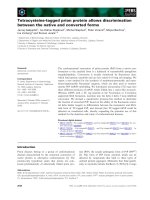
Fig. 2. In vitro phosphorylation of an SWTY
motif by Akt. (A) Sequence comparison of
an SWTY sequence with 14-3-3 binding
motifs and kinase recognition sites. (R) indi-
cates either one or both arginines are
allowed at this position. *C-terminus of the
protein (stop codon). (B) Phosphorylation of
an SWTY motif in vitro. GST fusion proteins
of the C-terminal domains of Kir2.1 (GST-
cKir2.1), GST-cKir2.1 with RKR-SWTY and
RKR-SWAY sequences (GST-cKir2.1-SWTY
and GST-cKir2.1-SWAY, respectively) were
generated as shown and affinity purified.
The GST fusion proteins were in vitro phos-
phorylated with recombinant PKA or Akt.
The reaction products were resolved by
SDS-PAGE and subjected to Coomassie
blue staining (left) and autoradiography
(right). (C) Akt recognition of )2 Thr of the
SWTY sequence. Purified GST-cKir2.1-RKR-
SWTY proteins were in vitro phosphorylated
with either Akt or PKA and analysed by an
anti-phospho-SWTY IgG (left) and an
anti-Kir2.1 IgG (right). (D) Akt-specific phos-
phorylation of an SWTY motif. After in vitro
phosphorylation with Akt, PKC and CaMK II,
biotinylated SWTY peptide was bound to a
streptavidin-coated 96-well plate and sub-
jected to ELISA with anti-SWpTY. The
absorbance at 405 nm (A405) was corrected
against signal with no kinase control. Values
are the means ± standard errors (SE) (bar)
of three independent experiments.
Student’s t-tests were used to analyse
differences; **P < 0.01.
14-3-3-mediated cell surface expression J J. Chung et al.
5550 FEBS Journal 276 (2009) 5547–5558 ª 2009 The Authors Journal compilation ª 2009 FEBS
ELISA using the anti-SWpTY IgG, further supporting
preferential phosphorylation by Akt (Fig. 2D).
PI3K ⁄ Akt-dependent regulation of
SWTY-mediated surface expression in vivo
To demonstrate the causal link between the activation
of Akt and SWTY-mediated surface expression, the
Akt activity in the cells was manipulated by the
co-expression of constitutively active or dominant neg-
ative Akt mutants. Figure 3A, B shows that the induc-
ible effects of fetal bovine serum were blocked by the
co-expression of a dominant negative K179M Akt
(dn-Akt). Furthermore, under serum starvation
conditions, the constitutively active Myr-Akt conferred
surface expression at a similar level to that of fetal
bovine serum-treated control cells (Fig. 3B, None).
Both Akt constructs masked the effect of fetal bovine
serum stimulation. These data provide evidence that
Akt activation is both necessary and sufficient for the
10
0
10
1
10
2
10
3
10
4
10
0
10
1
10
2
10
3
10
4
_
+
Fetal bovine serum
p110-CAAX
IP by -H A
FBS
IP
Inpu t
-SWpTY
Kir2.1
pAkt
Ak t
0
1
2
3
4
5
None p110-CAAX Myr-Akt
Co-transfection
Normalized fluorescence (A.U.)
None
Fetal bovine serum
Kir2.1-RKR-SWTY
+ dn-Ak t
Kir2.1-RKR-SWTY
+ p110-CAAX
Kir2.1-RKR-SWTY
dn-Akt
6
14-3- 3
14-3-3
Kir2.1-RKR-SWTY
+
––
+ + +
+ +
––
+ +
1 2 3 4
+ My r- -Akt
Kir2.1-RKR-SWTY
*
*
*
*
*
**
10
0
10
1
10
2
10
3
10
4
10
0
10
1
10
2
10
3
10
4
10
0
10
1
10
2
10
3
10
4
10
0
10
1
10
2
10
3
10
4
1
0
0
1
0
1
1
0
2
1
0
3
1
0
4
1
0
0
1
0
1
1
0
2
1
0
3
1
0
4
A
B
C
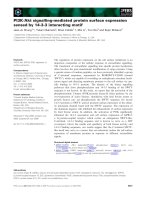
Fig. 3. Akt phosphorylation of SWTY and regulation of SWTY-mediated surface expression in vivo. (A) Flow cytometric analyses of an
HA-tagged Kir2.1-RKR-SWTY channel in HEK293 cells. Surface expression of either Kir2.1-RKR-SWTY alone or co-expressed with the domi-
nant negative form of Akt (dn-Akt), constitutively active form of Akt (Myr-Akt) or PI3K (p110-CAAX) was monitored under starvation and stim-
ulation by fetal bovine serum. (B) Normalized fluorescence of surface expression. Histogram displays the geometric means of the surface
staining signals of Kir2.1-RKR-SWTY normalized against the signal of no stimulation after 24 h of starvation. The results shown are the
means ± standard errors (SE) (bars) of three independent experiments. Student’s t-tests were used to analyse differences; *P < 0.05;
**P < 0.01. (C) Protein expression and binding induced by p110-CAAX. HA-tagged Kir2.1-RKR-SWTY was expressed alone or together with
p110-CAAX in HEK293 cells without fetal bovine serum for 24 h. The cells were then further incubated for 6 h in the absence (lanes 1 and
3) or presence (lanes 2 and 4) of serum. The top two panels show immunoblots by anti-SWpTY or anti-14-3-3 IgG after immunoprecipitation
with anti-HA IgG (IP). The bottom four panels show immunoblots of total protein lysates probed by anti-Kir2.1, anti-pAkt, anti-Akt and
anti-14-3-3 IgGs, respectively.
J J. Chung et al. 14-3-3-mediated cell surface expression
FEBS Journal 276 (2009) 5547–5558 ª 2009 The Authors Journal compilation ª 2009 FEBS 5551
fetal bovine serum-induced surface expression of the
SWTY-carrying Kir2.1 channel.
Because Akt is commonly activated by PI3K, a mem-
brane-targeted constitutively active catalytic subunit of
PI3K (p110-CAAX) was tested for its effects on SWTY-
mediated surface expression. Indeed, under serum star-
vation conditions, the co-expression of p110-CAAX
conferred a comparable surface expression to that by
fetal bovine serum treatment (Fig. 3A, B). Consistent
with its action on the Akt pathway, the co-expression of
p110-CAAX induced phosphorylation of Akt, as probed
by an anti-phospho-Akt IgG. Furthermore, both fetal
bovine serum treatment and p110-CAAX expression
induced an increase in anti-SWpTY signal, accompanied
by an enhanced interaction between Kir2.1-RKR-
SWTY and 14-3-3 (Fig. 3C, lanes 2 and 3). The inhibi-
tors for either PI3K (wortmannin) or Akt (API-2), but
not MEK inhibitor (PD98059), reduced the induction of
the surface expression by fetal bovine serum (Fig. S2).
Akt is a downstream Ser ⁄ Thr kinase of PI3K activated
by insulin and other growth factors, such as platelet-
derived growth factor (PDGF), epidermal growth factor
and insulin-like growth factor, in different cells [27]. To
investigate the specific signalling pathway(s) activating
Akt to direct SWTY-mediated surface expression,
HEK293 cells transfected with Kir2.1-RKR-SWTY were
starved and stimulated with different growth factors,
including PDGF and insulin. Insulin gave rise to the
highest phospho-Akt signal and surface expression,
whereas there were no changes in the total protein
expression of Akt and 14-3-3 on stimulation (Fig. 4).
Therefore, insulin signalling may be one pathway that
critically regulates SWTY-mediated surface expression
through the activation of the PI3K ⁄ Akt pathway.
Regulation of surface expression of GPR15 by
PI3K signalling pathway
Our discovery of the SWTY motif by random peptide
screening has led to the identification of proteins that
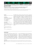
Fig. 4. Induction of SWTY-mediated surface expression by differ-
ent growth factors. (A) Phosphorylation of Akt by different growth
factors. HEK293 cells transfected with HA-tagged Kir2.1-RKR-SWTY
were cultured for 24 h in the absence of fetal bovine serum (FBS)
and stimulated either with fetal bovine serum (10%), PDGF
(60 ngÆmL
–1
) or insulin (100 ngÆmL
–1
) for 3 h. Lysates were sepa-
rated by SDS-PAGE and immunoblotted with antibodies for p-Akt,
Akt, 14-3-3 and Kir2.1. (B) Flow cytometric analyses of HA-tagged
Kir2.1-RKR-SWTY in HEK293 cells. (C) Normalized surface expres-
sion levels determined by fluorescence from flow cytometry in (B).
Values are means ± standard errors (SE) (bar) of four independent
experiments. Student’s t-tests were used to analyze differences;
*P < 0.05; **P < 0.01.
p-Akt
Akt
14-3-3
None
PDGF
Insulin
FBS
Kir2.1
1234
0.5
1.0
1.5
2.0
2.5
3.0
3.5
*
4.0
**
**
**
0
Stimulation
None
FBS
PDGF Insulin
Normalized fluorescence (A.U.)
None FBS
PDGF Insulin
10
0
10
1
10
2
10
3
10
4
10
0
10
1
10
2
10
3
10
4
10
0
10
1
10
2
10
3
10
4
10
0
10
1
10
2
10
3
10
4
A
B
C
14-3-3-mediated cell surface expression J J. Chung et al.
5552 FEBS Journal 276 (2009) 5547–5558 ª 2009 The Authors Journal compilation ª 2009 FEBS
require a C-terminal, SWTY-like signal for efficient
surface expression [8]. These include GPR15 (see
Fig. 5A for C-terminal sequence), a G protein-coupled
receptor that serves as a co-receptor for HIV and sim-
ian immunodeficiency virus (SIV) entry, and has also
been suggested to mediate regenerative enteropathy on
viral infection [28]. Mutation of )2 Ser to Ala (S359A)
abolishes the binding of 14-3-3 and substantially
reduces the surface expression of GPR15 ([8] and
Fig. 5B). To investigate whether the GPR15 C-terminal
sequence plays a similar role to SWTY by sensing
PI3K signalling, we tested the effects of the co-expres-
sion of active PI3K or stimulation with insulin or
serum on the surface expression of GPR15. As the
S359A mutant lacks the ability to bind to 14-3-3, the
change in surface expression level of the S359A mutant
caused by cell treatment would represent 14-3-3-inde-
pendent effects. Under the condition in which PI3K
expression and cell treatments all activated Akt (see
Fig. 5E, pAkt panel), the surface expression of wild-
type GPR15, but not S359A, was significantly elevated
by the over-expression of active PI3K (Fig. 5B, C).
Stimulation of cells with serum or insulin also caused a
significant increase in surface expression only for wild-
type GPR15. The enhancement by these treatments was
efficiently blocked by the PI3K inhibitor LY294002,
which supports the concept that PI3K activation is nec-
essary for the growth factor-induced surface expression
of GPR15 (Fig. 5D). Importantly, the quantification of
14-3-3 proteins co-immunoprecipitated with GPR15
demonstrated that PI3K co-expression and growth fac-
tor treatments all enhanced 14-3-3 binding to GPR15
by two- to four-fold (Fig. 5E, F). Together, these
results suggest that GPR15 surface expression may be
induced by the extracellular signals that activate the
PI3K pathway through the enhancement of 14-3-3
binding to the GPR15 C-terminal sequence.
Discussion
The recent identification of the SWTY (RGRSWTY-
COOH) sequence by genetic screening has reinforced
the concept of a new mode III 14-3-3 binding, which
is characterized by a restricted C-terminal location
and phosphorylation-dependent interaction with 14-3-
3 [8,15,29]. Although various kinases have been
implied in internal mode I and II 14-3-3 binding, a
kinase(s) recognizing a specific C-terminal sequence
and conferring 14-3-3 interaction has not been studied.
In this study, we provide evidence by both in vitro
and in vivo assays that Akt is responsible for the
phosphorylation of the SWTY sequence that leads to
14-3-3 binding.
Akt is a Ser ⁄ Thr kinase that is commonly activated
by PI3K. When extracellular signals, such as insulin
and other growth factors, activate PI3K through
receptor tyrosine kinases, the resultant phosphoinosi-
tide products recruit Akt to the cell membrane, where
Akt is phosphorylated and activated by phosphoinosi-
tide-dependent kinase 1. On activation, Akt phospho-
rylates a variety of proteins at the Ser ⁄ Thr residues of
a consensus sequence, RxRxxS ⁄ T, thereby inducing
diverse biological responses, such as the inhibition of
programmed cell death, promotion of cell proliferation
and regulation of protein trafficking [27,30,31].
Although Akt has been known to control the activity
of a number of cytosolic proteins by enabling 14-3-3
binding on phosphorylation [11,29], our study demon-
strates, for the first time, that C-terminal recognition
by Akt could lead to 14-3-3 binding and the promo-
tion of the cell surface expression of the membrane
protein. This provides a basis for the potential devel-
opment of assay technology and therapeutic interven-
tion by engineering membrane proteins of which
surface expression can be regulated by specific extra-
cellular signals. Functional positioning at the protein
C-terminus makes the SWTY motif suitable for such
purposes.
We have shown that PI3K ⁄ Akt activity or a signal-
ling pathway activating PI3K ⁄ Akt induces phosphory-
lation of the SWTY sequence, recruits 14-3-3 and
confers elevated surface expression of the chimeric
Kir2.1 channel. Because a variety of kinases can recog-
nize the 14-3-3 binding site, receptors with different
14-3-3 binding motifs may sense different signalling
pathways. It is of particular interest that many of the
proteins reported to carry C-terminal 14-3-3 binding
motifs are membrane proteins [4,8,15,32,33]. Among
them is GPR15, an orphan G protein-coupled receptor
that functions as a co-receptor for HIV and SIV entry
[28,34,35]. We have demonstrated previously that the
integrity of the )2 positioned Ser of GPR15 is impor-
tant for binding to 14-3-3, and hence surface expres-
sion [8]. In this study, we show that the surface
expression of GPR15 can be promoted by PI3K acti-
vation that enhances 14-3-3 binding to this receptor
(Fig. 5). It is known that HIV infection activates the
pro-survival PI3K ⁄ Akt pathway and thereby extends
the lifespan of infected macrophages [36]. Our results
suggest that the PI3K signalling pathway may play
further roles in HIV infection by regulating viral selec-
tivity and infection efficiency [34] through alteration of
the cell surface expression level of the HIV co-receptor
GPR15. GPR15 is also involved in the regenerative
enteropathy in HIV ⁄ SIV infection, where cell surface
GPR15 seems to transmit the apoptotic signal in the
J J. Chung et al. 14-3-3-mediated cell surface expression
FEBS Journal 276 (2009) 5547–5558 ª 2009 The Authors Journal compilation ª 2009 FEBS 5553
intestinal cell on contact with the virus [28,35]. Further
studies on endogenous GPR15 protein are necessary to
better understand the role of PI3K signalling in the
14-3-3-mediated surface expression of GPR15 and
HIV pathology.
The molecular mechanisms underlying 14-3-3-medi-
ated surface expression, for example how 14-3-3
protein binding can override the ER localization signal
and cell surface transport, are yet to be elucidated
[14]. Nevertheless, considering the wealth of 14-3-3
binding sequences [9,37], it is conceivable that there
are more membrane proteins than currently known
that are regulated by 14-3-3 binding for their specific
cellular localization. As multiple Ser ⁄ Thr kinases are
AB
CD
EF
Median fluorescence intensity
14-3-3-mediated cell surface expression J J. Chung et al.
5554 FEBS Journal 276 (2009) 5547–5558 ª 2009 The Authors Journal compilation ª 2009 FEBS
known to phosphorylate 14-3-3 binding sites [9,29],
different extracellular signals may confer selective
induction of 14-3-3-mediated surface expression of
membrane proteins. For example, the KCNK3
potassium channel has a C-terminal sequence of
RRSSV-COOH, which binds 14-3-3 in a phosphoryla-
tion-dependent manner and critically regulates surface
expression [5–8]. This sequence matches the consensus
site of PKA rather than Akt, but the identity of the
responsible kinases is not known. Identification and
investigation of more 14-3-3 binding membrane pro-
teins will facilitate our understanding of the molecular
mechanisms underlying the signal-induced 14-3-3-
mediated protein transport.
Materials and methods
Plasmids
Extracellularly HA-tagged mouse Kir2.1 was fused with the
C-terminal cytoplasmic tail of the Kir6.2 channel with mod-
ifications, as described previously [8]. These were
termed Kir2.1-RAA-SIS (Kir2.1-LLDALTLASSRG-
PLRAASVAVAKAKPKFSISPDSLS), Kir2.1-RKR-SWTY
(Kir2.1-LLDALTLASSRGPLRKRSVAVAKAKPKFRGR-
SWTY), Kir2.1-RKR-SWAY (Kir2.1-LLDALTLASSRGP-
LRKRSVAVAKAKPKFRGRSWAY). Human GPR15
plasmids with (S359A) or without (wild-type) Ala mutation
were cloned in pCDNA3.1(+) vector (Invitrogen, Carlsbad,
CA, USA), as described previously [8]. Plasmids expressing
a constitutively active PI3K (p110-CAAX, p110 subunit
fused with a C-terminal isoprenylation motif), constitutively
active Akt (Myr-Akt, Akt fused with an N-terminal myri-
stoylation motif) and kinase-dead dominant negative Akt
(K179M, mutation in lysine at amino acid 179) were identi-
cal to those described previously [38]. GST fusion con-
structs were prepared in pGEX-4T2 vector (GE Healthcare,
Piscataway, NJ, USA).
Antibodies
The rabbit anti-pan-Akt and anti-phospho-Akt IgGs were
purchased from Cell Signaling Technologies (Danvers, MA,
USA). Mouse monoclonal anti-HA and rabbit polyclonal
anti-14-3-3b (this antibody reacts with all seven 14-3-3 iso-
forms) were obtained from Santa Cruz Biotechnologies
(Santa Cruz, CA, USA). Mouse monoclonal anti-human
GPR15 IgG was purchased from R&D Systems
(Minneapolis, MN, USA) and labelled with R-phyco-
erythrin (Invitrogen) before use. The rabbit polyclonal
anti-Kir2.1 serum was raised against the C-terminal cyto-
plasmic region corresponding to amino acids 188–428. The
rabbit polyclonal serum specifically recognizing phosphory-
lated SWTY at the Thr residue was generated by Sigma-
Genosys (The Woodlands, TX, USA). Specificity to the
phosphorylated SWTY sequence was confirmed by ELISA
using N-terminally biotinylated FRGRSWpTY-COOH (pT,
phosphorylated Thr) and FRGRSWTY-COOH peptides
and alkaline phosphatase-conjugated goat anti-rabbit IgG
(Vector Laboratories, Burlingame, CA, USA) (see Fig. S1).
In vitro phosphorylation
The GST-fused C-terminal cytoplasmic tails of Kir2.1,
Kir2.1-RKR-SWTY and Kir2.1-RAA-SWTY were
expressed in BL21 Escherichia coli (Invitrogen) and purified
by glutathione-conjugated Sepharose beads (GE Health-
care). In vitro phosphorylation was performed by incubating
2 lg of each GST fusion protein with 50 ng of recombinant
Akt1 or PKA (Upstate Biotechnology, Lake Placid, NY,
Fig. 5. Regulation of surface expression of GPR15 by PI3K signalling. (A) C-terminal sequence of GPR15. C-terminal nine amino acid resi-
dues of human GPR15 wild-type and S359A mutant were aligned with the SWTY sequence. (B) Flow cytometric analyses of the surface
expression of GPR15. The GPR15 wild-type or S359A mutant was co-transfected with pCDNA3.1 (+) vector, full line with shaded area) or
p110-CAAX (+p110, full line with unshaded area) and cultured for 24 h in the absence of fetal bovine serum (FBS). For growth factor treat-
ment, cells were co-transfected with GPR15 and pCDNA3.1 plasmids, cultured for 24 h in the absence of fetal bovine serum, and treated
with insulin (100 ngÆmL
–1
) or fetal bovine serum (10%) for 6 h. Cells were stained with PE-labelled anti-GPR15 monoclonal IgG. The broken
line represents the background signal of antibody stain from the cells transfected with pCDNA3.1 vector alone. (C) Statistical analysis of the
effects of PI3K, insulin and fetal bovine serum on GPR15 surface expression. The cell populations that showed a positive signal for GPR15
were selected and analysed for the median fluorescence intensity by
FLOJO software. Values are means ± standard errors (SE) (bar) of tripli-
cate samples and were analysed for differences by Student’s t-test. *P < 0.05; **P < 0.005. (D) Effects of PI3K inhibitor on insulin- and fetal
bovine serum-induced surface expression of GPR15. LY294002 (10 l
M) was added to the cell culture 30 min prior to the cell treatment with
insulin or fetal bovine serum. Overlaid histograms are from GPR15-transfected cells with no stimulation (shaded area), with insulin (broken
line) and with LY294002 and insulin (full line with unshaded area). (E) Effects of PI3K, insulin and fetal bovine serum on 14-3-3 binding to
GPR15. Cells prepared as described in (B) were lysed and subjected to the co-immunoprecipitation of 14-3-3 proteins. Total cell lysate (Input)
and the eluates from the HA antibody immunoprecipitant (IP) were resolved by SDS-PAGE and immunoblotted with the corresponding anti-
bodies. In the pAkt panel, the arrow indicates the bands of the phosphorylated Akt signal. (F) Quantitative analysis of co-immunoprecipitated
14-3-3 proteins. The immunoblots developed by chemiluminescence using the imager were quantified for band intensity. The bar graph is a
representative of one of three experiments.
J J. Chung et al. 14-3-3-mediated cell surface expression
FEBS Journal 276 (2009) 5547–5558 ª 2009 The Authors Journal compilation ª 2009 FEBS 5555
USA) at 30 ºC for 60 min in dilution buffer containing
50 mm Tris ⁄ HCl (pH 7.5), 0.1 mm EGTA, 15 mm dith-
iothreitol, 10 m m MgCl
2
,1mm ATP and 0.125 CiÆmL
)1
[
33
P]ATP[cP]. Reaction products were resolved by SDS-
PAGE and subjected to Coomassie blue staining and autora-
diography. For immunoblotting with anti-SWpTY IgG, the
phosphorylation reaction was performed at 30 °C for 30 min
in the absence of [
33
P]ATP[cP].
Cell culture and transfection
HEK293 cells were maintained in 50% DMEM ⁄ 50% F12
medium containing 10% fetal bovine serum. For the stimu-
lation of cells, the transfected cells were serum starved for
24 h and then left untreated (control) or stimulated with
fetal bovine serum (10%), insulin (100 ngÆmL
–1
; Sigma,
St. Louis, MO, USA) or PDGF (60 ngÆmL
–1
; Sigma) for
the indicated times. For inhibitor studies, cells were pre-
treated with various concentrations of LY294002 (Sigma),
wortmannin (Sigma), API-2 (Calibiochem, La Jolla, CA,
USA) or PD098059 (Cell Signaling Technologies) for
30 min. The carrier concentration (0.1% dimethylsulfoxide)
was maintained constant for all cell treatments. Transient
transfections were performed using FuGENE6 (Roche
Applied Sciences, Indianapolis, IN, USA), 30 min after ini-
tiation of starvation.
Flow cytometry
Transfected HEK293 cells were harvested and washed with
Hanks’ balanced salt solution supplemented with 5 mm
Hepes (pH 7.3) and 2% fetal bovine serum (staining med-
ium). All antibody incubations and washes were performed
in staining medium at 4 °C. For the HA-tagged Kir2.1
channel, the cells were stained with anti-HA IgG for
30 min on ice, followed by 10 min staining with Alexa
Fluor 488-conjugated goat anti-mouse IgG (Molecular
Probes, Eugene, OR, USA). For GPR15, the cells were
stained with R-phycoerythrin-labelled anti-GPR15 IgG for
30 min on ice. The stained cells were examined for cell
surface fluorescence with FACSCalibur (BD Biosciences,
San Jose, CA, USA) and analysed with FloJo software
(Tree Star Inc., Ashland, OR, USA).
Immunoprecipitation and immunoblot
For immunoprecipitation, transfected cells were washed
with NaCl ⁄ P
i
once and lysed with lysis buffer (1% NP40,
25 mm Tris, 150 m m NaCl, pH 7.5) with protease inhibitor
cocktails (Sigma) for 20 min at 4 ° C. After centrifugation
for 20 min at 11 000 g, the supernatant was mixed with
protein A-conjugated agarose beads (Sigma) pre-incubated
with 1 lg of anti-HA IgG. After overnight incubation,
the beads were washed three times with lysis buffer, and
then the precipitated proteins were eluted with 2· sample
buffer for SDS-PAGE analysis and immunoblot, as
described previously [39]. The samples resolved in SDS-
PAGE gels were transferred to nitrocellulose and blotted
with the corresponding primary antibodies, followed by
horseradish peroxidase-conjugated secondary antibodies.
The immunoblots were developed with the ECL system
(GE Healthcare). In some experiments, the blots were
developed using Molecular Imager (Bio-Rad, Hercules, CA,
USA) for the quantification of the band intensity.
Statistical analysis
Values are expressed as the means ± standard error. The
significance of the difference between the means was calcu-
lated by Student’s t-test.
Acknowledgements
We thank members of the Shikano laboratory and
Li laboratory for valuable comments and discussions
on the manuscript. This work was supported by grants
from the National Institutes of Health (GM070959,
GM078579 and U54 MH084691 to ML), predoctoral
fellowship awards from the National Institutes of
Health (to J-JC) and the American Heart Association
(to BC).
References
1 Wallin E & von Heijne G (1998) Genome-wide analysis
of integral membrane proteins from eubacterial,
archaean, and eukaryotic organisms. Protein Sci 7,
1029–1038.
2 Viard P, Butcher AJ, Halet G, Davies A, Nurnberg B,
Heblich F & Dolphin AC (2004) PI3K promotes
voltage-dependent calcium channel trafficking to the
plasma membrane. Nat Neurosci 7 , 939–946.
3 Bezzerides VJ, Ramsey IS, Kotecha S, Greka A &
Clapham DE (2004) Rapid vesicular translocation and
insertion of TRP channels. Nat Cell Biol 6, 709–720.
4 O’Kelly I, Butler MH, Zilberberg N & Goldstein SA
(2002) Forward transport. 14-3-3 binding overcomes
retention in endoplasmic reticulum by dibasic signals.
Cell 111, 577–588.
5 Rajan S, Preisig-Muller R, Wischmeyer E, Nehring R,
Hanley PJ, Renigunta V, Musset B, Schlichthorl G, Derst
C, Karschin A et al. (2002) Interaction with 14-3-3 pro-
teins promotes functional expression of the potassium
channels TASK-1 and TASK-3. J Physiol 545, 13–26.
6 Zuzarte M, Heusser K, Renigunta V, Schlichthorl G,
Rinne S, Wischmeyer E, Daut J, Schwappach B &
Preisig-Muller R (2009) Intracellular traffic of the K+
14-3-3-mediated cell surface expression J J. Chung et al.
5556 FEBS Journal 276 (2009) 5547–5558 ª 2009 The Authors Journal compilation ª 2009 FEBS
channels TASK-1 and TASK-3: role of N- and C-termi-
nal sorting signals and interaction with 14-3-3 proteins.
J Physiol 587 (Pt5), 929–952.
7 O’Kelly I & Goldstein SA (2008) Forward transport of
K2p3.1: mediation by 14-3-3 and COPI, modulation by
p11. Traffic 9, 72–78.
8 Shikano S, Coblitz B, Sun H & Li M (2005) Genetic
isolation of transport signals directing cell surface
expression. Nat Cell Biol 7, 985–992.
9 Pozuelo Rubio M, Geraghty KM, Wong BH, Wood
NT, Campbell DG, Morrice N & Mackintosh C (2004)
14-3-3-affinity purification of over 200 human phospho-
proteins reveals new links to regulation of cellular
metabolism, proliferation and trafficking. Biochem J
379, 395–408.
10 Jin J, Smith FD, Stark C, Wells CD, Fawcett JP, Kulk-
arni S, Metalnikov P, O’Donnell P, Taylor P, Taylor L
et al. (2004) Proteomic, functional, and domain-based
analysis of in vivo 14-3-3 binding proteins involved in
cytoskeletal regulation and cellular organization. Curr
Biol 14, 1436–1450.
11 Fu H, Subramanian RR & Masters SC (2000) 14-3-3
proteins: structure, function, and regulation. Annu Rev
Pharmacol Toxicol 40, 617–647.
12 Yaffe MB (2002) How do 14-3-3 proteins work? – Gate-
keeper phosphorylation and the molecular anvil hypoth-
esis FEBS Lett 513, 53–57.
13 Coblitz B, Wu M, Shikano S & Li M (2006) C-terminal
binding: an expanded repertoire and function of 14-3-3
proteins. FEBS Lett 580, 1531–1535.
14 Ganguly S, Weller JL, Ho A, Chemineau P, Malpaux B
& Klein DC (2005) Melatonin synthesis: 14-3-3-depen-
dent activation and inhibition of arylalkylamine N-ace-
tyltransferase mediated by phosphoserine-205. Proc
Natl Acad Sci USA 102, 1222–1227.
15 Coblitz B, Shikano S, Wu M, Gabelli SB, Cockrell LM,
Spieker M, Hanyu Y, Fu H, Amzel LM & Li M (2005)
C-terminal recognition by 14-3-3 proteins for surface
expression of membrane receptors. J Biol Chem 280,
36263–36272.
16 Shikano S, Coblitz B, Wu M & Li M (2006) 14-3-3 pro-
teins: regulation of endoplasmic reticulum localization
and surface expression of membrane proteins. Trends
Cell Biol 16, 370–375.
17 Mrowiec T & Schwappach B (2006) 14-3-3 proteins in
membrane protein transport. Biol Chem 387, 1227–
1236.
18 Zerangue N, Schwappach B, Jan YN & Jan LY (1999)
A new ER trafficking signal regulates the subunit
stoichiometry of plasma membrane K(ATP) channels.
Neuron 22, 537–548.
19 Shikano S & Li M (2003) Membrane receptor traffick-
ing: evidence of proximal and distal zones conferred by
two independent endoplasmic reticulum localization
signals. Proc Natl Acad Sci USA 100, 5783–5788.
20 Zhou Y, Schopperle WM, Murrey H, Jaramillo A,
Dagan D, Griffith LC & Levitan IB (1999) A dynami-
cally regulated 14-3-3, Slob, and Slowpoke potassium
channel complex in Drosophila presynaptic nerve
terminals. Neuron 22, 809–818.
21 Thulin CD, Savage JR, McLaughlin JN, Truscott SM,
Old WM, Ahn NG, Resing KA, Hamm HE, Bitensky
MW & Willardson BM (2001) Modulation of the
G protein regulator phosducin by Ca
2+
⁄ calmodulin-
dependent protein kinase II phosphorylation and 14-3-3
protein binding. J Biol Chem 276, 23805–23815.
22 Chan HC, Wu WL, So SC, Chung YW, Tsang LL,
Wang XF, Yan YC, Luk SC, Siu SS, Tsui SK et al.
(2000) Modulation of the Ca(2+)-activated Cl(–) chan-
nel by 14-3-3epsilon. Biochem Biophys Res Commun
270, 581–587.
23 Kagan A, Melman YF, Krumerman A & McDonald
TV (2002) 14-3-3 amplifies and prolongs adrenergic
stimulation of HERG K+ channel activity. EMBO J
21, 1889–1898.
24 Savage JR, McLaughlin JN, Skiba NP, Hamm HE &
Willardson BM (2000) Functional roles of the two
domains of phosducin and phosducin-like protein.
J Biol Chem 275, 30399–30407.
25 Pozuelo Rubio M, Campbell DG, Morrice NA &
Mackintosh C (2005) Phosphodiesterase 3A binds to
14-3-3 proteins in response to PMA-induced phosphory-
lation of Ser428. Biochem J 392, 163–172.
26 Wischmeyer E & Karschin A (1996) Receptor stimula-
tion causes slow inhibition of IRK1 inwardly rectifying
K+ channels by direct protein kinase A-mediated phos-
phorylation. Proc Natl Acad Sci USA 93, 5819–5823.
27 Brazil DP, Yang ZZ & Hemmings BA (2004) Advances
in protein kinase B signalling: AKT ion on multiple
fronts. Trends Biochem Sci 29, 233–242.
28 Li Q, Estes JD, Duan L, Jessurun J, Pambuccian S,
Forster C, Wietgrefe S, Zupancic M, Schacker T, Reilly
C et al. (2008) Simian immunodeficiency virus-induced
intestinal cell apoptosis is the underlying mechanism of
the regenerative enteropathy of early infection. J Infect
Dis 197, 420–429.
29 Dougherty MK & Morrison DK (2004) Unlocking the
code of 14-3-3. J Cell Sci 117, 1875–1884.
30 Lawlor MA & Alessi DR (2001) PKB ⁄ Akt: a key medi-
ator of cell proliferation, survival and insulin responses?
J Cell Sci 114, 2903–2910.
31 Chae KS, Martin-Caraballo M, Anderson M & Dryer
SE (2005) Akt activation is necessary for growth factor-
induced trafficking of functional K(Ca) channels in
developing parasympathetic neurons. J Neurophysiol 93,
1174–1182.
32 Bodnar RJ, Gu M, Li Z, Englund GD & Du X (1999)
The cytoplasmic domain of the platelet glycoprotein
Ibalpha is phosphorylated at serine 609. J Biol Chem
274, 33474–33479.
J J. Chung et al. 14-3-3-mediated cell surface expression
FEBS Journal 276 (2009) 5547–5558 ª 2009 The Authors Journal compilation ª 2009 FEBS 5557
33 Sliva D, Gu M, Zhu YX, Chen J, Tsai S, Du X &
Yang YC (2000) 14-3-3zeta interacts with the alpha-
chain of human interleukin 9 receptor. Biochem J 345
Pt 3, 741–747.
34 Wade-Evans AM, Russell J, Jenkins A & Javan C
(2001) Cloning and sequencing of cynomolgus macaque
CCR3, GPR15, and STRL33: potential coreceptors for
HIV type 1, HIV type 2, and SIV. AIDS Res Hum
Retroviruses 17, 371–375.
35 Maresca M, Mahfoud R, Garmy N, Kotler DP, Fantini
J & Clayton F (2003) The virotoxin model of HIV-1
enteropathy: involvement of GPR15 ⁄ Bob and galacto-
sylceramide in the cytopathic effects induced by HIV-1
gp120 in the HT-29-D4 intestinal cell line. J Biomed Sci
10, 156–166.
36 Chugh P, Bradel-Tretheway B, Monteiro-Filho CM,
Planelles V, Maggirwar SB, Dewhurst S & Kim B
(2008) Akt inhibitors as an HIV-1 infected macrophage-
specific anti-viral therapy. Retrovirology 5 , 11.
37 Kakiuchi K, Yamauchi Y, Taoka M, Iwago M, Fujita
T, Ito T, Song SY, Sakai A, Isobe T & Ichimura T
(2007) Proteomic analysis of in vivo 14-3-3 interactions
in the yeast Saccharomyces cerevisiae . Biochemistry 46,
7781–7792.
38 Chen R, Kim O, Yang J, Sato K, Eisenmann KM,
McCarthy J, Chen H & Qiu Y (2001) Regulation of
Akt ⁄ PKB activation by tyrosine phosphorylation. J Biol
Chem 276, 31858–31862.
39 Chung JJ & Li M (2005) Biochemical characterization
of the native Kv2.1 potassium channel. FEBS J 272,
3743–3755.
Supporting information
The following supplementary material is available:
Fig. S1. Specificity of anti-phospho SWTY IgG.
Fig. S2. Effects of kinase inhibitors on fetal bovine
serum-induced s urface expression of Kir2.1-RKR-SWTY.
This supplementary material can be found in the
online version of this article.
Please note: As a service to our authors and readers,
this journal provides supporting information supplied
by the authors. Such materials are peer-reviewed and
may be re-organized for online delivery, but are not
copy-edited or typeset. Technical support issues arising
from supporting information (other than missing files)
should be addressed to the authors.
14-3-3-mediated cell surface expression J J. Chung et al.
5558 FEBS Journal 276 (2009) 5547–5558 ª 2009 The Authors Journal compilation ª 2009 FEBS