Báo cáo khoa học: Evolutionary changes to transthyretin: evolution of transthyretin biosynthesis doc
Bạn đang xem bản rút gọn của tài liệu. Xem và tải ngay bản đầy đủ của tài liệu tại đây (433.56 KB, 15 trang )
MINIREVIEW
Evolutionary changes to transthyretin: evolution of
transthyretin biosynthesis
Samantha J. Richardson
School of Medical Sciences, RMIT University, Bundoora, Vic., Australia
Introduction
Thyroid hormones (THs) are essential for normal
growth and development, and for regulation of the
basal metabolic rate. The two major thyroid hormones
are 5¢,3¢,5,3-tetraiodo-[L]-thyronine (thyroxine, T4)
and 3¢,5,3-triiodo-[L]-thyronine (T3). THs are synthe-
sized by the thyroid gland and then secreted into the
bloodstream (see Fig. 1). In mammals, most of the TH
produced by the thyroid gland is in the form of T4,
which has higher affinity than T3 for the TH distribu-
tor proteins (THDPs) in the blood [1]. However, T3
has higher affinity than T4 for the thyroid hormone
receptors (TRs) [2]. More than 99% of TH in blood is
bound to THDPs, which prevent avid nonspecific par-
titioning of THs into membranes. THs dissociate from
THDPs and can enter cells via TH transporters or by
passive diffusion as a result of their lipophilicity. THs
can be deiodinated by a family of deiodinases to either
activate (T4–T3) or deactivate [T4–rT3 (reverse T3),
T3–T2, etc.] THs [3]. Within cells, THs bind to specific
cytosolic TH-binding proteins before being translocat-
ed into the nucleus. THs elicit their effects by binding
to TR ⁄ RXR dimers in the nucleus, and together with
co-activator or co-repressor proteins, directly modulate
the expression of specific genes (see Fig. 1).
Many genes regulated by THs are involved in
growth and development, particularly of the brain [4].
Thus, normal growth and development requires tightly
regulated levels of THs to reach the nucleus of cells
throughout the body and brain, and a strong network
of buffering and regulatory feedback systems in order
Keywords
amphibians; birds; brain; choroid plexus;
eutherians; evolution; fish; gene regulation;
liver; marsupials; monotremes; reptiles;
thyroid hormones; transthyretin; vertebrates
Correspondence
S. J. Richardson, School of Medical
Sciences, RMIT University, PO Box 71,
Bundoora, Vic. 3083, Australia
Fax: +61 3 9925 7063
Tel: +61 3 9925 7897
E-mail:
(Received 2 February 2009, revised 11 June
2009, accepted 12 June 2009)
doi:10.1111/j.1742-4658.2009.07244.x
Thyroid hormones are involved in growth and development, particularly of
the brain. Thus, it is imperative that these hormones get from their site of
synthesis to their sites of action throughout the body and the brain. This
role is fulfilled by thyroid hormone distributor proteins. Of particular inter-
est is transthyretin, which in mammals is synthesized in the liver, choroid
plexus, meninges, retinal and ciliary pigment epithelia, visceral yolk sac,
placenta, pancreas and intestines, whereas the other thyroid hormone
distributor proteins are synthesized only in the liver. Transthyretin is syn-
thesized by all classes of vertebrates; however, the tissue specificity of trans-
thyretin gene expression varies widely between classes. This review
summarizes what is currently known about the evolution of transthyretin
synthesis in vertebrates and presents hypotheses regarding tissue-specific
synthesis of transthyretin in each vertebrate class.
Abbreviations
ApoAI, apolipoprotein AI; CSF, cerebrospinal fluid; LAMP-1, lysosome-associated membrane protein; RBP, retinol-binding protein; T3, 3¢,3,5-
triiodo-[L]-thyronine; T4, 3¢,5¢,3,5,-tetraiodo-[L]-thyronine; TBG, thyroxine-binding globulin; TBPA, thyroxine-binding prealbumin; TH, thyroid
hormone; THDP, thyroid hormone distributor protein; TLP, transthyretin-like protein; TRE, thyroid hormone response elements.
5342 FEBS Journal 276 (2009) 5342–5356 ª 2009 The Author Journal compilation ª 2009 FEBS
to maintain euthyroid homeostasis. For example,
insufficient TH during gestation in humans leads to
irreversible brain damage and mental retardation.
Many hormones affect neurogenesis in the adult brain
[5]. In rodents, THs are required for normal cycling of
adult neural stem cells in the subventricular zone [6].
A dramatic example of the effect of THs on develop-
ment is the metamorphosis of tadpoles into frogs: the
animal changes from an aquatic herbivore (with a long
intestine) with gills and a tail, to a terrestrial carnivo-
rous (with a short intestine) tetrapod with lungs. This
remarkable transition requires a finely regulated
co-ordination of gene-transcription events directing
apoptosis, resorption and tissue remodelling, which is
driven by THs [7]. This illustrates the importance of
the quantitative, temporal and spatial requirements of
TH distribution during development.
Often, the focus of TH-regulated events is on the
interaction of the THs with their receptors, co-modula-
tors and the thyroid hormone response elements
(TREs) in the target genes. However, this is just the
final step in a long chain of events that have been
quantitatively regulated at each step. The movement of
THs from the thyroid gland to a target cell is governed
by the THDPs in the blood and cerebrospinal fluid
(CSF). In humans (but not in all vertebrates or even in
all mammals), the THDPs in blood are albumin, trans-
thyretin and thyroxine-binding globulin (TBG). These
three proteins are synthesized by the liver and secreted
into the bloodstream. Transthyretin has intermediate
affinity for THs, between those for albumin (lower
affinity) and TBG (higher affinity). Together, they
form a buffering network system for TH distribution
in the blood [8]. The brain is separated from the rest
of the body by a set of interfaces often referred to as
‘the blood–brain barrier’, which actually consists of
four barrier interfaces [9]. Only one THDP is made in
the brain, namely transthyretin. Transthyretin is syn-
thesized by the epithelial cells of the choroid plexus
[10], which is the blood–CSF barrier and produces
most of the CSF. This transthyretin is secreted exclu-
sively into the CSF and is involved in the transport of
THs from the blood into the brain and throughout the
CSF [11]. This review will address the evolution of
transthyretin synthesis in vertebrates, specifically: the
sites of transthyretin synthesis; the evolution of tissue-
specific transthyretin synthesis in fish, amphibians, rep-
tiles, birds, monotremes, marsupials and eutherians;
the regulation of transthyretin gene expression; and
the change of transthyretin ligand in mammals.
Transthyretin
Transthyretin was discovered in 1942 in both human
CSF [12,13] and human serum [14]. It was originally
named ‘prealbumin’ because it was the only plasma
protein that migrated anodal to albumin during elec-
trophoresis. Transthyretin has a molecular mass of
about 55 kDa and is composed of four identical
subunits of about 14 kDa. It was not until Ingbar used
a Tris–malate buffer (rather than the then standard
barbital buffer) for the electrophoretic analysis of
serum that prealbumin was identified as a thyroid hor-
mone-binding protein [15] (barbital inhibits binding of
THs to transthyretin). Thus, the name was changed to
‘thyroxine-binding prealbumin’ (TBPA). A decade
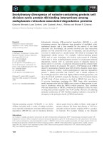
Fig. 1. Five classes of TH-binding proteins.
The thyroid gland secreted TH (predomi-
nantly T4 in mammals) into the blood,
where it binds THDPs (1). TH can dissociate
from THDPs and enter cells by passive
diffusion, or via TH transporter proteins (2).
Within the cell, THs can be deiodinated by
deiodinases (3) and bind cytosolic
TH-binding proteins (4). Within the nucleus,
T3 binds TH receptors (TRs) (5). NB:
deiodinases D1, D2 and D3 have different
locations with a cell; TRs change their
conformation upon binding to DNA.
,
albumin;
, transthyretin (TTR); , TBG;
, TH transporter; , deiodinase; ,
cytosolic TH-binding protein;
, TR; ,TR
bound to DNA. ([18]. Used with permission.)
S. J. Richardson Evolution of transthyretin biosynthesis
FEBS Journal 276 (2009) 5342–5356 ª 2009 The Author Journal compilation ª 2009 FEBS 5343
later, Raz and Goodman [16] discovered that TBPA
also bound retinol-binding protein (RBP). In 1981 the
name was finally changed again to ‘transthyretin’,
which describes its roles in the TRANSport of THY-
roid hormones and RETINol-binding protein [17]. For
details of the structure of transthyretin, see the review
in this series by Dr Hennebry.
Transthyretin synthesis has been identified in the
liver, in the choroid plexus of the brain, and in the
meninges, retinal and ciliary pigment epithelia, visceral
yolk sac, placenta, pancreas and intestine (see below),
whereas albumin synthesis and TBG synthesis have
only been identified in the liver.
Ligands of transthyretin
To assess the selection pressures governing the
regulation of tissue-specific transthyretin synthesis,
the functions of transthyretin must be considered.
Transthyretin has multiple ligands that can be divided
into two categories: ‘natural’ and ‘synthetic’. The natu-
ral ligands of transthyretin include: thyroid hormones
(T3 and T4) and RBP, which itself binds retinol, metal
ions, plant flavonoids, apolipoprotein AI (ApoAI) and
lysosome-associated membrane protein (LAMP-1). The
synthetic ligands include nonsteroidal anti-inflamma-
tory drugs, polychlorinated biphenols, industrial pollu-
tants and flame retardants [18]. As these synthetic
compounds can displace THs from transthyretin, they
can act as potent endocrine disruptors. Furthermore,
these endocrine disruptors can be transported into the
brain via binding to transthyretin synthesized by the
choroid plexus and have the potential to accumulate in
the brain. However, as this review is focused on the
evolution of transthyretin synthesis, only the natural
ligands of transthyretin will be discussed. For reviews
on non-TH ligands of transthyretin, readers are direc-
ted to excellent reviews published previously [19–24].
TH
In human blood, 99.97% of T4 and 99.70% of T3 is
bound to the THDPs albumin, transthyretin and TBG
[25]. Of these, TBG has the highest affinity for T4 and
T3 (1.0 · 10
10
and 4.6 · 10
8
m
)1
, respectively), trans-
thyretin has intermediate affinity (7.0 · 10
7
and
1.4 · 10
7
m
)1
, respectively) and albumin has the lowest
affinity (7.0 · 10
5
and 1.0 · 10
5
m
)1
, respectively).
Together, these three THDPs form a buffering net-
work for free T4 in blood (24 pm), which could assist
in protection against hypothyroidism (abnormally low
levels of free TH in blood) or hyperthyroidism (abnor-
mally high levels of free TH in blood) [8].
The function of THDPs is to ensure an even dis-
tribution of TH throughout tissues and to maintain
a circulating TH pool of sufficient size in the blood
and CSF [26]. To determine which of the three
THDPs contributes most effectively to the delivery
of THs to tissues, the dissociation rates and the cap-
illary transit times have to be considered. In brief,
the dissociation rates for T4 and T3 from TBG are
0.018 and 0.16 s
)1
, respectively; from transthyretin
are 0.094 and 0.69 s
)1
, respectively; and from albu-
min are 1.3 and 2.2 s
)1
, respectively [27]. Thus, given
the capillary transit times for various tissues [28],
transthyretin is responsible for much of the immedi-
ate delivery of THs to tissues [29]. An analogy by
Ingbar describes it quite nicely: ‘TBG is the savings
account for thyroxine and TBPA is the checking
account’ [30].
In mammals, transthyretin, albumin and TBG have
higher affinity for T4 than for T3 (see above), and, as
the concentrations of both free and total T4 are higher
than those of T3, T4 is often referred to as the ‘trans-
port form’ of TH. As T3 has higher affinity than T4
for the TH nuclear receptors [2], T3 is often referred
to as the ‘active form’ of TH. However, in birds, rep-
tiles, amphibians and fish, transthyretin has a higher
affinity for T3 than for T4 (see review in this series by
Dr Prapunpoj) and these animals do not have TBG in
their blood. Therefore, these animals could have a
potentially greater ratio of T3 to T4 in their blood
than mammals. By contrast, in mammals, transthyretin
and TBG distribute T4 (the precursor form) around
the blood rather than T3 (the ‘active’ form), which
binds to the nuclear receptors. This allows for tissue-
specific activation of T4–T3 by deiodinases, at the pre-
cise sites where T3 is required, giving a greater level of
control of TH action in mammals. This could be a
selection pressure for the change in ligand binding of
transthyretin from T3 (in fish, amphibians, reptiles and
birds) to T4 (in mammals).
RBP
RBP was first described by Kanai et al., in 1968 [31],
and was found to be bound to transthyretin in serum.
It was suggested that the transthyretin–RBP ⁄ retinol
complex (80 kDa) or the retinol ⁄ RBP–transthyretin–
RBP ⁄ retinol complex (100 kDa) prevented loss of
RBP–retinol (21 kDa) via glomerular filtration in the
kidneys [16]. The RBP–retinol complex has higher
affinity for transthyretin than apoRBP [32]. The X-ray
crystal structures of RBP–transthyretin complexes have
demonstrated that up to two molecules of RBP can
bind one tetramer of transthyretin [33].
Evolution of transthyretin biosynthesis S. J. Richardson
5344 FEBS Journal 276 (2009) 5342–5356 ª 2009 The Author Journal compilation ª 2009 FEBS
The hypothesis that RBP binds to transthyretin to
prevent loss of RBP and retinol by filtration in the kid-
neys may hold true for eutherians (‘placental mam-
mals’), but it is not immediately convincing when
considering other animals. For example, there are two
Orders of marsupials: the Diprotodonta (e.g. kanga-
roos, koalas and wombats) and the Polyprotodonta
(e.g. Tasmanian devil, dunnarts and Antechinus). Adult
Diprotodonta have transthyretin in their blood, whereas
adult Polyprotodonta do not have transthyretin in their
blood [34]. This raises the question as to whether there
is a difference in the glomerular filtration size cut-off in
diprotodont marsupials compared with that of polypro-
todont marsupials. Similarly, all of the species of sexu-
ally mature fish, amphibians, reptiles and monotremes
studied have RBP in their blood, but not transthyretin.
This raises questions as to whether the glomerular filtra-
tion cut-off is significantly smaller in noneutherians, or
if a plasma protein other than transthyretin fulfills the
role of binding RBP to prevent its loss via the kidneys.
If the function of transthyretin was to prevent loss of
RBP–retinol through the kidneys, one might speculate
that hepatic transthyretin synthesis would have
co-evolved with hepatic RBP synthesis and that genes
for both transthyretin and RBP would have similar
developmental and evolutionary expression patterns.
Metal ions, plant flavonoids, ApoAI and LAMP-I
The vast majority of data on transthyretin binding to
metal ions [35], plant flavonoids [20], ApoAI [36] and
LAMP-I [37] pertain to eutherian transthyretins.
Therefore, this data set is not broad enough to build
hypotheses regarding selection pressures leading to the
binding of these compounds by transthyretins during
evolution. Thus, it is not yet possible to produce a sec-
tion on the influence of these ligands on the evolution
of transthyretin synthesis.
Sites of transthyretin synthesis
Liver
Transthyretin is synthesized by the liver and secreted
into the blood [38], where it binds THs and RBP ⁄ reti-
nol. However, transthyretin–RBP ⁄ retinol can also be
secreted from the liver as a complex [39]. Thus, hepatic
transthyretin is involved in the distribution of THs
and retinol throughout the body via the blood. The
protein-bound pool of THs is believed to counteract
the avid partitioning of the lipophilic THs into the
lipid membranes and to maintain a circulating pool of
THs in the bloodstream [26]. Very recently, it has been
revealed that transthyretin is also involved in periph-
eral nerve regeneration [40].
Choroid plexus
Transthyretin is synthesized by the choroid plexus epi-
thelial cells and secreted into the CSF [10]. At least in
rodents, this transthyretin is involved in the movement
of T4 (but not of T3) from the blood into and within the
brain, as previously reviewed [18]. In addition, transthy-
retin synthesized by the choroid plexus and secreted into
the CSF and interstitial fluid is involved in the delivery
of TH to stem cells and progenitor cells within the sub-
ventricular zone of the brain [41], which requires TH for
cell cycle regulation [6]. The absence of transthyretin
synthesized by the choroid plexus results in reduced
apoptosis of progenitor cells in the subventricular zone
of the adult mouse brain [41], spatial reference memory
impairment [42], increased exploratory activity and
reduced depressive behaviour [43], and overexpression
of the neuropeptide Y phenotype [44]. Reduced levels of
transthyretin have been reported in the CSF of patients
suffering from depression, Alzheimer’s disease and
Down’s syndrome [18]. In the light of reports of
decreased transthyretin synthesis and secretion in the
brains of ageing mammals [45], the role of transthyretin
in the ageing brain requires further investigation.
Visceral yolk sac
Transthyretin and RBP synthesized in the visceral yolk
sac of rodents has been suggested to be involved in the
transport of THs and retinol from the maternal circu-
lation to the developing fetus [46,47]. Further support
for this came from a previous publication [48] in which
it was demonstrated that both transthyretin and RBP
are secreted across the basolateral membrane towards
the fetal circulation; the report also suggested that the
visceral yolk sac could be the source of plasma pro-
teins for the fetus before the fetal liver is functional.
Placenta
Transthyretin synthesis by the eutherian placenta has
been suggested indirectly [49] and more recently dem-
onstrated directly [50], where it has been proposed to
be involved with the transfer of THs from the mother
to the fetus.
Retinal and ciliary pigment epithelia of the eye
Transthyretin is synthesized by the retinal pigment
epithelium of the eye in several eutherian species [51]
S. J. Richardson Evolution of transthyretin biosynthesis
FEBS Journal 276 (2009) 5342–5356 ª 2009 The Author Journal compilation ª 2009 FEBS 5345
and is secreted across the apical membrane into the
extracellular matrix, together with RBP that is also
synthesized by the retinal pigment epithelium [52].
Transthyretin and RBP synthesized by the retinal pig-
ment epithelium have been proposed to be involved in
the delivery of retinol to Mu
¨
ller and amacrine cells
[52], where it is converted to retinal, which is required
for photoreceptor function. More recently, transthyre-
tin synthesis by the ciliary pigment epithelium was
identified, at about one-third of the levels found in the
retinal pigment epithelium [53].
Intestine
Transthyretin synthesis has been identified in human
intestines during fetal development [54], but not in the
intestine of adult rats [55]. A function for transthyretin
synthesized by the intestine has not yet been defined.
However, as the intestines are extrahepatic tissue with
the highest concentration of THs [56], a role for TH
distribution or transport seems likely.
Pancreas
Transthyretin synthesis in the islets of Langerhans of
rat pancreas has previously been described [57].
Recently, a role for transthyretin in promoting glu-
cose-induced increases in cytoplasmic calcium ion
concentration and insulin release in pancreatic beta
cells has been proposed [58]. A role for the transthy-
retin tetramer in protection against beta cell apoptosis
was also proposed, having implications for type 1
diabetes in humans.
Other tissues
A single observation of extremely low levels of trans-
thyretin synthesis by the meninges in rat brain has
been reported [59]. Transthyretin synthesis (detected by
PCR) has also been identified in the skin, heart, skele-
tal muscle, kidney, testis, gills and pituitary in a species
of adult fish (sea bream, Sparus aurata) [60]. Functions
for transthyretin synthesized in these tissues have not
yet been identified.
Sites of transthyretin synthesis
throughout vertebrate evolution
Fish
Among teleost fish, transthyretin synthesis in the whole
animal has been reported during early embryogenesis
in sea bream (S. aurata) [60]. Masu salmon (Oncorhyn-
chus massou) synthesize transthyretin in their liver only
during smoltification (a process driven by THs) [61],
and subsequently Atlantic salmon (Salmo salar) and
Chinook salmon (Oncorhynchus tshawytscha) were
reported to undergo hepatic transthyretin synthesis
only during smoltification [62]. Hepatic transthyretin
synthesis was also detected in 3-year-old tuna (Thun-
nus orientalis) [63].
A comprehensive survey of tissues in adult sea
bream revealed a wide distribution of transthyretin
transcripts after PCR analysis (which is a more sensi-
tive method than those used in other studies refer-
enced) in liver, intestine, whole brain, kidney, testis,
gills and pituitary. However, only the signal in the liver
could be confirmed by northern blotting analysis [60].
Until now, there have been no published data on
transthyretin synthesis by the choroid plexus of teleost
fish. There is an unpublished report that fish choroid
plexus does not synthesize transthyretin (G. Schreiber,
personal communication); however, using PCR, Santos
and Power [60] amplified transthyretin transcript from
the whole brain of adult sea bream, which presumably
contains the choroid plexus. Whether this transthyretin
was synthesized by the choroid plexus remains to be
investigated.
Of the agnathan fish, two species (from two different
genera) of lamprey have been studied [64]. Transthyre-
tin cDNAs were cloned and sequenced from Petromy-
zon marinus and Lampetra appendix. These are the first
transthyretin sequences from vertebrates basal to tele-
ost fish. The N-terminal regions of transthyretin
subunits from both species were longer than those from
other vertebrates. Transthyretin was found to be syn-
thesized in the liver of lampreys throughout their life
cycles and the synthesis of transthyretin was elevated
during metamorphosis. In other vertebrates, a transient
increase in transthyretin ⁄ THDP coincides with the
increase in TH levels during development (mammals),
metamorphosis (amphibians) or smoltification (fish)
[62]. These processes are (at least in part) driven by
THs. However, in these two species of lampreys, the
increase in transthyretin gene expression coincides with
a decrease in plasma TH levels [64]. The Agnatha are
at least 530 Myr old, and the function of THs in lam-
preys appears to be different from that in most other
vertebrates, as a decrease in TH triggers metamorpho-
sis, rather than an increase in TH concentrations [65].
Accordingly, lamprey metamorphosis can also be
induced by goitrogens [66]. It is intriguing that in
amphibians an increase in hepatic transthyretin gene
expression coincides with an increase in TH concentra-
tion in the blood, which drives metamorphosis, whereas
in lampreys an increase in hepatic transthyretin gene
Evolution of transthyretin biosynthesis S. J. Richardson
5346 FEBS Journal 276 (2009) 5342–5356 ª 2009 The Author Journal compilation ª 2009 FEBS
expression is concurrent with a decrease in plasma TH
concentration, which drives metamorphosis.
It appears that fish have a wider variety of patterns
of hepatic transthyretin synthesis compared with other
classes of vertebrates. These patterns include: hepatic
transthyretin synthesis only during times of increased
TH levels in serum; hepatic transthyretin synthesis
throughout the life cycle; or hepatic transthyretin syn-
thesis throughout the life cycle but an increase during
times of decreased TH levels in serum. Fish comprise
an extremely diverse class of vertebrates, including sev-
eral highly derived lineages, which could explain the
diversity of hepatic transthyretin synthesis patterns.
The evolutionary structural precursor to transthyre-
tin is the transthyretin-like protein (TLP) (see the
review in this series by Dr Hennebry). TLPs have been
identified in all Kingdoms, but transthyretins have
only been identified in the Phylum Chordata [67]. The
transthyretin gene probably arose as a duplication of
the TLP gene around the stage of divergence of the
echinoderms (see the review by Dr Hennebry). TLPs
do not bind THs, and at least some are involved in
uric acid degradation [68]. Of the vertebrate trans-
thyretins, lamprey transthyretins are most closely
related to TLPs.
Amphibians
There are three Orders within the Class Amphibia:
Anura (frogs and toads), Urodela (newts and salaman-
ders) and Gymnophiona (caecilians). Of these, only a
few species of Anura have been investigated regarding
transthyretin synthesis.
For the amphibian species studied thus far, hepatic
transthyretin synthesis occurs around the time of meta-
morphosis, which is driven by increased TH levels in
plasma. In Rana catesbieana, hepatic transthyretin syn-
thesis was only detected just before the climax of meta-
morphosis [69,70], whereas in Xenopus laevis, hepatic
transthyretin gene expression occurs only during meta-
morphosis [71].
Transthyretin is not synthesized in the choroid
plexus of adult or metamorphosing frogs (Limno-
dynastes dumerili), cane toads (Bufo marinus) [72],
in X. laevis tadpole brain [71], or in R. catesbeiana
tadpole choroid plexus [70].
Reptiles
There are four Orders of extant reptiles: Squamata (liz-
ards and snakes), Chelonia (turtles and tortoises),
Crocodilia (crocodiles, alligators and caimans) and
Rhynchocephalia (the tuatara).
Transthyretin was not detected in the blood of adult
tuatara (Sphenodon punctatus), Kreft’s tortoise (Emy-
dura kreftii), saltwater crocodile (Crocodylus porosus),
stumpy-tailed lizard (Tiliqua rugosa), garden skink
(Lampropholis guichenoti), or bearded dragon (Amphib-
olurus barbatus) [34]. Transthyretin synthesis has only
been detected in reptilian liver during development [62]
(see the review by Dr Yamauchi in this miniseries).
All four species of reptiles that were investigated –
stumpy-tailed lizards (T. rugosa) [73], the red-eared sli-
der turtle (Trachemys scripta), the common snapping
turtle (Chelydra serpentine) [74] and the salt-water
crocodile (C. porosus) [75] – were found to synthesize
transthyretin in their choroid plexus.
Transthyretin mRNA was detected in the eyes of
1-year-old salt-water crocodiles (C. porosus), but not in
the liver or heart [75]. Transthyretin mRNA was not
detected in the liver, eye, brain (excluding choroid
plexus), heart or kidney of adult stumpy-tailed lizards
(T. rugosa) [73].
Birds
Transthyretin synthesis was detected in both the cho-
roid plexus and the liver of chickens (Gallus gallus),
pigeons (Columba livia), quails (Coturnix japonica) and
ducks (Anas platyrhynchos) at all ages investigated
from hatching until adult [76]. Transthyretin is also
synthesized in the liver of adult geese (Anser anser)
[34], zebra finch (Taeniopygia guttata), budgerigar
(Melopsittacus undulatus), peafowl (Pavo cristatus
) and
penguin (Eudyptula minor novaehollandiae) (S. Rich-
ardson, unpublished observations).
Adult chickens (G. gallus) were studied in further
detail, with transthyretin mRNA detected in RNA
extracts from liver, choroid plexus and eye, but not
detected in lung, brain (without choroid plexus), heart,
spleen, intestine, kidney or skeletal muscle [77].
The group of extant birds that are believed to have
branched earliest from the common lineage with reptiles
are the ratites. These include the emu, cassowary, ostrich
and rhea. Transthyretin was detected in the serum from
adult emu ( Dromaius novahollandiae), ostrich (Stru-
thio camelus) [78] and rhea (Rhea americana), and also
from ostrich chicks (S. Richardson, unpublished obser-
vations). This suggests that as soon as the avian lineage
diverged from the reptilian lineage, the transthyretin
gene was expressed in the liver of adult animals.
Monotremes
Unfortunately, there are no large breeding colonies of
monotremes, which renders animals available for
S. J. Richardson Evolution of transthyretin biosynthesis
FEBS Journal 276 (2009) 5342–5356 ª 2009 The Author Journal compilation ª 2009 FEBS 5347
investigation as scarce. Hence, only adult animals have
been investigated thus far. Transthyretin was not
detected in the serum of adult echidnas (Tachyglos-
sus aculeatus), either during hibernation or during
arousal, or from platypus (Ornithorhynchus anatinus)
[34] or zaglossus (Zaglossus bruijni) (S. Richardson,
unpublished observation). However, transthyretin was
found to be synthesized by the choroid plexus of the
only monotreme investigated: the echidna [34].
Marsupials
Australian marsupials can be divided into two Orders:
the evolutionarily older Polyprotodonta (e.g. Tasma-
nian devil, dunnart) and the younger Diprotodonta
(koala and kangaroo). Polyprotodonta are carnivores
and have many teeth on their upper and lower jaws
that are suitable for tearing and chewing flesh, whereas
Diprotodonta are herbivores and have two large teeth
on their upper and lower jaws that are suitable for
grazing. Concordant with their diets, Polyprotodonta
have relatively short digestive tracts, whereas Dip-
rotodonta have longer digestive tracts. (These points
will be referred to later in the review.)
Australian polyprotodont marsupials synthesize
transthyretin in their livers only during development
[62] (see the review by Dr Yamauchi in this miniseries)
and not as adults [34,79,80], whereas transthyretin was
synthesized by the choroid plexus of all ages of marsu-
pials investigated [34,79]. By contrast, diprotodont
marsupials synthesize transthyretin in their liver and
choroid plexus throughout life [34,79].
All American marsupials are polyprotodont and are
believed to be closer to the ancestral marsupial than
the Australian marsupials. Synthesis of transthyretin
by the choroid plexus has only been studied in one
American marsupial species: the short-tailed grey opos-
sum (Monodelphis domestica). Synthesis of transthyre-
tin by the choroid plexus was detected during
development from the day of birth [81] and in the
adult [79].
In 1973, Davis and Jurgelski reported that 177
Virginia opossums (Didelphis virginiana) did not have
transthyretin in their serum [82]. However, a more
recent study has shown that M. domestica, D. virgini-
ana, Caluromys lanatus (woolly opossum) and Dromici-
ops australis (monito del monte) do have transthyretin
in their blood [83]. By contrast, transthyretin was not
detected in serum from Marmosa sp., Metachirus sp.,
Chironectes sp. or Philander sp. However, positive con-
trols were not available for these latter species, so these
results are inconclusive as they could be false negatives
[83]. Similarly to the situation in eutherians, transthy-
retin is a negative acute-phase plasma protein in mar-
supials (see below), and thus individuals that were not
healthy or were stressed at the time of blood or liver
collection could have yielded negative results because
of the acute-phase response. This could also explain
the absence of transthyretin in D. virginiana serum,
investigated by David and Jurgelski.
For a summary of the evolutionary history and
phylogenetic relationships of marsupials, see below.
Eutherians
Eutherians are the group of vertebrates in which trans-
thyretin biology has been most intensively studied, in
particular rodents and humans. Rats were used for the
bulk of basic research carried out on transthyretin,
whereas humans have been investigated in detail for
normal transthyretin physiology and in particular for
transthyretin-related diseases, namely the transthyretin
amyloidoses. Furthermore, in the past 15 years a pleth-
ora of genetically engineered mouse models for human
transthyretin-related diseases have been created and
investigated. Perhaps some of the tissues synthesizing
transthyretin in eutherians also synthesize transthyretin
in other species, but this has yet to be investigated.
Tissues in adult eutherians known to synthesize
transthyretin include: liver, choroid plexus, visceral
yolk sac, placenta, retinal and ciliary pigment epithelia,
pancreas and meninges. Functions for transthyretin
synthesized by these tissues (where known) are
described above.
From the evolutionary perspective, a study investi-
gating hepatic transthyretin synthesis in the eutherian
Order Insectivora was carried out, as these animals are
believed to be most similar to the common ancestors
of eutherians and marsupials. Hepatic transthyretin
synthesis was detected in each species studied: shrews
(Sorex ornatus californicus and Sorex araneus), hedge-
hogs (Erinaceus europaeus) and moles (Talpa euro-
paea). This indicates that hepatic transthyretin gene
expression in eutherians probably appeared before the
diversification of eutherian lineages [84].
Negative acute-phase regulation of the
transthyretin gene in the liver but not
in the choroid plexus
Transthyretin is a typical ‘negative acute-phase plasma
protein’ (i.e. following trauma, surgery, inflammation
or malnutrition, the transthyretin gene in the liver is
down-regulated and consequently the transthyretin
concentration in the blood decreases) [85]. This is also
the case for the albumin gene. As there is only one
Evolution of transthyretin biosynthesis S. J. Richardson
5348 FEBS Journal 276 (2009) 5342–5356 ª 2009 The Author Journal compilation ª 2009 FEBS
transthyretin gene per haploid genome in rats (and
now known to be the situation for several other spe-
cies), the question arose as to whether the transthyretin
gene was also under negative acute-phase regulation in
the choroid plexus. Intriguingly, the transthyretin gene
in the choroid plexus was not under negative acute-
phase regulation (i.e. the transthyretin gene is regu-
lated independently in the liver and in the choroid
plexus) [86].
As transthyretin synthesis is involved in transport-
ing THs into the brain, as the brain is dependent on
THs for normal development, as the developing and
adult brain are sensitive to the effects of THs and as
hepatic albumin and transthyretin are negative acute-
phase plasma proteins (resulting in a reduction of
total circulating TH in blood during the acute phase),
it was proposed that when the body is experiencing
trauma or inflammation, normal rates of transthyretin
gene transcription in the choroid plexus would ensure
that the brain would be protected against hypothy-
roidism [86].
As some marsupials synthesize transthyretin in
their liver, an investigation into whether hepatic
transthyretin gene regulation was also under negative
acute-phase regulation in marsupials was carried out.
Following either brain surgery or injection of lipo-
polysaccharide, hepatic transthyretin synthesis was
down-regulated in M. domestica, a South American
opossum [87]. As the common ancestor of eutherians
and marsupials is presumably more closely related to
American marsupials than to Australian marsupials
or to eutherians, this suggests that (at least in mam-
mals) as soon as transthyretin is synthesized in the
liver, its gene is under negative acute-phase regula-
tion [87].
A summary of the data from Costa and colleagues
on the transcription factors governing tissue-specific
regulation of transthyretin gene transcription in rats
has been previously published [18].
Transthyretin gene regulation during
evolution
In this section, only adult animals are considered (for
regulation of the transthyretin gene during develop-
ment in various classes of vertebrates, see the minire-
view in this series by Dr Yamauchi). The choroid
plexus and liver have been investigated for transthyre-
tin synthesis in all classes of adult vertebrates. Trans-
thyretin synthesis by other tissues has not been studied
as thoroughly (usually only in eutherians or fish),
hence there are insufficient data to make generaliza-
tions about the evolution of transthyretin synthesis in
tissues other than the choroid plexus and the liver.
Liver
For a comprehensive analysis, serum from adult indi-
viduals from about 150 species was analysed for the
presence of THDPs. All species studied were found to
have albumin, and in some species (e.g. fish, amphibi-
ans, reptiles and some mammals) albumin was the only
THDP [34]. Therefore, it was concluded that albumin is
the phylogenetically oldest THDP in adult vertebrates.
Birds and eutherians had transthyretin in addition
to albumin, and an interesting situation became appar-
ent amongst the Australian marsupials: some had albu-
min as their only THDP, and others had transthyretin
in addition to albumin. Those that had transthyretin
in serum belonged to the Order Diprotodonta, whereas
those that did not have transthyretin in their serum
belonged to the Order Polyprotodonta [34,80] (for an
evolutionary tree based on the fossil record, see
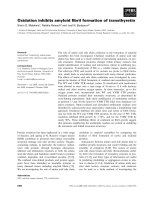
Fig. 2). TBG-like proteins were detected in serum from
Fig. 2. Evolutionary ⁄ developmental tree for
transthyretin synthesis in the choroid plexus
and liver of vertebrates. Evolutionary tree
showing approximate divergence times for
vertebrate groups, based on the fossil
record. Superimposed are symbols indicat-
ing the onset of transthyretin synthesis in
vertebrates. ++, onset of transthyretin syn-
thesis in the choroid plexus, in juveniles and
in adults of extant species; LD, hepatic
transthyretin synthesis during development
only; ?LD, possible onset of hepatic trans-
thyretin synthesis during development only;
+, hepatic transthyretin synthesis during
development and in adult. MYA, million
years ago. ([62]. Used with permission.)
S. J. Richardson Evolution of transthyretin biosynthesis
FEBS Journal 276 (2009) 5342–5356 ª 2009 The Author Journal compilation ª 2009 FEBS 5349
various mammalian species, but no clear phylogeny
was apparent.
Diprotodont marsupials have two large teeth on
their upper and lower jaws and are herbivores (e.g.
kangaroos and wombats), whereas polyprotodont
marsupials have many teeth on their upper and lower
jaws and are carnivores (e.g. Tasmanian devils and
dunnarts).
According to the fossil record, marsupials originated
in the region of Laurasia, which is now North America,
and were polyprotodont [88]. From there, they
migrated to what is now South America (for a sche-
matic diagram of the positions of these continents
about 150 Ma, see Fig. 3) and those in the northern
region died out. From South America, some marsupials
migrated back to (what is now) North America and
others migrated across Gondwanaland. About 45 Ma,
Gondwanaland began to break up into South America,
Antarctica and Australia [89]. There are fossils of mar-
supials in Antarctica (e.g. Seymour island) [90], and
many marsupials were isolated on the Australian conti-
nent. Shortly after the separation of Gondwanaland,
there was a radiation of marsupials in Australia, which
included the divergence of diprotodont marsupials from
polyprotodont marsupials [88]. (See Figs 2 and 3.) It
was previously suggested that in marsupials, the trans-
thyretin gene was turned on in the liver when the
‘younger’ Diprotodonta had diverged from the ‘older’
Polyprotodonta [34,83], whereas transthyretin was syn-
thesized in the liver as soon as the avian and eutherian
lineages evolved [34,78,84].
The digestive tracts of herbivorous marsupials
(diprotodont) are longer than those of carnivorous
marsupials (polyprotodont) [91]. The intestines are the
extrathyroidal tissue with the highest TH content [56],
and it has been suggested that the THDPs may be
responsible for the regulation of delivery of THs into
the intestines [92]. It was previously proposed that the
increase in lipid pool (e.g. length of intestine) was a
selection pressure for ‘turning on’ adult hepatic trans-
thyretin gene expression. It was argued that as the
transthyretin gene was already being expressed in the
choroid plexus of all reptiles, birds and mammals, the
onset of adult hepatic transthyretin gene expression
would have simply required a change in distribution of
transcription factors [8,34,93].
However, more recent data on hepatic transthyretin
synthesis during development [61,62,69–71], revealed
that all species studied had hepatic transthyretin syn-
thesis at some stage during development, often coincid-
ing with an increase in serum TH concentrations. In
some species, hepatic transthyretin synthesis continued
into adult life, whereas in other species the gene was
turned off during late stages of development. This led
to a re-evaluation of the data and hypotheses regard-
ing selection pressures for what was previously
described as the ‘onset of adult hepatic transthyretin
synthesis’, which should now be viewed as selection
pressure for ‘maintaining hepatic transthyretin synthe-
sis throughout life’. In light of this, the revised hypoth-
eses for selection pressures for maintaining hepatic
transthyretin synthesis throughout life are as follows.
Hypothesis 1. Maintaining hepatic transthyretin gene
expression in adulthood is related to the increase in lipid
pool to body mass ratio. A study by Hulbert and Else
[94] compared many physiological parameters of rep-
tiles (which do not have transthyretin in their blood)
and eutherians (which do have transthyretin in their
blood) of similar body mass. Amongst other data, they
showed that internal organs were larger in adult euthe-
rians, which therefore had larger lipid pools and conse-
quently a greater lipid volume to body mass ratio,
than reptiles of a similar body weight. As THs are
lipophilic and preferentially partition into the lipid
phase rather than the aqueous phase [95,96], the
increase in the relative size of the lipid pool could have
been a selection pressure for maintaining hepatic trans-
thyretin synthesis during adult life. As transthyretin
has higher affinity than albumin for THs, the presence
of transthyretin in the blood would contribute to
ensuring a circulating pool of THs, thereby counteract-
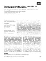
Fig. 3. Marsupial migration in relation to the movement of tectonic
plates. The positions of the land masses currently known as North
America (N.A.), South America (S.A.), Africa (Afr), Antarctica (Ant),
India (Ind) and Australia (Aus) about 150 Ma. Arrows indicate the
directions of three major marsupial migrations over about 100 Myr:
1., from North America to South America; 2., from South America
to North America and via Antarctica to Australia; 3., extensive radia-
tion of marsupials within Australia. (Data from [88–90]. Figure from
[18], used with permission.)
Evolution of transthyretin biosynthesis S. J. Richardson
5350 FEBS Journal 276 (2009) 5342–5356 ª 2009 The Author Journal compilation ª 2009 FEBS
ing the increased sink (lipid pool) for TH to poten-
tially partition into. Another example is comparison of
adult Australian marsupials. Diprotodont marsupials
(with longer intestines) have hepatic transthyretin syn-
thesis, whereas adult polyprotodont marsupials (with
shorter intestines) do not (intestines are the extrahe-
patic tissue with the highest concentration of TH) [56].
Hypothesis 2. Maintaining hepatic transthyretin syn-
thesis in adulthood is related to homeothermy. Transthy-
retin was found in serum from all studied species of
birds and eutherians, which are known homeotherms
(i.e. they maintain their body temperature at or near
37 °C by metabolic means). However, transthyretin
was not detected in serum from adult fish, amphibians,
or reptiles (including members from all four extant
Orders: Crocodilia, Squamata, Chelonia and Rhyncho-
cephalia), which are ectotherms (and in whom body
temperature is determined by a combination of behav-
iour and the environment) [8]. Marsupials and mono-
tremes are ‘poor endotherms’ (i.e. their body
temperatures are 25–32 °C, but when placed in cold
environments, cannot maintain their body tempera-
tures as well as ‘true endotherms’) [97]. THs are intri-
cately involved with the control of basal metabolic
rate, oxygen consumption and homeothermy. The
basal metabolic rates for monotremes, marsupials
and eutherians are approximately 140, 200 and
290 kJÆkg
)0.75
, respectively [97]. A selection pressure
for maintaining hepatic transthyretin synthesis through-
out life could have been to enable the appropriate
distribution of THs throughout the body to maintain
homeothermy.
Choroid plexus
Transthyretin is the major protein synthesized and
secreted by the choroid plexus of reptiles, birds, mono-
tremes, marsupials and eutherians, but is not synthe-
sized by the choroid plexus of amphibians [8] or fish
(G. Schreiber, unpublished observations). [However,
more recently, transthyretin mRNA has been detected
in whole-brain homogenates of some fish (see above).
It remains to be elucidated if this transthyretin gene
expression is in the choroid plexus]. It appears that the
transthyretin gene in the choroid plexus was turned on
once, at the stage of the stem-reptiles (the closest com-
mon ancestor to reptiles, birds and mammals), but not
of amphibians and fish (see Fig. 2). The early reptiles
were the first to develop traces of a cerebral neocortex
[98], thereby increasing their brain volume. As THs are
lipophilic and readily partition into cell membranes,
the increase in brain size may have been the selection
pressure for ‘turning on’ the transthyretin gene in the
choroid plexus. This resulted in transthyretin assisting
movement of THs from the blood across the blood–
CSF barrier into the brain, and also acting as a THDP
in the CSF [8].
Because hepatic transthyretin synthesis is present in
all extant classes of vertebrates (including fish, amphib-
ians and reptiles) during development, it is possible
that the stem-reptiles had the transthyretin gene in
their genomes, which may have been expressed in the
liver during development, then a change in specificity
of transcription factors could have been all that was
required to activate transthyretin synthesis in the cho-
roid plexus.
The major protein synthesized and secreted by the
choroid plexus of juvenile and adult amphibians is the
lipocalin prostaglandin D synthetase [72], also known
as beta-trace [99] and Cpl1 [100]. Prostaglandin D syn-
thetase is a monomeric 20 kDa protein that belongs to
the lipocalin superfamily of proteins. Lipocalins have a
calyx (cup) structure and are specialized in binding
small molecules. This raises the question of whether
this lipocalin was the evolutionary functional precursor
to transthyretin in the choroid plexus. [This should not
be confused with TLP, which is probably the evolu-
tionary structural precursor of transthyretin (see the
review in this miniseries by Dr Hennebry)].
Implications of transthyretin evolving
from distributing T3 to T4
It has been demonstrated that 100% of transthyretin
synthesized by the choroid plexus is secreted into the
CSF, and that none is secreted into the blood [11]. In
rats, this transthyretin was shown to transport
125
I-T4
but not
125
I-T3 from the blood across the blood–CSF
barrier into the brain [96]. However, if the transthyre-
tin synthesized by the choroid plexus binds T3 with
higher affinity than T4, as is presumably the case for
birds and reptiles [78] (fish and amphibians do not syn-
thesize transthyretin in the choroid plexus), the ques-
tion then arises as to whether in birds and reptiles, T3
(rather than T4) is transported across the blood–CSF
barrier into the brain. This also raises questions about
the evolution of deiodinases in the body, and in partic-
ular in specific regions of the brain.
The selection pressure leading to the change from
transthyretin preferentially binding T3 to T4 could be
from transporting the ‘active’ form of the hormone, to
transporting a ‘precursor’ form of the hormone. This
would allow greater flexibility and specificity at the local
tissue level to either activate the T4 by deiodinating it to
T3, or to inactivate the T4 by deiodinating it to rT3.
This could be especially true in the brain, as in the rat
S. J. Richardson Evolution of transthyretin biosynthesis
FEBS Journal 276 (2009) 5342–5356 ª 2009 The Author Journal compilation ª 2009 FEBS 5351
brain the percentage of T3 caused by local deiodination
of T4 is very specific to the region of the brain: 65% in
the cortex, 51% in the cerebellum, 35% in the pons,
32% in the hypothalamus, 30% in the medulla oblon-
gata and 22% in the spinal cord [101]. It could be con-
sidered ‘safer’ to distribute a precursor form of TH
around the body and into the CSF and brain, than to
distribute the active form. Thus, a change from binding
the ‘active’ form of the hormone (T3) to the precursor
form of the hormone (T4) could allow for more precise
control of TH action (activation and deactivation) in
specific regions of the body and brain.
Concluding remarks and future
directions
In general, transthyretin synthesis appears to be corre-
lated to a demand for an increase in capacity for TH
distribution. This includes the need to counteract
increasing lipid pools in both the body and the brain,
and for establishment of homeothermy. During devel-
opment, transient transthyretin ⁄ THDP gene expression
is correlated with an increase in vascular TH concen-
tration and in TH-driven developmental events. A
notable exception to this is represented by the
lampreys, where an increase in hepatic transthyretin
synthesis is accompanied by a decrease in vascular TH
concentration.
The change in ligand specificity of mammalian trans-
thyretins is intriguing. In this regard, mammals are the
exception rather than the rule. This highlights the
importance of comparative biology in understanding
TH metabolism. As the majority of laboratory animal
models are mammalian, our thinking is often skewed
by the disproportionate volume of data coming from
mammalian species – especially eutherian species. The
shift from distributing T3 to distributing T4 could
have the advantage of giving a greater level of control
over the regulation of TH-responsive genes, by distrib-
uting the precursor form of the hormone throughout
the blood and CSF followed by local deiodination to
either activate or deactivate the hormone. The marsu-
pial transthyretins represent a ‘transition’ between
eutherian transthyretins and nonmammalian trans-
thyretins in terms of ligand preference and strength of
binding. For this reason, it would be interesting to
analyse the distribution of deiodinases in marsupials.
In addition, the regulation of nongenomic effects of
TH in noneutherians should be considered.
Transthyretin synthesis in the choroid plexus is
believed to have begun at the stage of the stem rep-
tiles, about 320 Ma, which developed the first traces of
a cerebral neocortex (i.e. involved an increase in brain
volume). It would be interesting to compare the pat-
tern and distribution of deiodinases in the brains of
adult animals not synthesizing transthyretin in choroid
plexus with those that do synthesize transthyretin in
the choroid plexus, during development and in adult-
hood. Similarly, comparison of patterns of deiodinases
in brains of animals synthesizing transthyretin that
preferentially binds T3 with those in animals where
transthyretin preferentially binds T4 could give
valuable insights into the evolution of cerebral TH
metabolism.
An alternative hypothesis concerning the onset of
transthyretin synthesis by the choroid plexus considers
the implications of animals moving out of the water
onto the land (i.e. fish and amphibians do not synthesize
transthyretin in their choroid plexus, whereas reptiles,
birds and mammals do). Iodine must be derived from
the diet, and the main source of iodine is seaweed.
Iodine is more scarce on land than in the sea, especially
in mountainous regions far from the ocean. Could the
onset of cerebral transthyretin synthesis be a mechanism
for ensuring delivery of TH to the brain under condi-
tions of potentially restricted iodine supply? Could the
change in ligand binding from T3 to T4 be also attrib-
uted to increased storage of iodine as T4?
The change in temporal and spatial regulation of
transthyretin gene expression throughout evolution
required the evolution of sophisticated families of tran-
scription factors, co-modulator proteins and deiodinas-
es, rendering transthyretin an excellent model for the
study of the evolution of tissue-specific expression
throughout the vertebrate classes.
References
1 Robbins J & Edelhoch H (1986) Peripheral hormone
metabolism. Hormone transport in blood. In Werner’s
The Thyroid (Ingbar SH & Braverman LE, eds), pp.
116–127. Lippincott Co, Philadelphia.
2 Sandler B, Webb P, Apriletti JW, Huber BR, Togashi
M, Cunha Lima ST, Juric S, Nilsson S, Wagner R,
Fletterick RJ et al. (2004) Thyroxine-thyroid hormone
receptor interactions. J Biol Chem 279, 55801–55808.
3 Kohrle J (2000) The deiodinase family: selenoenzymes
regulating thyroid hormone availability and action.
Cell Mol Life Sci 57, 1853–1863.
4 Morreale de Escobar G, Obregon MJ & Escobar del
Rey F (2004) Role of thyroid hormone during early
brain development. Eur J Endocrinol 151(Suppl 3),
U25–U37.
5 Richardson SJ, Almeida OF & Demeneix BA (2007)
Hormones and adult neurogenesis in mammals. Expert
Rev Endocrinol Metab 2 , 261–276.
Evolution of transthyretin biosynthesis S. J. Richardson
5352 FEBS Journal 276 (2009) 5342–5356 ª 2009 The Author Journal compilation ª 2009 FEBS
6 Lemkine GF, Raj A, Alfama G, Turque N, Hassani Z,
Alegria-Prevot O, Samarut J, Levi G & Demeneix BA
(2005) Adult neural stem cell cycling in vivo requires
thyroid hormone and its alpha receptor. FASEB J 19,
863–865.
7 Shi Y-B (1999) Amphibian Metamorphosis. From Mor-
phology to Molecular Biology. John Wiley & Sons,
New York.
8 Schreiber G & Richardson SJ (1997) The evolution of
gene expression, structure and function of transthyretin.
Comp Biochem Physiol B Biochem Mol Biol 116, 137–
160.
9 Saunders NR, Habgood MD & Dziegielewska KM
(1999) Barrier mechanisms in the brain, I. Adult brain.
Clin Exp Pharmacol Physiol 26, 11–19.
10 Stauder AJ, Dickson PW, Aldred AR, Schreiber G,
Mendelsohn FA & Hudson P (1986) Synthesis of trans-
thyretin (pre-albumin) mRNA in choroid plexus epithe-
lial cells, localized by in situ hybridization in rat brain.
J Histochem Cytochem 34, 949–952.
11 Schreiber G, Aldred AR, Jaworowski A, Nilsson C,
Achen MG & Segal MB (1990) Thyroxine
transport from blood to brain via transthyretin
synthesis in choroid plexus. Am J Physiol 258,
R338–R345.
12 Kabat EA, Landow H & Moore DH (1942) Electro-
phoretic patterns of concentrated cerebrospinal fluid.
Proc Soc Exp Biol Med 49, 260–263.
13 Kabat EA, Moore DH & Landow H (1942) An elec-
trophoretic study of the protein components in cere-
brospinal fluid and their relationship to the serum
proteins. J Clin Invest 21, 571–577.
14 Siebert FB & Nielson JW (1942) Electrophoretic study
of the blood protein response in tuberculosis. J Biol
Chem 143, 29–38.
15 Ingbar SH (1958) Pre-Albumin – a thyroxine-binding
protein of human plasma. Endocrinology 63, 256–259.
16 Raz A & Goodman DS (1969) The interaction of thy-
roxine with human plasma prealbumin and with the
prealbumin-retinol-binding protein complex. J Biol
Chem 244, 3230–3237.
17 (1981) Nomenclature. NCotIUoBI-IJCoB. J Biol Chem
256, 12–14.
18 Richardson SJ (2007) Cell and molecular biology of
transthyretin and thyroid hormones. Int Rev Cytol 258,
137–193.
19 Craik DJ, Duggan BM & Munro SL (1996) Conforma-
tions and binding interactions of thyroid hormone
analogues. In Biological Inhibitors Studies in Medicinal
Chemistry (Choudhary MI ed), pp. 255–302. Harwood
Academic Publishers, Amsterdam.
20 Koehrle J, Spanka M, Irmscher K & Hesch RD (1988)
Flavonoid effects on transport, metabolism and action
of thyroid hormones. In Plant Flavonoids in Biology
and Medicine II: Biochemical, Cellular and Medicinal
Properties (Cody V, Harborne JB & Beretz A ed.), pp
323–340. Alan R Liss, New York.
21 Koehrle J (2004) Low dose competition of flavonoids
with endogenous thyroid transport proteins: Potential
relevance to the thyroid hormone axis. In Functional
Food: Safety Aspects (SKLM SCoFS, ed.), pp. 112–
137. Wiley-VCH Verlag, Berlin.
22 Kohrle J (2000) Flavonoids as a risk factor for goitre
and hypothyroidism. In The Thyroid and Environment
(Peter F, Wiersinga W & Hostalek U ed.), pp. 41–63.
Schattauer, Stuttgart, New York.
23 Kohrle J (1992) The trace components – selenium and
flavonoids – affect iodothyronine deiodinases, thyroid-
hormone transport and Tsh regulation. Acta Med
Austriaca 19, 13–17.
24 Van der Heide D, Kastelijn J & Schroder-van der Elst JP
(2003) Flavonoids and disease. Biofactors 19
, 113–119.
25 Mendel CM (1989) The free hormone hypothesis: a
physiologically based mathematical model. Endocr Rev
10, 232–274.
26 Mendel CM, Weisiger RA, Jones AL & Cavalieri RR
(1987) Thyroid hormone binding proteins in plasma
facilitate uniform distribution of Thyroxine within Tis-
sues – A Perfused Rat Liver Study. Endocrinology 120,
1742–1749.
27 Mendel CM & Weisiger RA (1990) Thyroxine uptake
by perfused rat liver. No evidence for facilitation by
five different thyroxine-binding proteins. J Clin Invest
86, 1840–1847.
28 Mendel CM, Cavalieri RR, Gavin LA, Pettersson T &
Inoue M (1989) Thyroxine transport and distribution
in Nagase Analbuminemic Rats. J Clin Invest 83, 143–
148.
29 Robbins J (2000) Thyroid hormone transport proteins
and the physiology of hormone binding. In The Thy-
roid – A Fundamental and Clinical Text (Braverman LE
& Utiger RD, eds), pp. 105–120. Lippincott Williams
& Wilkins, Philadelphia.
30 Robbins J (2002) Transthyretin from discovery to now.
Clin Chem Lab Med 40, 1183–1190.
31 Kanai M, Raz A & Goodman DS (1968) Retinol-bind-
ing protein: the transport protein for vitamin A in
human plasma. J Clin Invest 47, 2025–2044.
32 Noy N, Slosberg E & Scarlata S (1992) Interactions of
retinol with binding proteins: studies with retinol-bind-
ing protein and with transthyretin. Biochemistry 31,
11118–11124.
33 Monaco HL, Rizzi M & Coda A (1995) Structure of a
complex of two plasma proteins: transthyretin and reti-
nol-binding protein. Science 268, 1039–1041.
34 Richardson SJ, Bradley AJ, Duan W, Wettenhall RE,
Harms PJ, Babon JJ, Southwell BR, Nicol S, Donne-
llan SC & Schreiber G (1994) Evolution of marsupial
and other vertebrate thyroxine-binding plasma
proteins. Am J Physiol 266, R1359–R1370.
S. J. Richardson Evolution of transthyretin biosynthesis
FEBS Journal 276 (2009) 5342–5356 ª 2009 The Author Journal compilation ª 2009 FEBS 5353
35 Wilkinson-White LE & Easterbrook-Smith SB (2007)
Characterization of the binding of Cu(II) and Zn(II) to
transthyretin: effects on amyloid formation. Biochemis-
try 46, 9123–9132.
36 Liz MA, Faro CJ, Saraiva MJ & Sousa MM (2004)
Transthyretin, a new cryptic protease. J Biol Chem
279, 21431–21438.
37 Chang MH, Hua CT, Isaac EL, Litjens T, Hodge G,
Karageorgos LE & Meikle PJ (2004) Transthyretin
interacts with the lysosome-associated membrane pro-
tein (LAMP-1) in circulation. Biochem J 382, 481–489.
38 Schreiber G (1987) Synthesis, processing, and secretion
of plasma proteins by the liver and other organs and
their regulation. In The Plasma Proteins (Putnam FW,
ed.), pp. 293–363. Academic Press, New York.
39 Wei SH, Episkopou V, Piantedosi R, Maeda S, Shi-
mada K, Gottesman ME & Blaner WS (1995) Studies
on the metabolism of retinol and retinol-binding pro-
tein in Transthyretin-Deficient Mice Produced by
Homologous Recombination. J Biol Chem 270, 866–
870.
40 Fleming CE, Saraiva MJ & Sousa MM (2007) Trans-
thyretin enhances nerve regeneration. J Neurochem
103, 831–839.
41 Richardson SJ, Lemkine GF, Alfama G, Hassani Z &
Demeneix BA (2007) Cell division and apoptosis in
the adult neural stem cell niche are differentially
affected in transthyretin null mice. Neurosci Lett 421,
234–238.
42 Sousa JC, Marques F, Dias-Ferreira E, Cerqueira JJ,
Sousa N & Palha JA (2007) Transthyretin influences
spatial reference memory. Neurobiol Learn Mem 88,
381–385.
43 Sousa JC, Grandela C, Fernandez-Ruiz J, de Miguel
R, de Sousa L, Magalhaes AI, Saraiva MJ, Sousa N &
Palha JA (2004) Transthyretin is involved in depres-
sion-like behaviour and exploratory activity. J Neuro-
chem 88, 1052–1058.
44 Nunes AF, Saraiva MJ & Sousa MM (2006) Transthy-
retin knockouts are a new mouse model for increased
neuropeptide Y. FASEB J 20, 166–168.
45 Chen RL, Athauda SB, Kassem NA, Zhang Y, Segal
MB & Preston JE (2005) Decrease of transthyretin
synthesis at the blood-cerebrospinal fluid barrier of
old sheep. J Gerontol A Biol Sci Med Sci 60, 852–
858.
46 Soprano DR, Soprano KJ & Goodman DS (1986) Ret-
inol-binding protein and transthyretin mRNA levels in
visceral yolk sac and liver during fetal development in
the rat. Proc Natl Acad Sci USA 83 , 7330–7334.
47 Sklan D & Ross AC (1987) Synthesis of retinol-binding
protein and transthyretin in yolk sac and fetus in the
rat. J Nutr 117, 436–442.
48 Thomas T, Southwell BR, Schreiber G & Jaworowski
A (1990) Plasma protein synthesis and secretion in the
visceral yolk sac of the fetal rat: gene expression,
protein synthesis and secretion. Placenta 11, 413–430.
49 Schroder-van der Elst JP, van der Heide D, Rokos H,
Morreale de Escobar G & Kohrle J (1998) Synthetic
flavonoids cross the placenta in the rat and are found
in fetal brain. Am J Physiol 274, E253–E256.
50 McKinnon B, Li H, Richard K & Mortimer R (2005)
Synthesis of thyroid hormone binding proteins trans-
thyretin and albumin by human trophoblast. J Clin
Endocrinol Metab 90, 6714–6720.
51 Cavallaro T, Martone RL, Dwork AJ, Schon EA &
Herbert J (1990) The retinal pigment epithelium is the
unique site of transthyretin synthesis in the rat eye.
Invest Ophthalmol Vis Sci 31, 497–501.
52 Ong DE, Davis JT, O’Day WT & Bok D (1994) Syn-
thesis and secretion of retinol-binding protein and
transthyretin by cultured retinal pigment epithelium.
Biochemistry 33
, 1835–1842.
53 Kawaji T, Ando Y, Nakamura M, Yamamoto K,
Ando E, Takano A, Inomata Y, Hirata A & Tanihara
H (2005) Transthyretin synthesis in rabbit ciliary pig-
ment epithelium. Exp Eye Res 81 , 306–312.
54 Loughna S, Bennett P & Moore G (1995) Molecular
analysis of the expression of transthyretin in intestine
and liver from trisomy 18 fetuses. Hum Genet 95, 89–
95.
55 Dickson PW, Aldred AR, Marley PD, Tu GF, Howlett
GJ & Schreiber G (1985) High Prealbumin and Trans-
ferrin Messenger RNA Levels in the Choroid Plexus of
Rat Brain. Biochem Biophys Res Commun 127, 890–
895.
56 Nguyen TT, Distefano JJ, Yamada H & Yen YM
(1993) Steady-state organ distribution and metabolism
of thyroxine and 3,5,3¢-Triiodothyronine in intestines,
liver, kidneys, blood, and residual carcass of the rat in-
Vivo. Endocrinology 133, 2973–2983.
57 Kato M, Kato K, Blaner WS, Chertow BS & Good-
man DS (1985) Plasma and cellular retinoid-binding
proteins and transthyretin (prealbumin) are all local-
ized in the islets of Langerhans in the rat. Proc Natl
Acad Sci USA 82, 2488–2492.
58 Refai E, Dekki N, Yang SN, Imreh G, Cabrera O, Yu
L, Yang G, Norgren S, Rossner SM, Inverardi L et al.
(2005) Transthyretin constitutes a functional compo-
nent in pancreatic beta-cell stimulus-secretion coupling.
Proc Natl Acad Sci USA 102, 17020–17025.
59 Blay P, Nilsson C, Owman C, Aldred A & Schreiber G
(1993) Transthyretin expression in the rat brain: effect
of thyroid functional state and role in thyroxine trans-
port. Brain Res 632, 114–120.
60 Santos CRA & Power DM (1999) Identification of
transthyretin in fish (Sparus aurata): cDNA cloning
and characterisation. Endocrinology 140, 2430–2433.
61 Yamauchi K, Nakajima J, Hayashi H & Hara
A (1999) Purification and characterization of
Evolution of transthyretin biosynthesis S. J. Richardson
5354 FEBS Journal 276 (2009) 5342–5356 ª 2009 The Author Journal compilation ª 2009 FEBS
thyroid-hormone-binding protein from masu salmon
serum – A homolog of higher-vertebrate transthyretin.
Eur J Biochem 265, 944–949.
62 Richardson SJ, Monk JA, Shepherdley CA, Ebbesson
LO, Sin F, Power DM, Frappell PB, Kohrle J &
Renfree MB (2005) Developmentally regulated thyroid
hormone distributor proteins in marsupials, a reptile,
and fish. Am J Physiol Regul Integr Comp Physiol 288,
R1264–R1272.
63 Kawakami Y, Seoka M, Miyashita S, Kumai H &
Ohta H (2006) Characterization of transthyretin in the
Pacific bluefin tuna, Thunnus orientalis. Zoolog Sci 23,
443–448.
64 Manzon RG, Neuls TM & Manzon LA (2007) Molec-
ular cloning, tissue distribution, and developmental
expression of lamprey transthyretins. Gen Comp Endo-
crinol 151, 55–65.
65 Wright GM & Youson JH (1977) Serum thyroxine
concentrations in larval and metamorphosing anadro-
mous sea lamprey, Petromyzon marinus L. J Exp Zool
202, 27–32.
66 Manzon RG & Youson JH (2002) KClO(4) inhibits
thyroidal activity in the larval lamprey endostyle in
vitro. Gen Comp Endocrinol 128, 214–223.
67 Hennebry SC, Wright HM, Likic VA & Richardson SJ
(2006) Structural and functional evolution of
transthyretin and transthyretin-like proteins. Proteins
64, 1024–1045.
68 Hennebry SC, Law RH, Richardson SJ, Buckle AM &
Whisstock JC (2006) The crystal structure of the trans-
thyretin-like protein from Salmonella dublin, a
prokaryote 5-hydroxyisourate hydrolase. J Mol Biol
359, 1389–1399.
69 Yamauchi K, Kasahara T, Hayashi H & Horiuchi R
(1993) Purification and characterization of a 3,5,3¢-L-
triiodothyronine-specific binding protein from bullfrog
tadpole plasma: a homolog of mammalian transthyre-
tin. Endocrinology 132, 2254–2261.
70 Yamauchi K, Takeuchi H, Overall M, Dziadek M,
Munro SL & Schreiber G (1998) Structural characteris-
tics of bullfrog (Rana catesbeiana) transthyretin and its
cDNA – comparison of its pattern of expression during
metamorphosis with that of lipocalin. Eur J Biochem
256, 287–296.
71 Prapunpoj P, Yamauchi K, Nishiyama N, Richardson
SJ & Schreiber G (2000) Evolution of structure, ontog-
eny of gene expression, and function of Xenopus laevis
transthyretin. Am J Physiol – Regul Integr Comp Phys-
iol 279, R2026–R2041.
72 Achen MG, Harms PJ, Thomas T, Richardson SJ,
Wettenhall RE & Schreiber G (1992) Protein synthesis
at the blood-brain barrier. The major protein secreted
by amphibian choroid plexus is a lipocalin. J Biol
Chem 267, 23170–23174.
73 Achen MG, Duan W, Pettersson TM, Harms PJ, Rich-
ardson SJ, Lawrence MC, Wettenhall RE, Aldred AR
& Schreiber G (1993) Transthyretin gene expression in
choroid plexus first evolved in reptiles. Am J Physiol
265, R982–R989.
74 Richardson SJ, Hunt JL, Aldred AR, Licht P & Schre-
iber G (1997) Abundant synthesis of transthyretin in
the brain, but not in the liver, of turtles. Comp Bio-
chem Physiol B-Biochem Mol Biol 117 , 421–429.
75 Prapunpoj P, Richardson SJ & Schreiber G (2002)
Crocodile transthyretin: structure, function, and evolu-
tion. Am J Physiol – Regul Integr Comp Physiol 283,
R885–R896.
76 Southwell BR, Duan W, Tu GF & Schreiber G
(1991) Ontogenesis of transthyretin gene expression in
chicken choroid plexus and liver. Comp Biochem
Physiol – B: Comp Biochem 100, 329–338.
77 Duan W, Achen MG, Richardson SJ, Lawrence MC,
Wettenhall RE, Jaworowski A & Schreiber G (1991)
Isolation, characterization, cDNA cloning and gene
expression of an avian transthyretin. Implications
for the evolution of structure and function of trans-
thyretin in vertebrates. Eur J Biochem 200
, 679–
687.
78 Chang L, Munro SLA, Richardson SJ & Schreiber G
(1999) Evolution of thyroid hormone binding by trans-
thyretins in birds and mammals. Eur J Biochem 259,
534–542.
79 Duan W, Richardson SJ, Babon JJ, Heyes RJ, South-
well BR, Harms PJ, Wettenhall RE, Dziegielewska
KM, Selwood L, Bradley A et al. (1995) Evolution of
transthyretin in marsupials. Eur J Biochem 227, 396–
406.
80 Richardson SJ, Bradley A, Duan W, Southwell BR,
Selwood L & Schreiber G (1993) The expression of the
transthyretin gene in liver evolved during the radiation
of diprotodont marsupials in Australia. Gen Comp
Endocrinol 90, 177–182.
81 Li ZS, Dziegielewska KM & Saunders NR (1997)
Transthyretin distribution in the developing choroid
plexus of the South American opossum (Monodelphis
domestica). Cell Tissue Res 287, 621–624.
82 Davis PJ & Jurgelski W Jr (1973) Thyroid hormone-
binding in opossum serum: evidence for polymorphism
and relationship to haptoglobin polymorphism. Endo-
crinology 92, 822–832.
83 Richardson SJ, Wettenhall RE & Schreiber G (1996)
Evolution of transthyretin gene expression in the liver
of Didelphis virginiana and other American marsupials.
Endocrinology 137, 3507–3512.
84 Prapunpoj P, Richardson SJ, Fumagalli L & Schreiber
G (2000) The evolution of the thyroid hormone distrib-
utor protein transthyretin in the order insectivora, class
mammalia. Mol Biol Evol 17, 1199–1209.
S. J. Richardson Evolution of transthyretin biosynthesis
FEBS Journal 276 (2009) 5342–5356 ª 2009 The Author Journal compilation ª 2009 FEBS 5355
85 Schreiber G & Howlett G (1983) Synthesis and
secretion of acute-phase proteins. In Plasma Protein
Secretion by the Liver (Glaumann H, Peters T & Red-
man C, ed.), pp. 423–449. Academic Press, London.
86 Dickson PW, Aldred AR, Marley PD, Bannister D &
Schreiber G (1986) Rat choroid plexus specializes in
the synthesis and the secretion of transthyretin (preal-
bumin). Regulation of transthyretin synthesis in cho-
roid plexus is independent from that in liver. J Biol
Chem 261, 3475–3478.
87 Richardson SJ, Dziegielewska KM, Andersen NA,
Frost S & Schreiber G (1998) The acute phase response
of plasma proteins in the polyprotodont marsupial
Monodelphis domestica. Comp Biochem Physiol B Bio-
chem Mol Biol 119, 183–188.
88 Tyndale-Biscoe H (1973) Life of Marsupials. Arnold,
London.
89 Talent J (1984) Australian biogeography past and pres-
ent: determinants and implications. In Phanerozoic
Earth History of Australia (Veevers J, ed.), pp. 57–93.
Oxford University Press (Clarendon), London.
90 Woodburne MO & Zinsmeister WJ (1982) Fossil Land
Mammal from Antarctica. Science 218, 284–286.
91 Hume ID (1982) Digestive Physiology and Nutrition of
Marsupials. Cambridge University Press, Cambridge.
92 Distefano JJ, Nguyen TT & Yen YM (1993) Transfer
kinetics of 3,5,3¢-Triiodothyronine and Thyroxine from
rat blood to large and small intestines, liver, and kid-
neys in vivo. Endocrinology 132, 1735–1744.
93 Richardson SJ (2002) The evolution of
transthyretin synthesis in vertebrate liver, in primitive
eukaryotes and in bacteria. Clin Chem Lab Med 40,
1191–1199.
94 Hulbert AJ & Else PL (1989) Evolution of mammalian
endothermic metabolism – mitochondrial activity and
cell composition. Am J Physiol 256, R63–R69.
95 Hillier AP (1970) The binding of thyroid hormones to
phospholipid membranes. J Physiol 211, 585–597.
96 Dickson PW, Aldred AR, Menting JG, Marley PD,
Sawyer WH & Schreiber G (1987) Thyroxine transport
in choroid plexus. J Biol Chem 262, 13907–13915.
97 Dawson TJ & Hulbert AJ (1970) Standard metabolism,
body temperature, and surface areas of Australian
Marsupials. Am J Physiol 218, 1233–1238.
98 Kent GC (1987) Comparative Anatomy of the Verte-
brates. Time Mirror ⁄ Mosby College, St Louis.
99 Beuckmann CT, Aoyagi M, Okazaki I, Hiroike T,
Toh H, Hayaishi O & Urade Y (1999) Binding of
biliverdin, bilirubin, and thyroid hormones to lipoca-
lin-type prostaglandin D synthase. Biochemistry 38,
8006–8013.
100 Lepperdinger G (2000) Amphibian choroid plexus li-
pocalin, Cpl1. Biochim Biophys Acta 1482, 119–126.
101 van Doorn J, Roelfsema F & van der Heide D (1985)
Concentrations of thyroxine and 3,5,3¢-triiodothyronine
at 34 different sites in euthyroid rats as determined by
an isotopic equilibrium technique. Endocrinology 117,
1201–1208.
Evolution of transthyretin biosynthesis S. J. Richardson
5356 FEBS Journal 276 (2009) 5342–5356 ª 2009 The Author Journal compilation ª 2009 FEBS