Báo cáo khoa học: A novel metallocarboxypeptidase-like enzyme from the marine annelid Sabellastarte magnifica – a step into the invertebrate world of proteases pdf
Bạn đang xem bản rút gọn của tài liệu. Xem và tải ngay bản đầy đủ của tài liệu tại đây (990.39 KB, 16 trang )
A novel metallocarboxypeptidase-like enzyme from the
marine annelid Sabellastarte magnifica – a step into the
invertebrate world of proteases
Maday Alonso-del-Rivero
1
, Sebastian A. Trejo
3
,Mo
´
nica Rodrı
´
guez de la Vega
3
, Yamile Gonza
´
lez
1
,
Silvia Bronsoms
3
, Francesc Canals
2
, Julieta Delfı
´
n
1
, Joaquin Diaz
1
, Francesc X. Aviles
3
and Marı
´
a
A. Cha
´
vez
1
1 Centro de Estudio de Proteı
´
nas, Facultad de Biologı
´
a, Universidad de la Habana, Cuba
2 Institut de Recerca Hospital Vall d’Hebron, Barcelona, Spain
3 Institut de Biotecnologı
´
a i Biomedicina and Departament de Bioquı
´
mica i Biologı
´
a Molecular, Universitat Autonoma de Barcelona, Spain
Introduction
Natural evolution has frequently generated a large
adaptative variety of forms among protein functional
families, and metallocarboxypeptidases (MCPs) have
also followed this trend. Such enzymes are exopeptid-
Keywords
enzyme specificity; marine annelid;
metallocarboxypeptidases; metalloproteins;
Sabellastarte magnifica
Correspondence
F. X. Aviles, Institut de Biotecnologı
´
ai
Biomedicina (IBB) and Departament de
Bioquı
´
mica i Biologia Molecular, Universitat
Autonoma de Barcelona, 08193 Bellaterra
(Barcelona), Spain
Fax: +34 93 581 2011
Tel: +34 93 581 1231
E-mail:
(Received 16 March 2009, revised 16 June
2009, accepted 30 June 2009)
doi:10.1111/j.1742-4658.2009.07187.x
After screening 25 marine invertebrates, a novel metallocarboxypeptidase
(SmCP) has been identified by activity and MS analytical approaches, and
isolated from the marine annelid Sabellastarte magnifica. The enzyme,
which is a minor component of the molecularly complex animal body, as
shown by 2D gel electrophoresis, has been purified from crude extracts to
homogeneity by affinity chromatography on potato carboxypeptidase inhib-
itor and by ion exchange chromatography. SmCP is a protease of
33792 Da, displaying N-terminal and internal sequence homologies with
M14 metallocarboxypeptidase-like enzymes, as determined by MS and auto-
mated Edman degradation. The enzyme contains one atom of Zn per mole-
cule, is activated by Ca
2+
and is drastically inhibited by the metal chelator
1,10-phenanthroline, as well as by excess Zn
2+
or Cu
2+
, but moderately so
by EDTA. SmCP is also strongly inhibited by specific inhibitors of metallo-
carboxypeptidases, such as benzylsuccinic acid and the protein inhibitors
found in potato and leech (i.e. recombinant forms, both at nanomolar
levels). The enzyme displays high peptidase efficiency towards pancreatic
carboxypeptidase-A synthetic substrates, such as those with hydrophobic
residues at the C-terminus but, remarkably, also towards the acidic ones.
This property, previously described as for carboxypeptidase O-like activity,
has been shown on long peptide substrates by MS. The results obtained in
the present study indicate that SmCP is a novel member of the M14 metal-
locarboxypeptidases family (assignable to the M14A or pancreatic-like
subfamily) with a wider specificity that has not been described previously.
Abbreviations
AAFP, N-(4-methoxyphenylazoformyl)-
L-phenyl-alanine; AAFR, N-(4-methoxyphenylazoformyl)-L-Arg; ACTH fragment (18–39),
adrenocorticotropic hormone (RPVKVYPNGAEDESAEAFPLEF); BAEE, benzoyl arginyl ethyl ester; BTEE, benzoyl tyrosine ethyl ester; CP,
carboxypeptidase; CPA, carboxypeptidase A; CPB, carboxypeptidase B; CPO, carboxypeptidase O; DIGE, difference gel electrophoresis;
E-64,
L-carboxy-trans-2,3-epoxypropyl-leycylamido (4-guanidino) butane; FAAK, [3-(2-furyl)acryloyl]-L-alanyl-L-lysine; FAPP, N-(3-[2-
furyl]acryloyl)-Phe-Phe; Hippuryl-Phe, N-benzoyl-Gly-Phe; MCP, metallocarboxypeptidase; rLCI, recombinant leech carboxypeptidase inhibitor;
rPCI, recombinant potato carboxypeptidase inhibitor; V15E, synthetic substrate [VKKKARKAAGC(Amc)AWE].
FEBS Journal 276 (2009) 4875–4890 ª 2009 The Authors Journal compilation ª 2009 FEBS 4875
ases that catalyze the hydrolysis of peptide bonds at
the C-terminus of peptides and proteins. They belong
to the catalytic classes of either metalloproteases (clan
MC, family M14) or serine proteases (clan SC, family
S10) [1] and their action causes strong effects in the
biological activity of their peptide and protein sub-
strates [2]. M14 MCPs, including those from animals,
plants and bacteria, have been divided into three main
subfamilies based on structural similarity and sequence
homology. The first one, which includes the digestive
enzymes carboxypeptidase (CP) A (CPA) 1, CPA2,
carboxypeptidase B (CPB) 1 and mast cell CPA3, as
well as CPA4, CPA5 CPA6 and carboxypeptidase O
(CPO) (known at the gene level), has been termed sub-
family M14A or A ⁄ B; the second one, including the
bioactive peptide-processing or regulatory enzymes
(e.g. carboxypeptidases N, E, M and D, amongst oth-
ers) has been termed subfamily M14B or N ⁄ E [3]. Very
recently, a novel subfamily composed of enzymes of
larger size and apparently with a predominant cyto-
solic location, termed M14D, Nna-like or CCPs, has
been proposed [4]. Furthermore, three main classes
may be distinguished according to their substrate spec-
ificity: (a) for aromatic ⁄ hydrophobic residues (A-like),
(b) for basic residues (B-like) and (c) for acidic resi-
dues (O-like) [3,5].
MCP enzymes have been isolated from different
sources [3,5,6], mainly from vertebrates, but a few of
them have come from marine invertebrate organisms:
the digestive crayfish carboxypeptidase (CPB) [7], the
carboxypeptidase E-like enzyme from the sea hare
Aplysia californica, with important regulatory func-
tions in this organism [8], two CPs (A and B types)
from the hepatopancreas of the crab Paralithodes cam-
tschatica [9], the CPA-like protease from squid hepato-
pancreas of Illex illecebrosus [10], and CPs (two A and
one B type) isolated from the pyloric ceca of the starf-
ishes Asterias amurensis [11,12] and Asterina pectinifera
[13].
More than 95% of the Earth’s animal species are
invertebrates [14]. The ecological services provided by
invertebrates are immeasurable; life as we know it
would be quite different or decline without them (see
Center for Applied Biodiversity Science; http://sci-
ence.conservation.org). Overall, our knowledge about
MCPs in invertebrates is very limited given the tremen-
dous variety of such organisms and compared to the
much larger number of characterized CP from verte-
brates [6]. In the present study, we screened for the
presence of CP activity in marine invertebrates belong-
ing to the Phyla Cnidaria, Annelida, Mollusca, Echi-
nodermata, Arthropoda and Chordata, amongst
others, collected on the coasts of Havana, Cuba. The
study has been based on the use of N-(4-meth-
oxyphenylazoformyl)-l-phenylalanine (AAFP), a sensi-
tive, specific and known colorimetric substrate for
CPA enzymes. One of the highest activity levels was
detected in extracts from the marine annelid S. magni-
fica. This marine invertebrate, also termed ‘magnificent
feather duster’, was obtained from coral reefs. It
belongs to the Phylum Annelida, Class Polychaeta,
which shows a clear delimitation between its tentacle
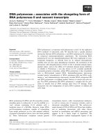
crown and its body (Fig. 1) [15]. Some studies per-
formed on another annelid, belonging to the Sabellidae
family, have only detected proteolytic activity assign-
able to serine proteases, which appeared to be involved
in reproduction [16] despite their digestive origin.
The presence of a carboxypeptidase-like enzyme in
Annelida marine invertebrates has not been described
so far.
The present study describes the enzymatic activity
and MS detection of a novel MCP (termed SmCP)
from S. magnifica, and its occurrence as a minor com-
ponent within the animal body extracts by 2D- PAGE.
The enzyme has been isolated and purified, and then
characterized by size, metal content, location, basic
interactions, sequence analysis of different regions of
the enzyme, and by a description of the main parame-
ters related to enzyme kinetics, specificity and inhibi-
tion ranges, as well as other basic molecular features.
From this, it is apparent that SmCP is a novel M14
MCP (belonging to the pancreatic-like subfamily),
showing simultaneous CPA- and CPO-like activities,
which is an unusual feature. The present study com-
prises an attempt to expand the growing field of the
M14 family of proteolytic enzymes, which is now quite
diverse and contains more than 25 different variants
Fig. 1. S. magnifica Phylum Annelida, Class Polychaeta, Subclass
Palpata, Order Canalipalpata, Suborder Sabellida, Family Sabellidae,
Genus Sabellastarte [14] The ‘tentacle crown’ and the ‘body’ parts
of the animal are clearly visible.
A novel metallocarboxypeptidase from S. magnifica M. Alonso-del-Rivero et al.
4876 FEBS Journal 276 (2009) 4875–4890 ª 2009 The Authors Journal compilation ª 2009 FEBS
[4–6], but for which only very few members from
invertebrates have been characterized until now.
Results
Detection of MCP activities in marine organisms
Twenty-five marine species belonging to different
invertebrate Phyla were screened for CPA activity
using AAFP as a substrate: four species of Mollusca
(Aplysia dactylomela, Aplysia juliana, Isognomun radia-
tus and Lima scabra); four species of Chordata (Pallu-
sia nigra, Microcosmus gamus, Molgula occidentalis
and Pyura vittata); 11 species of Cnidaria (Bartholo-
mea annulata, Budonosoma granulifera, Cassiopea
xamachana, Condylactys gigantea, Gorgonia ventalina,
Lebrunia danae, Palythoa caribaeorum, Physalia phy-
salis, Plexaura homomalla, Stichodactyla helianthus and
Zoanthus pulchellus); two species of Annelida (Sabellas-
tarte magnifica and Hermodice carunculata); two species
of Echinodermata (Holothuria mexicana and Isostisch-
opus badionotus); and two species of Arthropoda (Lito-
peaeus schmitti and Litopenaeus vannamei).
Among them, only the three species S. magnifica
(Phyllum Annelida), B. granulifera (Phyllum Cnidaria)
and P. vittata (Phyllum Chordata) gave rise to positive
results, with specific activity values of 56.0, 1.6 and
1.8 UÆ100 mg
)1
extract, respectively. In these three
cases, we found a linear relationship between CP-like
activity and the quantity of extract used in the assay.
Given that the material of the annelid S. magnifica
showed by far the highest specific activity, it was
selected for further characterization studies. In this
case, it was also found that extracts from the ‘body’
showed CP activity, whereas the feather-like ‘crown’
was devoid of it.
‘Intensity fading’ MALDI-TOF MS
Once we focused our attention on S. magnifica body
extracts, we found there direct evidence of at least one
MCP enzyme, of approximately 35 kDa by ‘intensity
fading’ MALDI-TOF MS [17]. In the present study,
the added ‘binder’ was the recombinant form of potato
carboxypeptidase inhibitor (rPCI) (4.5 kDa), immobi-
lized on agarose beads, with the aim of both perturb-
ing the MS spectrum and capturing the MCP in the
body extract. The control spectra, as well as the ‘per-
turbed’ one (by rPCI addition, followed by removal of
the captured targets by sedimentation of the beads),
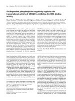
are shown in Fig. 2A,B. It is apparent that some of
the ion signals of the spectra were faded when the
extract was treated with immobilized PCI. Subse-
quently, MS analysis of the protein eluted from the
beads (Fig. 2C) detected a molecular ion of 34 kDa.
This molecular species, which is able to strongly inter-
act with PCI, presumably represents the CP-like
enzyme activity found in S. magnifica body extract.
The experiment indicates not only the occurrence in
the extract of the strong ligand (the enzyme SmCP) for
the added protease inhibitor, but also that this ligand
is probably functional in the very complex extract (i.e.
not in the zymogen state). It is worth noting that the
apparent simplicity of the MALDI-TOF spectrum of
the extract shown in Fig. 2C is most likely caused not
only by the low expansion scale used, but also by
1000
1500
Control MS (body extract)
A
Intens. (a.u.)Intens. (a.u.)Intens. (a.u.)
0
500
2000
+PCI
0
500
1000
1500
100
150
Elution
0
50
10 000 15 000 20 000 25 000 30 000 35 000
m/z
B
C
Fig. 2. MALDI-TOF MS of the ‘intensity
fading’ experiment (A) Mass spectra of the
S. magnifica body extract (control sample)
before rPCI-agarose addition (B) Unbound
proteins mass spectra obtained after rPCI-
agarose addition (C) MS spectra of recov-
ered m ⁄ z signal after elution of the sample,
corresponding to CP-like enzyme The arrow
indicates the ‘perturbed’ signal by rPCI-aga-
rose addition.
M. Alonso-del-Rivero et al. A novel metallocarboxypeptidase from S. magnifica
FEBS Journal 276 (2009) 4875–4890 ª 2009 The Authors Journal compilation ª 2009 FEBS 4877
‘signal suppression effects’; such phenomena usually
affect visualization of signals in media crowded in mol-
ecules [17–19], as will be reported and discussed subse-
quently.
Molecular complexity of the S. magnifica body
extract by 2D-PAGE
The molecular complexity of the S. magnifica extracts
(both from the body and from the crown, or mixed)
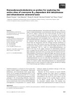
was demonstrated by 2D-PAGE analysis (Fig. 3). A
great number of visible protein bands [as revealed
either by staining with silver or using difference gel
electrophoresis (DIGE)] appeared in the analysis of
both parts of the animal, with a major presence of
bands in the body (upper part) versus the crown (lower
part). In Fig. 3, we show, in the uncombined
(Fig. 3A,B) or in the combined way (Fig. 3C), the pro-
tein components of both parts of the animal labeled
with fluorescent dyes using the DIGE approach. That
is, the different materials (i.e. crown and body extracts,
purified enzyme) were pre-labeled independently with
DIGE reagents before they were mixed and run simul-
taneously in a single 2D-PAGE separation. The inde-
pendent labeling of the crown and body extracts was
performed not only to allow the differential tracking
of their components, but also to deal with the very
high content of dyes and interfering materials from the
crown, which required a harsh cleaning (and denatur-
ing) procedure. Such interfering materials strongly per-
turbed the electrophoretic separation, and also gave
rise to severe band strikes and decreased resolution.
Only after testing several pre-cleaning and staining
procedures (not shown), and selecting an adequate
one, were we able to unveil the real band complexity
of the extracts (see Experimental procedures). We hope
that this experience might be useful for the analysis of
other invertebrates with a high content in dyes and
other similar problems.
Overall, more than 200 protein species are detected
by this procedure, among which those in the
17–37 kDa range are the most prominent. To facilitate
identification, we repeated the 2D-PAGE with three
different initial samples from the body, after passing
them through microcolumns with immobilized protein-
aceous inhibitors of serine (soya bean protease inhibi-
tor, SBTI), cysteine (chicken cystatin) and aspartic
(pepstatin) proteases. The intact, flow-through
(depleted) and captured (released) materials were deriv-
atized with DIGE and run in the same 2D-PAGE gel
for each case (see Experimental procedures). The anal-
ysis of the ‘captured’ spots allowed us to potentially
A
B
C
Fig. 3. 2D gel electrophoresis of pre-labeled protein extracts from S. magnifica The gel contained 30 lg of total protein, separated by IEF
using a pH 3–10 IPG strip in the first dimension and 15% SDS ⁄ PAGE in the second dimension The gel was first stained with the DIGE
approach (see Experimental procedures), and subsequently checked by silver staining (A) Labeling with Cy5 fluorofor for the tentacle crown
(B) Labeling with Cy2 fluorofor for the body (C) Body and tentacle crown alltogether (overlapped images) In the light box, the corresponding
position of SmCP enzyme is shown when it was run in an individual 2D-PAGE (and visualized by immunostaining) The spots labeled with
numbers correspond to molecular species affected by affinity capture on the immobilized inhibitors cystatin C (3, 4, 5, 6, 7 and 14) and
soybean trypsin inhibitor (8, 9, 10, 11, 12 and 13), or on both (1 and 2).
A novel metallocarboxypeptidase from S. magnifica M. Alonso-del-Rivero et al.
4878 FEBS Journal 276 (2009) 4875–4890 ª 2009 The Authors Journal compilation ª 2009 FEBS
identify at least 14 proteins captured differentially for
the first two microcolumns, which are labeled with
numbers in Fig. 3B (1 and 2 by both; 3, 4, 5, 6, 7 and
14 by the cystatin one; and 8, 9, 10, 11, 12 and 13 by
the SBTI one). An initial validation of these assign-
ments as proteolytic enzymes (awating MS ⁄ MS analy-
sis) was made by ‘intensity fading’ MALDI-TOF MS
using the mentioned set of immobilized inhibitors,
employing a strategy similar to the one for PCI
described above.
It is important to note that the band corresponding
to the SmCP enzyme, the target of the present study,
did not appear at around 34 kDa, which is the mass
assigned to it as a potential MCP (see MALDI-TOF
MS analysis and below), when the extracts (either from
the body or body + crown) were analyzed. However,
such a band is clearly visible when the enzyme is puri-
fied, concentrated and subsequently applied to the 2D-
PAGE (Fig. 3, encircled region). We assume that such
a difference is a result of the very low abundance of
SmCP in the animal. Also, it is relevant that the use of
an antibody raised against the sequence around
Asn144-Arg145, preserved in CPs [4], gave rise to a
spot in the same location by immunostaining (not
shown), confirming its assignment.
Purification and partial molecular characterization
of SmCP
After detection of carboxypeptidase activity in the
annelid worm (‘bodies’) of S. magnifica, SmCP was
fractionated to homogeneity using affinity chromatog-
raphy on a PCI-Sepharose column as the first step of
purification. The enzymatic activity was detected in the
eluted fraction with a 79% yield and a 286-fold purifi-
cation with respect to the crude extract (Table 1). The
second step of purification comprised anion exchange
chromatography on a TSK-DEAE 5PW column
(FPLC) (Tosoh Bioscience LLC, Montgomeryville,
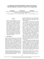
PA, USA) (Fig. 4A). SmCP eluted in a single fraction
with a specific activity of 322 UÆmg
)1
and 1150-fold
purification (Table 1). The purified enzyme was submit-
ted to metal analysis by inductive coupled plasma-MS,
which indicated that it contains 0.96 atoms of Zn per
molecule.
A single band with a molecular mass of 34 kDa was
detected by SDS ⁄ PAGE (Fig. 4B). This result agrees
with the molecular mass of 33 792 Da that was obtained
when it was analyzed by MALDI-TOF MS (Fig. 4C).
In addition, Edman degradation analysis revealed a
unique N-terminal sequence, confirming the homogene-
ity of SmCP at this end of the molecule. Despite the
rather limited size of the N-terminal region sequenced
(19 residues: AFDLNDFNTLEDTYDQMNV), a
blast search for this sequence revealed a consistent
Table 1. Summary of a typical purification procedure for SmCP
The assays were carried out as described in the Experimental
procedures. Substrate AAFP at 0.1 m
M, pH 7.5, 25 °C.
Step
Protein
(mg)
Enzymatic
activity
(U)
Specific
sctivity
(UÆmg
)1
)
Yield
(%)
Purification
(n-fold)
Extract 404 114 0.28 100 1
Affinity
chromatography
1.12 90 80.3 79 286
Ion exchange
chromatography
0.23 74 322 65 1150
14.2
12
28
34.1
51
90
120
203
I
II
III
(mAU)
20.0
UV1/
280 nm
Conc
CP activity
15.0
10.0
5.0
–5.0
0 20 40 60 80 100
0
100
200
300
Unit·m
–1
400
500
mL
0.0
0
200
400
600
800
15 000 20 000 25 000 30 000 35 000
m/z
40 000
Intens. (a.u.)
16 928.956
33 792.855.
A
B
C
Fig. 4. Purification of SmCP from the body extract of S. magnifica
and its molecular weight (A) Ion exchange chromatography on a
TSK-DEAE gel (7.5 · 7.5 cm) column Buffer A: 20 m
M Tris–HCl (pH
8.0); buffer B: 1
M Tris–HCl (pH 8.0) (I) Equilibration: 0% B for
45 min; (II) 60% B for 20 min; and (III) gradient 60% to 80% B for
170 min; flow rate: 68 cmÆh
)1
–––, A
280
; ,EnzAct; –––, Conc
NaCl (B) SDS ⁄ PAGE gel (125%) of the purified enzyme Lane 1,
Standard molecular weights [myosin (203 kDa), galactosidase
(120 kDa), bovine serum albumin (90 kDa), ovoalbumin (51 kDa),
carbonic anhydrase (34.1 kDa), soybean trypsin inhibitor (28 kDa)
and lysosyme (14.2 kDa)] Lane 2: Fraction of S. magnifica purified
by PCI-Sepharose and anionic exchange chromatography (C) MS
spectrum (MALDI-TOF) of SmCP.
M. Alonso-del-Rivero et al. A novel metallocarboxypeptidase from S. magnifica
FEBS Journal 276 (2009) 4875–4890 ª 2009 The Authors Journal compilation ª 2009 FEBS 4879
homology with other MCPs, such as porcine and bovine
carboxypeptidase A1 precursor, mosquito Aedes ae-
gipty CPA, and the carboxypeptidase homolog from
Bothrops jaraca, amongst others (Fig. 5). Subsequently,
and as a result of SmCP trypsin digestion followed by
LC-MS ⁄ MS analyses, we identified nine internal pep-
tides (termed T1–T9), which showed identity to internal
sequences of different CPs (Fig. 5). Some of them
include important ‘canonical’ residues of the catalytic
site of these enzymes [3]. Thus, in peptides T2 and T6,
respectively, His69 and His196 (using canonical num-
bering) were found, which are tetrahedrally coordinated
to the catalytic zinc ion in all MCPs (i.e. the numbering
system corresponds to bovine pancreatic CPA and is
used throughout). The other three most important resi-
dues found in the sequenced peptides are Glu270 (T9),
Asn144 and Arg145 (T2). Glu270, in the S1 subsite, acts
as a general base for catalysis, whereas Asn144 and
Arg145, in the S1¢ subsite, bind the C-terminal carboxyl-
ate group of the substrate. The peptide T6 appears to
contain Tyr198, which usually belongs to the S2 CP sub-
site. In addition, peptides T4 and T5 appear to contain
two cysteine residues conserved in all members of MCP
A ⁄ B subfamily, forming the disulfide bridge Cys138-
Cys161 [1]. Any peptide assignable to the putative speci-
ficity site [3] was found. Overall, these results indicate
that SmCP represents a CP-like enzyme of the M14A
subfamily [1,4].
Fig. 5. Alignment of the amino terminal and internal sequences of SmCP with the sequences of carboxypeptidases from other organisms
SmCP sequences were derived after trypsin treatment of the purified enzyme followed by LC-MS ⁄ MS (de novo sequencing) and bioinfor-
matics analyses (see Experimental procedures) Similar and identical residues are shown in light and dark grey, respectively ‘Canonical’ resi-
dues of CP (based on bovine CPA1) that are present in the trypsin peptides of SmCP are labeled with an asterisk The sequences are CPA
from Aedes aegypti (yellow fever mosquito) (Q9U9K2 AEDAE); Carboxypeptidase A1 precursor from Mus musculus (CBPA1 MOUSE); car-
boxypeptidase A2 from Paralichthys olivaceus (Japanese flounder) (Q8QAXN5 PAROL); carboxypeptidase A1 precursor from Sus scrofa
(CBPA1 PIG); carboxypeptidase A1 precursor from Bos taurus (CPBPA1 BOVIN); carboxypeptidase homolog from B. jaraca (Q9PUF2 BOT-
JA); CPO from Homo sapiens (CBPO HUMAN); CPB from Astacus fluviatilis (broad-fingered crayfish) (CBPB ASTFL); CPA precursor from
H. armigera (cotton bollworm) (097434_HELAM); carboxypeptidase precursor from H. armigera (cotton bollworm) (Q6H962_HELAM); MCP
from Culicoides sonorensis (Q5QBL3_9DIPT); and carboxypeptidase A2 precursor from H. sapiens (CBPA2_HUMAN).
A novel metallocarboxypeptidase from S. magnifica M. Alonso-del-Rivero et al.
4880 FEBS Journal 276 (2009) 4875–4890 ª 2009 The Authors Journal compilation ª 2009 FEBS
Kinetic characterization of SmCP
Kinetic analyses for isolated SmCP was performed using
different types of standard synthetic substrates for carb-
oxypeptidases that were clearly cleaved by the enzyme.
The K
m
, k
cat
and k
cat
⁄ K
m
determined for the enzyme
against AAFP, N -benzoyl-Gly-Phe (Hippuryl-Phe) and
N-(3-[2-furyl]acryloyl)-Phe-Phe (FAPP) as substrates
are shown in Table 2. Such kinetic parameters indicate
that SmCP is highly efficient against the three CPA type
substrates used. On the other hand, we found that
SmCP is unable to cleave CPB type substrates such as
[3-(2-furyl)acryloyl]-L-alanyl-l-lysine (FAAK) or N-(4-
methoxyphenylazoformyl)-l-Arg (AAFR). Therefore,
SmCP appears to be more related to the A-type than to
the B-type MCPs [1–4].
The influence of pH on SmCP activity was also ana-
lyzed using the AAFP substrate, and indicated an opti-
mum pH value in the range 7.0–7.5. The effect of
various protease inhibitors on the SmCP enzymatic
activity is shown in Table 3. Inhibitors of cysteine
proteases (l-carboxy-trans-2,3-epoxypropyl-leycylami-
do (4-guanidino) butane, E-64; cystatin), aspartic pro-
teases (pepstatin) and serine proteases (Pefabloc,
soybean trypsin–chymotrypsin inhibitor, soybean tryp-
sin inhibitor, aprotinin) did not have noticeable effects
on SmCP activity. The enzyme was drastically inhib-
ited by the chelating agent 1,10-phenanthroline at
1mm. However, EDTA at 10 mm, which might act by
metal chelation, did not produce any inhibition at sim-
ilar concentrations and inhibitor ⁄ enzyme (I
o
⁄ E
o
) rela-
tionships (3 · 10
5
m). Nevertheless, EDTA partial
inhibitory effects were observed when preincubation
times were increased. By contrast, benzylsuccinic acid,
a well-known organic inhibitor of A-type carboxypep-
tidases, fully cancelled the enzyme activity, at 1 mm.
Furthermore, the addition of the protein inhibitor of
carboxypeptidases PCI (in fact rPCI, a recombinant
form, reactive towards CPA and CPB type enzyme) at
0.4 lm produced a 70% inhibition of SmCP activity.
The apparent K
i
value for this inhibitor towards SmCP
was 7.37 · 10
)8
m; however, the adjusted value
considering the substrate-induced dissociation was
2.45 · 10
)8
m. Another protein inhibitor from leech
(rLCI, also recombinant) at 13.5 lm produced a 70%
inhibition of SmCP activity. The estimated K
i
value
for rLCI was 2.95 · 10
)8
m, and its adjusted value
considering the substrate induced dissociation was
1.45 · 10
)8
m (Table 4). Preincubation of the inhibi-
tors with the enzymes for various periods of time did
not affect its inhibitory activity, suggesting that rLCI
and rPCI are fast tight binding inhibitors.
Table 2. Kinetic parameters for substrate hydrolysis catalyzed by SmCP in comparison with data reported for bovine pancreatic CPA (bCPA)
The assays were carried out under the same conditions as those described for AAFP Substrate concentrations in the range 0.11–1.2 m
M
(3.29 nM of the enzyme in assay), 0.1–2 mM (24 lM of the enzyme in assay) and 0.02–0.25 nM (3.29 nM of the enzyme in assay) were used
for AAFP, Hippuryl-Phe and FAPP, respectively.
Enzyme
AAFP Hippuryl-Phe FAPP
K
m
(mM) k
cat
s
)1
k
cat
⁄ K
m
M
)1
Æs
)1
K
m
(mM) k
cat
s
)1
k
cat
⁄ K
m
M
)1
Æs
)1
K
m
(mM) k
cat
s
)1
k
cat
⁄ K
m
M
)1
Æs
)1
SmCP 0.05 ± 0.01 42.5 79 · 10
5
0.36 ± 0.03 145 3.8 · 10
5
0.14 ± 0.01 15 1.7 · 10
5
bCPA 0.11 ± 0.01
a
44.0 41 · 10
5
0.88 ± 0.05
b
60 6.8 · 10
4
0.05 ± 0.01
b
340 6.8 · 10
6
a
Mock et al [23].
b
Cho et al [24]
Table 3. Effect of protease inhibitors on the relative activity of
SmCP SmCP: 3.29 n
M; AAFP: 0.1 mM; pH 7.5, 25 °C The enzyme
was preincubated with the inhibitors for 10 min at 25 °C.
Inhibitor Concentration
% Enzymatic
activity I
o
⁄ E
o
E-64 0.1 mM 100 3.0 · 10
4
M
Pefabloc 10 mM 100 3.03 · 10
6
M
Pepstatin A 50 lM 94 1.51 · 10
4
M
Trypsin-chymotrysin
inhibitor (soybean)
3mM 100 9.1 · 10
5
M
1,10-Phenanthroline 1 mM 21 3.03 · 10
5
M
Benzylsuccinic acid 1 mM < 1 3.03 · 10
5
M
EDTA 10 mM 117 3.03 · 10
5
M
PCI 0.4 lM 28.5 1.21 · 10
2
M
LCI 13.5 lM 30 4.1 · 10
2
M
Aprotinin 3 mM 100 9.1 · 10
5
M
Trypsin inhibitor
(soybean)
2mM 100 6.0 · 10
5
M
Table 4. K
i
values of rPCI and rLCI against SmCP compared to pre-
vious data obtained for bovine pancreatic CPA (bCPA) SmCP:
3.29 n
M; AAFP: 0.1 mM; pH 7.5, 25 °C The enzyme was preincu-
bated with the inhibitors for 10 min at 25 °C.
Carboxypeptidase
K
i
(nM)
rPCI rLCI
SmCP 24.5 ± 03 14.5 ± 05
bCPA 1.5 ± 02
a
1.6 ± 01
b
a
Ryan et al [25].
b
Reverter et al [27].
M. Alonso-del-Rivero et al. A novel metallocarboxypeptidase from S. magnifica
FEBS Journal 276 (2009) 4875–4890 ª 2009 The Authors Journal compilation ª 2009 FEBS 4881
On the other hand, we evaluated the effect on SmCP
of metal ions after overnight dialysis against EDTA at
10 mm (followed by the removal of excess EDTA by
dialysis against metal-free buffers; see Experimental
procedures). After this, SmCP only retains 40% of its
initial activity. This apoform subsequently was used as
a control for the studies with metals. We observed that
1mm Ca
2+
,Mn
2+
or Mg
2+
enhanced the enzyme
activity of apoSmCP above 100% of the control activ-
ity, whereas the addition of Cd
2+
at 1 mm or Co
2+
at
1mm or 10 mm did not affect the enzymatic activity
of the control (Fig. 6). However, Cu at 1 mm and
10 mm reduced the apoenzyme activity to 11% and
15% of its residual activity. Noteworthy, under our
conditions, the addition of Zn
2+
at 1 mm or 10 mm
brought the activity to 100% (full rescue) and to 70%,
respectively, with the latter assignable to inhibition by
this metal.
Specificity of cleavage
Two different long peptides were used as substrate
models to analyze the ability of SmCP to cleave differ-
ent kinds of residues at the C-terminus, in comparison
Fig. 6. Effect of divalent metals on SmCP
activity The concentrations used in the
assays were 329 n
M for the enzyme SmCP
and 0.1 m
M for the substrate AAFP, at pH
7.5 and 25 °C The enzyme, after EDTA
treatment and dialysis against metal-free
buffer (see Experimental procedures), was
preincubated with the different ion metal
salts at 1 m
M, for 10 min at 25 °C The
assays were also performed, under the
same conditions, at 10 m
M for Zn
2+
,Co
2+
and Cu
2+
.
SmCP vs ACTH
A
B
SmCP vs V15E
E F
E
A
W
E
A
W
E
E F
E F
F
F
F
2188
2317
2466
1427
1529
1541
1563
1587
1619
1693
1716
1748
ACTH control 60 min
bCPA vs ACTH
2466
2317
ACTH control 60 min
bCPA vs V15E
1793
1716
1748
V15E control 60 min
V15E control 60 min
SmCP + PCI 60 min
15 min
30 min
60 min
bCPA + PCI 60 min
15 min
30 min
60 min
Fig. 7. Determination of SmP specificity for
C-terminal substrate residues. Comparative
analysis by MALDI-TOF MS of the degrada-
tion of two synthetic substrates by SmCP
and bovine pancreatic CPA (bCPA). The
assays were performed in 10 m
M Tris–HCl
buffer (pH 8.0) with 1 l
M of peptides and
2.19 n
M of SmCP or 1 nM of bCPA in 10 lL
of final volume for 60 min. (A) represents
the enzymatic activity of SmCP against the
ACTH fragment and V15E peptide, whereas
(B) represents the enzymatic activity of
bCPA against the same substrate.
Sequence of the ACTH fragment (residues
18–39): RPVKVYPNGAEDESAEAFPLEF,
MW: 2466 Da; ACTHdes-F, MW: 2317 Da;
ACTHdes-EF, MW: 2188 Da; V15E peptide
sequence, VKKKARKAAGC(Amc)AWE: MW
1716 Da; V15Edes-E, MW: 1587 Da;
V15Edes-WE, MW: 1400 Da;
V15Edes-AWE, MW: 1329 Da.
A novel metallocarboxypeptidase from S. magnifica M. Alonso-del-Rivero et al.
4882 FEBS Journal 276 (2009) 4875–4890 ª 2009 The Authors Journal compilation ª 2009 FEBS
with bovine pancreatic CPA (a reference enzyme in the
field). After 15 min of incubation of SmCP with the
adrenocorticotropic hormone (ACTH) fragment used
as substrate (residues 18–39, 2466 Da), the enzyme was
able to release phenylalanine (ACTHdes-F, 2317 Da)
and glutamic acid (ACTHdes-EF, 2188 Da) residues
from the substrate C-terminus (Fig. 7A). No further
amino acids were released after a 30-min incubation
period. Under the same conditions, bovine pancreatic
CPA was only able to hydrolyze the C-terminal phen-
ylalanine residue from ACTH to obtain the ACTHdes-
F (2317 Da). The addition of the protein inhibitor
rPCI prevented cleavage in all cases.
To confirm the capability of SmCP to hydrolyze
acidic residues from the C-terminus of peptides, the
specificity of SmCP against synthetic substrate
[VKKKARKAAGC(Amc)AWE] (V15E peptide) (resi-
due 15, 1716 Da) was evaluated (Fig. 7B). After
15 min of incubation, the release of glutamic acid from
the peptide was observed and, after 60 min, the new
C-terminus residues formed and tryptophan and ala-
nine were further released, as shown by the trimming
scale: 1716, 1587 and 1329 Da. However, bovine pan-
creatic CPA was unable to hydrolyze the first of such
C-terminal residues, glutamic acid, even after 60 min
of incubation. Again, the addition of rPCI prevented
any kind of hydrolysis by the enzyme. The release of a
glutamic acid residue from the C-terminus of peptides
is a very unusual capability of a CPA-like enzyme and
is reminiscent of the so-called CPO forms [3,5].
Discussion
The growing application of genomics and related tech-
nologies is facilitating an expanding view of the pres-
ent enzymatic families, including proteases [20] and
CPs in particular [4]. However, such an advance is lim-
ited in the invertebrate world because of the great
diversity of organisms within it, which complicates the
study, but has the potential to generate enzyme vari-
ants of great biological and biotechnological values.
To gain insight into the field of MCPs, one of the
most unknown among proteases in invertebrates, we
have used a mix of both modern and more classical
approaches to identify and characterize them, estab-
lishing comparisons with the vertebrate species (i.e. the
reference ones). The present study started with a sys-
tematic screening in extracts from 25 invertebrates
from marine Caribbean species, using a specific and
sensitive enzymatic assay; this allowed us to detect the
presence of CPA-like activity in the body extract of
the marine annelid S. magnifica. Given that we previ-
ously reported the successful use of MALDI-TOF MS
for the initial detection of CPs and carboxypeptidase
inhibitors in other crude biological extracts [17–19], we
have applied such approaches to the S. magnifica case.
The use of affinity capture on microbeads or microcol-
umns derivatized with a recombinant carboxypeptidase
inhibitor from potatoes, specific for such class of
enzymes, and the use of MALDI-TOF MS signal anal-
ysis approaches, allowed us to quickly identify in this
annelid a 35-kDa species as a potential MCP, which
we named SmCP.
Different fractionation methods have been per-
formed to purify SmCP from the body extract of
S. magnifica. In initial attempts, using anion exchange
and gel filtration chromatographies, we found a frac-
tion with clear carboxypeptidase activity, which, inter-
estingly, conveyed two additional activities against
typical substrates for trypsin-like (benzoyl arginyl ethyl
ester; BAEE) and chymotrypsin-like (benzoyl tyrosine
ethyl ester; BTEE) serine proteases (data not shown).
This suggests that, in the fractionation, SmCP could
co-elute with serine proteases, perhaps establishing bin-
ary or ternary complexes with such enzymes, as shown
in other organisms [21,22]. Nevertheless, the substitu-
tive use of affinity chromatography on rPCI-agarose,
in subsequent experiments, allowed the selective cap-
ture of SmCP and contributed to its separation from
the other enzymes. Potentially, rPCI could promote
the dissociation of SmCP from ‘complexes with serine
proteases’ that it might establish in the crude extracts.
This is an issue that merits further research.
The 2D-PAGE analysis of the crude extracts indi-
cates that they are very complex in protein species,
and that a stainable band at around 35 kDa, attribut-
able to SmCP, is not directly visible with such
approach unless high sensitivity approaches (i.e immu-
nostaining) are employed. This is probably a result of
the low representation of this enzyme in the animal
extracts, in agreement with its subsequent analysis and
visualization in the purified form.
Additionally, we obtained evidence by affinity cap-
ture on three different kinds of immobilized proteina-
ceous inhibitors (soybean trypsin inhibitor, cystatin,
pepstatin), indicating that different main 2D-PAGE
protein bands around 20–55 kDa correspond to cyste-
ine and serine protease enzymes present in the S. mag-
nifica body extract. At least 14 species that gave
stainable and clearly visible bands were detected by
this approach. They were provisionally validated by
‘intensity fading’ MALDI-TOF MS perturbation stud-
ies carried out by the addition of such protein inhibi-
tors on the extracts. Full validation would require
either direct isolation or MS ⁄ MS analyses. The later
type of study is under way in our laboratory, but is
M. Alonso-del-Rivero et al. A novel metallocarboxypeptidase from S. magnifica
FEBS Journal 276 (2009) 4875–4890 ª 2009 The Authors Journal compilation ª 2009 FEBS 4883
proving more difficult than expected because of the
very low homologies shown by S. magnifica proteases
with respect to equivalent ones found in databases.
Given the poor representation of invertebrate proteases
in databases, this is not an unexpected problem when
carrying out identification proteomics.
It is worth noting that the preliminary detection of
serine and cysteine proteases species in the body
extracts correlates with the measure of their activities
by enzymatic analysis of the crude samples. Interest-
ingly, neither approach revealed evidence of the occu-
rence of aspartic proteases. Overall, although the
presence of pigments and other interfering products
initially constituted a very serious problem, once this
was technically solved, the feasibility and data genera-
tion capability of both the 2D-PAGE and ‘intensity
fading’ MALDI-TOF MS of this annelid indicated
that such proteomic-like approaches (and probably
related ones) are very promising for the analysis of
proteolytic enzymes in marine invertebrates.
A central question in the analysis of novel MCPs
from biological sources is whether they occur in their
precursor or mature forms [2–5]. In the present study,
using direct extracts from S. magnifica, we found only
a monomeric and activated form of SmCP, as shown
by its enzymatic activity, molecular mass, derived
N-terminal sequence and homology analysis. Procarb-
oxypeptidases are usually activated by proteolytic
removal of their activation segment by serine prote-
ases, mostly trypsin. Studies on procarboxypeptidases
from several species have indicated that its activation
is dependent of the environmental ionic conditions
and, sometimes, the influence of quaternary structure
[2,5]. Under our experimental conditions, quick activa-
tion of SmCP by autologous serine-like proteases,
which appeared to be present in large quantities in the
extract, could be favored. On the other hand, the
coincidence between the N-terminal sequences of
SmCP and those from several other MCPs included in
alignments (Fig. 5) also suggests that SmCP has been
purified in the active mature form. In addition, we
found that the sequences of a number of SmCP inter-
nal peptides included important residues that belong to
catalytic site and domain of this enzyme family,
confirming our interpretation.
All the experimental data reported in the present
study indicate that SmCP belongs to the M14A sub-
family of metalloproteases [6], the so-called pancreatic-
like forms (or A ⁄ B), favoring its potential digestive
function in the marine annelid. Its molecular weight
(33.7 kDa), N-terminal sequence and behavior towards
a panel of substrates and inhibitors are similar to those
of mammalian pancreatic CP (i.e. the best known).
These types of enzymes have molecular masses close to
35 kDa after the removal of the propeptide, whereas
the regulatory CPs (or N ⁄ E) display higher mass val-
ues as a result of the presence of other domains in
addition to the CP domain [2,3]. On the other hand,
SmCP shows sequence homology with some CPs iso-
lated from different vertebrates and invertebrates,
belonging to the A ⁄ B subfamily with CPA substrate
preferences. Only a few CPs have been isolated from
marine invertebrates, and in not one case have the
whole or extended sequences been disclosed. This
would be the case for the two CPAs and CPBs isolated
from the hepatopancreas of the crab P. camtschatica
[9] and the CPA-like enzyme from the squid hepato-
pancreas of I. illecebrosus [10].
SmCP is able to cleave different types of CPA sub-
strates such as AAFP, Hippuryl-Phe and FAPP, with
an overall efficiency similar to bovine pancreatic CPA,
but with some significant differences in k
cat
, K
m
and
k
cat
⁄ K
m
for certain substrates [23,24]. In addition,
SmCP has a maximum activity at pH 7.5, in agreement
with the optimum pH activity of almost all M14A CP-
like forms, including marine enzymes [7–13], which lie
in the neutral range (pH 6.5–8.5), and is consistent
with the pH at their sites of biological action [1,2].
As previously shown for mammalian CPs [25–27],
potato and leech proteinaceous inhibitors efficiently
inhibit SmCP, displaying similar K
i
values. In addition,
two smaller organic molecules (benzylsuccinic acid and
1,10-phenantroline) known to act on MCPs are also
able to inhibit the enzyme. By contrast, EDTA, which
chelates metal ions, at 10 mm, failed to inhibit SmCP
activity significantly after 10 min of preincubation,
which is in agreement with the reported properties of
other invertebrate MCPs isolated from the gut of Tion-
ela bisselliella [28] and from Helicoverpa armigera larvae
[29] for which EDTA effects are also time dependent.
The capability of divalent metal ions to substitute
the essential active site Zn
2+
of MCPs [30,31], or bind
a second atom nearby [32], interfering with the cata-
lytic mechanism, is well known. We also observed
diverse effects by the addition of such metals to SmCP.
After its dialysis against EDTA at 10 mm, SmCP
reduced its activity to 40% of initial activity. Starting
from this state, the capacity of different metal ions to
regenerate SmCP activity demonstrated that, in certain
cases [Mn, Mg and Ca], there is an enhancement of
activity of the enzyme; in others [Cd and Co], no
changes are observed; and, in a third case [Cu], a clear
inhibition is produced. Such results are quite congru-
ent with the well-known properties of mammalian CPs
[33]. In the case of Zn, an enhancement of SmCP
activity was observed when added at 1 mm, whereas,
A novel metallocarboxypeptidase from S. magnifica M. Alonso-del-Rivero et al.
4884 FEBS Journal 276 (2009) 4875–4890 ª 2009 The Authors Journal compilation ª 2009 FEBS
at 10 mm, little recovery of the initial activity
occurred.
The sense and intensity of the changes in the enzy-
matic parameters show different degrees of fitting with
what has been described for other invertebrate CPs,
such as the sea hare A. californica [8], the squid I. ille-
cebrosus [10] and the larvae Helicoverpa armiguera [29],
as well as for other mammalian CPs [2,34]. Notewor-
thy, the rather homogeneous and common inhibitory
action of Zn is presently the only one that can be eas-
ily rationalized, given the well-known effects of this
metal on the structure and enzymatic properties of
CPs [32,35].
One of the most interesting features of SmCP is
its wide specificity on both synthetic and long pep-
tide substrates (Fig. 7), particularly the hydrophobic
ones characteristic of a CPA-like specificity, although
it is unable to hydrolyze those assignable to a CPB-
like specificity (i.e. with Arg or Lys at the C-termi-
nus). By contrast, SmCP is also able to hydrolyze
acidic C-terminal residues, such as glutamic acid.
The latter type of specificity, now termed CPO [5],
was recently described for a CP isolated from the
insect H. armiguera [36], which is unable to hydro-
lyze either CPA or CPB substrates. The strict speci-
ficity of CPO has been proposed to be a result of
the presence of a basic residue at the substrate rec-
ognition pocket [5,37], which is different than those
for the other two general types (A and B). SmCP is
the first marine invertebrate CP to be described with
this specificity.
In conclusion, SmCP shares many similarities with
the M14A MCPs isolated from other sources, such as
molecular mass, N-terminal sequence, the presence of
key catalytic residues, optimum pH, the effect of some
metal ions and salts and the inhibition pattern. On the
other hand, it shows a broad capability for releasing
C-terminus substrate residues, being able to hydrolyze
both CPA and CPO substrates, comprising a mixed
specificity not previously described for CP-like
enzymes. It may be a digestive requirement of the ani-
mal (i.e. the S. magnifica annelid). Before an ample
characterization of other proteolytic enzymes present
in this invertebrate is achieved (several other proteases,
such as serine proteases, appear to be there by
2D-PAGE and MS analyses; not shown), such require-
ments can only be a matter of guesswork. We are still
far from a consistent characterization of the ‘degra-
domes’ of invertebrates (i.e. the genomically and prote-
omically related complement of proteolytic enzymes),
as it is performed nowadays in higher eukaryotes [38].
Surely, this would prove interesting, both biologically
and biotechnologically, given the tremendous richness
and diversity of invertebrates in natural products and
species. We hope that our results can stimulate interest
in this a field and contribute to its advancement.
Experimental procedures
Chemical reagents
All chemicals were of reagent grade and obtained from the
following sources: E-64, Pefabloc Sc and aprotinin from
Boehringer (Mannheim, Germany). ACTH fragment (RPV
KVYPNGAEDESAEAFPLEF) and V15E peptides [VKK
KARKAAGC(Amc)AWE] were synthesized by DiverDrugs
(Barcelona, Spain). Chromatographic columns were supplied
by Tosoh Bioscience LLC (Montgomeryville, PA, USA).
rPCI and rLCI were prepared by one of our groups (at the
Universitat Autonoma de Barcelona, Spain), which was the
first to clone and produce them in recombinant form [3].
Soybean trypsin–chymotrypsin inhibitor, soybean trypsin
inhibitor, benzylsuccinic acid, 1,10 phenanthroline; pepstatin
A, Hippuryl-Phe, FAPP, FAAK, BAEE and BTEE were
supplied by Sigma (St Louis, MO, USA); AAFP and AAFR
were supplied by Bachem (Weil am Rhein, Germany).
Preparation of extracts
The marine organisms belonging to the kingdom Methazoa
(Phyla: Annelida, Urochordata, Echinodermata, Cnidaria,
Mollusca, Artropoda) were collected in the north coast of
Havana and classified by Cuban specialists at the National
Institute of Oceanology (Havana, Cuba). The organisms
were homogenized in their own sea water liquid (1 : 2,
w ⁄ v). The homogenates were centrifuged at 10 000 g for
30 min at 4 °C. In the case of the marine invertebrate
S. magnifica, belonging to the Phylum Annelida, the ani-
mals were separated into two parts, tentacle crowns and
bodies, which were homogeneized as described above.
Carboxypeptidase assays
The general assay for CPA-like activity was carried out
using AAFP as substrate [23]. It was prepared at 10 mm in
dimethylsulfoxide. From this solution, 10 lL of substrate
was added to 50 lL of extract or enzyme samples in
940 lLof50mm Tris–HCl, 0.5 m NaCl (pH 7.5) for a final
concentration of 0.1 mm in the assay. The hydrolysis of the
chromogenic substrate caused a decrease in A
350
, which was
followed at 15-s intervals for 10 min at 25 °C in a kinetic
spectrophotometer Pharmacia-Biotech (Uppsala, Sweden).
The amount of residual substrate was determined using an
absorption coefficient of 19 · 10
)6
lm
)1
Æcm
)1
for AAFP.
One unit of CP activity is equivalent to the amount of
enzyme able to hydrolyze 1 lmolÆmin
)1
of AAFP under the
specified conditions. SmCP enzymatic activity was also
M. Alonso-del-Rivero et al. A novel metallocarboxypeptidase from S. magnifica
FEBS Journal 276 (2009) 4875–4890 ª 2009 The Authors Journal compilation ª 2009 FEBS 4885
determined using other CPA substrates, such as Hippuryl-
Phe [39] and FAPP [40]. The activity of CPB was determined
with two different substrates, FAAK [41] and AAFR [42].
For CPO-like activity evaluation, Hippuryl-Glu was used as
substrate [43]. In all cases, the experimental conditions were
similar to those described for the CPA substrate assays.
Protein concentration
Protein concentration was determined by the bicinchoninic
acid method [44], using the BCA kit (Pierce Ltd, Rockford,
IL, USA) and bovine serum albumin as standard. For a
mixture, the concentration was determined by measuring
A
280
, assuming A
280 (1%)
= 10.
Determination of Zn
The presence and amount of this metal in the purified
SmCP enzyme was determined by inductive coupled
plasma-MS, using QEXcell equipment (Thermal Elemental,
Windsford, UK). Standard procedures using Chellex100
(Bio-Rad, Hercules, CA, USA) and O-phenantroline [30],
were followed to remove divalent metal ions from water
and buffers, which were used in all stages of the analysis.
MALDI-TOF MS identification and interaction
with inhibitors
Enzyme identification in the S. magnifica crude extracts and
interactomic experiments with protein inhibitors of CPs
were carried out using the ‘intensity fading’ MALDI-
TOF MS approach, as previously reported [17–19]. In the
experiment, 1 lLofS. magnifica body extract was mixed
with 2 lL of rPCI immobilized on agarose microbeads and
incubated for 3 min at room temperature. To eliminate the
unbound proteins, the rPCI-agarose matrix was washed
with 10 mm Tris–HCl buffer (pH 7.5) three times. The elu-
tion from rPCI-agarose microbeads was carried out mixing
the matrix with 2 lL of 0.1% formic acid. After 3 min of
incubation, 0.5 lL of the drop was pipetted to be analyzed
by MALDI-TOF MS, as described below.
Affinity capture by immobilized protease
inhibitors
Microcolumns based on agarose matrices (0.1–1 mL) with
immobilized cystatin C (generously provided by M. Abra-
hamson, Division of Clinical Chemistry and Pharmacology,
Lund University, Sweden), SBTI (reference T0637 and pep-
statin A (reference P2032), both from Sigma, were used to
capture proteases from S. magnifica extracts. Extracts were
loaded in 100 m m ammonium bicarbonate (pH 8.5) in the
first two cases, and in 100 mm sodium acetate (pH 5.5) in
the last case. The captured proteins were released by trifluo-
roacetic acid 0.2% (pH 2) in the former cases, and with 1 m
NaCl (pH 5.5) in the latter case, and precipitated by addition
of 10% trichloroacetic acid ⁄ five volumes of acetone
()20 °C) before 2D-PAGE analysis.
SDS/PAGE
A 12.5% SDS ⁄ PAGE was performed, according to the
method of Laemmli [45]. The gel was stained with Coomassie
blue R-250. Prestained molecular weight standards were used.
2D gel electrophoresis
Given the high content of interfering materials in marine
invertebrate extracts, two of the different assayed proce-
dures are described. In the milder one, the pieced body of
S. magnifica was resuspended at 100 mgÆmL
)1
in solution
contained 5 mm dithithreitol and 5 mm of EDTA. The solid
was disaggregated by a 0.6 mm needle and then sonicated
three times for 10 min. The extract was centrifuged at
10 000 g for 10 min and the supernatant was precipitated
by adding precooled acetone (20% final, v ⁄ v) at )20 °C for
30 min. The pellet was eliminated and the new supernatant
was precipitated with four volumes of acetone ⁄ 10% trichlo-
roacetic acid. The precipitate was collected by centrifuga-
tion at 10 000 g for 10 min. The pellet was washed two
times with acetone and it was resuspended in 100 lLof
lysis buffer (7 m urea, 2 m thiourea, 4% Chaps, 30 mm
Tris–HCl, pH 8.5). Protein quantification was performed
using RC-DC Protein Assay Kit from Bio-Rad. In the
stronger procedure, an additional clarification step was car-
ried out using the 2D Clean up kit (Amersham Biosciences
Piscataway, NJ, USA) in accordance with the manufac-
turer’s instructions. Then the precipitate was resuspended
in lysis buffer. For the 2D-DIGE approach, the samples
were labeled with two different CyDye DIGE fluorofors
(Cy2 for body extract and Cy5 for tentacle crown extract)
before performing the 2D-PAGE. Each sample was labeled
with 200 pmol (1 lL) of CyDye per 30 lg of protein, incu-
bated on ice for 30 min in the dark and quenched with
1 lLof10mm lysine and then incubated on ice for 10 min
in the dark, according to the manufacturer’s instructions.
2D-PAGE with immobilized pH gradient was carried out
according to Go
¨
rg et al. [46]. Samples were loaded in the first
dimension IEF, using the cup-loading method, onto previ-
ously rehydrated 11 cm IPG drystrips (GE Healthcare,
Milwaukee, WI, USA) that contain an immobilized linear
gradient in the range pH 3–10. Approximately 30 lgof
tentacles crown and body extracts, after prelabeling, were
loaded and run either independently or jointly in this first
dimension; in the latter case, after a previous mix and load of
equal amounts of extracts from the two parts of the animal.
IEF was performed at 300 V for 1 h, followed by three gradi-
ent steps (1000 V for 30 min; 5000 V for 80 min and 8000 V
A novel metallocarboxypeptidase from S. magnifica M. Alonso-del-Rivero et al.
4886 FEBS Journal 276 (2009) 4875–4890 ª 2009 The Authors Journal compilation ª 2009 FEBS
for 30 min) and, finally, 8000 V for 2 h. After focusing, the
strips were equilibrated and proteins separated on 15% poly-
acrylamide gels. Electrophoresis was carried out at 4 °C until
the front of fast migrating ions reached the bottom of the gel.
2D-PAGE gels were stained with silver nitrate [47]. The
stained gels were immediately scanned using a Umax Astra
4000U scanning device (Umax Systems GmbH, Willich,
Germany), and digitalized images were evaluated using
ImageMaster 2D-PAGE 5.0 (GE Healthcare).
For 2D-DIGE, fluorescence images of the gels were
obtained on a Typhoon 9400 scanner (GE Healthcare). Cy2
and Cy5 images were scanned at excitation ⁄ emission wave-
lengths of 488 ⁄ 520 nm and 633 ⁄ 670 nm, respectively, at a
resolution of 100 lm. To facilitate visualization and print-
ing, all figures have been processed to obtain a negative
copy of the initial image (i.e. converting the black back-
ground of the fluorescent images into a white background)
and to visualize the spots in a single colour (black,
although we used the initial colours for differential identifi-
cations); however, we strictly maintained the initial raw
intensities of the spots along the whole image.
Purification procedure of SmCP
In order to use an affinity chromatography as the first step
of the purification procedure, rPCI was immobilized on
BrCN activated Sepharose (3 mgÆmL
)1
of gel) [45]. The
body extract dissolved in 50 mm Tris–HCl, 0.5 m NaCl (pH
7.5) was loaded into a 6 mL PCI-Sepharose column
(1.3 · 5 cm). The column was washed with the same buffer
at a flow rate of 19 cmÆh
)1
and the elution was carried out
in one step by increasing the pH to 12.0 with 0.05 m
sodium phosphate buffer. Fractions (1 mL each) were
mixed with 100 lLof1m Tris–HCl buffer (pH 6.0) to
adjust the eluates to pH 8.0. Fractions with CP activity
were concentrated and dialyzed against 20 mm Tris–HCl
buffer (pH 8.0) and applied to an ion exchange column of
TSK-DEAE (0.75 · 7.5 cm), previously equilibrated with
20 mm Tris–HCl buffer (pH 8.0), at a flow rate of
68 cmÆh
)1
. After extensive washing with the equilibration
buffer (45 min, six column volumes), the column was
washed with 60% of buffer B (1 m Tris–HCl, pH 8.0) for
20 min (three column volumes). Bound enzyme was eluted
with a linear gradient from 60% to 80% of buffer B over
170 min (25 column volumes) at the same flow rate.
MS, N-terminal sequence analysis and proteolytic
cleavage
A MALDI-TOF spectrometer was used to analyze the
molecular mass of peptides and proteins (Ultraflex MS;
Bruker, Ettlingen, Germany). Ionization was accomplished
with a 337 nm pulsed nitrogen laser and spectra were
acquired in the linear positive ion mode, using 25 kV acceler-
ation voltage. The analysis of proteins or peptide fragments
was carried out using 3,5-dimethoxy-4-hydroxycinnamic acid
(sinapinic acid) and a-cyano-4-hydroxicinnamic acid as
matrices. Samples were prepared by mixing them with equal
volumes of a saturated solution of the matrices. From this
mixture, 1 lL was spotted on the sample slide and allowed to
evaporate to dryness.
N-terminal amino acid sequence analysis was performed
by automated Edman degradation on an Applied Biosystems
(Applied Biosystems, Foster City, CA, USA) protein
sequencer (Procise 492) using reagents and solvents from the
supplier. For MS sequence analyses, the enzyme was dena-
tured and reduced by addition of 6 m guanidinium HCl and
3mm EDTA in 0.3 m Tris–HCl buffer (pH 8.6) with 50 mm
of dithiothreitol for 4 h at 40 °C in the dark under a N
2
atmosphere. The solution was diluted three times with Milli-
Q grade water (Millipore Co., Billerica, MA, USA) and trea-
ted with trypsin (sequence grade) for 24 h at 37 °C and at
10 : 1 ratio (w ⁄ w). The reactions were stopped by adding the
same volume of 0.1% of trifluoroacetic acid in water and
ZipTip C18 (Millipore Co.) was used to remove the salts.
Finally, peptides were dissolved in 0.5% acetic acid in water
and they were then further sequenced by nLC-ESI-MS ⁄ MS.
The interpretation of MS ⁄ MS spectra was performed manu-
ally, but was assisted by the software packages data
explorer 4.4 (Applied Biosystems, Foster City, CA, USA)
and peaks studio 4.5 SP2 (Bioinformatics Solutions Inc.,
Waterloo, Canada).
Enzyme kinetic characterization
Kinetic parameters
The K
m
and V
max
values for the purified enzyme were eval-
uated using different CPA substrates such as AAFP [23],
Hippuryl-Phe [39] and FAPP [40] in accordance with the
experimental conditions described above for the CP assays.
Kinetic parameters were graphically calculated by adjusting
the experimental data to the rectangular hyperbola curve,
using origin software (OriginLab, Northampton, MA,
USA).
pH optimum of SmCP activity
The optimum pH was determined using the AAFP substrate
by measuring the activity of SmCP (1.95 nm in assay) at var-
ious pH values using the buffers: 20 mm sodium phosphate
buffer (pH 11.0 and 12.0); 20 mm Hepes buffer (pH 7.0, 7.5,
8.0 and 8.5); 20 mm carbonate-bicarbonate buffer (pH 9.0,
9.5, 10.0 and 10.5); and 20 mm Tris–HCl buffer (pH 7.0,
7.5, 8.0, 8.5 and 9.0). All other experimental conditions were
as described for the CP assay using AAFP as substrate [23].
Effect of inhibitors and metal cations
Inhibition studies of SmCP by proteinaceous inhibitors was
evaluated against pepstatin A, rPCI, rLCI, aprotinin,
M. Alonso-del-Rivero et al. A novel metallocarboxypeptidase from S. magnifica
FEBS Journal 276 (2009) 4875–4890 ª 2009 The Authors Journal compilation ª 2009 FEBS 4887
soybean trypsin inhibitor, soybean trypsin–chymotrypsin
inhibitor and by synthetic inhibitors, such as E-64, EDTA,
benzylsuccinic acid, 1,10-phenanthroline and Pefabloc. The
effect of divalent metals such as Ca, Mn, Cd, Cu, Mg, Co
and Zn was analyzed using Milli-Q water that had been
passed through Chelax 100 columns according to establish
procedures (Bio-Rad). The enzyme was dialysed against
10 mm EDTA, in 20 mm Tris–HCl (pH 7.5) (metal-free),
overnight at 4 °C. EDTA was removed using a PD10 col-
umn and metal-free buffer. The apoenzyme was then incu-
bated for 10 min with divalent metals before activity was
assayed. The final activity was reported as the percentage
of apoenzyme activity.
Measurement of equilibrium dissociation constant (K
i
)
The time needed for SmCP to attain inhibition equilibrium
by PCI and LCI was determined by preincubation of the
inhibitors at 5 nm and 7.5 nm, respectively with SmCP
(3.29 nm and 22.9 nm in assay, respectively) for 2, 5, 10
and 20 min before substrate addition. To estimate the K
i
values of the inhibitors for SmCP, the enzyme was preincu-
bated for 10 min with increasing amounts of PCI (from
18 nm to 1.8 lm) and LCI (from 17.2 nm to 0.17 lm). Both
experiments were performed under conditions of
E
o
⁄ K
i
= 10. At each point, the activity (v
i
) was measured
against AAFP as substrate (0.05 mm, equivalent to 1 K
m
).
The residual activity was defined as v
i
⁄ v
o
, where v
o
repre-
sents the enzymatic activity in the absence of the inhibitor.
The experimental points were adjusted to the equation
described for tight binding inhibition [48] by employing
nonlinear fitting using statistica software (StatSoft, Tulsa,
OK, USA), as described previously [49].
Specificity against peptides substrates
The specificity of SmCP was studied using a fragment
derived from ACTH (18–39) as substrate for CPA-like
activity and V15E peptide as substrate for CPO-like activ-
ity. The reaction mixture contained 2.19 nm of SmCP and
1 lm of the peptides in 10 lLof10mm Tris–HCl buffer
(pH 8.0). The assays were monitored by MALDI-TOF MS
at 37 °C for 15, 30 and 60 min. Inhibition assays were per-
formed using PCI (5.6 lm) in the mixture. Similar experi-
ments were performed in parallel with bovine pancreatic
CPA (1 n m).
Acknowledgements
This work was supported by the International Founda-
tion of Science, Sweden (Grants F3342-1 and F3276-1),
by grant BIO2007-6846 (Ministerio de Educacio
´
ny
Ciencia-CICYT ⁄ MCINN, Spain) and by Xarxa de
Refere
`
ncia en Biotecnologia (XeRBa, Generalitat de
Catalunya). M.A.C. acknowledges a Visitor Grant from
AGAUR (Generalitat de Catalunya). Professor Magnus
Abrahamson and colleagues (Lund, Sweden) are
acknowledged for kindly providing immobilized cysta-
tin. The authors are grateful for technical support pro-
vided by Dagmara Diaz and Rachel Lopez, as well as
from ProteoRed-Instituto Nacional de Proteo
´
mica, and
particularly from Drs A. Paradela (ProteoRed Madrid-
node) and M. Carrascal (ProteoRed Barcelona-node).
The authors are also grateful to Kamela Alegre (IBB,
Universitat Autonoma de Barcelona, Spain), for the
kind revision of this article.
References
1 Barrett AJ, Rawlings ND & Woessner JF, eds (2004)
‘Handbook of Proteolytic Enzymes’ , 2nd edn. Elsevier
Academic Press, London.
2 Skidgel RA (1996) Structure and function of mamma-
lian zinc carboxypeptidases. In Zinc Metalloproteases in
Health and Disease (Hooper NM ed.), pp. 241–283.
Taylor and Francis, London.
3 Arolas JL, Vendrell J, Aviles FX & Fricker LD (2007)
Metallocarboxypeptidases: emerging drug targets in bio-
medicina. Curr Pharm Des 13, 347–364.
4 Rodriguez de la Vega RM, Sevilla RG, Hermoso A,
Lorenzo J, Tanco S, Diez A, Fricker LD, Bautista JM
& Avile
´
s FX (2007) Nna1-like proteins are active metal-
locarboxypeptidases of a new and diverse M14 sub-
family. FASEB J 20, 851–865.
5 Vendrell J, Aviles FX & Fricker LD (2004) Metallo-
carboxypeptidases. In Handbook of metalloproteins
(Messerschmidt A, Bode W & Cygler M eds), Vol. 3,
pp. 176–189. John Wiley and Sons, Ldt, Chichester.
6 Rawlings ND, Morton FR & Barrett AJ (2006) MER-
OPS: the peptidase database. Nucleic Acids Res 34,
D270–D272.
7 Zwilling R, Jacob F, Bauer H, Neurath H & Enfield
DL (1979) Crayfish carboxypeptidase. Affinity chroma-
tography, characterization and amino acid sequence.
Eur J Biochem 94, 223–229.
8 Juvvadi S, Fan X, Nagle GT & Fricker DL (1997)
Characterization of Aplysia carboxypeptidase E. FEBS
Lett 408, 195–200.
9 Sakharov II & Prieto GA (2000) Purification and some
properties of two carboxypeptidases from the hepato-
pancreas of the crab Paralithodes camtschatica. Mar
Biotechnol (NY) 2, 259–266.
10 Raksakulthai R & Haard NF (2001) Purification and
characterization of a carboxypeptidase from squid hepa-
topancreas (Illex illecebrosus). J Agric Food Chem 49,
5019–5030.
11 Kishimura H & Hayashi K (2002) Isolation and charac-
terization of carboxypeptidase B from the pyloric ceca
A novel metallocarboxypeptidase from S. magnifica M. Alonso-del-Rivero et al.
4888 FEBS Journal 276 (2009) 4875–4890 ª 2009 The Authors Journal compilation ª 2009 FEBS
of the starfish Asterias amurensis. Comp. Biochem Phys-
iol Biochem Mol Biol 133, 183–189.
12 Kishimura H & Hayashi K (1991) Purification and
properties of carboxypeptidase A-like enzyme from the
starfish Asterias amurensis. Nippon Suisan Gakkaishi 57,
1939–1944.
13 Kishimura H, Hayashi K & Ando S (2006) Characteris-
tics of carboxypeptidase B from pyloric ceca of the
starfish Asterina pectinifera. Food Chem, 95, 264–269.
14 Brusca RC & Brusca GJ (2003) Invertebrates. 2 edn.
Sinauer Associates Inc., Sunderland, Massashusetts.
15 Knight-Jones P & Mackie ASY (2003) A revision of
Sabellastarte (Polychaeta: Sabellidae). J Nat Hist 37,
no. 19, 2269–2301.
16 Peaucellier G (1983) Purification and characterization
of proteases from the polychaete annelid Sabellaria alve-
olata. Eur J Biochem 136, 435–445.
17 Villanueva J, Yanes O, Querol E, Serrana L & Aviles
FX (2003) Identification of protein ligand in complex
biological samples using intensity-fading MALDI TOF
mass spectrometry. Anal Chem 75, 3053–3063.
18 Yanes O, Villanueva J, Querol E & Aviles FX (2005)
Functional screening of serine proteases inhibitors in
the medical leech Hirudo medicinalis. Monitored by
intensity fading MALDI TOF MS. Mol Cell Proteomics
4, 1602–1612.
19 Yanes O, Villanueva J, Querol E & Avile
´
s FX (2007)
Detection of non covalent protein interactions by ‘inten-
sity fading’ MALDI-TOF mass spectrometry: applica-
tions to proteases and proteases inhibitors. Nat Protoc
2, 119–130.
20 Puente XS, Gutie
´
rrez-Ferna
´
ndez A, Ordo
´
n
˜
ez GR,
Hillier LW & Lo
´
pez-Otı
´
n C (2005) Comparative geno-
mic analysis of human and chimpanzee proteases.
Genomics 86, 638–647.
21 Kobayashi R, Kobayashi Y & Hirs CHW (1978) Identi-
fication of a binary complex of procarboxypeptidase A
and a precursor of protease E in porcine pancreatic
secretion. J Biol Chem 253, 5526–5530.
22 Gomis-Ruth FX, Go
´
mez M, Bode W, Huber R &
Avile
´
s FX (1995) The three-dimensional structure of the
native ternary complex of bovine pancreatic procarb-
oxypeptidase A with proproteinase E and chymotrysi-
nogen C. EMBO J 14, 4387–4394.
23 Mock WL, Liu Y & Stanford DJ (1996) Arazoformyl
peptide surrogates as spectrophotometric kinetic assay
substrates for carboxypeptidase. Anal Biochem 239,
218–222.
24 Cho JH, Kim DH, Lee KJ, Kim DH & Choi KY (2001)
The role of Tyr248 probed by mutant bovine carboxy-
peptidase A: insight into the catalytic mechanism of
carboxypeptidase A. Biochemistry 40, 10197–10203.
25 Ryan CA, Hass GM & Kuhn RW (1974) Purification
and properties of a carboxypeptidase inhibitor from
potatoes. J Biol Chem 249, 5495–5499.
26 Hass GM & Ryan CA (1981) Carboxypeptidase inhibi-
tor from potatoes. Methods Enzymol 80, 778–791.
27 Reverter D, Vendrell J, Canals F, Horstmann J, Avile
´
s
FX, Fritz H & Sommerhoff CP (1998) A carboxypepti-
dase inhibitor from the medical leech. Hirudo medici-
nalis. J Biol Chem 273(49), 32927–32933.
28 Ward CW (1976) Properties of the major carboxypepti-
dase in the larvae of the webbing clothes moth Tineolla
bisselliella. Biochim Biophys Acta 429, 564–572.
29 Bown DP, Wilkinson HS & Gatehouse JA (1998)
Midgut carboxypeptidase from Helicoverpa armigera
(Lepidoptera: noctuidae) larvae: enzyme characteriza-
tion, cDNA cloning and expression. Insect Biochem Mol
Biol 28, 739–749.
30 Auld DS (1995) Removal and replacement of metal ions
in metallopeptidases. Methods Enzymol 248, 228–242.
31 Auld DS (2009) The ins and outs of biological zinc
sites. Biometals 22, 141–148.
32 Gomez-Ortiz M, Gomis-Ruth FX, Huber R & Aviles
FX (1997) Inhibition of carboxypeptidase A by excess
zinc: analysis of the structural determinants by X-ray
crystallography. FEBS Lett 400, 336–340.
33 Coleman JE & Vallee BL (1961) Metallocarboxypeptid-
ases: stability constants and enzymatic characteristics.
J Biol Chem 236, 2244–2249.
34 Tan AK & Eaton DL (1995) Activation and character-
ization of procarboxypeptidase B from human plasma.
Biochemistry 34, 5811–5816.
35 Alvarez-Santos S, Gonza
´
les-Lafont A & Iluch JM
(1996) Theoretical study of the mechanism of carboxy-
peptidase A inhibition by zinc ions. New J Chem, 20,
979–984.
36 Bown DP & Gatehouse JA (2004) Characterization of a
digestive carboxypeptidase from the insect pest corn
earworm (Helicoverpa armigera) with novel specific
towards C terminal glutamate residues. Eur J Biochem
271, 2000–2011.
37 Wei S, Segura S, Vendrell J, Aviles FX, Lanoue E, Day
R, Feng Y & Fricker LD (2002) Identification of three
members of the human metallocarboxypeptidase gene
family . J Biol Chem 277, 14954–14964.
38 Overall CM & Dean RA (2006) Degradomics: systems
biology of the protease web. Pleiotropic roles of MMPs
in cancer. Cancer Metastasis 25, 69–75.
39 Bergmeyer HU, Gawehn K & Grassl M (1974) Methods
of Enzymatic Analysis (Bergmeyer HU, ed), Vol. 1, 2nd
edn, pp. 436–437, Academic Press, Inc., New York, NY.
40 Peterson LM, Holmquist B & Bethume JL (1982)
Unique activity assay for carboxypeptidase A in human
serum. Anal Biochem 125, 420–426.
41 Plummer TR Jr & Kimmel MT (1980) An improved
spectrophotometric assay for human plasma carboxy-
peptidase N1 Anal Biochem 108, 348–354.
42 Mock WL & Xu D (1999) Catalytic activity of
carboxypeptidase B and of carboxypeptidase Y with
M. Alonso-del-Rivero et al. A novel metallocarboxypeptidase from S. magnifica
FEBS Journal 276 (2009) 4875–4890 ª 2009 The Authors Journal compilation ª 2009 FEBS 4889
anisylazoformyl substrate Bioorg Med Chem Lett 9,
187–192.
43 Edge M, Forder C, Hennam J, Lee I, Tonge D, Hard-
ern I, Fitton J, Eckersley K, East S, Shufflebotham A
et al. (1998) Engineered human carboxypeptidase B
enzymes that hydrolyse hippuryl-L-glutamic acid:
reversed-polarity mutants Protein Eng 11, 1229–1234.
44 Smith PK, Krohn RI, Hermanson GT, Mallia AK,
Gartner FH, Provenzano MD, Fujimoto EK, Goeke
NM, Olson BJ & Klenk DC (1985) Measurement of pro-
tein using bicinchoninic acid Anal Biochem 150, 76–85.
45 Laemmli UK (1970) Cleavage of structural proteins
during the assembly of the head of bacteriophage T4
Nature 227, 680–685.
46 Gorg A, Weiss W & Dunn MJ (2004) Current two-
dimensional electrophoresis technology for proteomics
Proteomics 4, 3665–3685.
47 Blum H, Beier H & Gross HJ (1987) Silver staining of
proteins in polyacrylamide gels Electrophoresis 8, 93–99.
48 Williams JW & Morrison JF (1979) The kinetics of
reversible tight-binding inhibition Methods Enzymol 63,
437–467.
49 Gonzalez Y, Pons T, Gil J, Besada V, Alonso del Rive-
ro M, Tanaka AS, Araujo MS & Cha
´
vez MA (2007)
Characterization and comparative 3D modeling of
CmPI-II, a novel ‘non-classical’ Kazal-type inhibitor
from the marine snail Cenchritis muricatus (Mollusca)
Biol Chem 388, 1183–1194.
A novel metallocarboxypeptidase from S. magnifica M. Alonso-del-Rivero et al.
4890 FEBS Journal 276 (2009) 4875–4890 ª 2009 The Authors Journal compilation ª 2009 FEBS