Báo cáo khoa học: Xenopus Rbm9 is a novel interactor of XGld2 in the cytoplasmic polyadenylation complex pptx
Bạn đang xem bản rút gọn của tài liệu. Xem và tải ngay bản đầy đủ của tài liệu tại đây (374.79 KB, 14 trang )
Xenopus Rbm9 is a novel interactor of XGld2 in the
cytoplasmic polyadenylation complex
Catherine Papin*, Christel Rouget* and Elisabeth Mandart
Centre de Recherche en Biochimie Macromole
´
culaire, Universite
´
Montpellier II, France
Translational regulation of mRNA is often linked to
the control of the poly(A) tail length, as its cytoplasmic
lengthening can stabilize mRNA and activate transla-
tion. During early development, control of the poly(A)
tail length by cytoplasmic polyadenylation is critical
for the regulation of specific mRNA expression [1].
The molecular mechanisms that underlie the regula-
tion of polyadenylation-dependent translation are well
Keywords
cytoplasmic polyadenylation; Gld2; poly(A)
polymerase; Rbm9; Xenopus oocyte
Correspondence
C. Papin, Centre de Recherche en Biochimie
Macromole
´
culaire, UMR 5237 Universite
´
Montpellier II CNRS, 1919, Route de
Mende, 34293 Montpellier Cedex 5, France
Fax: +33 4 99 61 99 01
Tel: +33 4 99 61 99 59
E-mail:
E. Mandart, Centre de Recherche en
Biochimie Macromole
´
culaire, UMR 5237
Universite
´
Montpellier II CNRS, 1919, Route
de Mende, 34293 Montpellier Cedex 5,
France
Fax: +33 467 521559
Tel: +33 467 613339
E-mail:
*Present address
Re
´
gulation des ARNm et De
´
veloppement,
Institut de Ge
´
ne
´
tique Humaine, Montpellier,
France
Database
AM419007, AM419008, AM419009,
AM419010
(Received 20 June 2007, revised 15 Novem-
ber 2007, accepted 28 November 2007)
doi:10.1111/j.1742-4658.2007.06216.x
During early development, control of the poly(A) tail length by cytoplas-
mic polyadenylation is critical for the regulation of specific mRNA expres-
sion. Gld2, an atypical poly(A) polymerase, is involved in cytoplasmic
polyadenylation in Xenopus oocytes. In this study, a new XGld2-interacting
protein was identified: Xenopus RNA-binding motif protein 9 (XRbm9).
This RNA-binding protein is exclusively expressed in the cytoplasm of
Xenopus oocytes and interacts directly with XGld2. It is shown that
XRbm9 belongs to the cytoplasmic polyadenylation complex, together with
cytoplasmic polyadenylation element-binding protein (CPEB), cleavage and
polyadenylation specificity factor (CPSF) and XGld2. In addition, tethered
XRbm9 stimulates the translation of a reporter mRNA. The function of
XGld2 in stage VI oocytes was also analysed. The injection of XGld2 anti-
body into oocytes inhibited polyadenylation, showing that endogenous
XGld2 is required for cytoplasmic polyadenylation. Unexpectedly, XGld2
and CPEB antibody injections also led to an acceleration of meiotic matu-
ration, suggesting that XGld2 is part of a masking complex with CPEB
and is associated with repressed mRNAs in oocytes.
Abbreviations
CPE, cytoplasmic polyadenylation element; CPEB, cytoplasmic polyadenylation element-binding protein; CPSF, cleavage and polyadenylation
specificity factor; Gal4AD, Gal4 activation domain; Gal4BD, Gal4 DNA-binding domain; GVBD, germinal vesicle breakdown; MAPK, mitogen-
activated protein kinase; mPR, membrane progestin receptor; PABP, poly(A)-binding protein; PAP, poly(A) polymerase; PARN, poly(A)-
specific ribonuclease; PAT, polyadenylation test; Rbm9, RNA-binding motif protein 9; RRL, rabbit reticulocyte lysate; RRM, RNA recognition
motif.
490 FEBS Journal 275 (2008) 490–503 ª 2008 The Authors Journal compilation ª 2008 FEBS
documented, especially in Xenopus oocytes. Elements
located in the 3¢-UTR have been implicated in the reg-
ulation of cytoplasmic polyadenylation of maternal
mRNAs. Of these, the cytoplasmic polyadenylation
element (CPE) is bound by CPEB (CPE-binding pro-
tein) [2], a critical regulator of cytoplasmic polyadeny-
lation that can display opposite roles in the regulation
of translation. On the one hand, CPEB represses the
translation of CPE-containing mRNAs via its interac-
tion with other partners, including Maskin, the RNA
helicase Xp54 and Pumilio [3–5]. Maskin interacts
simultaneously with both CPEB and the eukaryotic
initiation factor eIF4E. This interaction interferes with
the formation of eIF4F, a complex required for trans-
lational initiation, and therefore represses translation.
On the other hand, CPEB has a positive role in pro-
moting the translational activation of target RNAs by
cytoplasmic polyadenylation. CPEB belongs to a com-
plex with the cleavage and polyadenylation specificity
factor (CPSF), which binds to another essential cis ele-
ment, the hexanucleotide AAUAAA, with the scaffold
protein Symplekin, the poly(A) polymerase (PAP) and
the poly(A)-specific ribonuclease (PARN) deadenylase
[6,7]. Meiotic reactivation by progesterone addition
leads to CPEB phosphorylation and activation of the
complex, which allow PAP to elongate the poly(A) tail
[6,7]. Then, the poly(A) tail binds to the poly(A)-bind-
ing protein (PABP), which brings in eIF4G, thus
allowing the positioning of the 40s ribosomal subunit
on the 5¢-end of the mRNA, and the translation of
specific mRNAs [8]. Additional proteins binding to
other specific sequences at the 3¢-UTR of mRNAs
have also been characterized [5,9], suggesting that
other transcript-specific complexes are present at the
3¢-UTR of regulated mRNAs.
Although cytoplasmic polyadenylation is regulated
by a protein complex at the 3¢-end of the mRNA, PAP
is the only known enzyme capable of elongating the
poly(A) tail. This activity was thought to be performed
only by canonical PAPs present in Xenopus oocytes
[10,11]. Yet, PAPs from another family, called Gld2,
and distinct from canonical PAPs, have been character-
ized in yeast, Caenorhabditis elegans, Xenopus and
mammals [12–14]. CeGLD-2 is required for progression
through meiotic prophase and promotes entry into mei-
osis from the mitotic cell cycle [15]. Its polymerase
activity is stimulated by interaction with an RNA-bind-
ing protein, GLD-3, forming a heterodimeric PAP with
GLD-2 as the catalytic subunit [16]. GLD-2 homo-
logues displaying polyadenylation activity have also
been identified in mice and humans [17–19]. In Xeno-
pus, XGld2 has been identified as a component of the
cytoplasmic polyadenylation complex, together with
CPEB, CPSF, Symplekin and CstF-77 [6,19,20].
Interestingly, XGld2 does not interact with the repres-
sor factors Maskin and Pumilio, implying that PAP is
not associated with this repressive complex [19]. There-
fore, CPEB and CPSF appear to be factors that are
important in recruiting XGld2 to CPE-containing
mRNA, although other RNA-binding proteins may
also be involved. In vitro studies have shown that
XGld2 is involved in cytoplasmic polyadenylation [6],
but its role in stage VI oocytes and during oocyte mei-
otic maturation has not been addressed.
The RNA-binding protein Rbm9 (RNA-binding
motif protein 9; also known as Fox2, fxh and RTA) is
part of a family of proteins that includes A2BP1 (also
called Fox1) and HRNbp3. Several of these homo-
logues have been identified in mammals, zebrafish,
Drosophila and worm [21,22]. A2BP1 and Rbm9 are
involved in the regulation of alternative splicing in
muscle and the nervous system, and operate through
their binding to an intronic splicing enhancer in mam-
mals [22–25]. RTA has been shown to act as a negative
regulator of the transcriptional activity of the human
oestrogen receptor [26].
In this study, XRbm9 was identified as a new
XGld2-interacting protein. This RNA-binding protein
is only detected in the cytoplasm of Xenopus oocytes,
and belongs to the cytoplasmic polyadenylation com-
plex with CPEB, CPSF and XGld2. In addition, teth-
ered XRbm9 stimulates the translation of a reporter
mRNA. The function of XGld2 was also analysed in
stage VI oocytes. Using specific antibody, it was shown
that endogenous XGld2 is required for cytoplasmic
polyadenylation, and is probably part of a masking
complex with CPEB in oocytes.
Results
Identification of Rbm9 as a Gld2-interacting
protein
Like other members of the Gld2 family, XGld2 lacks
any recognizable RNA-binding domain, suggesting
that other factors associate with the polymerase to
determine which RNAs will undergo polyadenylation.
To identify XGld2-associated proteins, a yeast two-
hybrid screen was performed. Because of the lack of a
good quality Xenopus cDNA library, a human embry-
onic cDNA library was used as prey. Both the N-ter-
minal (hGld2N) and C-terminal (hGld2C) parts of
hGld2 (Fig. 1A) were fused to the Gal4 DNA-binding
domain (Gal4BD) and used as baits. After sequencing
the putative Gld2-interacting candidates, three inde-
pendent cDNA clones were shown to correspond to
C. Papin et al. XRbm9, a novel XGld2 interactor
FEBS Journal 275 (2008) 490–503 ª 2008 The Authors Journal compilation ª 2008 FEBS 491
a putative RNA-binding protein encoded by Rbm9.
Therefore, our isolated cDNA was designated as
hRbm9. The mammalian Rbm9 gene has multiple pro-
moters and numerous alternative splicing events that
give rise to a large family of proteins with variable
N- and C-termini and internal deletions. Information
relevant to its sequence is presented as supplementary
Fig. S1. Only those yeast strains co-expressing hGld2
or hGld2N and hRbm9 were able to grow in medium
lacking histidine, whereas hGld2C ⁄ hRbm9 co-transfor-
mants did not elicit any growth (Fig. 1B). These results
indicate that the N-terminal part of hGld2 (amino
acids 1–185) interacts directly with hRbm9 in a yeast
two-hybrid assay. Co-immunoprecipitation experi-
ments in rabbit reticulocyte lysate (RRL) and Xenopus
oocytes using HA-tagged hRbm9 showed that hRbm9
associates with XGld2 and CPEB in RRL and in ovo
(data not shown).
On the basis of these results, it was surmised that the
Rbm9 protein might be present in Xenopus oocytes.
Using the hRbm9 sequence in a blast search,
Xenopus laevis expressed sequence tags (ESTs) were
identified that yielded a complete ORF. A cDNA
sequence containing the full-length ORF was isolated by
hGld2
hGld2 N
hGld2 C
Gal4
BD
Gal4
BD
NLS CatalyticCentral PAP/25A
AB
CD
E
hGld2 N
hGld2 C
hGld2
pADGal4
Aurora A
pGBT9
hRbm9 (cl12)
cl 5
Maskin p17
+
+
Gal4 BD
Gal4 AD
Growth in
-W-L-H medium
+
RRM
1 411
1 401
RNP1RNP2
Ab
98%similarity:
RGG
RGG
RGG
XRbm9
hRbm9
47.5
Oocyte stages
62
StVIStII StVStIVStIII MII
XRbm9
Embryo stages
47.5
62
Tubulin
XRbm9
Cyto N
XRbm9
XRbm9
XGld2
RPA
12345
StVI MII
47.5
62
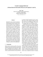
Fig. 1. Identification of Rbm9, a novel Gld2-interacting protein. (A) Schematic representation of the hGld2 fusion proteins used for the two-
hybrid screen. The amino- and carboxy-terminal moieties of hGld2 (hGld2N and hGld2C, respectively) were expressed as fusion proteins with
Gal4BD, and used together for the screen. (B) Growth of transformed yeast in selective medium. Bait plasmids (left) were mated with prey
plasmids (top). In addition to hGld2N and hGld2C, bait plasmids included the empty bait plasmid (pGBT9), full length hGld2 and the kinase
AuroraA. Prey plasmids included the empty prey plasmid (pADGal4), hRbm9 (clone 12 from the screen), a negative control obtained from
the screen (clone 5) and Maskin p17. AuroraA–Maskin p17 interaction served as a positive control. Double transformants growing on med-
ium lacking tryptophan, leucine and histidine (–W–L–H), indicating an interaction, are designated by ‘+’. Double transformants which did not
grow are indicated by ‘)’. (C) Schematic representation of the XRbm9 sequence and comparison with hRbm9 isolated in the screen. The
two proteins carry two different carboxy-terminal domains (dark and light grey, respectively) as a result of alternative splicing. The sequence
similarity in RRM is indicated (%). For sequence comparison between XRbm9 and hRbm9, see supplementary Fig. S1. (D) Total, nuclear and
cytoplasmic protein extracts were analysed by western blotting with the indicated antibodies. RPA, exclusively expressed in the nucleus,
served as enucleation control. (E) Immunoblot analyses of Xenopus oocyte extracts (left panel) and embryo extracts (right panel) with
XRbm9 and b-tubulin antibodies. b-Tubulin, consistently expressed throughout oocyte maturation and embryogenesis, served as a loading
control. (D, E) XRbm9: in vitro translated XRbm9 served as migration size control. Protein sizes are indicated (kDa).
XRbm9, a novel XGld2 interactor C. Papin et al.
492 FEBS Journal 275 (2008) 490–503 ª 2008 The Authors Journal compilation ª 2008 FEBS
RT-PCR from oocyte total RNA. The 411-amino-acid
ORF contains a single central RNA recognition motif
(RRM)-type RNA-binding domain with two RNP
domains (Fig. 1C and supplementary Fig. S1). In addi-
tion, the ORF contains two arginine ⁄ glycine-rich
(RGG) motifs that are characteristic of RNA-binding
proteins, and an alanine-rich carboxy-terminal sequence
that could be involved in protein–protein interactions.
Interestingly, this alanine-rich sequence, generated by
alternative splicing, is not present in the hRbm9 isolated
in the screen (supplementary Fig. S1). Sequence com-
parison with hRbm9 shows an overall 59% similarity,
which increases to 98% for the RNA-binding domain
(Fig. 1C). Therefore, this cDNA is referred to as
XRbm9.
To study the biological role of XRbm9 in Xenopus
oocytes, an XRbm9 antibody was raised (supple-
mentary Fig. S2) and used to examine the abundance
and localization of endogenous XRbm9 in oocytes
(Fig. 1D). A single endogenous protein of about
55 kDa, co-migrating with the in vitro-translated
XRbm9 protein (lane 1), was present in stage VI and
mature oocytes (lanes 2 and 3). Interestingly, XRbm9
was exclusively detected in the oocyte cytoplasm
(lane 4). Western blot analysis showed that XRbm9 is
expressed throughout oogenesis, oocyte maturation
and during embryogenesis up to stage 33 (Fig. 1E).
These data identify a novel Gld2-interacting protein,
XRbm9, which is expressed in the oocyte cytoplasm.
XRbm9 is a component of the cytoplasmic
polyadenylation complex
Next, the interactions between XGld2 and XRbm9
were investigated by yeast two-hybrid analyses.
Human and Xenopus Rbm9 and Gld2 can interact with
each other reciprocally (Fig. 2A). Deletion constructs
showed that the Gld2–Rbm9 interaction is mediated
by the Gld2 N-terminal domain. Interestingly, the
N-terminal parts of Xenopus and human Gld2 share
only 36% similarity. Moreover, XGld2D4, a splice var-
iant (shown in supplementary Fig. S4), interacts with
Rbm9, but the N-terminal part of this variant
(XGld2D4N) does not. These data suggest that the
interacting domain in the N-terminal region of Gld2 is
more likely to be conformational than a definite
sequence. Conversely, Rbm9 N-terminal-most residues
are not required for the Gld2–Rbm9 interaction
(Fig. 2B). However, the RRM-containing central
domain of hRbm9 (amino acids 48–269) and amino
acids 269–350 of hRbm9 are not able to mediate the
binding. These data suggest that a domain surrounding
amino acid 269 is important for the interaction, or that
most of the Rbm9 sequence is required for the inter-
action. However, the possibility that smaller regions of
hRbm9 (amino acids 48–269 or 269–350) are not suffi-
ciently expressed in yeast to detect an interaction
cannot be ruled out.
To test whether XRbm9 interacts with polyadenyla-
tion factors in ovo, co-immunoprecipitation experi-
ments were performed under various conditions using
our specific XRbm9 antibody. The injection of HA-
tagged XRbm9DN (55–411) into oocytes and precipita-
tion with HA antibody in the presence of RNaseA
showed that endogenous XGld2 and CPEB were
specifically immunoprecipitated with overexpressed
XRbm9 (Fig. 2C, top panel). Overexpressed HA-
XRbm9 was also detected in XGld2 and CPEB immu-
noprecipitates (Fig. 2C, bottom panel). Alternatively,
HA-tagged XGld2 was overexpressed in oocytes, and
the lysates were immunoprecipitated with XRbm9,
XGld2, CPEB antibodies or a control IgG in the pres-
ence of RNaseA (Fig. 2D). This condition allowed us
to co-precipitate endogenous XRbm9 with XGld2 and
CPEB. Reciprocally, overexpressed XGld2 and endo-
genous CPSF100 and CPEB were co-precipitated
with the XRbm9, CPEB and XGld2 antibodies.
Finally, in oocytes that did not overexpress exogenous
proteins, endogenous XRbm9 was co-immunoprecipi-
tated with the XGld2 and CPEB antibodies (Fig. 2E).
Reciprocally, CPEB was present in the XRbm9 and
XGld2 precipitates.
Together, these results show that endogenous
XRbm9 belongs to a complex with XGld2, CPEB and
CPSF independent of an RNA intermediate and possi-
bly through its direct interaction with XGld2.
XRbm9 stimulates translation in Xenopus
oocytes
As XRbm9 is associated with the polyadenylation com-
plex, its requirement for cytoplasmic polyadenylation
was investigated. XRbm9 antibody was injected into
oocytes in order to interfere with the endogenous
protein, and mos mRNA polyadenylation was scored
using a polyadenylation test (PAT). XRbm9 antibody
injection did not affect the progesterone-induced poly-
adenylation extent of the reporter RNA (supplementary
Fig. S3) and had no effect on meiotic maturation (data
not shown). These data indicate that either XRbm9
is not required for cytoplasmic polyadenylation in
oocytes, or that the XRbm9 antibody was not able to
prevent XRbm9 function.
The role of XRbm9 was investigated using the teth-
ered approach that has been employed to study the
function of proteins involved in mRNA stability or
C. Papin et al. XRbm9, a novel XGld2 interactor
FEBS Journal 275 (2008) 490–503 ª 2008 The Authors Journal compilation ª 2008 FEBS 493
translation [27–29]. XRbm9 protein was fused to the
MS2 coat protein to allow the tethering of XRbm9 to
a reporter mRNA bearing a tandem pair of MS2-bind-
ing sites. Oocytes were first injected with the
MS2-XRbm9-encoding mRNA, or MS2 alone and
MS2-U1A as negative controls. As positive
control, MS2-PABP, known to stimulate translation in
oocytes, was also injected [28]. After 6 h of incubation
to allow protein synthesis, two reporter mRNAs were
co-injected: a firefly luciferase mRNA bearing MS2-
binding sites in its 3¢-UTR and an internal control
mRNA encoding the Renilla luciferase. After another
16 h of incubation, both luciferase activities were
determined. MS2-XRbm9 expression stimulated the
XRbm
9
N (55-411)
hRbm9 48-269
hRbm9 269-350
hRbm
9
N (48-401)
hRbm
9
+
pADGal4
+
+
+
XRbm9
+
+
++
+
+
+
+
+
+
+
+
Maskin p17
A
B
hGld2
XGld2N
XGld2
XGld2
XGld2N
hGld2
XGld2 4N
hGld2C
hGld2N
XGld2 4
Gal4 BD
Gal4 BD
Gal4 AD
Gal4 AD
HA (XRbm9)
C
Input
XGld2
HA IgG
IP
XRbm9
CPEB
HA-XRbm9 N overexpression
Input
XGld2
XRbm9
CPEB
IgG
IP
D
HA-XGld2 overexpression
Input
XGld2
XRbm9
CPEB
IgG
XGld2
CPEB
CPSF100
IP
XRbm9
XRbm9
endogenous proteins
E
CPEB
Input
XGld2
Rbm9
CPEB
IgG
IP
Growth in -W-L-H medium
Growth in
-W-L-H
medium
NLS
Gld2
Catalytic
Central
PAP/25A
RRM
Rbm9
RGG
RNP
Cter
Fig. 2. XRbm9 is part of a complex with
XGld2, CPEB and CPSF. (A, B) Gld2–Rbm9
interaction in yeast two-hybrid system. The
two-hybrid system was used to determine
interactions between the indicated con-
structs. Gld2 constructs were expressed as
fusion proteins with Gal4BD and Rbm9 con-
structs were expressed as fusion proteins
with Gal4AD. Double transformants growing
on medium lacking tryptophan, leucine and
histidine, indicating an interaction, are desig-
nated by ‘+’. Double transformants which
did not grow are indicated by ‘)’. (C–E)
Co-immunoprecipitation experiments in the
presence of RNaseA. Oocyte extracts alone
(E), overexpressing HA-tagged XRbm9DN
(C) and HA-tagged XGld2 (D) were immuno-
precipitated as indicated, and the immuno-
precipitates were analysed by western
blotting as indicated. The equivalent of one
oocyte was loaded as input.
XRbm9, a novel XGld2 interactor C. Papin et al.
494 FEBS Journal 275 (2008) 490–503 ª 2008 The Authors Journal compilation ª 2008 FEBS
luciferase activity by about sixfold compared with the
MS2 protein alone (Fig. 3A). This activation was
comparable with that obtained with MS2-PABP. This
activation was cis-dependent, as MS2-XRbm9 and
MS2-PABP fusion proteins did not affect the expres-
sion of firefly luciferase reporter mRNA lacking the
MS2-binding sites (LucDMS2). As expected, the con-
trol MS2-U1A did not stimulate translation regardless
of whether MS2-binding sites were or were not pres-
ent. Moreover, similar levels of all MS2 fusion proteins
were expressed in the oocytes (Fig. 3B). These experi-
ments show that tethered XRbm9 is able to activate
the translation of reporter mRNA in oocytes.
We then investigated how the tethering of an
XRbm9 protein to an mRNA could stimulate transla-
tion. As an XGld2-interacting protein, XRbm9 could
enhance translation by targeting XGld2 to the mRNA
and allowing its polyadenylation, which would enhance
its translation. To assess this issue directly, a tethered
assay was performed in which MS2-XRbm9 was co-
injected with the HA-tagged catalytically inactive form
of XGld2 (XGld2 D242A). As shown in Fig. 3C, over-
expression of XGld2 D242A (Fig. 3D) did not affect
the translational activation by MS2-XRbm9. More-
over, the overexpression of the wild-type form of
XGld2 did not potentiate the stimulation of the
U1A
MS2
MS2-PAB
MS2-XRbm9
MS2-U1A
Tubulin
Reticulocytes Oocytes
XRbm9
PABP
U1A
XRbm9
PABP
BA
CD
MS2 MS2
U1A
MS2
XRbm9
MS2
PABP
Luc-MS2
Luc-
MS2
1
0
2
4
6
7
8
5
3
HA
Tubulin
MS2 XGld2
XGld2 D24
2A
XGld2
W
T
HA MS2-XGld2
HA XGld2
MS2
XGld2
MS2
XRbm9
+
XGld2
D242A
MS2
XRbm9
+
XGld2WT
MS2
XRbm9
MS2
1
0
2
4
5
3
Fig. 3. Tethered XRbm9 stimulates translation in Xenopus oocytes. (A) Oocytes expressing MS2, MS2-U1A, MS2-XRbm9 or MS2-PABP
fusion proteins were injected with either Luc-MS2 and Renilla luciferase mRNAs (dark grey) or Luc-DMS2 and Renilla luciferase mRNAs
(light grey). The translation of the reporter mRNAs was determined by a dual luciferase assay. Luciferase activity was plotted (the firefly ⁄
Renilla luciferase activity ratios in the presence of the fusion proteins are shown relative to the activity with MS2 alone, set at unity). The
mean values of three different experiments are shown. For each experiment, three to five pools, each containing three to five oocytes, were
assayed per experimental point, and the mean values and standard deviations were determined. (B) Expression of MS2 fusion proteins in
reticulocytes (RRL) and oocytes by western blotting using MS2 antibody. In oocytes, MS2-PABP co-migrates with a non-specific band (star)
when compared with the migration of in vitro-translated MS2-PABP. (C) Oocytes expressing MS2, MS2-XRbm9 and HA-MS2-XGld2 fusion
proteins, or coexpressing MS2-XRbm9 and HA-XGld2 D242A or MS2-XRbm9 and HA-XGld2WT, were injected with Luc-MS2 and Renilla
luciferase mRNAs. The translation of the reporter mRNAs was determined by a dual luciferase assay. (D) HA-MS2-XGld2, HA-XGld2DA and
HA-XGld2WT protein expression in oocytes by western blotting using HA antibody.
C. Papin et al. XRbm9, a novel XGld2 interactor
FEBS Journal 275 (2008) 490–503 ª 2008 The Authors Journal compilation ª 2008 FEBS 495
luciferase activity by MS2-XRbm9. These data suggest
that the translational activation by tethered XRbm9 is
not dependent on XGld2. This experiment also shows
that the translational activation by MS2-XRbm9 is
comparable with that obtained with MS2-XGld2.
XGld2 antibody injection accelerates the
G2 ⁄ M transition in Xenopus oocytes
During the course of our experiments, it was noticed
that the XGld2 antibody was able to affect endoge-
nous XGld2 function (supplementary Fig. S3). XGld2
interacts with the polyadenylation factors CPEB and
CPSF in oocytes [6,19]. However, so far, an antibody
directed against XGld2 has not been used to study
Gld2 function in oocytes. The difficulty in visualizing
endogenous XGld2 with a specific antibody may be
caused by its small amounts in frog’s eggs. Using our
specific XGld2 antibody (supplementary Fig. S4A–C),
the endogenous (Fig. 4A, lanes 2 and 3), overexpressed
(lane 4) and HA-tagged (lane 1) XGld2 proteins were
detected by western blotting. The antibody can also
specifically immunoprecipitate endogenous XGld2
protein (Fig. 4A, lane 7). In addition, CPEB and
CPSF160 were detected in the XGld2 immunoprecipi-
tates, showing that, consistent with the overexpression
studies, immunoprecipitated endogenous XGld2 is
associated with CPEB and CPSF (Fig. 4A,
lanes 11–13).
Advantage was taken of this specific XGld2 anti-
body to address the function of XGld2 in meiotic
maturation. The antibody was injected into oocytes
induced to maturate with progesterone. Unexpectedly,
XGld2 antibody injection accelerated the G2 ⁄ M transi-
tion when compared with control (i.e. IgG-injected or
uninjected) oocytes (Fig. 4B). XGld2 antibody-injected
oocytes underwent 50% germinal vesicle breakdown
(GVBD) 2 h before control oocytes, suggesting an
acceleration of the G2 ⁄ M transition in meiosis I. This
hastening of maturation was correlated with a preco-
cious synthesis of Mos and AuroraA proteins, and
with the activation of the mitogen-activated protein
kinase (MAPK) (Fig. 4C). To confirm these findings,
the function of CPEB, another protein involved in
mRNA masking, was inhibited. Injection of CPEB
antibody led to similar results on progesterone-induced
oocyte maturation and on the molecular markers
(Fig. 4D, E). Moreover, CPEB antibody injection in
oocytes without progesterone treatment led to a mild
but reproducible activation of extracellular signal-regu-
lated kinase (ERK) (Fig. 4F, see Discussion).
Thus, affected XGld2 or CPEB function leads to
accelerated progesterone-induced oocyte maturation,
suggesting that XGld2, as well as CPEB and CstF-77
[20], belong to a masking complex in oocytes.
XGld2 antibody inhibits cytoplasmic
polyadenylation in Xenopus oocytes
It was tested whether the activity of endogenous
XGld2 polymerase was required for cytoplasmic poly-
adenylation in Xenopus oocytes using XGld2 antibody.
In vitro PAT assay in egg extracts was not possible as
XGld2 antibody was not able to deplete the polymer-
ase from the extracts. Therefore, XGld2 or CPEB anti-
body was injected into oocytes and exogenous
mos mRNA polyadenylation was scored using PAT
assay. Although progesterone induced robust poly-
adenylation of the reporter RNA (Fig. 5A, lanes 2
and 3, and supplementary Fig. S3), a decrease in both
the length of the poly(A) tail and the overall extent of
polyadenylation was observed when XGld2 antibody
was injected (lane 5). Injection of CPEB antibody also
prevented poly(A) tail elongation (lane 4). Inhibition
of polyadenylation by XGld2 antibody was also
detected during the kinetics of maturation (Fig. 5B),
with the decrease in the poly(A) tail length being
observed as soon as 1 h after progesterone addition
(compare lanes 3 and 8). These data represent direct
evidence that endogenous XGld2 is required for cyto-
plasmic polyadenylation in maturing oocytes.
Taken together, these results demonstrate that
endogenous XGld2 is a component of the cytoplasmic
polyadenylation machinery and is required for this
regulatory event.
Discussion
In this study, a new XGld2 interactor, the RNA-bind-
ing protein XRbm9, was identified. It was demon-
strated that it is part of a complex with Gld2, CPEB
and CPSF, and that tethered XRbm9, via the
MS2 protein, stimulates translation. Moreover, it was
shown that endogenous XGld2 is required for cyto-
plasmic polyadenylation, and is probably part of a
masking complex with CPEB in stage VI oocytes.
Using a specific antibody, endogenous XGld2 was
inhibited and, for the first time, its function was
assessed in vivo. XGld2 antibody injection led to the
inhibition of mos mRNA cytoplasmic polyadenylation,
corroborating the significant role of XGld2 in cyto-
plasmic polyadenylation during meiotic maturation.
Intriguingly, XGld2 or CPEB antibody injection also
led to an acceleration of progesterone-induced oocyte
maturation. This dual effect of an antibody has previ-
ously been reported during the study of p82, the clam
XRbm9, a novel XGld2 interactor C. Papin et al.
496 FEBS Journal 275 (2008) 490–503 ª 2008 The Authors Journal compilation ª 2008 FEBS
CPEB homologue [30], where it was proposed that
p82 has two functions: the first involving masking in
immature oocytes and the second involving the activa-
tion of translation by cytoplasmic polyadenylation. It
has been reported previously that CPEB antibody
injection leads to an inhibition of meiotic maturation
[31]. However, later studies have implicated CPEB
in mRNA masking in oocytes [3,30,32,33], and this
Uninjected
Ig G
CPEB Ab
Uninjected
Ig G
XGld2 Ab
0
20
40
60
80
100
% GVBD
0
20
40
60
80
100
% GVBD
hours in progesterone
3
10. 5
hours in progesterone
2.5
3.5 1085.54.5
0 0.5 4321.5 6
Mos
Tubulin
IgG
CPEB Ab
IgG
CPEB Ab
PP ERK
Tubulin
PP ERK
IgG
CPEB Ab
Aurora A
IgG
CPEB Ab
hours in Pg
0 0.5 1 54321.5 6
Mos
Tubulin
IgG
XGld2 Ab
IgG
XGld2 Ab
PP ERK
IgG
XGld2 Ab
Aurora A
IgG
XGld2 Ab
hours in Pg
XGld2
CPEB
IgG
CPSF160
XGld2
IP
CPEB
IgG
Input
HA-XGld2
XGld2
StVI
MII
HA
XGld2
XGld2
XGld2
1
10 11 12 13
4657
15
56 7 8 9
234
Input
XGld2
XGld2
XGld2
IgG
IgG
IP
overexp.
XGld2
endogen.
XGld2
XG
ld2 Ab
CPEB
Ab
IgG
M
II
C
AB
DE
F
Fig. 4. XGld2 and CPEB antibody injections accelerate the G2 ⁄ M transition in oocytes. (A) Characterization of the XGld2 antibody. Top panel:
western blot analysis of overexpressed HA-XGld2 or XGld2 in oocytes or endogenous XGld2 in stage VI (StVI) or mature (MII) oocytes with
the XGld2 antibody. Middle panel: immunoprecipitates from XGld2-overexpressing (overexp. XGld2) or stage VI (endogen. XGld2) oocytes
with XGld2 antibody or a control IgG were analysed by western blotting as indicated. The star indicates a non-specific band. Bottom panel:
oocyte extracts were immunoprecipitated and analysed by western blotting as indicated. The equivalent of one oocyte was loaded as input.
(B) Oocytes were injected with XGld2 or non-specific (IgG) antibodies, or left uninjected. After 1 h of incubation, maturation was induced with
progesterone (Pg) and the percentage of GVBD was scored at the indicated time and plotted. This graph is representative of five experiments.
(C) Immunoblot analysis of Mos, AuroraA, activated MAPK (PP ERK) and b-tubulin levels in oocytes collected during an experiment depicted
in (A). A significant increase in Mos and AuroraA protein synthesis and ERK biphosphorylation was observed in XGld2 antibody-injected
oocytes (XGld2 Ab) as early as 1.5 h after progesterone treatment, compared with 4 h for control oocytes (IgG). (D, E) Similar experiments as
in (B) and (C), respectively, using the CPEB antibody. (F) Oocytes were injected with XGld2, CPEB or non-specific (IgG) antibodies. After 16 h
of incubation without progesterone, the activation status of MAPK (PP ERK) was assessed by western blot. The mature oocyte (MII) served as
a control of ERK activation. It should be noted that XGld2 antibody injection did not trigger MAPK activation in the absence of progesterone.
C. Papin et al. XRbm9, a novel XGld2 interactor
FEBS Journal 275 (2008) 490–503 ª 2008 The Authors Journal compilation ª 2008 FEBS 497
is confirmed by the present data which show an
acceleration of meiotic maturation by CPEB antibody
injection. The discrepancy with regard to the effect of
CPEB antibody injection on oocyte maturation may
be the result of the use of different CPEB antibodies
that do not recognize the same epitopes in the CPEB
protein. As XGld2 associates with CPEB in stage VI
oocytes [this study and 6,19,20], the data presented
here are consistent with the presence of XGld2 in
a masking complex with CPEB in oocytes. The
antibodies, by interacting with their target proteins,
could disrupt this masking complex, alleviate the
repression and allow the translation of maturation-
required proteins before the requirement of cytoplasmic
polyadenylation. In agreement with this, ERK activa-
tion (reflecting Mos synthesis) by CPEB antibody
injection without progesterone treatment (Fig. 4F)
strengthens the idea that perturbation of the repressive
complex leads to the synthesis of Mos without the need
for poly(A) tail elongation. Therefore, the complex
bearing XGld2 and CPEB, already present in stage VI
oocytes, could be considered as a masking complex.
CeGLD-2 polymerase activity is stimulated by inter-
action with the RNA-binding protein GLD-3 [16]. In
Xenopus oocytes, previous studies have shown that
CPEB and CPSF are RNA-binding proteins that bring
XGld2 to the 3¢-end of mRNAs regulated by cytoplas-
mic polyadenylation [6,19]. In this study, XRbm9 was
identified as a new RNA-binding protein that interacts
with XGld2. It was shown that XRbm9 is a component
of the polyadenylation complex with CPEB and CPSF.
Hence, three RNA-binding proteins interact directly
with XGld2 and are present in the same complex. How-
ever, the possibility that XRbm9 and XGld2 are in com-
plexes independent of CPEB cannot be ruled out. More
generally, different RNA-binding proteins, interacting
with Gld2, could connect the PAP to different types of
RNA target. It was shown that tethered XRbm9 stimu-
lates the translation of a reporter mRNA. This stimula-
tion does not seem to be dependent on the presence of
XGld2, as the overexpression of wild-type or catalyti-
cally inactive XGld2 together with MS2-XRbm9 does
not affect the translational activation by XRbm9. How-
ever, it cannot be excluded that, under physiological or
specific conditions, XRbm9 is able to target XGld2 to
specific mRNA. The molecular mechanism underlying
XRbm9-dependent translational activation is unclear
and awaits further investigations.
The subcellular localization of mammalian Rbm9 is
unclear and is dependent on the isoform and the tissue
examined; however, it appears to be mainly nuclear in
cell lines and brain where, nevertheless, there is addi-
tional cytoplasmic expression [23,24]. In this study, an
XRbm9 isoform expressed at steady state in the oocyte
cytoplasm was identified. The amino-terminal-most
sequence of XRbm9 is particular, as it is extended in
comparison with the amino-terminal sequences identi-
fied in X. tropicalis, mammals, C. elegans and zebra-
fish. This peculiar sequence could be the mark of
an oocyte-specific XRbm9 isoform. It is probable,
however, that other XRbm9 isoforms are present in
A
500 bp
400 bp
350 bp
300 bp
220 bp
200 bp
M
poly(A) tail
Ab t0
no Ab
IgG
XGld2 Ab
CPEB Ab
400 bp
300 bp
mos
S22
B
IgG XGld2 Ab
M
hours in
progesterone
poly(A) tail
500 bp
400 bp
350 bp
300 bp
220 bp
200 bp
mos
S22
400 bp
300 bp
1423 5
0.5 1 531.5 0.5 1
53
1.5
1423 567 1089 11
Ab t0
Fig. 5. XGld2 is required for cytoplasmic polyadenylation. (A, B)
Polyadenylation assay in oocytes. (A) Oocytes were injected with
mos 3¢-UTR RNA and, 30 min later, with XGld2 or CPEB antibodies
(Ab), nonspecific IgG (IgG) or left uninjected (no Ab). After 1 h of
incubation, maturation was induced with progesterone. Total RNA
was extracted from pools of five oocytes collected at the time of
progesterone addition (Ab t0) or when 30% of control oocytes had
undergone GVBD. Total RNA was submitted to mos polyadenyla-
tion analysis (PAT) using specific primers. This gel is representative
of five experiments. (B) Kinetics of mos 3¢-UTR polyadenylation.
Oocytes were injected with XGld2 antibody or nonspecific IgG and
treated as in (A). The mos 3¢-UTR polyadenylation status was
assessed at the indicated time after progesterone (Pg) addition. In
this experiment, 30% of oocytes underwent GVBD at the 5 h time
point. The polyadenylation status of the endogenous S22 RNA in
the same samples was not affected by the injection of antibodies
(negative control). Fragment sizes (M) are indicated on the right in
base pairs (bp).
XRbm9, a novel XGld2 interactor C. Papin et al.
498 FEBS Journal 275 (2008) 490–503 ª 2008 The Authors Journal compilation ª 2008 FEBS
embryonic and adult tissues, and that they display
nuclear localization. XGld2 is expressed in both the
nucleus and cytoplasm, whereas XRbm9 is only
detected in the cytoplasm. The nuclear function of
XGld2 remains unstudied, but its role could be
related to the function of the Saccharomyces cerevisae
Trf4 protein in RNA quality control. However, this
XGld2 nuclear function should be independent of the
XRbm9 isoform isolated in this study.
Interestingly, recent studies have shown that proteins
involved in splicing, as well as the exon junction com-
plex, may mediate the enhancing effect of splicing on
mRNA translation [34–36]. Rbm9, as a splicing factor
interacting with a PAP, may also participate in the
translational enhancement mediated by introns.
Indeed, the presence of the PAP Gld2 on the messen-
ger, targeted by a protein of the Rbm9 family, may
allow the polyadenylation of the messenger regulated
by Rbm9, hence enhancing its translation. Further
studies are needed to determine a potential role for
Rbm9 in this type of translational regulation.
In mammals, Rbm9 has been identified as a repres-
sor of tamoxifen activation of the oestrogen receptor
and as a gene upregulated by androgens [26,37].
Moreover, Underwood et al. [23] have shown that
mRbm9 is expressed in the ovary, whereas mA2BP1 is
not, and other particular Rbm9 splice variants appear
to be specific to breast, ovary or other oestrogen-
sensitive tissues. Therefore, it would be of interest
to examine whether, in oocytes, XRbm9 activity or
localization could be regulated by progesterone.
hRbm9 has been shown to interact directly with the
oestrogen receptor [26]. In Xenopus, different recep-
tors have been described to mediate oocyte matura-
tion [38–40]. However, these steroid receptors are not
detected in the membrane where progesterone signal-
ling is initiated. More recently, a membrane progestin
receptor (mPR) unrelated to nuclear steroid receptors
has been identified [41]. Investigating the possible
interaction between XRbm9 and the progesterone
receptor could lead us to uncover a link between pro-
gesterone and the cytoplasmic polyadenylation
machinery.
Experimental procedures
Xenopus oocytes and embryos
Oocyte manipulations in MMR buffer (5 mm Hepes pH 7.8,
100 mm NaCl, 2 mm KCl, 1 mm MgSO
4
, 0.1 mm EDTA,
2mm CaCl
2
) and oocyte extracts in lysis buffer were per-
formed as described in [42]. Manual enucleation of oocytes
was performed as described in [20]. Progesterone was used
at 10 mgÆmL
)1
. For microinjections, the usual injected vol-
ume for antibodies and RNA was 20–40 nL per oocyte, and
the number of injected oocytes was 35 for each condition.
In vitro fertilization and embryo cultivation were performed
as described in [43].
Cloning of XGld2 and hGld2
The CeGld2 cDNA sequence was used as a reference in our
blast search of databases from the X. laevis EST project
(http://). This search yielded multi-
ple overlapping ESTs that produced a complete ORF.
Stage VI oocyte total RNA was used to perform an oli-
go(dT)-primed reverse transcription employing the Super-
script
TM
II reverse transcriptase (Invitrogen, Cergy-Pontoise,
France). PCR using the primers 74 (5¢-GTCGCTGTGTT
GTTCTGTCAGGC-3¢) and 75 (5¢-GGCCACCGTTTTT
AGCATTTCTCCC-3¢) was performed, and the amplified
PCR products were cloned into a TA cloning vector
(pCRII) (Invitrogen) and sequenced. The longest clone cor-
responded to the XGld2 cDNA described in Barnard et al.
[6]. The shortest corresponded to an alternatively spliced
form of XGld2 missing exon 4 (XGld2D4, see supplemen-
tary Fig. S4). A blast search of the human genome data-
base was conducted using the XGld2 coding sequence to
identify homologous human cDNAs. Primers encompassing
a putative ORF were designed as follows: 89, 5¢-ATCGAT
ATGTTCCCAAACTCAATTTTGGGTCG-3¢; 90, 5¢-TAG
AGACCAGTTATCTTTTCAG-3¢. Oligo(dT)-primed cDNA
from SW80 cell line RNA was used to perform a PCR
using the above primers. The PCR products were cloned
into a TA cloning vector (Invitrogen) and sequenced. Three
human cDNAs corresponding to those described in
Rouhana et al. [19] were isolated. The cDNA used for the
two-hybrid screen was the alternatively spliced variant lack-
ing exon 8 (hGld2D8).
Cloning of Xenopus Rbm9
With the hRbm9 cDNA sequence isolated during the
two-hybrid screen as the query sequence, a blast search
was run on databases from the X. laevis EST pro-
ject (). This search generated
multiple overlapping ESTs that yielded a complete ORF.
Stage VI oocyte total RNA was used to perform an
oligo(dT)-primed reverse transcription employing the
Superscript
TM
II reverse transcriptase (Invitrogen). PCR
using the primers 123 (5¢-CCCTTTCCTGTTAG
CAGTGTG-3¢) and 120 (5¢-GGGACAATAGGCTTA
CGTCACT-3¢) was performed, and the amplified PCR
products were cloned into a TA cloning vector (Invitro-
gen) and sequenced. An alternatively spliced exon was
also isolated during the course of the XRbm9 cloning
(supplementary Fig. S1).
C. Papin et al. XRbm9, a novel XGld2 interactor
FEBS Journal 275 (2008) 490–503 ª 2008 The Authors Journal compilation ª 2008 FEBS 499
DNA constructs and RNA synthesis
XGld2
Expression vectors.
pCS2 XGld2, pCSH XGld2: the
XGld2 ORF from pCRII XGld2 was inserted into the
ClaI-EcoRI sites of the pCS2+ and pCSH vectors (in
frame with the HA tag [20]).
Two-hybrid vectors. pGBT9 XGld2: the XGld2 ORF from
pCRII XGld2 was inserted into the Cla I-KpnI sites of the
pBSKS+ vector. The XGld2 ORF from pKS XGld2 was
then inserted in frame with Gal4BD at the EcoRI site of the
pGBT9 vector. pGBT9 XGld2N: the EcoRI-PstI fragment
(XGld2 amino acids 1–149) from pKS XGld2 was inserted
in frame with Gal4BD into the pGBT9 vector. pGBT9
XGld2D4, pGBT9 XGld2D4N : these were generated from
pCRII XGld2D4 as described for full-length XGld2.
hGld2
Two-hybrid vectors.
pGBT9 hGld2: the hGld2 ORF from
pCRII hGld2D8 was inserted into the EcoRI site of the
pGBT9 vector in frame with Gal4BD. pGBT9 hGld2N: the
EcoRI-PstI fragment (hGld2D8 amino acids 1–185) from
pCRII hGld2D8 was inserted in frame with Gal4BD into
the pGBT9 vector. pGBT9 hGld2C: the PstI fragment
(hGld2D8 amino acids 184–480) from pCRII hGld2D8 was
inserted in frame with Gal4BD into the pGBT10 vector.
XRbm9
pCRII XRbm9ORF.
The XRbm9 ORF from the ATG
to the stop codon of the protein was generated by PCR
using the following primers: 122 (5¢-ATCGATATGGC
AGATGCTGTAATGTC-3¢) and 120 (as above). The
amplified PCR products were cloned into a TA cloning
vector (Invitrogen) and sequenced.
Expression vectors. pCS2 XRbm9: the XRbm9 ORF from
pCRII XRbm9ORF was inserted into the ClaI-EcoRI sites
of the pCS2+ vector. pCSH XRbm9DN: the XRbm9DN
fragment corresponding to amino acids 55–411 was inserted
into the ClaI-EcoRI sites of the pCSH vector [20] in frame
with the HA tag.
Two-hybrid vectors. pAD XRbm9: the XRbm9 ORF from
pCRII XRbm9ORF was inserted in frame with the Gal4
activation domain (Gal4AD) into the EcoRI site of the
pADGal4 vector. pAD XRbm9DN: the XRbm9DN frag-
ment was inserted in frame with Gal4AD into the EcoRI
site of the pADGal4 vector.
MS2 fusion protein. cDNA encoding XRbm9 was cloned
as a PCR product using the primers 5¢-TGCTAGCATGG
CAGATGCTGTAATG-3¢ and 5¢-CCTCGAGTCAGTAC
GGAGCAAATCG-3¢ containing NheI and XhoI restric-
tion sites (italic) into the NheI-XhoI-restricted pMSP
vector.
hRbm9
Two-hybrid vectors.
pAD hRbm9DN: the hRbm9 frag-
ment corresponding to amino acids 48–401 from pADGal4
hRbm9 was inserted into the EcoRI-SmaI sites of the
pBSKS+ vector to generate pKS hRbm9DN. The EcoRV-
SmaI fragment from this plasmid was inserted in frame
with Gal4AD into the SmaI site of the pADGal4 vector.
pAD hRbm9 48-269: the hRbm9 fragment corresponding to
amino acids 48–269 from pADGal4 hRbm9 was inserted
into the EcoRI-PstI sites of the pBSKS+ vector to gener-
ate pKS hRbm9 48-269. The EcoRV-PstI fragment from
this plasmid was inserted in frame with Gal4AD into the
SmaI-PstI sites of the pADGal4 vector. pAD hRbm9 269-
350: the hRbm9 fragment corresponding to amino acids
269–350 from pADGal4 hRbm9 was inserted in frame with
Gal4AD into the Pst I site of the pADGal4 vector.
pGBT9 AuroraA and pGADGH-p17 were provided
by Y. Arlot-Bonnemains [45]. The wild-type mos 3¢-UTR
reporter RNA construct was obtained from [20]. Capped
mRNA encoding the different constructs was prepared by
linearizing the pCS2- or pCSH-encoding ORFs [20] with
NotI and carrying out transcription reactions according to
[46]. It was used at an initial concentration of 400 ngÆmL
)1
.
Yeast two-hybrid screen
A directional human embryonic cDNA library (gift from
N. Bonneaud, CNRS, Montpellier, France) was used for
screening [44]. The human hGld2 bait plasmids were con-
structed by cloning the amino-terminal (amino acids 1–185)
and carboxy-terminal (amino acids 184–480) parts of
hGld2D8 in frame with Gal4BD. The yeast two-hybrid
screen was performed using the mating procedure described
in [44]. After 3–5 days, the histidine-positive clones were
analysed. PCR amplification of the inserts using pADGal4-
specific primers was performed on the yeast colonies, and
the PCR products were sequenced. Approximately
7 · 10
5
clones were screened and 90 clones were found to
grow in the absence of histidine. Inserts from 24 clones
were sequenced and analysed by blast comparison with the
translated nucleotide sequence database.
Antibodies and immunoblot analysis
XGld2 and XRbm9 antibodies were directed against the pep-
tides NH
2
-NTARAVYEKQKFD-COOH and NH
2
-SQG-
NQEPTATPDT-COOH, respectively. These antibodies were
raised by injection of the thyroglobulin-coupled peptides
(Sigma-Aldrich, St Louis, MO, USA) into rabbits (New
XRbm9, a novel XGld2 interactor C. Papin et al.
500 FEBS Journal 275 (2008) 490–503 ª 2008 The Authors Journal compilation ª 2008 FEBS
Zealand), followed by affinity purification. The antibody
against Xenopus CPEB is an affinity-purified rabbit poly-
clonal antiserum [20]. The RPA and AuroraA antibodies
were provided by J. M. Lemaıˆ tre (CNRS, Montpellier,
France) and C. Prigent (CNRS, Remes, France), respec-
tively. The CPSF100 and MS2 antibodies were provided by
E. Wahle (University of Halle, Germany) and P. G. Stockley
(University of Leeds, UK), respectively. The anti-CPSF160,
anti-Mos and IgG antibodies were obtained from Santa Cruz
Technology (Santa Cruz, CA, USA) (SC-28872, SC-086 and
SC-2027, respectively). The pTpY ERK-MAPK antibody
was obtained from New England Biolabs (Ipswich, MA,
USA) (9106S). The b-tubulin and HA antibodies were
obtained from E7 (Iowa Hybridoma Bank, Iowa City,
IA, USA) and 12CA5 (Abcam, Cambridge, MA, USA)
hybridomas, respectively. For microinjections, purified anti-
bodies were dialysed and used at 1 lgÆlL
)1
. Western blots
were performed as described in [42].
Immunoprecipitations
Protein oocyte extracts corresponding to 30 oocytes,
RNaseA treatment and immunoprecipitations were per-
formed as described in [20]. Depending on the size of the
protein, the samples were boiled or not, separated by
SDS-PAGE and analysed by western blotting.
Polyadenylation assay
Oocyte total RNA was extracted using the Mini RNA Isola-
tion II
TM
Kit (Zymo Research, Cambridge, UK), and PAT
was carried out according to [47]. Subsequent PCRs were
carried out as described in Rouget et al. [20]. The polyadeny-
lation status of the mRNA encoding the X. laevis ribosomal
protein S22 was analysed using the dT-PAT primer and a
specific upstream primer (5¢-GGGATCGTTTCCAGAT
GCG-3¢). The PCR products were resolved in a 2.5%
agarose gel and visualized by ethidium bromide staining.
Tethering
Control plasmids (pMSP, MS2-U1A and MS2-PABP) and
the Luc-MS2 reporter were supplied by N. Minshall and
N. Standart (University of Cambridge, UK) [29]. The plas-
mid encoding HA-MS2-XGld2 was supplied by L. Rouhana
and M. Wickens (University of Wisconsin, Madison, WI,
USA) [19]. Plasmids encoding the fusion proteins were linear-
ized with HindIII. Luc-MS2 was linearized with BglII or SpeI
to obtain LucDMS2 mRNA. Linearized DNA was tran-
scribed using T7 RNA polymerase. Oocyte manipulation
was performed as described above. Microinjections and lucif-
erase assay were performed as described in Minshall et al.
[29], except that oocytes were homogenized in 40 lL per
oocyte of lysis buffer (Promega, Charbonnieres, France).
Acknowledgements
We thank N. Bonneaud for reagents, advice and assis-
tance during the two-hybrid screen. We are grateful to
Nicola Minshall and Nancy Standart for the tethering
assay constructs, and to Marvin Wickens and Labib
Rouhana for the gifts of HA-MS2-XGld2 and HA-
MS2-XGld2 D242A, respectively. We thank Claude
Prigent, Jean-Marc Lemaıˆ tre and Peter G. Stockley for
the gift of antibodies. We are grateful to E. Wahle and
U. Kuehn for the gift of the unpublished CPSF100
antibody. We thank Y. Arlot-Bonnemains for the
Maskinp17 and AuroraA two-hybrid constructs. We
thank M. Simonelig for critical reading of the manu-
script, and J. M. Donnay and G. Herrada for technical
assistance. This work was supported by the Centre
National de la Recherche Scientifique and the Associa-
tion pour la Recherche sur le Cancer (contract no.
4469 and 3147 to E.M.). C.R. is supported by the
Ministe
`
re de la Jeunesse de l’Education Nationale et
de la Recherche and by the Association pour la
Recherche sur le Cancer.
References
1 Vasudevan S, Seli E & Steitz JA (2006) Metazoan
oocyte and early embryo development program: a pro-
gression through translation regulatory cascades. Genes
Dev 20, 138–146.
2 Hake LE & Richter JD (1994) CPEB is a specificity fac-
tor that mediates cytoplasmic polyadenylation during
Xenopus oocyte maturation. Cell 79, 617–627.
3 Stebbins-Boaz B, Cao Q, de Moor CH, Mendez R &
Richter JD (1999) Maskin is a CPEB-associated factor
that transiently interacts with elF-4E. Mol Cell 4, 1017–
1027.
4 Minshall N & Standart N (2004) The active form of
Xp54 RNA helicase in translational repression is an
RNA-mediated oligomer. Nucleic Acids Res 32, 1325–
1334.
5 Nakahata S, Kotani T, Mita K, Kawasaki T, Katsu Y,
Nagahama Y & Yamashita M (2003) Involvement of
Xenopus Pumilio in the translational regulation that is
specific to cyclin B1 mRNA during oocyte maturation.
Mech Dev 120, 865–880.
6 Barnard DC, Ryan K, Manley JL & Richter JD (2004)
Symplekin and xGLD–2 are required for CPEB-medi-
ated cytoplasmic polyadenylation. Cell 119, 641–651.
7 Kim JH & Richter JD (2006) Opposing polymerase-
deadenylase activities regulate cytoplasmic polyadenyla-
tion. Mol Cell 24, 173–183.
8 Cao Q & Richter JD (2002) Dissolution of the maskin-
eIF4E complex by cytoplasmic polyadenylation and
C. Papin et al. XRbm9, a novel XGld2 interactor
FEBS Journal 275 (2008) 490–503 ª 2008 The Authors Journal compilation ª 2008 FEBS 501
poly(A)-binding protein controls cyclin B1 mRNA
translation and oocyte maturation. EMBO J 21, 3852–
3862.
9 Charlesworth A, Wilczynska A, Thampi P, Cox LL &
MacNicol AM (2006) Musashi regulates the temporal
order of mRNA translation during Xenopus oocyte mat-
uration. EMBO J 25, 2792–2801.
10 Gebauer F & Richter JD (1995) Cloning and character-
ization of a Xenopus poly(A) polymerase. Mol Cell Biol
15, 1422–1430.
11 Ballantyne S, Bilger A, Astrom J, Virtanen A & Wic-
kens M (1995) Poly (A) polymerases in the nucleus and
cytoplasm of frog oocytes: dynamic changes during
oocyte maturation and early development. RNA 1,
64–78.
12 Wang SW, Toda T, MacCallum R, Harris AL & Nor-
bury C (2000) Cid1, a fission yeast protein required for
S-M checkpoint control when DNA polymerase delta
or epsilon is inactivated. Mol Cell Biol 20, 3234–3244.
13 Read RL, Martinho RG, Wang SW, Carr AM & Nor-
bury CJ (2002) Cytoplasmic poly(A) polymerases medi-
ate cellular responses to S phase arrest. Proc Natl Acad
Sci USA 99, 12079–12084.
14 Saitoh S, Chabes A, McDonald WH, Thelander L,
Yates JR & Russell P (2002) Cid13 is a cytoplasmic
poly(A) polymerase that regulates ribonucleotide reduc-
tase mRNA. Cell 109, 563–573.
15 Kadyk LC & Kimble J (1998) Genetic regulation of
entry into meiosis in Caenorhabditis elegans. Develop-
ment 125, 1803–1813.
16 Wang L, Eckmann CR, Kadyk LC, Wickens M & Kim-
ble J (2002) A regulatory cytoplasmic poly(A) polymer-
ase in Caenorhabditis elegans. Nature 419, 312–316.
17 Kwak JE, Wang L, Ballantyne S, Kimble J & Wickens
M (2004) Mammalian GLD–2 homologs are poly(A)
polymerases. Proc Natl Acad Sci USA 101, 4407–4412.
18 Nakanishi T, Kubota H, Ishibashi N, Kumagai S,
Watanabe H, Yamashita M, Kashiwabara S, Miyado K
& Baba T (2006) Possible role of mouse poly(A)
polymerase mGLD-2 during oocyte maturation. Dev
Biol 289, 115–126.
19 Rouhana L et al. (2005) Vertebrate GLD2 poly(A)
polymerases in the germline and the brain. RNA 11,
1117–1130.
20 Rouget C, Papin C & Mandart E (2006) Cytoplasmic
CstF-77 protein belongs to a masking complex with
cytoplasmic polyadenylation element-binding protein in
Xenopus oocytes. J Biol Chem 281, 28687–28698.
21 Meyer BJ (2000) Sex in the worm counting and compen-
sating X-chromosome dose. Trends Genet 16, 247–253.
22 Jin Y, Suzuki H, Maegawa S, Endo H, Sugano S, Ha-
shimoto K, Yasuda K & Inoue K (2003) A vertebrate
RNA-binding protein Fox-1 regulates tissue-specific
splicing via the pentanucleotide GCAUG. EMBO J 22,
905–912.
23 Underwood JG, Boutz PL, Dougherty JD, Stoilov P &
Black DL (2005) Homologues of the Caenorhabditis
elegans
Fox-1 protein are neuronal splicing regulators
in mammals. Mol Cell Biol 25, 10005–10016.
24 Nakahata S & Kawamoto S (2005) Tissue-dependent
isoforms of mammalian Fox-1 homologs are associated
with tissue-specific splicing activities. Nucleic Acids Res
33, 2078–2089.
25 Ponthier JL et al. (2006) Fox-2 splicing factor binds to a
conserved intron motif to promote inclusion of protein
4.1R alternative exon 16. J Biol Chem 281, 12468–12474.
26 Norris JD, Fan D, Sherk A & McDonnell DP (2002) A
negative coregulator for the human ER. Mol Endocrinol
16, 459–468.
27 Coller JM, Gray NK & Wickens MP (1998) mRNA
stabilization by poly(A) binding protein is independent
of poly(A) and requires translation. Genes Dev 12,
3226–3235.
28 Gray NK, Coller JM, Dickson KS & Wickens M (2000)
Multiple portions of poly(A)-binding protein stimulate
translation in vivo. EMBO J 19, 4723–4733.
29 Minshall N, Thom G & Standart N (2001) A conserved
role of a DEAD box helicase in mRNA masking. RNA
7, 1728–1742.
30 Minshall N, Walker J, Dale M & Standart N (1999)
Dual roles of p82, the clam CPEB homolog, in cyto-
plasmic polyadenylation and translational masking.
RNA 5, 27–38.
31 Stebbins-Boaz B, Hake LE & Richter JD (1996) CPEB
controls the cytoplasmic polyadenylation of cyclin,
Cdk2 and c-mos mRNAs and is necessary for oocyte
maturation in Xenopus. EMBO J 15, 2582–2592.
32 Barkoff AF, Dickson KS, Gray NK & Wickens M
(2000) Translational control of cyclin B1 mRNA during
meiotic maturation: coordinated repression and cytoplas-
mic polyadenylation. Dev Biol 220, 97–109.
33 de Moor CH & Richter JD (1999) Cytoplasmic poly-
adenylation elements mediate masking and unmasking
of cyclin B1 mRNA. EMBO J 18, 2294–2303.
34 Sanford JR, Gray NK, Beckmann K & Caceres JF
(2004) A novel role for shuttling SR proteins in mRNA
translation. Genes Dev 18, 755–768.
35 Wiegand HL, Lu S & Cullen BR (2003) Exon junction
complexes mediate the enhancing effect of splicing on
mRNA expression. Proc Natl Acad Sci USA 100,
11327–11332.
36 Nott A, Le Hir H & Moore MJ (2004) Splicing
enhances translation in mammalian cells: an additional
function of the exon junction complex. Genes Dev 18,
210–222.
37 Lieberman AP, Friedlich DL, Harmison G, Howell
BW, Jordan CL, Breedlove SM & Fischbeck KH (2001)
Androgens regulate the mammalian homologues of
invertebrate sex determination genes tra-2 and fox-1.
Biochem Biophys Res Commun 282, 499–506.
XRbm9, a novel XGld2 interactor C. Papin et al.
502 FEBS Journal 275 (2008) 490–503 ª 2008 The Authors Journal compilation ª 2008 FEBS
38 Tian J, Kim S, Heilig E & Ruderman JV (2000) Identi-
fication of XPR-1, a progesterone receptor required for
Xenopus oocyte activation. Proc Natl Acad Sci USA 97,
14358–14363.
39 Bayaa M, Booth RA, Sheng Y & Liu XJ (2000) The
classical progesterone receptor mediates Xenopus oocyte
maturation through a nongenomic mechanism. Proc
Natl Acad Sci USA 97, 12607–12612.
40 Lutz LB, Cole LM, Gupta MK, Kwist KW, Auchus RJ
& Hammes SR (2001) Evidence that androgens are the
primary steroids produced by Xenopus laevis ovaries
and may signal through the classical androgen receptor
to promote oocyte maturation. Proc Natl Acad Sci
USA 98, 13728–13733.
41 Zhu Y, Bond J & Thomas P (2003) Identification, clas-
sification, and partial characterization of genes in
humans and other vertebrates homologous to a fish
membrane progestin receptor. Proc Natl Acad Sci USA
100, 2237–2242.
42 Papin C, Rouget C, Lorca T, Castro A & Mandart E
(2004) XCdh1 is involved in progesterone-induced
oocyte maturation. Dev Biol 272, 66–75.
43 Newport J & Kirschner M (1982) A major developmen-
tal transition in early Xenopus embryos: I. Characteriza-
tion and timing of cellular changes at the midblastula
stage. Cell 30, 675–686.
44 Zhou R, Bonneaud N, Yuan CX, de Santa Barbara
P, Boizet B, Schomber T, Scherer G, Roeder RG,
Poulat F & Berta P (2002) SOX9 interacts with a
component of the human thyroid hormone receptor-
associated protein complex. Nucleic Acids Res 30,
3245–3252.
45 Pascreau G, Delcros JG, Cremet JY, Prigent C &
Arlot-Bonnemains Y (2005) Phosphorylation of
maskin by Aurora-A participates in the control
of sequential protein synthesis during Xenopus
laevis oocyte maturation. J Biol Chem 280, 13415–
13423.
46 Papin C & Smith JC (2000) Gradual refinement of acti-
vin-induced thresholds requires protein synthesis. Dev
Biol 217, 166–172.
47 Salles FJ & Strickland S (1995) Rapid and sensitive
analysis of mRNA polyadenylation states by PCR.
PCR Methods Appl 4 , 317–321.
Supplementary material
The following supplementary material is available
online:
Fig. S1. Sequence alignment of XRbm9, hRbm9 and
RTA.
Fig. S2. XRbm9 antibody specificity.
Fig. S3. Effect of XRbm9 antibody injection on cyto-
plasmic polyadenylation.
Fig. S4. XGld2 antibody characterization and XGld2
D4 predicted amino acid sequence.
This material is available as part of the online article
from
Please note: Blackwell Publishing are not responsible
for the content or functionality of any supplementary
materials supplied by the authors. Any queries (other
than missing material) should be directed to the corre-
sponding author for the article.
C. Papin et al. XRbm9, a novel XGld2 interactor
FEBS Journal 275 (2008) 490–503 ª 2008 The Authors Journal compilation ª 2008 FEBS 503