Báo cáo khoa học: Kinetic properties of catecholoxidase activity of tarantula hemocyanin pot
Bạn đang xem bản rút gọn của tài liệu. Xem và tải ngay bản đầy đủ của tài liệu tại đây (395.07 KB, 11 trang )
Kinetic properties of catecholoxidase activity
of tarantula hemocyanin
Elmar Jaenicke and Heinz Decker
Institut fu
¨
r Molekulare Biophysik, Johannes Gutenberg Universita
¨
t, Mainz, Germany
Hemocyanins are oxygen transport proteins in the
hemolymph of many arthropod and mollusk species.
Although the overall molecular architecture of arthro-
pod and mollusk hemocyanins seems to be unrelated,
and to represent two different protein folds, their
active site and immediate surroundings are very similar
[1–4]. Both types of hemocyanin bind dioxygen in
l:g
2
-g
2
coordination at type 3 copper centers [5,6].
Type 3 copper centers cannot only reversibly bind
oxygen, as in hemocyanins, but can also activate and
metabolize oxygen in enzymes such as tyrosinase (EC
1.14.18.1) and catecholoxidase (EC 1.10.3.1). Tyrosi-
nase catalyzes the o-hydroxylation of monophenols
and the subsequent oxidation of the resulting o-diphe-
nols to o-quinones, whereas catecholoxidase only cata-
lyzes the latter reaction [7–9]. The common name
phenoloxidase is used for both tyrosinases and cat-
echoloxidase of arthropod origin. Both enzymes are
found in almost all organisms, and serve diverse func-
tions, such as pigmentation, arthropod cuticle scleroti-
zation after molting, wound healing and the innate
immune response, where they kill pathogens by either
encapsulating them with melanin or exposing them to
highly reactive o-quinones [10,11]. The production of
o-quinones by phenoloxidase is the first step in mela-
nin biosynthesis [7,12,13]. The structure of phenol-
oxidases involves two different types of molecular
architecture. Arthropod phenoloxidases are very simi-
lar to arthropod hemocyanins in terms of sequence,
whereas phenoloxidases found in all other phyla seem
to be more closely related to the protein fold of mol-
lusk hemocyanin [3,14,15].
Keywords
Eurypelma; hemocyanin; micelle;
phenoloxidase; SDS
Correspondence
E. Jaenicke, Institut fu
¨
r Molekulare
Biophysik, Johannes Gutenberg Universita
¨
t,
Jakob Welder Weg 26, 55128 Mainz,
Germany
Fax: +49 6131 3923557
Tel: +49 6131 3923570
E-mail:
(Received 2 January 2008, revised 22
January 2008, accepted 25 January 2008)
doi:10.1111/j.1742-4658.2008.06311.x
Phenoloxidases occur in almost all organisms, being essentially involved in
various processes such as the immune response, wound healing, pigmenta-
tion and sclerotization in arthropods. Many hemocyanins are also capable
of phenoloxidase activity after activation. Notably, in chelicerates, a pheno-
loxidase has not been identified in the hemolymph, and thus hemocyanin is
assumed to be the physiological phenoloxidase in these animals. Although
phenoloxidase activity has been shown for hemocyanin from several cheli-
cerate species, a characterization of the enzymatic properties is still lacking.
In this article, the enzymatic properties of activated hemocyanin from the
tarantula Eurypelma californicum are reported, which was activated by
SDS at concentrations above the critical micellar concentration. The acti-
vated state of Eurypelma hemocyanin is stable for several hours. Dopamine
is a preferred substrate of activated hemocyanin. For dopamine, a K
M
value of 1.45 ± 0.16 mm and strong substrate inhibition at high substrate
concentrations were observed. Typical inhibitors of catecholoxidase, such
as l-mimosine, kojic acid, tyramine, phenylthiourea and azide, also inhibit
the phenoloxidase activity of activated hemocyanin. This indicates that the
activated hemocyanin behaves as a normal phenoloxidase.
Abbreviations
k
cat
, turnover number; K
I
, inhibition constant; K
M
, Michaelis–Menten constant; L-DOPA, L-dihydroxyphenylalanine; NADA, N-acetyldopamine;
V
max
, maximum velocity.
1518 FEBS Journal 275 (2008) 1518–1528 ª 2008 The Authors Journal compilation ª 2008 FEBS
Hemocyanins as oxygen carriers normally do not
exhibit phenoloxidase activity. However, some are also
latent phenoloxidases, as phenoloxidase activity can be
induced by at least three different methods: firstly, by
proteolytic cleavage of the N-terminus, resembling the
activation of phenoloxidase by the phenoloxidase cas-
cade [16,17]; secondly, by complex formation with either
antimicrobial peptides or clotting enzyme in the horse-
shoe crab [18,19]; thirdly, by interaction with amphi-
philic compounds, such as SDS, which has recently been
shown to induce a conformational change in hemocya-
nin resulting in the activation of phenoloxidase activity
[20]. This anionic synthetic compound is widely used to
activate latent enzymatic activities, not only in pheno-
loxidase, but also, for example, in the proteasome [21–
31]. In the proteasome, a reversible conformational
change is induced in the presence of SDS, which mimics
activation by fatty acids – a mode of activation that has
also been demonstrated in phenoloxidases [26,27,32].
Most arthropods possess a phenoloxidase in their
hemolymph, hemocytes or cuticle [11]. However, in
some chelicerates, such as scorpions, tarantulas and
horseshoe crabs, no phenoloxidase has been found to
date, even though hemocytes from a tarantula were
specifically examined for the presence of immune-
related gene transcripts [33]. Because of their close
relationship and physiological similarity with other
arthropods, it seems unlikely that these animals totally
lack a phenoloxidase, which is needed for important
functions, such as cuticle sclerotization after molting
and the immune response. Thus, it is proposed that, in
these animals, hemocyanin substitutes for phenoloxi-
dase, fulfilling its physiological function [2,3]. The acti-
vation of hemocyanin by either antimicrobial peptides
or clotting enzyme, as observed in the horseshoe crab
Tachypleus tridentatus, supports this idea [18,19].
Although, in the last years, phenoloxidase activity
has been reported for various hemocyanins, a thor-
ough kinetic characterization of its activity is still lack-
ing as, especially in crustaceans, the activity is very
weak and transient, making characterization very diffi-
cult [2]. In contrast, hemocyanin of the chelicerate
Eurypelma californicum forms a stable activated state
and its enzymatic properties are reported for the first
time in this article.
Results
Activation of hemocyanin
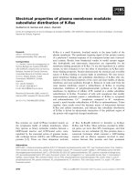
In the first experiment, the activation kinetics, i.e. the
time dependence of the development of phenoloxidase
activity after the addition of SDS, were studied
(Fig. 1). After the addition of SDS, the catecholoxi-
dase activity of Eurypelma hemocyanin developed
gradually over a period of 5 min until a constant activ-
ity was reached (Fig. 1). As an equilibrium condition
(i.e. constant activity) is a prerequisite for the measure-
ment of enzyme kinetics, the conditions with constant
activity were established. This was accomplished by
preincubating hemocyanin with 5 mm SDS for 5 min
before adding the substrate. Using this condition,
which was employed for all further experiments, the
reaction proceeded at a constant rate for at least 5 min
more (Fig. 1).
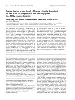
Dependence of activation on SDS concentration
SDS has a complex behavior when dissolved in aque-
ous medium, as it exists as a monomer at low concen-
trations and forms micelles at concentrations higher
than the critical micellar concentration. To determine
how the SDS concentration influences the catecholoxi-
dase activity of hemocyanin, the catecholoxidase activ-
ity was measured at different SDS concentrations and,
furthermore, under conditions with different critical
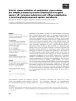
micellar concentrations (Fig. 2). The critical micellar
concentration of SDS in sodium phosphate buffer
depends almost exclusively on the sodium concentra-
tion; therefore, sodium phosphate buffers with differ-
ent concentrations were used to set different critical
micellar concentrations [34]. Critical micellar concen-
trations of 1.3 ± 0.1, 1.9 ± 0.1 and 4.1 ± 0.1 mm
were observed by conductivity measurements for
phosphate buffers with concentrations of 0.1, 0.05 and
Fig. 1. Activation kinetics of hemocyanin by SDS. Eurypelma
hemocyanin (0.7 mgÆmL
)1
) was activated with SDS in 0.1 M phos-
phate buffer (pH 7.0) at 30 °C by incubating hemocyanin with 5 m
M
SDS for periods between 15 s and 8 min. The reaction was started
by the addition of 2 m
M dopamine, and the activity was determined
by measuring the absorption increase at 475 nm for 10 s.
E. Jaenicke and H. Decker Catecholoxidase activity of tarantula hemocyanin
FEBS Journal 275 (2008) 1518–1528 ª 2008 The Authors Journal compilation ª 2008 FEBS 1519
0.01 m, respectively (Fig. 3). For the activation of
hemocyanin, a sigmoidal concentration dependence
was observed, and the concentration of half-maximum
activation always coincided with the critical micellar
concentration of the respective buffer concentration
(Fig. 2). This observation proves that hemocyanin is
activated by SDS micelles, as suggested recently [20].
The lowest SDS concentration at which full activation
is reached under all conditions is 5 mm. Consequently,
this concentration was used for further experiments.
Under these conditions, hemocyanin is activated by
SDS, which induces a conformational switch in cheli-
cerate hemocyanins, without any destructive effect on
protein structure [20].
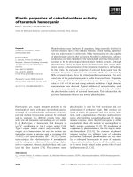
Stability of the activated state
Many hemocyanins, especially those of crustaceans,
exhibit only very weak or transient activity when acti-
vated by SDS. This can be attributed to the fact that,
unlike chelicerate hemocyanins, the conformational
change evoked by activation by SDS harms crustacean
hemocyanin and leads to a loss of activity within a few
minutes [35]. To ensure that the catecholoxidase activ-
ity of activated tarantula hemocyanin remains con-
stant, the activity of hemocyanin was measured after
exposure to 5 mm SDS for several hours (Fig. 4). The
enzymatic activity remained almost unchanged within
a 12 h period in the presence of SDS, indicating a
stable activated state.
100
Activity (%)
80
60
012345
SDS concentration (m
M)
67
40
20
0
Fig. 2. Dependence of activation on the SDS concentration.
The dependence of activation on the SDS concentration and the
critical micellar concentration of SDS was assayed in phosphate
buffer (pH 7.0) at 20 °C. The hemocyanin concentration was
0.25 mgÆmL
)1
and 1 mM dopamine was used as substrate. First,
hemocyanin was preincubated with SDS for 5 min, and then the
reaction was started by the addition of substrate. The reaction was
followed for 5 min by measuring the absorption at 475 nm. The
critical micellar concentration of SDS was varied using sodium
phosphate buffer at three concentrations:
, 0.01 M; , 0.05 M;
d, 0.1
M. The activity is given as the percentage of the maximum
activity measured at any SDS concentration in the experiment.
Fig. 3. Determination of the critical micellar concentration of SDS.
The critical micellar concentration was determined by conductivity
measurements in phosphate buffer (pH 7.0). Critical micellar
concentrations of 1.3 ± 0.1, 1.9 ± 0.1 and 4.1 ± 0.1 m
M were
observed for phosphate buffers with concentrations of 0.1
M (A),
0.05
M (B) and 0.01 M (C), respectively. The critical micellar concen-
tration (broken line) is defined as the intersection between the
extrapolated conductivity slope of monomers and micelles (full
lines), which differ in their electrophoretic mobility because of their
different size.
Catecholoxidase activity of tarantula hemocyanin E. Jaenicke and H. Decker
1520 FEBS Journal 275 (2008) 1518–1528 ª 2008 The Authors Journal compilation ª 2008 FEBS
Substrate turnover and substrate specificity
of activated hemocyanin
After establishing the activation conditions, the cat-
echoloxidase activity of activated Eurypelma hemocya-
nin was characterized more closely with respect to
substrate turnover and substrate specificity. The experi-
ments concerning substrate specificity used only
diphenolic substrates, as monophenolase activity was
never observed under the experimental conditions used
(results not shown).
The concentration dependence of substrate turn-
over was characterized using dopamine as substrate
and 5 mm SDS for activation (Fig. 5). At high dopa-
mine concentration, a decrease in activity was
observed, indicating substrate inhibition. The highest
activity was observed at a dopamine concentration
of 6 mm. The Michaelis–Menten constant (K
M
) was
determined by nonlinear fitting using normal Micha-
elis–Menten kinetics in the concentration range below
4mm where no substrate inhibition was observed
(Fig. 5). In this way, the K
M
value was determined
to be 1.45 ± 0.16 mm, the maximum velocity (V
max
)
5.5 ± 0.4 lmÆs
)1
and the resulting turnover number
(k
cat
) 6.0 ± 0.4 s
)1
. When the data were fitted to
the model for substrate inhibition (Eqn 2), similar
values were obtained, i.e. K
M
= 1.22 ± 0.20 mm,
V
max
= 5.25 ± 0.34 lmÆs
)1
and k
cat
= 5.7 ± 0.3 s
)1
(Fig. 5). The inhibition constant ( K
I
) was deter-
mined to be 29.64 ± 4.98 mm, thus revealing that
the substrate molecules at the active site bind with
a 25-fold higher affinity than inhibitory substrate
molecules.
Substrate specificity
To investigate the substrate specificity of activated
hemocyanin, the turnover of several diphenols was
compared using an oxygen electrode (Fig. 6). For this
comparison, a set of physiologically important diphe-
nols, such as dopamine, l-dihydroxyphenylalanine
(l-DOPA), N-acetyldopamine (NADA), epinephrine
and norepinephrine, was used. Catechol, although
normally not of physiological significance, was also
included, because it is the smallest diphenol. The high-
est substrate concentration tested was limited by the
solubility of the respective substrate. Thus, because of
their low solubility, l-DOPA, NADA, epinephrine and
norepinephrine could not be measured at concentra-
tions at which the maximum velocity is expected; con-
sequently, some of the curves did not approach
maximum velocity closely enough to yield a reliable
nonlinear fit of K
M
and V
max
. Thus, it was only possi-
ble to determine the catalytic efficiency (k
cat
⁄ K
M
) (Eqn
1). The best substrate was dopamine, with a catalytic
efficiency at least five-fold higher than that of the
other substrates (Table 1). Dopamine is an important
metabolite for sclerotization and melanization of the
1.0
0.8
0.6
0.4
0.2
0.0
0 10 20 30 40 50 60
Incubation time (min)
v
0
(M·min
–1
) × 10
–4
200 400 600
Fig. 4. Stability of the activated state of hemocyanin. Eurypelma
hemocyanin (0.19 mgÆmL
)1
) was preincubated with 5 mM SDS for
periods between 5 min and 12 h in 0.1
M phosphate buffer (pH 7.0)
at 25 °C. The reaction was started by the addition of 1 m
M dopa-
mine as substrate, and the activity was determined by measuring
the absorbance at 475 nm.
300
250
200
150
100
50
0
0
10 20
Dopamine concentration (m
M)
30 40 50
v
0
(µM·min
–1
)
Fig. 5. Substrate inhibition of activated Eurypelma hemocyanin.
The enzymatic activity of Eurypelma hemocyanin (0.4 mgÆmL
)1
)
was assayed at an SDS concentration of 5 m
M in 0.1 M phosphate
buffer (pH 7.0). To measure at a constant rate, Eurypelma hemo-
cyanin was incubated with SDS for 5 min before addition of the
substrate (Fig. 1). Data analysis in the substrate range 0.3–4 m
M
(broken line) revealed a K
M
value of 1.45 ± 0.16 mM and a k
cat
value of 6.0 ± 0.4 s
)1
. At substrate concentrations above 4 mM,
substrate inhibition was observed and the data were fitted to a sim-
ple model for substrate inhibition (Eqn 2). K
M
= 1.22 ± 0.20 mM,
k
cat
= 5.7 ± 0.3 s
)1
and K
I
= 29.64 ± 4.98 mM were obtained for
the inhibitory binding site (full line).
E. Jaenicke and H. Decker Catecholoxidase activity of tarantula hemocyanin
FEBS Journal 275 (2008) 1518–1528 ª 2008 The Authors Journal compilation ª 2008 FEBS 1521
cuticle, giving physiological importance to this obser-
vation [10,36]. NADA, epinephrine, norepinephrine
and l-DOPA had quite similar but lower catalytic effi-
ciencies. The lowest catalytic efficiency was observed
for catechol. This is probably a result of the fact that
catechol is the smallest diphenol and is therefore diffi-
cult to maintain in a stable position at the active site
needed for oxidation.
Inhibition of activated hemocyanin
The effect of typical inhibitors of phenoloxidase on
activated tarantula hemocyanin was assayed (Fig. 7).
For this experiment, dopamine was used as substrate
and l-mimosine, kojic acid, tyramine, phenythiourea
and azide were used as inhibitors. All five inhibitors
are known to inhibit phenoloxidases, which bind to
the active site or in the substrate binding pocket. As a
result of their structural similarity to the substrates,
this is easily rationalized for l-mimosine, kojic acid
and tyramine, which inhibit by competitive inhibition
[37]. It should be noted that tyramine is also a sub-
strate for tyrosinase. However, as the tyrosinase activ-
ity of activated hemocyanin was not observed under
our assay conditions, we were able to use tyramine as
a substrate analog (i.e. competitive inhibitor). Phenyl-
thiourea is a strong inhibitor, binding with its sulfur
atom between the copper atoms of the active site, as
observed in the crystal structure of sweet potato cat-
echoloxidase [8]. Azide interacts with type 3 copper
centers and inhibits tyrosinase, although the mode of
inhibition has not yet been clearly established [38,39].
All five inhibitors inhibited the phenoloxidase
activity of activated Eurypelma hemocyanin (Fig. 7).
Fitting the enzymatic data using a competitive mecha-
nism (Eqn 3) yielded K
I
values of 11.9 ± 1.3 and
47.7 ± 5.2 lm for kojic acid and l-mimosine, respec-
tively. Inhibition by tyramine was one order of magni-
tude weaker, with a K
I
value of 278.2 ± 30.6 lm.
Fitting attempts revealed that inhibition by phenylthio-
urea and azide was not in accordance with a competi-
tive binding model. Thus, the binding affinity of the
inhibitors could not be determined by fitting the data.
300
1000
800
600
400
200
0
250
200
150
100
50
0
0123
Substrate concentration (m
M)
Dopamine
L-dopa
NADA
Epinephrine
Norepinephrine
Catechol (m
M)
Catechol
4 5 6 7 8 9 0 20406080100
v
0
(µM·min
–1
)
v
0
(µM·min
–1
)
Fig. 6. Substrate specificity of activated hemocyanin. The turnover of several different diphenols by activated Eurypelma hemocyanin
(0.42 mgÆmL
)1
) was measured with a Clark-type oxygen electrode in 0.1 M phosphate buffer (pH 7.0). Hemocyanin was preincubated with
5m
M SDS for 5 min before addition of the substrate (Fig. 1) to ensure measurement at a constant rate. The highest substrate concentration
used was limited by the solubility of the respective substrate. Catechol, which is much more soluble than the other diphenols, was mea-
sured to concentrations up to 100 m
M (inset). For each substrate concentration, two independent measurements were made and the mean
is given in the graph. Data were analyzed by fitting the catalytic efficiency (k
cat
⁄ K
M
) in the linear part of the curve (Table 1). The lines con-
necting the data are shown to facilitate the identification of the different substrate curves and do not represent a fit.
Table 1. Catalytic efficiency of activated hemocyanin for different
substrates. Data from the comparison of different substrates
(Fig. 6) were analyzed using linear fitting, as described in the text,
to obtain the catalytic efficiency (k
cat
⁄ K
M
).
Substrate k
cat
⁄ K
M
(mM
)1
Æs
)1
)
Dopamine 3.91 ± 0.55
NADA 0.68 ± 0.08
Norepinephrine 0.66 ± 0.10
Epinephrine 0.60 ± 0.09
L-DOPA 0.59 ± 0.08
Catechol 0.20 ± 0.03
Catecholoxidase activity of tarantula hemocyanin E. Jaenicke and H. Decker
1522 FEBS Journal 275 (2008) 1518–1528 ª 2008 The Authors Journal compilation ª 2008 FEBS
Nevertheless, phenylthiourea is obviously the strongest
inhibitor by far as it inhibits strongly at the micro-
molar concentrations used. The binding affinities of
l-mimosine and kojic acid to the active site were quite
similar, but they inhibited the phenoloxidase activity
of hemocyanin to a much lesser degree than did phen-
ylthiourea. The binding affinity of the monophenol
tyramine as an inhibitor was much weaker than that
of l-mimosine and kojic acid, and azide was the weak-
est inhibitor of all five inhibitors tested. Therefore,
activated Eurypelma hemocyanin is inhibited by the
same inhibitors and in the same way as other phenol-
oxidases.
Discussion
Phenoloxidase, especially catecholoxidase, activity has
been observed in many arthropod hemocyanins. Sev-
eral chelicerate species, such as tarantulas, horseshoe
crabs and scorpions, seem to lack a phenoloxidase,
although considerable effort has been made to identify
one. Given the fact that these animals need a phenol-
3.0
L-Mimosine
A
D E
B C
Phenylthiourea Azide
Kojic acid Tyramine
2.5
2.0
1.5
1.0
0.5
0.0
0 1 2
Dopamine concentration (m
M) Dopamine concentration (mM)
Do
p
amine concentration
(
mM
)
Do
p
amine concentration
(
mM
)
Dopamine concentration (m
M)
3 4 5 6
0 1 2 3 4 5 6 7 8 0 1 2 3 4 5 6 7 8
0 1 2 3 4 5 6 0 1 2 3 4 5 6
3.0
2.5
2.0
1.5
1.0
0.5
0.0
3.0
2.5
2.0
1.5
1.0
0.5
0.0
3.0
2.5
2.0
1.5
1.0
0.5
0.0
3.0
2.5
2.0
1.5
1.0
0.5
0.0
v
0
(M·min
–1
) × 10
–4
v
0
(M·min
–1
) × 10
–4
Fig. 7. Inhibition of activated hemocyanin. The inhibition of SDS-activated hemocyanin (0.42 mgÆmL
)1
) by five inhibitors of catecholoxidase
was assayed in 0.1
M phosphate buffer (pH 7.0). Before initiation of the reaction by the addition of the substrate dopamine, hemocyanin
was preincubated for 5 min with 5 m
M SDS and the indicated concentration of inhibitor. For each inhibitor ⁄ substrate concentration, one
experiment was performed. The data for the three competitive inhibitors
L-mimosine, kojic acid and tyramine were fitted in parallel according
to Eqn (3). Kojic acid and
L-mimosine inhibited enzymatic activity with K
I
values of 11.9 ± 1.3 and 47.7 ± 5.2 lM respectively. Inhibition by
tyramine was one order of magnitude weaker with K
I
= 278.2 ± 30.6 lM. Note that tyramine is not a substrate of activated hemocyanin
under the conditions used. Data for the inhibitors phenylthiourea and azide were not analyzed further because of the lack of a suitable inhibi-
tion model. (A)
L-Mimosine: d,0mM; , 100 lM; , 250 lM; r, 500 lM; (B) kojic acid: d,0mM; ,20lM; ,50lM; r, 150 lM; (C) tyra-
mine: d,0m
M; , 0.2 mM; , 0.6 mM; r, 1.6 mM; (D) phenylthiourea: d,0mM; ,3lM; ,6lM; r,9lM; (E) azide: d,0mM;
, 3.0 mM; , 4.5 mM; r, 6.0 mM.
E. Jaenicke and H. Decker Catecholoxidase activity of tarantula hemocyanin
FEBS Journal 275 (2008) 1518–1528 ª 2008 The Authors Journal compilation ª 2008 FEBS 1523
oxidase, as all known arthropods do, it seems that
their hemocyanin replaces the phenoloxidase in their
hemolymph, as shown for the horseshoe crab Tachyp-
leus [18,19]. Arthropod phenoloxidases generally are
produced as inactive prophenoloxidases, which can be
activated by limited proteolysis [11,40,41]. Interaction
with fatty acids and phospholipids can also activate
prophenoloxidase, although the physiological signifi-
cance of this method of activation has not been estab-
lished in arthropods [32,42].
Hemocyanin can also be considered as a propheno-
loxidase, as it is activated by the same mechanisms as
those which activate phenoloxidase, and can be inhib-
ited by the same inhibitors [25,35]. An additional
method of activation was found for the hemocyanin of
the horseshoe crab Tachypleus by binding of anti-
microbial peptides and proteins of the hemolymph
coagulation cascade to hemocyanin [18,19].
In the laboratory, activation by SDS mimics the
activation by fatty acids and phospholipids, and is
accepted as the standard method for prophenoloxidase
and hemocyanin activation [21–31]. At first sight, SDS,
as an unnatural synthetic agent, may not seem to be
the compound of choice to study the physiological
function of a protein. Nevertheless, for practical rea-
sons (i.e. solubility, availability), it has been used to
activate proenzymes, including even the proteasome
[26,27]. In addition, the real physiological activators
are often unknown, and thus the use of an activator
such as SDS is warranted to learn more about the
functional properties of an enzyme. Generally, binding
of SDS during activation causes a conformational
change, as shown for the proteasome, phenoloxidases
and, recently, for hemocyanin [20,26,27,31,43].
In our experiments, the activation curves for hemo-
cyanin by SDS showed a sigmoidal behavior, with a
midpoint which always coincided with the critical
micellar concentration of SDS. Recently, the hypothe-
sis that SDS micelles are needed to induce a conforma-
tional change, which activates hemocyanin, was
suggested [20]. Our observation that the critical micel-
lar concentration and SDS midpoint concentrations of
activation strictly coincide confirms this hypothesis,
and indicates that free SDS monomers are not suffi-
cient for activation under our experimental conditions
(Figs 2 and 3). The nature of the interaction between
the hemocyanin multimer and SDS micelles is not
known, and it is unclear why the conformational
change induced by this interaction does not occur
instantly but requires several minutes for completion
(Fig. 1). However, the activated state is stable, as only
small variations in activity were observed over the time
course of several hours (Fig. 4).
Although tyrosinase and catecholoxidase from
plants and fungi have been characterized in great
detail, only very few enzymatic data are available on
phenoloxidases from arthropods, where most studies
have focused only on establishing the enzymatic activ-
ity. Even less is known about the enzymatic parame-
ters of activated hemocyanin. Most studies on the
phenoloxidase activity of hemocyanin in the past have
focused on the activity in the species investigated
and ⁄ or the way in which hemocyanin is activated.
Only for a few crustacean hemocyanins have the enzy-
matic properties been reported to a small extent
[17,44,45]. In all of these cases, the activity was very
weak and the activated state was not stable, therefore
limiting the enzymatic analysis. Therefore, the enzy-
matic properties of the phenoloxidase activity of a
chelicerate hemocyanin have not been investigated
thoroughly previously. We have attempted to close this
gap with this study. The enzymatic parameters of acti-
vated hemocyanin and selected parameters reported in
the literature are compared in Table 2.
Activated hemocyanin is able to turn over a wide
range of diphenolic substrates. Activated hemocyanin
only accepts o-diphenols as substrates (i.e. catecholase
activity) under our experimental conditions, and lacks
the ability to hydroxylate monophenols in the ortho-
position (i.e. monophenolase activity). The reason why
Table 2. Enzymatic parameters of activated hemocyanin in com-
parison with other phenoloxidases.
Activated
hemocyanin Other phenoloxidases
K
M
1.45 ± 0.16 mM
(dopamine)
0.72 ± 0.08 m
M
(dopamine, Agaricus [53])
0.28 ± 0.01 m
M
(L-DOPA, Agaricus [53])
1.04 m
M (L-DOPA,
Neurospora [54])
8.9 m
M (L-DOPA,
Streptomyces [37])
0.54 ± 0.05 m
M (tyramine,
Agaricus [53])
k
cat
6.0 ± 0.4 s
)1
(dopamine)
475 ± 17 s
)1
(dopamine,
Agaricus [53])
107 ± 2 s
)1
(L-DOPA,
Agaricus [53])
1070 s
)1
(L-DOPA,
Neurospora [54])
25 ± 1 s
)1
(tyramine,
Agaricus [53])
K
I
(L-mimosine) 47.7 ± 5.2 lM 30 ± 3 lM
(Streptomyces [37])
K
I
(kojic acid) 11.9 ± 1.3 lM 3.4 ± 0.3 lM
(Streptomyces [37])
Catecholoxidase activity of tarantula hemocyanin E. Jaenicke and H. Decker
1524 FEBS Journal 275 (2008) 1518–1528 ª 2008 The Authors Journal compilation ª 2008 FEBS
activated hemocyanin is unable to catalyze the mono-
hydroxylation reaction is not known. With regard to
diphenolic substrates, the best substrate by far is dopa-
mine, whereas other diphenols are converted at a much
lower catalytic efficiency, with almost identical values
for all diphenols except catechol (Table 1). Dopamine
is an important substrate in arthropods for cuticle
sclerotization after molting [46,47]. This observation is
in good agreement with the fact that, in the tarantula
Eurypelma, hemocyanin has been identified as part of
the protein components of the cuticle, and similar
observations have been made in crustaceans [44,48,49].
The substrate affinity for dopamine is within the nor-
mal range observed for other phenoloxidases, which
varies over almost one order of magnitude depending
on the substrate (Table 2). Unfortunately, no data are
available on physiological diphenol levels in chelicer-
ates, and thus a comparison of how substrate affinity
and physiological substrate levels relate to one another
is not possible. The turnover rate for dopamine is
almost two orders of magnitude lower than the dopa-
mine turnover in other phenoloxidases. Nevertheless,
turnover rates as low as 25 s
)1
have been reported for
the monophenol tyramine, and this turnover rate is
only four-fold higher than the dopamine turnover by
activated hemocyanin. At first sight, the low turnover
rate observed for activated hemocyanin seems to make
a physiological role for this molecule less likely. How-
ever, it must be considered that hemocyanin, unlike
other phenoloxidases, is present at concentrations of at
least 40 mgÆmL
)1
in the hemolymph. These high con-
centrations, which are orders of magnitude higher than
the concentrations found for other phenoloxidases,
could make up for the lower turnover rates.
Unexpectedly, strong substrate inhibition was
observed for the substrate dopamine, which has not
been reported previously for other phenoloxidases.
Recently, the first study on the detailed enzymatic
parameters of a hexameric arthropod phenoloxidase
has also reported substrate inhibition [50]. Thus, it is
possible that substrate inhibition is common amongst
arthropod phenoloxidases, but has not been reported
previously simply because of a lack of studies. The
inhibition of activated hemocyanin by typical inhibi-
tors of phenoloxidase was similar to the inhibition
observed in phenoloxidase (Table 2).
In conclusion, it has been shown that tarantula
hemocyanin exhibits the properties of a common phe-
noloxidase, and consequently may function as such
when activated appropriately in the animal. In the
future, further efforts will be made to determine the
physiological activators and regulatory mechanisms
in vivo.
Experimental procedures
Reagents
All reagents used were of the highest purity and were pur-
chased from Sigma (Steinheim, Germany). Ultrapure water
(Milli-Q-Plus-PF; Millipore, Eschborn, Germany) was used
for all solutions.
Purification of hemocyanin
Tarantulas (Eurypelma californicum) were obtained from
the North Carolina Biological Supply (Charlotte, NC,
USA). Hemolymph was collected by dorsal puncturing of
the pericard and immediately diluted 1 : 1 with stabilization
buffer (0.1 m Tris ⁄ HCl, 5 mm MgCl
2
,5mm CaCl
2
,
pH 7.8). On average, 100 lL of hemolymph was collected
from one spider. Therefore, it was necessary to pool the
hemolymph of five or six spiders to obtain a sufficient
hemolymph volume for the purification procedure. The
hemolymph was centrifuged at 15 000 g for 10 min at 4 °C
to remove cellular debris. The supernatant containing
hemocyanin was applied to a Sephacryl S-300 16 ⁄ 60 HR size
exclusion column (GE Healthcare Biosciences, Uppsala,
Sweden). The column was eluted with stabilization buffer
at a flow rate of 0.6 mLÆmin
)1
at room temperature.
Hemocyanin-containing fractions were identified by their
absorbance at 340 nm and stored at 4 °C. Only fractions
containing 24-meric hemocyanin were used for the
experiments.
The protein concentration of hemocyanin samples was
determined by measuring the absorbance at 278 nm using the
molar extinction coefficient [e
278
(nm) = 1.1 mLÆmg
)1
Æcm
)1
]
for chelicerate hemocyanin. Hemocyanin samples were
concentrated in Biomax30K centrifugal filters (Millipore),
when necessary.
Determination of the critical micellar
concentration
The critical micellar concentration of SDS was determined
by conductivity measurements using a Model 712 Conduc-
tometer (Metrohm, Herisau, Switzerland) [34]. During the
measurement, the temperature of the sample was kept con-
stant at 20 °C in a water bath.
Enzymatic assays
The phenoloxidase activity of hemocyanin was followed
spectroscopically or with a Clark-type oxygen electrode.
For the spectroscopic assay, the formation of dopa-
chrome was measured at 475 nm with a Hitachi U-3000
photometer (Hitachi, Tokyo, Japan) [51]. Quartz cuvettes
(Hellma, Mu
¨
llheim, Germany) with an optical path length
of 1 cm were used. Dopachrome is unstable and reacts to
E. Jaenicke and H. Decker Catecholoxidase activity of tarantula hemocyanin
FEBS Journal 275 (2008) 1518–1528 ª 2008 The Authors Journal compilation ª 2008 FEBS 1525
derivatives after a few minutes; thus, only the first 5 min
after initiation of the reaction were measured. All measure-
ments were performed at 25 °C in 0.1 m phosphate buffer
(pH 7.0) unless noted otherwise. Three different protocols
were used. For the first protocol, hemocyanin and
dopamine were mixed and the reaction was initiated by the
addition of activator (SDS). For the second protocol,
hemocyanin was preincubated with activator (SDS) for
5 min and the reaction was then initiated by the addition of
dopamine. The third protocol was used for inhibition
experiments. The inhibitor was preincubated with hemo-
cyanin and activator (SDS) for 5 min and the reaction was
initiated by the addition of dopamine. The dead time
between initiation of the reaction and the measurement was
less than 15 s in all protocols. The unstable dopamine
solutions were prepared fresh daily and kept on ice in the
dark to minimize the auto-oxidation of dopamine.
A Clark-type oxygen electrode (Hansatech Oxygen-Elec-
trode DW1, Saur Laborbedarf, Reutlingen, Germany) was
used to compare the turnover of different substrates by fol-
lowing the oxygen consumption in the reaction mixture.
The same conditions and protocols as described for the
spectroscopic measurements were used.
In all experiments, dopamine was used as substrate unless
noted otherwise, as it was the best substrate when the sub-
strate specificity was tested; furthermore, its good solubility
allowed experiments at high substrate concentrations.
At the beginning of the enzymatic measurements, the
experimental error of the enzymatic assays was determined
for both the spectroscopic and oxygen electrode assays by
measuring the turnover ten times under identical condi-
tions. The experimental error amounted to ± 5%.
Data analysis
Enzyme kinetic data were analyzed using the program
Sigma-Plot 2000 (Systat Software, Erkrath, Germany).
Data for uninhibited kinetics were fitted by nonlinear
regression to uninhibited Michaelis–Menten kinetics.
Some of the substrates showed poor solubility, and there-
fore the kinetic data only covered the part of the curve at
low concentration relative to the respective K
M
value,
where saturation had not yet been reached. In these cases,
K
M
and k
cat
could not be determined with confidence.
However, at substrate concentrations lower than K
M
, the
catalytic efficiency (k
cat
⁄ K
M
) could be determined from the
initial slope of the curve:
v
0
¼
k
cat
K
M
½E
T
½Sð1Þ
where v
0
is the initial enzymatic rate, [S] is the substrate
concentration, k
cat
is the turnover number and [E
T
] is the
enzyme concentration.
Substrate inhibition was fitted according to the simplest
model, assuming that a second substrate molecule can bind
to the enzyme–substrate complex and render it inactive
[52]. Data were fitted according to:
v
0
¼
v
max
½S
½SþK
M
þ
½S
2
K
I
ð2Þ
where K
I
is the binding constant for the inhibitory substrate
molecule.
The data for the three competitive inhibitors kojic acid,
l-mimosine and tyramine were fitted in parallel for all three
inhibitors according to:
v
0
¼
V
max
½S
½SþK
M
1 þ
½I
1
K
IM
þ
½I
2
K
IK
þ
½I
3
K
IT
ð3Þ
where K
IM
is the inhibition constant for l-mimosine, K
IK
is
the inhibition constant for kojic acid and K
IT
is the inhibi-
tion constant for tyramine. Although no combinations of
inhibitors were tested, all the enzymatic data for the three
competitive inhibitors were fitted in parallel to ensure that
only one K
M
value and one V
max
value were obtained for
the uninhibited reaction. Fitting the data for the three
inhibitors individually would have resulted in three K
M
and
three V
max
values.
For the calculation of the enzyme concentration, it was
assumed that only subunit types b and c, i.e. four of the 24
subunits which make up the native hemocyanin molecule,
possess enzymatic activity [16,25].
Acknowledgements
We thank Claudia Dietze for performing the conduc-
tivity measurements. This work was supported by the
‘Fonds der chemischen Industrie’ (EJ) and the ‘Deut-
sche Forschungsgemeinschaft’ (HD).
References
1 Burmester T (2001) Molecular evolution of the arthro-
pod hemocyanin superfamily. Mol Biol Evol 18, 184–
195.
2 Decker H & Jaenicke E (2004) Recent findings on phe-
noloxidase activity and antibacterial activity of hemo-
cyanins. Dev Comp Immunol 28, 673–687.
3 Jaenicke E & Decker H (2004) Functional changes in
the family of type 3 copper proteins in evolution. Chem
Biochem 5, 163–169.
4 van Holde K, Miller K & Decker H (2001) Hemocya-
nins and invertebrate evolution. J Biol Chem 276,
15563–15566.
5 Cuff M, Miller K, van Holde K & Hendrickson W
(1998) Crystal structure of a functional unit from
Octopus hemocyanin. J Mol Biol 278, 855–870.
6 Magnus K, Hazes B, Ton-That H, Bonaventura C,
Bonaventura J & Hol W (1994) Crystallographic
Catecholoxidase activity of tarantula hemocyanin E. Jaenicke and H. Decker
1526 FEBS Journal 275 (2008) 1518–1528 ª 2008 The Authors Journal compilation ª 2008 FEBS
analysis of oxygenated and deoxygenated states of
arthropod hemocyanin shows unusual differences.
Proteins 19, 302–309.
7 Sanchez-Ferrer A, Rodriguez-Lopez J, Garcia-Canovas
F & Garcia-Carmona F (1995) Tyrosinase: a compre-
hensive review of its mechanism. Biochim Biophys Acta
1247, 1–11.
8 Klabunde T, Eicken C, Sacchettini J & Krebs B (1998)
Crystal structure of a plant catechol oxidase containing
a dicopper center. Nat Struct Biol 5, 1084–1090.
9 Matoba Y, Kumagai T, Yamamoto A, Yoshitsu H &
Sugiyama M (2006) Crystallographic evidence that
dinuclear copper center of tyrosinase is flexible during
catalysis. J Biol Chem 281, 8981–8990.
10 Sugumaran M (1991) Molecular mechanisms for mam-
malian melanogenesis: comparison with insect cuticular
sclerotization. FEBS Lett 293, 4–10.
11 Cerenius L & So
¨
derha
¨
ll K (2004) The prophenoloxi-
dase-activating system in invertebrates. Immunol Rev
198, 116–126.
12 Sugumaran M (2002) Comparative biochemistry of
eumelanogenesis and the protective roles of phenoloxi-
dase and melanin in insects. Pigment Cell Res 15, 2–9.
13 Land E, Ramsden C & Riley P (2004) Quinone chemis-
try and melanogenesis. Methods Enzymol 378, 88–109.
14 Decker H & Terwilliger N (2000) Cops and robbers:
putative evolution of copper oxygen-binding proteins.
J Exp Biol 203, 1777–1782.
15 Decker H, Jaenicke E, Hellmann N, Lieb B, Meissner
U & Markl J (2007) Minireview: recent insights in the
structure, function and evolution of hemocyanins.
Integr Comp Biol 47, 631–644.
16 Decker H & Rimke T (1998) Tarantula hemocyanin
shows phenoloxidase activity. J Biol Chem 273, 25889–
25892.
17 Lee S, Lee B & Soderhall K (2004) Processing of cray-
fish hemocyanin subunits into phenoloxidase. Biochem
Biophys Res Commun 322, 490–496.
18 Nagai T, Osaki T & Kawabata S (2001) Functional
conversion of hemocyanin to phenoloxidase by horse-
shoe crab antimicrobial peptides. J Biol Chem 276,
27166–27170.
19 Nagai T & Kawabata S (2000) A link between blood
coagulation and prophenoloxidase activation in arthro-
pod host defense. J Biol Chem 275, 29264–29267.
20 Baird S, Kelly S, Price N, Jaenicke E, Meesters C, Nil-
lius D, Decker H & Nairn J (2007) Hemocyanin confor-
mational changes associated with SDS-induced phenol
oxidase activation. Biochim Biophys Acta 1774, 1380–
1394.
21 Moore B & Flurkey W (1990) Sodium dodecyl sulfate
activation of a plant polyphenol oxidase. J Biol Chem
265, 4982–4988.
22 Chazarra S, Cabanes J, Escribano J & Garcia-Carmona
F (1997) Kinetic study of the suicide inactivation of
latent polyphenoloxidase from iceberg lettuce (Lattuca
sativa) induced by 4-tert-butylcatechol in the presence
of SDS. Biochim Biophys Acta 1339, 297–303.
23 Jimenez M & Garcia-Carmona F (1996) The effect of
sodium dodecyl sulfate on polyphenoloxidase. Phyto-
chemistry 42, 1503–1509.
24 Escribano J, Cabanes J & Garcia-Carmona F (1997)
Characterisation of latent polyphenol oxidase in table
beet: effect of sodium dodecyl sulfate. J Sci Food Agric
73, 34–38.
25 Decker H, Ryan M, Jaenicke E & Terwilliger N (2001)
SDS-induced phenoloxidase activity of hemocyanins
from Limulus polyphemus, Eurypelma californicum, and
Cancer magister. J Biol Chem 276, 17796–17799.
26 Dalmann B, Rutschmann M, Kuehn L & Reinauer H
(1985) Activation of the multicatalytic proteinase from
rat skeletal muscle by fatty acids or sodium dodecyl
sulfate. Biochem J 228, 171–177.
27 Shibatani T & Ward W (1995) Sodium dodecyl sulfate
(SDS) activation of the 20S proteasome in rat liver.
Arch Biochem Biophys 321, 160–166.
28 Flurkey W (1986) Polyphenoloxidase in higher plants:
immunological detection and analysis of in vitro trans-
lation products. Plant Physiol 81, 614–618.
29 Kenten R (1958) Latent phenolase in extracts of broad-
bean (Vicia faba L.) leaves. 2. Activation by anionic
wetting agents. Biochem J 68, 244–251.
30 Robb D, Mapson L & Swain T (1964) Activation of
latent tyrosinase of broad bean. Nature 201, 503–504.
31 Swain T, Mapson L & Robb DA (1966) Activation of
Vicia faba (L.) tyrosinase as effected by denaturing
agents. Phytochemistry 5, 469–482.
32 Sugumaran M & Nellaiappan K (1991) Lysolecithin – a
potent activator of prophenoloxidase from the hemo-
lymph of the lobster, Homarus americanus. Biochem
Biophys Res Commun 176, 1371–1376.
33 Lorenzini D, da Silva P Jr, Soares M, Arruda P, Setubal
J & Daffre S (2006) Discovery of immune-related genes
expressed in hemocytes of the tarantula spider Acantho-
scurria gomesiana. Dev Comp Immunol 30, 545–556.
34 Dutkiewicz E & Jakubowska A (2002) Effect of electro-
lytes on the physicochemical behaviour of sodium dode-
cyl sulphate micelles. Colloid Polym Sci 280, 1009–1014.
35 Jaenicke E & Decker H (2004) Conversion of crusta-
cean hemocyanin to catecholoxidase. Micron 35,
89–90.
36 Andersen S, Hojrup P & Roepstorff P (1995) Insect
cuticular proteins. Insect Biochem Mol Biol 25, 153–176.
37 Bubacco L, Vijgenboom E, Gobin C, Tepper A, Salgado
J & Canters G (2000) Kinetic and paramagnetic
NMR investigations of the inhibition of Streptomyces
antibioticus tyrosinase. J Mol Catal 8B, 27–35.
38 Salvato B & Beltramini M (1990) Hemocyanins: mole-
cular architecture, structure and reactivity of the binu-
clear copper site. Life Chem Rep 8, 1–47.
E. Jaenicke and H. Decker Catecholoxidase activity of tarantula hemocyanin
FEBS Journal 275 (2008) 1518–1528 ª 2008 The Authors Journal compilation ª 2008 FEBS 1527
39 Healey D & Strothkamp K (1981) Inhibition of the cat-
echolase and cresolase activity of mushroom tyrosinase
by azide. Arch Biochem Biophys 211, 86–91.
40 Chosa N, Fukumitsu T, Fujimoto K & Ohnishi E
(1997) Activation of prophenoloxidase A1 by an
activating enzyme in Drosophila melanogaster. Insect
Biochem Mol Biol 27, 61–68.
41 Robinson S & Dry I (1992) Broad bean leaf polypheno-
loxidase is a 60-kilodalton protein susceptible to proteo-
lytic cleavage. Plant Physiol 99, 317–323.
42 Nellaiappan K & Sugumaran M (1996) On the presence
of phenoloxidase in the hemolymph of the horseshoe
crab, Limulus. Comp Biochem Physiol 113B, 163–168.
43 Kanade S, Paul B, Rao A & Gowda L (2006) The con-
formational state of polyphenol oxidase from field bean
(Dolichos lablab) upon SDS and acid-pH activation.
Biochem J 395, 551–562.
44 Terwilliger N & Ryan M (2006) Functional and phylo-
genetic analyses of phenoloxidases from Brachyuran
(Cancer magister) and Branchiopod (Artemia francis-
cana, Triops longicaudatus) crustaceans. Biol Bull 210,
38–50.
45 Pless D, Aguilar M, Falcon A, Lozano-Alvarez E &
Heimer de la Cotera E (2003) Latent phenoloxidase
activity and N-terminal amino acid sequence of hemo-
cyanin from Bathynomus giganteus, a primitive crusta-
cean. Arch Biochem Biophys 409, 402–410.
46 Andersen S & Roepstorff P (2007) Aspects of cuticular
sclerotization in the locust, Schistocerca gregaria, and
the beetle, Tenebrio molitor. Insect Biochem Mol Biol
37, 223–234.
47 Suderman R, Dittmer N, Kanost M & Kramer K
(2006) Model reactions for insect cuticle sclerotization:
cross-linking of recombinant cuticular proteins upon
their laccase-catalyzed oxidative conjugation with catec-
hols. Insect Biochem Mol Biol 36, 353–365.
48 Adachi K, Endo H, Watanabe T, Nishioka T & Hirata
T (2005) Hemocyanin in the exoskeleton of crustaceans:
enzymatic properties and immunolocalization. Pigment
Cell Res 18, 136–143.
49 Paul R, Bergner B, Pfeffer-Seidl A, Decker H, Efinger
R & Storz H (1994) Gas transport in the haemolymph
of arachnids. I. Oxygen transport and the physiological
role of haemocyanin. J Exp Biol 188, 25–46.
50 Brack A, Hellmann N & Decker H (2008) Kinetic prop-
erties of hexameric tyrosinase from the crustacean Pal-
inurus elephas. Photochem Photobiol (in press).
51 Mason H (1948) The chemistry of melanin. III: Mecha-
nism of the oxidation of dihydroxyphenylalanine by
tyrosinase. J Biol Chem 172, 83–99.
52 Cornish-Bowden A (1995) Fundamentals of Enzyme
Kinetics. Portland Press, London.
53 Fenoll L, Rodrı
´
guez-Lo
´
pez J, Varo
´
n R, Garcı
´
a-Ruiz P,
Garcı
´
a-Ca
´
novas F & Tudela J (2002) Kinetic characteri-
sation of the reaction mechanism of mushroom tyrosi-
nase on tyramine ⁄ dopamine and l-tyrosine methyl
esther ⁄ l-dopa methyl esther. Int J Biochem Cell Biol 34,
1594–1607.
54 Wilcox D, Porras A, Hwang Y, Lerch K, Winkler M &
Solomon E (1985) Substrate analogue binding to the
coupled binuclear copper active site in tyrosinase. JAm
Chem Soc 107, 4015–4027.
Catecholoxidase activity of tarantula hemocyanin E. Jaenicke and H. Decker
1528 FEBS Journal 275 (2008) 1518–1528 ª 2008 The Authors Journal compilation ª 2008 FEBS