Báo cáo khoa học: Endogenous tetrahydroisoquinolines associated with Parkinson’s disease mimic the feedback inhibition of tyrosine hydroxylase by catecholamines doc
Bạn đang xem bản rút gọn của tài liệu. Xem và tải ngay bản đầy đủ của tài liệu tại đây (469.66 KB, 13 trang )
Endogenous tetrahydroisoquinolines associated with
Parkinson’s disease mimic the feedback inhibition
of tyrosine hydroxylase by catecholamines
Joachim Scholz
1,2,
*, Karen Toska
3,
*, Alexander Luborzewski
1
, Astrid Maass
4
, Volker Schu
¨
nemann
5
,
Jan Haavik
3
and Andreas Moser
1
1 Neurochemistry Research Group, Department of Neurology, University of Lu
¨
beck, Germany
2 Neural Plasticity Research Group, Department of Anesthesia and Critical Care, Massachusetts General Hospital and Harvard Medical
School, Charlestown, MA, USA
3 Department of Biomedicine, Section of Biochemistry and Molecular Biology, University of Bergen, Norway
4 Fraunhofer-Institute for Algorithms and Scientific Computing (SCAI), Sankt Augustin, Germany
5 Department of Physics, Technical University Kaiserslautern, Germany
Keywords
enzyme stability; feedback inhibition;
Parkinson’s disease; tetrahydroisoquinolines;
tyrosine hydroxylase
Correspondence
J. Scholz, Neural Plasticity Research Group,
Department of Anesthesia and Critical Care,
Massachusetts General Hospital and
Harvard Medical School, 149 13th Street,
Room 4309, Charlestown, MA 02129, USA
Fax: +1 617 7243632
Tel: +1 617 7243623
E-mail:
*These authors contributed equally to this
work
(Received 14 November 2007, revised 23
January 2008, accepted 28 February 2008)
doi:10.1111/j.1742-4658.2008.06365.x
N-methyl-norsalsolinol and related tetrahydroisoquinolines accumulate in
the nigrostriatal system of the human brain and are increased in the cere-
brospinal fluid of patients with Parkinson’s disease. We show here that
6,7-dihydroxylated tetrahydroisoquinolines such as N-methyl-norsalsolinol
inhibit tyrosine hydroxylase, the key enzyme in dopamine synthesis, by
imitating the mechanisms of catecholamine feedback regulation. Docked
into a model of the enzyme’s active site, 6,7-dihydroxylated tetrahydroiso-
quinolines were ligated directly to the iron in the catalytic center, occupy-
ing the same position as the catecholamine inhibitor dopamine. In this
position, the ligands competed with the essential tetrahydropterin cofactor
for access to the active site. Electron paramagnetic resonance spectros-
copy revealed that, like dopamine, 6,7-dihydroxylated tetrahydroisoquino-
lines rapidly convert the catalytic iron to a ferric (inactive) state.
Catecholamine binding increases the thermal stability of tyrosine hydroxy-
lase and improves its resistance to proteolysis. We observed a similar
effect after incubation with N-methyl-norsalsolinol or norsalsolinol. Fol-
lowing an initial rapid decline in tyrosine hydroxylation, the residual
activity remained stable for 5 h at 37 °C. Phosphorylation by protein
kinase A facilitates the release of bound catecholamines and is the most
prominent mechanism of tyrosine hydroxylase reactivation. Protein
kinase A also fully restored enzyme activity after incubation with
N-methyl-norsalsolinol, demonstrating that tyrosine hydroxylase inhibition
by 6,7-dihydroxylated tetrahydroisoquinolines mimics all essential aspects
of catecholamine end-product regulation. Increased levels of N-methyl-
norsalsolinol and related tetrahydroisoquinolines are therefore likely to
accelerate dopamine depletion in Parkinson’s disease.
Abbreviations
CSF, cerebrospinal fluid; DA, dopamine; hTH, human tyrosine hydroxylase;
L-DOPA, L-3,4-dihydroxyphenylalanine; MPTP, 1-methyl-4-phenyl-
1,2,3,6-tetrahydropyridine; NMNorsal, N-methyl-norsalsolinol; NMSal, N-methyl-salsolinol; NMTIQ, N-methyl-1,2,3,4-tetrahydroisoquinoline;
Norsal, norsalsolinol; PD, Parkinson’s disease; PKA, protein kinase A; ROS, reactive oxygen species; TH, tyrosine hydroxylase;
TIQ, tetrahydroisoquinoline.
FEBS Journal 275 (2008) 2109–2121 ª 2008 The Authors Journal compilation ª 2008 FEBS 2109
N-methyl-norsalsolinol, salsolinol and N-methyl-salso-
linol are endogenous tetrahydroisoquinolines (TIQs)
formed through non-enzymatic condensation of dopa-
mine (DA) with aldehydes or pyruvic acid. Increased
concentrations of these TIQs are found in the cerebro-
spinal fluid (CSF) of patients with Parkinson’s disease
(PD) [1–3]. Accumulation of N-methylated TIQs in the
substantia nigra and the corpus striatum of the human
brain [2] and their structural similarity to 1-methyl-
4-phenyl-1,2,3,6-tetrahydropyridine (MPTP) (Fig. 1)
have led to the hypothesis that TIQs are directly
involved in the degeneration of dopaminergic neurons.
Like MPTP, TIQs inhibit mitochondrial respiration.
However, the toxicity of TIQs is low because of their
limited ability to cross the mitochondrial membrane
[4]. High concentrations of N-methyl-salsolinol
(NMSal) are required to induce apoptosis of dopami-
nergic cells in vitro [5], and NMSal causes a loss of
tyrosine hydroxylase-immunoreactive neurons in the
rat substantia nigra in vivo only after repeated stereo-
taxic injections [6]. Some TIQs even have neuroprotec-
tive effects [7,8]. Rather than provoking neuronal
degeneration, endogenous TIQs may interfere with DA
synthesis. N-methyl-norsalsolinol (NMNorsal) [9] and
salsolinol [10,11] inhibit tyrosine hydroxylase (TH;
tyrosine 3-monooxygenase, EC 1.14.16.2), the key
enzyme in DA synthesis, in vitro, and a single injection
of NMSal into the rat corpus striatum markedly
reduces TH activity in vivo, leading to an almost com-
plete loss of DA in the absence of neuronal degenera-
tion [6].
The CSF levels of TIQs increase in early PD and
decrease as the disease progresses [12]. TH inhibition
by endogenous TIQs may therefore be most prominent
at a critical time, when surviving substantia nigra neu-
rons are challenged by the necessity to increase DA
synthesis and release in order to uphold the functional
integrity of the nigrostriatal pathway [13–15]. Such
adaptive neurochemical changes are likely to delay the
appearance of clinical signs in PD, which lags several
years behind the onset of dopaminergic neuron degen-
eration in the substantia nigra [16]. Animal models of
PD have demonstrated the plasticity of the nigrostria-
tal system. For example, near-complete recovery of
motor function is achieved when striatal DA levels are
restored by concomitant virus-mediated transfer of the
genes encoding TH and GTP cyclohydrolase, the rate-
limiting synthetic enzyme for the essential TH cofactor
6(R)-l-erythro-5,6,7,8-tetrahydrobiopterin (BH
4
) [17].
Thus, understanding how TIQs block TH activity is
important in order to develop treatment strategies that
help to sustain dopaminergic nigrostriatal signaling in
early PD.
TH catalyzes the hydroxylation of tyrosine to l-3,4-
dihydroxyphenylalanine (l-DOPA), which is the rate-
limiting step in synthesis of the catecholamines DA,
norepinephrine and epinephrine. TH consists of four
identical subunits that contain a C-terminal catalytic
domain (residues 156–498) and an N-terminal regula-
tory domain. The active site of the enzyme is a 17 A
˚
deep crevice with a ferrous iron atom located in its
center [18]. Alternative mRNA splicing of a single pri-
mary transcript generates at least four isoforms of
human TH (hTH) that are differentially expressed in
tissues; the most prominent isoforms in the brain are
hTH1 and hTH2. The isoforms differ only in the
N-terminal regulatory region; the C-terminal domain
with the active site is identical in all hTH isoforms,
and the catalytic domain is also highly conserved
across animal species and in other aromatic amino
acid hydroxylases [19]. TH activity is subject to intri-
cate regulation. Transcriptional control, modulation of
RNA stability, translational regulation and enzyme
stability establish a steady-state level of TH protein
[20,21]. Short-term regulation of TH activity includes
feedback inhibition by catecholamine end products,
allosteric modulation and phosphorylation-dependent
activation by various kinases [22,23].
Using recombinant hTH1 and hTH4, we have exam-
ined the inhibitory effect of NMNorsal and structur-
ally related TIQs on TH. Molecular docking revealed
that 6,7-dihydroxylated TIQs associated with PD
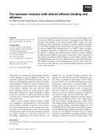
Fig. 1. Chemical structures of DA and the TIQs examined in this
study. The 6,7-dihydroxylated TIQs Norsal, NMNorsal and NMSal
have an intact catechol moiety. NMNorsal and NMSal are endoge-
nous compounds with structural similarity to MPTP.
TIQs imitate mechanisms of TH feedback inhibition J. Scholz et al.
2110 FEBS Journal 275 (2008) 2109–2121 ª 2008 The Authors Journal compilation ª 2008 FEBS
compete with the essential tetrahydropterin cofactor of
hTH for access to the enzyme’s active site, whereas
MPTP has low affinity for the amino acid binding site.
By binding directly to the ferrous iron atom in the cat-
alytic center and converting the iron to a ferric state,
6,7-dihydroxylated TIQs block hTH activity through a
mechanism that mimics the physiological feedback
inhibition by catecholamines. Unlike DA, which
caused a near complete loss of hTH activity over time,
NMNorsal stabilized hTH, but with a reduced level of
activity.
Results
6,7-Dihydroxylated TIQs inhibit human TH
The recombinant isoforms hTH1 and hTH4 produced
539 ± 41 nmolÆmin
)1
Æmg
)1
and 564 ± 40 nmolÆ
min
)1
Æmg
)1
l-DOPA, respectively. Human TH activity
decreased in the presence of NMNorsal and NMSal,
two 6,7-dihydroxylated TIQs (Fig. 1) that have previ-
ously been identified in the CSF of patients with PD
[1,3,12]. NMNorsal inhibited hTH almost as strongly
(IC
50
= 0.3 lm) as the catecholamine end product DA
(IC
50
= 0.2 lm), whereas higher concentrations of
NMSal (IC
50
= 4.0 lm) were required to reduce hTH
activity (Fig. 2A). A kinetic analysis indicated that
NMNorsal reduced hTH activity by competing with
the essential pterin cofactor (Fig. 2B); hTH inhibition
by NMNorsal was noncompetitive with respect to
tyrosine (data not shown).
We compared the inhibitory effects of NMNorsal
and NMSal with those of two other TIQs, norsalsolin-
ol (Norsal) and N-methyl-1,2,3,4-tetrahydroisoquino-
line (NMTIQ). Norsal has an intact catechol moiety
like NMNorsal and NMSal, but its piperidine nitrogen
is unmethylated; NMTIQ lacks the two hydroxyl resi-
dues at positions 6 and 7 of its benzene ring (Fig. 1).
Norsal decreased enzymatic l -DOPA synthesis with an
IC
50
of 10.0 lm (Fig. 2A). In contrast, hTH activity
remained unchanged in the presence of the non-
hydroxylated NMTIQ (Fig. 2A). Consequently, hTH
inhibition depends critically on the catechol moiety of
6,7-dihydroxylated TIQs. Methylation of the piperidine
nitrogen or a neighboring carbon modulates the effi-
cacy of 6,7-dihydroxylated TIQs in reducing hTH
activity, but is not responsible for the overall inhibi-
tory effect.
Molecular docking
TH belongs to a family of tetrahydropterin-dependent
amino acid hydroxylases that also includes phenylala-
nine hydroxylase and tryptophan hydroxylase. These
enzymes are composed of four identical subunits, each
containing a divalent iron atom in its catalytic domain
that is required for activity [24]. To explore the mecha-
nism of TH inhibition by 6,7-dihydroxylated TIQs, we
identified potential binding sites of NMNorsal, NMSal
and Norsal in the crystal structure of the enzyme’s cat-
alytic domain (Protein Data Bank identification code
2toh) [18] using molecular docking. We also deter-
mined the energetically favored docking sites for
NMTIQ and MPTP, and compared all conformations
with the binding site of the physiological feedback
inhibitor DA.
The most favorable placements of NMNorsal,
NMSal and Norsal overlapped almost completely and
were identical to that of DA (Fig. 3). This common
binding mode for ligands with a catechol moiety was
characterized by a tight bidentate bonding of the cate-
chol oxygen atoms to the catalytic iron. The mean
Fig. 2. TH inhibition by NMNorsal and structurally related TIQs. (A) Activity of recombinant hTH in the presence of DA and TIQs. Data are
shown as the percentage of the activity level in the absence of inhibitors (n = 4). (B) A Lineweaver–Burk plot of hTH activity in the presence
of NMNorsal (1.0 l
M) at various concentrations of the pterin cofactor DPH
4
(n = 3).
J. Scholz et al. TIQs imitate mechanisms of TH feedback inhibition
FEBS Journal 275 (2008) 2109–2121 ª 2008 The Authors Journal compilation ª 2008 FEBS 2111
distance between the catechol oxygen atoms and the
iron was 1.74 A
˚
for both NMNorsal and Norsal and
1.72 A
˚
for NMSal, compared to 1.71 A
˚
for DA
(Table 1). The oxygen atoms were placed opposite the
e nitrogen atoms of His331 and His336, creating a
plane perpendicular to the benzene ring of Phe300.
The two oxygen atoms thus formed an intrinsic part of
the iron coordination sphere, with the piperidine rings
of the TIQs and the aminoethyl moiety of DA project-
ing from the binding pocket (Fig. 3). A potential inter-
action between the DA nitrogen and the backbone
oxygen of Leu294 was outweighed by loss of rotational
entropy of the DA side chain. Table 1 summarizes the
energy components that characterize the most favor-
able conformations of NMNorsal, NMSal, Norsal and
DA. Electrostatic (Coulomb) interactions with the
active site iron and the surrounding TH amino acids
were the largest energy contribution in all conforma-
tions. Separate docking runs for the (R) and (S)
enantiomers of protonated NMNorsal and NMSal,
respectively, revealed no differences in their binding
sites or conformational energy components.
The binding site of the 6,7-dihydroxylated TIQs and
DA interfered with that of the essential pterin cofactor
[18,25], preventing the cofactor from gaining access to
the active site. In contrast, the energetically favored
positions of the non-catechol compounds NMTIQ and
MPTP (Fig. 3) indicated a placement corresponding to
the binding site of the amino acid substrate in the crys-
tal structure of phenylalanine hydroxylase [26]. In
these conformations, hydrogen bonds formed between
the positively charged nitrogen atoms of NMTIQ and
MPTP and the backbone oxygen of Ser324. The dis-
tances between the nitrogen atoms and the oxygen of
Ser324 were 2.01 A
˚
for NMTIQ and 2.26 A
˚
for
MPTP, respectively. Substantially greater distances
(5.49 A
˚
for NMTIQ and 4.92 A
˚
for MPTP) hindered
an alternative formation of hydrogen bonds between
the nitrogen atoms of the ligands and the backbone
oxygen of Pro325. Although we did not directly com-
pare the molecular interaction energies of NMTIQ and
MPTP with those of the physiological substrate tyro-
sine, we hypothesize that the binding affinity of both
ligands will be much lower because NMTIQ and
MPTP lack the carboxylate group of the amino acid,
which is likely to interact electrostatically with Arg316.
Therefore, competition between NMTIQ or MPTP
and tyrosine for the common binding site seems
improbable.
6,7-Dihydroxylated TIQs oxidize the catalytic iron
The divalent state of the iron atom in the center of the
catalytic site is an essential requirement for TH activity
[24]. Catecholamine inhibitors trap the iron in a ferric
state, leading to inactivation of the enzyme [27]. Using
low-temperature electron paramagnetic resonance
(EPR) spectroscopy, we examined the oxidation status
and spin of the iron in hTH in the presence of DA
and four structurally distinct TIQs.
The divalent iron of the unbound enzyme was EPR-
silent. Addition of DA produced a signal with g values
of 7.1 and 4.8 (Fig. 4A). This signal originated from
Fig. 3. 6,7-Dihydroxylated TIQs and DA bind at identical sites in
the catalytic center of TH. The ball and stick view shows the ener-
getically most favorable conformations of NMNorsal (red), NMSal
(yellow) and Norsal (green) in the crystal structure of the enzyme’s
catalytic domain (Protein Data Bank identification code 2toh) [18]
as determined by molecular docking; superposed is the binding
position of DA (dark blue) [25]. In these bidentate conformations,
the immediate environment surrounding the active site iron (amber)
was a slightly distorted octahedral shape, formed by the two cate-
chol oxygens of the TIQs, a water molecule, and the TH residues
His331, His336 and Glu376. NMTIQ (light blue) and the exogenous
neurotoxin MPTP (orange) bound at a greater distance from the
catalytic iron. The nitrogen atoms of TH residues are colored blue,
oxygen atoms are shown in red; hydrogen atoms are omitted for
clarity.
TIQs imitate mechanisms of TH feedback inhibition J. Scholz et al.
2112 FEBS Journal 275 (2008) 2109–2121 ª 2008 The Authors Journal compilation ª 2008 FEBS
the ground state Kramers’ doublet of a trivalent
S =5⁄ 2 spin system with a rhombicity parameter of
E ⁄ D = 0.05, indicating that the iron had been oxi-
dized to Fe(III). The same characteristic signal was
detected in the presence of NMNorsal (Fig. 4A),
NMSal and Norsal. We determined the proportion of
oxidized iron after adding DA or these TIQs at a con-
centration equimolar to the hTH subunit concentration
(220 ± 7.5 lm). Equimolar DA concentrations led to
the generation of 80% high-spin Fe(III); NMNorsal
produced 64% high-spin Fe(III), NMSal 78% and
Norsal 76% (Fig. 4B). Oxidation of the iron strongly
indicates that 6,7-dihydroxylated TIQs, like DA, coor-
dinate directly to the active site iron. In contrast,
adding an equimolar concentration of the non-hydrox-
ylated NMTIQ caused only formation of nonspecific
high-spin ferric iron, which accounted for less than 4%
of the total hTH iron content (Fig. 4B).
TH reactivation by protein kinase A
The primary mechanism of short-term TH regulation
is post-translational modification of the catecholamine-
bound enzyme by protein kinases, which phosphory-
late TH at serine residues of the N-terminal domain
[22,23,28]. Phosphorylation by protein kinase A (PKA)
at Ser40, the most prominent of these regulatory sites,
increases the dissociation rate of bound catecholamine
inhibitors [29,30]. Catecholamine removal facilitates
Fe(III) reduction by tetrahydropterin, leading to an
increase in V
max
of the enzyme reaction [31,32].
We compared the effects of PKA on TH activity
after inhibition with either NMNorsal or DA. PKA
did not change the basal enzyme activity when hTH
was fully reconstituted with Fe(II) and concentrations
of the pterin cofactor DPH
4
were saturating (Fig. 4C).
However, tyrosine hydroxylation increased when PKA
was added after hTH inhibition by 0.1 lm DA
(Fig. 4C). PKA likewise reactivated hTH after inhibi-
tion by NMNorsal. Incubation of the enzyme with
0.1 lm NMNorsal reduced its activity to approxi-
mately 50%. When PKA was added after the incuba-
tion, hTH activity was fully restored (Fig. 4C).
TIQs stabilize TH, albeit at a reduced level
of activity
DA and other catecholamines have a stabilizing effect
on the conformation of TH [33]. We therefore com-
pared the thermostability of hTH at 37 °C in the pres-
ence of NMNorsal, Norsal and DA. In the absence of
these ligands, the specific activity of hTH decreased
slowly but continuously. After 20 min, the hTH activ-
ity was 69 ± 4% compared to baseline; after 5 h, the
remaining activity was reduced to 3% (Fig. 5A). DA
(20 lm) markedly accelerated the initial loss of hTH
activity and decreased tyrosine hydroxylation to 23%
within 10 min. In contrast to the uninhibited enzyme,
the activity remained steady at this level for 90 min
(Fig. 5A). Similar to DA, NMNorsal and Norsal
(20 lm each) also provoked a fast initial decline of
hTH activity but stabilized the activity at 59% and
48%, respectively, for 5 h. Even after 20 h at 37 °C,
the enzyme activity was not completely lost, with resid-
ual activities of 28% and 23%, respectively, compared
to 2% after 20 h of incubation with DA. Surprisingly,
a small transient increase in hTH activity occurred
after 1 h incubation in the presence of NMNorsal and
Norsal (Fig. 5A).
DA is an unstable neurotransmitter (Fig. 5B). Its
autoxidation leads to the formation of DA quinone
and is accompanied by the generation of hydrogen
Table 1. Molecular interaction energies of DA, TIQs and MPTP in the crystal structure of TH (Protein Data Bank identification code 2toh).
ND, not determined.
Ligand DA NMNorsal NMSal Norsal NMTIQ MPTP
FlexX rank 9 21 21 23 57 101
FlexX score )16.21 )10.67 )10.57 )12.71 0 0
Forcefield rank 1 10 8 11 16 15
Forcefield score 498.81 499.07 498.16 488.3 471.11 463.13
Number of comparable placements (rmsd < 1.4 A
˚
)29 11 9 22 6 1
Final LIECE score (kcalÆmol
)1
) )29.43 )27.39 )28.99 )25.88 )2.96 )3.83
Iron oxygen distance (mean) 1.71 1.74 1.72 1.74 ND ND
Van der Waals interactions (kcalÆmol
)1
) )4.29 )9.42 )10.53 )7.98 )22.86 )27.87
Coulomb interactions (kcalÆmol
)1
) 338.05 163.33 237.04 175.12 101.19 )58.7
Polar solvation contribution (kcalÆmol
)1
) 309.58 145.36 218.51 157.65 123.67 85.06
Nonpolar solvation contribution (kcalÆmol
)1
) )2.99 )2.46 )2.3 )2.6 )2.44 )3.09
Intramolecular contribution (kcalÆmol
)1
) )0.69 )0.33 )0.43 )0.64 )0.16 )0.62
J. Scholz et al. TIQs imitate mechanisms of TH feedback inhibition
FEBS Journal 275 (2008) 2109–2121 ª 2008 The Authors Journal compilation ª 2008 FEBS 2113
peroxide and reactive oxygen species (ROS) [34,35].
Accumulation of DA quinone and ROS may contrib-
ute to the rapid initial loss of hTH activity that we
observed during the incubation with DA [36]. Hydro-
gen peroxide may also be generated by partial uncou-
pling of the pterin oxidation from the tyrosine
hydroxylation [37]. In the presence of iron, hydrogen
peroxide is converted to a hydroxyl radical and
hydroxide through the Fenton reaction [34,38]. We
examined the possible involvement of ROS in hTH
inhibition by DA, NMNorsal and Norsal using cata-
lase (EC 1.11.1.6), which converts hydrogen peroxide
to oxygen and water. Catalase (0.05 mgÆmL
)1
) slowed
the initial DA-induced decrease in hTH activity with-
out preventing the overall activity loss (Fig. 5C). Cata-
lase did not alter the hTH inhibition by NMNorsal or
Norsal (Fig. 5C), nor did it have an effect on the
decline of hTH activity in the absence of inhibitors
(data not shown). We conclude that hydrogen peroxide
is formed and accelerates TH inhibition in the presence
of DA; in contrast, hydrogen peroxide appears not to
be involved in the TH inhibition by NMNorsal and
Norsal, which are stable compounds compared to DA
(Fig. 5B).
Discussion
The catecholamines DA, norepinephrine and epineph-
rine regulate TH activity through two types of inhibi-
tion: reversible competition with the essential
Fig. 4. Oxidation of the active site iron. (A)
Rapid freeze-quench EPR spectra of hTH in
the presence of equimolar concentrations of
DA or NMNorsal exhibited a characteristic
signal at g values of 7.1 and 4.8 (arrow-
heads), indicating the formation of Fe(III).
The signal at g = 4.3 stemmed from non-
specific high-spin ferric iron; a Cu(II) impurity
caused the signal at g = 2. The redox state
of the iron remained unchanged after addi-
tion of NMTIQ. (B) To determine the propor-
tion of enzyme-bound iron converted to
Fe(III), we compared the integrated absorp-
tion spectra with a 1 m
M Fe(III) cytochrome
P450cam standard. The assays contained
220 ± 7.5 l
M hTH subunits fully reconsti-
tuted with Fe(II) and equimolar concentra-
tions of NMNorsal, NMSal and Norsal.
Nonspecific high-spin ferric iron formed in
the presence of the non-hydroxylated
NMTIQ accounted for less than 4% of the
total iron. **P < 0.01 compared to DA or
any of the other TIQs in a one-way
ANOVA
followed by Tukey’s test. (C) Activity of
hTH phosphorylated by PKA after inhibition
with DA (0.1 l
M) or NMNorsal (0.1 lM).
*P < 0.05 for the difference between hTH
activities in the absence and presence of
PKA (unpaired t test).
TIQs imitate mechanisms of TH feedback inhibition J. Scholz et al.
2114 FEBS Journal 275 (2008) 2109–2121 ª 2008 The Authors Journal compilation ª 2008 FEBS
tetrahydropterin cofactor and an almost irreversible
blockade of TH activity by facilitating oxidation of the
catalytic iron [21,23]. As catecholamine-bound TH is
thermally stable and resists proteolytic cleavage [33],
the enzyme becomes trapped in an inactive state.
Using recombinant human TH, we show here that
endogenous TIQs associated with PD mimic the mech-
anisms of catecholamine feedback inhibition: TIQs
both compete with the tetrahydropterin for access to
the active site and form a tight bidentate ligation to
the iron atom in the center of the catalytic site, the
latter prompting oxidation of the iron and consequent
hTH inactivation. TH inhibition by endogenous TIQs
depends critically on 6,7-dihydroxylation of the ben-
zene ring. Only NMNorsal, NMSal and Norsal, which
possess an intact catechol moiety, are inhibitors of
hTH; hTH activity does not decrease in the presence
of the non-hydroxylated NMTIQ. NMNorsal was the
strongest inhibitor among the 6,7-dihydroxylated TIQs
studied. Its IC
50
of 0.3 lm nearly equals that of DA,
suggesting that even small intracellular concentrations
of NMNorsal are sufficient to produce a major effect
on neurotransmitter synthesis. In comparison, up to
10
4
-fold higher concentrations of TIQs are required to
cause cytotoxic blockade of the mitochondrial respira-
tory chain [4], indicating that 6,7-dihydroxylated TIQs
primarily interfere with DA synthesis in PD rather
than provoking neuronal degeneration. Although the
levels of 6,7-dihydroxylated TIQs in the substantia
nigra and corpus striatum of patients with PD are
unknown, a recent analysis indicated that the average
concentration of NMSal in the substantia nigra,
caudate nucleus and putamen of individuals without
neurological or psychiatric disease is between 65 and
110 pmolÆg
)1
[2]. Salsolinol and NMNorsal are nor-
mally not detected in the CSF, but elevated levels of
these TIQs of up to 60 pmolÆmL
)1
were found in
patients with PD [3,12], and the concentration of
Fig. 5. Thermostability of hTH increases after DA and TIQ chelation. (A) Recombinant hTH was incubated with 20 lM DA, NMNorsal or Nor-
sal for 20 h at 37 °C. Aliquots of the assay were removed at the indicated intervals to measure enzyme activity. We carried out six indepen-
dent measurements for the uninhibited enzyme and four for each inhibitor at every interval. (B) Stability of DA, NMNorsal and Norsal during
incubation at 37 °C in Hepes buffer containing 1.5 l
M Fe(II) sulfate (n = 4). (C) Activity of hTH incubated with DA, NMNorsal or Norsal
(20 l
M each) in the presence of catalase (50 lgÆmL
)1
). Catalase slowed the initial loss of hTH activity caused by DA but had no effect on
hTH inhibition by NMNorsal or Norsal (n = 4).
J. Scholz et al. TIQs imitate mechanisms of TH feedback inhibition
FEBS Journal 275 (2008) 2109–2121 ª 2008 The Authors Journal compilation ª 2008 FEBS 2115
NMSal in the CSF of patients in PD is twice as high
as in control individuals of a similar age [1]. The
increased CSF levels probably reflect a rise in the
nigrostriatal concentration of 6,7-dihydroxylated TIQs
that is sufficient to provoke TH inhibition.
Similar to the kinetics of TH inhibition by catechol-
amines [10,39], enzyme inhibition by NMNorsal is
competitive with respect to the tetrahydropterin cofac-
tor and noncompetitive with respect to tyrosine. Previ-
ous molecular docking studies [25,40] and X-ray
crystallography [18] indicate that BH
4
and analogue
pterins coordinate close to the active-site iron in TH,
forming an aromatic p-stacking interaction with
enzyme residue Phe300. In the modeled complex of the
enzyme’s catalytic domain, NMNorsal ligation inter-
fered with the docking site for BH
4
. By preventing the
pterin cofactor from binding, NMNorsal blocks tyro-
sine hydroxylation at a critical reaction step [41]. The
natural pterin BH
4
is considered to be the first sub-
strate to bind at the TH active site, followed by mole-
cular oxygen and tyrosine [42]. Furthermore, electron
transfer from the BH
4
carbonyl oxygen to the mole-
cular oxygen and the generation of a hydroxylating
intermediate are presumably rate-limiting for the
enzyme reaction [43,44].
Direct binding of NMNorsal, NMSal and Norsal to
the iron at the center of the catalytic site enabled bid-
entate ligation between the two hydroxyl residues of
their catechol moiety and the iron. The unbound ends
of these molecules projected from the binding pocket.
Potentially, they interact with the enzyme’s regulatory
domain, which has not been crystallized yet and was
not incorporated in our model. The coordination mode
and actual binding site of the 6,7-dihydroxylated TIQs
are identical with those for the catecholamine feedback
inhibitor DA in a previous docking model [25] and an
X-ray absorption fine-structure study [27]. In our
model, these TIQs had almost the same binding affini-
ties to the catalytic center as DA. In contrast,
NMTIQ, which lacks a catechol moiety, occupied the
binding site of the amino-acid substrate tyrosine [26]
when docked into the catalytic center of TH. The same
conformation was obtained with MPTP. However, nei-
ther NMTIQ nor MPTP formed electrostatic interac-
tions with the surrounding TH residues, resulting in a
low binding affinity. NMTIQ is therefore unlikely to
compete with tyrosine in vivo, which may explain why
NMTIQ does not have an inhibitory effect on TH.
However, molecular docking in our model was limited
to the catalytic center of TH, and the lack of a strong
docking conformation here does not exclude the
existence of allosteric binding sites for NMTIQ or
MPTP.
Tight ligation of DA and other catecholamine end
products to the active-site iron of TH has two major
consequences. First, catecholamine binding increases
the proportion of oxidized iron bound to the enzyme
[10,27,39,45], leading to loss of TH activity [46,47].
Second, thermal stability of the enzyme increases and
its resistance to proteolysis improves [33]. TH prepara-
tions from animal tissues are inevitably contaminated
with catecholamines and thus contain a sizable propor-
tion of bound Fe(III) [45]. In our study, we used
recombinant hTH reconstituted with Fe(II), which
allowed us to accurately quantify the formation of
Fe(III). We found that equimolar concentrations of
NMNorsal, NMSal or Norsal cause a rapid increase in
Fe(III). In the presence of these TIQs, between 64 and
78% of the hTH iron was oxidized, compared to 80%
in the presence of DA. The precise mechanism respon-
sible for the oxidation of enzyme-bound Fe(II) is
unclear. Most likely, molecular oxygen is the actual
oxidant [48]. Formation of a TH–Fe(III)–catechol-
amine complex induces an absorbance change at
700 nm that can be detected using visible spectroscopy.
Under anaerobic conditions, the absorbance change is
only 50% of that observed in the presence of molecu-
lar oxygen [27]. An important effect of catechols
appears to be a shift in the equilibrium of bound iron
towards the ferric state and prevention of its reduction
to Fe(II). Consequently, catechols, which themselves
are reducing agents, trap oxidized iron in the complex
with the enzyme.
To study the effect of DA and 6,7-dihydroxylated
TIQs on TH stability, we incubated the enzyme with
DA, NMNorsal or Norsal at 37 °C for up to 20 h.
In the absence of inhibitors, hTH activity gradually
declined. DA rapidly reduced hTH activity by 77%,
but the residual activity remained stable for 90 min.
NMNorsal and Norsal also caused an initially rapid
decrease in hTH activity; however, in the presence of
these TIQs, residual activity levels of 59 and 48%,
respectively, were sustained over several hours, exceed-
ing the activity of the uninhibited enzyme. DA is an
unstable neurotransmitter and its metabolites are likely
to contribute to the more profound loss of hTH activ-
ity during the incubation. DA autoxidation produces
DA quinone, which inhibits TH through covalent
modification of its cysteinyl residues [36,49]. Both
autoxidation and enzymatic DA metabolism lead to
the generation of hydrogen peroxide. In the iron-rich
environment of the substantia nigra, hydrogen perox-
ide is readily converted to ROS such as superoxide
and hydroxyl radicals [34,38]. Partial uncoupling of
the hydroxylase reaction caused by ligands binding to
the enzyme active site and changing its geometry may
TIQs imitate mechanisms of TH feedback inhibition J. Scholz et al.
2116 FEBS Journal 275 (2008) 2109–2121 ª 2008 The Authors Journal compilation ª 2008 FEBS
provide another source of ROS [37]. Catalase protects
against the formation of oxygen radicals by converting
hydrogen peroxide to oxygen and water. Catalase
attenuated the loss of hTH activity during incubation
with DA, but had no effect on hTH activity in the
presence of NMNorsal or Norsal. We conclude that
hydrogen peroxide formation accelerates TH inhibition
by DA, but is not involved in inhibition of the enzyme
by 6,7-dihydroxylated TIQs. The similar residual activ-
ity levels of the enzyme after 20 h of incubation with
DA, NMNorsal or Norsal indicate that hydrogen per-
oxide is not required for the overall inhibitory effect of
catechols, including DA.
Reactivation of catecholamine-bound TH is medi-
ated by phosphorylation at serine residues within the
N-terminal domain [22]. Cyclic AMP-dependent phos-
phorylation at Ser40 by PKA does not directly regu-
late the reduction of Fe(III) [32], but strongly increases
the dissociation rate of catecholamines and decreases
the K
M
for the pterin cofactor [29,30,50,51]. Phosphor-
ylation at Ser40 also increases the affinity of TH for
14-3-3 proteins, which protect the enzyme from
dephosphorylation [51]. Catecholamine removal allows
the pterin cofactor to regain access to the enzyme
active site and reduce the iron to its active ferrous
form [32,48]. PKA also restored hTH activity after
inhibition by NMNorsal. Because NMNorsal and DA
bind at identical sites in the catalytic center, we
hypothesize that hTH phosphorylation by PKA results
in a conformational change that facilitates the release
of NMNorsal in the same way as it promotes the
dissociation of catecholamine inhibitors. However, the
precise conformational changes that are induced by
the phosphorylation of TH are unknown. Phosphory-
lation at Ser40 may provide a negative charge that
interacts with the amino group of DA and the pyridine
moiety of NMNorsal in opposition to the bidentate
ligation of the catalytic iron, pulling the ligands away
from the iron and allowing them to leave the catalytic
center; alternatively, phosphorylation-induced confor-
mational changes may mimic protonation of an N-ter-
minal TH residue that interacts with positively charged
ligand groups, reducing their binding affinity [29,52].
The increase in endogenous TIQs is most prominent
in early PD [12,53], when approximately two-thirds of
the dopaminergic neurons in the substantia nigra have
been lost [54]. Supported by other, non-dopaminergic
compensatory mechanisms [55], the remaining neurons
need to increase DA synthesis and release in order to
balance the shortfall caused by their degenerating
counterparts [14,15]. We propose that blockade of cat-
echolamine synthesis by NMNorsal and related endog-
enous TIQs enhances DA depletion. Dopaminergic
neurons of the substantia nigra are likely to be primar-
ily affected, because endogenously formed 6,7-dihydr-
oxylated TIQs accumulate in this region of the human
midbrain [2]. TH inhibition occurred at TIQ concen-
trations substantially lower than those required for
blockade of mitochondrial respiration and induction of
neuronal cell death [4–6]. Even though the remarkable
stabilizing effect of TIQs on purified TH may translate
into preservation of a reduced enzyme activity in vivo,
the predominant effect of NMNorsal and other 6,7-
dihydroxylated TIQs present in PD is likely to be a
decrease in DA synthesis.
Experimental procedures
Chemicals
Chemical compounds were purchased from Sigma-Aldrich
(Munich, Germany) unless otherwise indicated. NMNorsal
was synthesized by demethylation of 2-methyl-6,7-dimeth-
oxy-1,2,3,4-tetrahydroisoquinoline using 47% hydrogen
bromide [56]. The purity of the product was > 98% as
determined by NMR spectroscopy.
Recombinant human TH isozymes
Complementary DNAs of the coding sequences for hTH1
and hTH4 were inserted into a pET vector and transcribed
in a BL21(DE3) strain of Escherichia coli engineered to con-
tain an isopropyl b-d-thiogalactopyranoside (IPTG)-induc-
ible T7 RNA polymerase gene [47]. After incubation in the
presence of 0.4 mm IPTG for 2 h at 37 °C, the bacteria were
harvested and stored at )20 °C until use. We lysed the bac-
teria using a French press (Thermo Scientific, Waltham,
MA, USA), and, after centrifugation at 35 000 g for 1 h,
purified the hTH isoforms from the supernatant as previ-
ously described [47], using a combination of diethylamino-
ethyl (DEAE)–Sepharose anion exchange chromatography,
heparin–Sepharose affinity chromatography and size-exclu-
sion chromatography on a Sephacryl S-300 gel column (GE
Healthcare, Uppsala, Sweden). We verified by N-terminal
amino acid sequence analysis that the isoforms were pure
and had the predicted sequences [19] except for the N-termi-
nal methionine residue, which was missing in 96% of hTH1
and 90% of hTH4 samples. We concentrated the purified
enzymes and stored them in liquid nitrogen. The hTH iso-
forms typically contained less than 0.1 iron atoms per sub-
unit, had a high catalytic activity when reconstituted with
Fe(II), and were stable at neutral pH [47].
TH activity assay
We reconstituted recombinant hTH1 and hTH4 (subunit
concentration 0.1 lm) using 0.1 mm Fe(II) sulfate before
J. Scholz et al. TIQs imitate mechanisms of TH feedback inhibition
FEBS Journal 275 (2008) 2109–2121 ª 2008 The Authors Journal compilation ª 2008 FEBS 2117
we preincubated hTH with DA or TIQs for 15 min in
100 mm Hepes buffer (pH 7.0) containing 1 mg ÆmL
)1
bovine catalase. The enzyme reaction was started by addi-
tion of 0.1 mml-tyrosine (Merck, Darmstadt, Germany),
0.1 mm 6,7-dimethyl-5,6,7,8-tetrahydropterin (DPH
4
),
0.1 UÆmL
)1
dihydropteridine reductase and 0.1 mm
NADH. After incubation for 2 min at 30 °C under aerobic
conditions, we terminated the reaction by adding 1.1% per-
chloric acid. For hTH activation by PKA, we reconstituted
the protein samples with Fe(II) as described above and
added 0.2 mgÆmL
)1
bovine PKA, 0.4 mm MgCl
2
and
0.1 mm ATP 5 min before starting the reaction [57]. The
concentration of the reaction product l-DOPA was mea-
sured by HPLC with electrochemical detection. We used
2-methyl-3-(3,4-dihydroxyphenyl)-dl-alanine (50 nm)asan
internal chromatography standard. HPLC was performed
at 30 °C using a C18 column (Eurospher RP18, particle
size 5 lm, column size 250 · 4.0 mm; Knauer, Berlin,
Germany) and pre-column (35 · 4.0 mm; Knauer). The
mobile phase consisted of a degassed solution containing
0.3 mm Na
2
-EDTA, 0.52 mm 1-Na-octane sulfate, 11.5%
methanol and 0.1 m citrate buffer, pH 3.0. The detector cell
operated at 0.8 V. Nonenzymatic l-DOPA formation was
determined using 0.1 mmd-tyrosine as substrate in the
presence of the TH inhibitor a-methyl-l -para-tyrosine
(0.1 mm). Enzymatic synthesis of l-DOPA was determined
by subtracting the concentration of nonenzymatically
formed l-DOPA from the total concentration [9,10].
Molecular docking
Ligand–protein complexes were based on the crystal struc-
ture of the enzyme’s catalytic domain (Protein Data Bank
identification code 2toh) [18] after removal of co-crystal-
lized 7,8-dihydrobiopterin and all water molecules except
for HOH601, which completes the iron coordination sphere
as a counterpart of Glu376. Residue 300 of TH was
reverted to phenylalanine [58]. We employed the software
corina (version F; Molecular Networks, Erlangen,
Germany) to generate 3D structures of the ligands, flexx
(version 2.0.2; BioSolveIT, Sankt Augustin, Germany) for
the ligand docking, and amber (version 8; Department of
Pharmaceutical Chemistry, University of California, San
Francisco, CA, USA) to optimize complexes by force-field
energy minimization [25]. General amber force-field atom
types and Gasteiger atomic charges were assigned to the
ligand atoms [59]. We limited the output to a maximum of
200 placements per ligand; the (R) and (S) enantiomers of
NMNorsal and NMSal were treated separately.
The docking runs included all active site atoms within a
radius of 12.0 A
˚
around the catalytic iron. The maximum
distance between the hydroxyl oxygen atoms of the ligands
and the active site iron was set at 5.0 A
˚
, allowing docking
modes that included monodentate and bidentate binding
[25]. Ligand–protein complexes were subjected to 50 steps
of steepest-descent energy minimization, followed by 350
steps of conjugated-gradient energy minimization, applying
a distance-dependent dielectric constant of 2 r. In order to
focus on the most plausible placements, the resulting con-
formations were clustered based on their rmsd values and
force-field energies. Starting from the energetically most
favorable conformation as the reference placement, all con-
formations with an rmsd of less than 1.4 A
˚
with respect to
the reference placement were considered identical and
excluded from further analyses. This continued with the
next best conformation of the remaining placements until
no further placements were left. We ranked alternative
ligand placements according to the energy score of each
conformation, and determined interaction energies for the
20 energetically most favorable conformations. To account
for aqueous solvation effects, we assessed electrostatic inter-
actions using the generalized Born method. The linear inter-
action energy with continuum electrostatics (LIECE) [60]
was calculated as the sum of unweighted differences of van
der Waals energies, electrostatic energies, electrostatic and
nonpolar solvation energies, and an entropically reasoned
penalty of 1.4 kJÆmol
)1
per rotatable bond in order to esti-
mate the relative binding affinities. For iron, the surface
parameters for the generalized Born calculations were una-
vailable and had to be estimated.
Electron paramagnetic resonance spectroscopy
We reconstituted recombinant hTH samples with Fe(II) and
incubated them with equimolar concentrations of DA or
TIQs for 2 min under aerobic conditions at room tempera-
ture. We recorded rapid freeze-quench EPR spectra at a tem-
perature of 10 K and a microwave frequency of 9.6456 GHz
using a conventional X-Band spectrometer (Bruker 200D
SRC, Karlsruhe, Germany) equipped with a helium-flow
cryostat (ESR 910, Oxford Instruments, Witney, UK) [27].
The microwave power was 80 mW. The modulation ampli-
tude was 0.5 mT and the modulation frequency was
100 kHz. Spin quantifications were performed by integration
of the experimental absorption spectra and comparison with
a1mm Fe(III) cytochrome P450cam (camphor 5-monooxy-
genase) standard from Pseudomonas putida. The integrated
areas were weighted using Aasa correction factors [27].
TH thermostability
Recombinant hTH (2 lm) was incubated with 20 lm DA,
NMNorsal or Norsal at 37 °C in the presence of 1.5 lm
Fe(II) sulfate. Catalase was included in the assay as indi-
cated. At defined intervals, we removed aliquots of 5 lLto
measure TH activity. We incubated the aliquots for 2 min
at 30 °C in a reaction mixture containing 50 mm Hepes
buffer (pH 7), 0.1 mm Fe(II) sulfate, 25 lm
3
H-tyrosine and
50 lgÆmL
)1
catalase. The enzyme reaction was started by
the addition of 0.5 mm BH
4
in 5 mm dithiothreitol, and
TIQs imitate mechanisms of TH feedback inhibition J. Scholz et al.
2118 FEBS Journal 275 (2008) 2109–2121 ª 2008 The Authors Journal compilation ª 2008 FEBS
stopped using 7.5% charcoal in 1 m hydrochloric acid.
Using HPLC, we also determined the stability at 37 °Cof
DA, NMNorsal and Norsal during incubation for up to
20 h in Hepes buffer containing 1.5 lm Fe(II) sulfate.
Statistical analysis
Data are given as mean ± SEM. Mean differences between
hTH activities in the absence and presence of PKA were
analyzed by an unpaired t test. We used a one-way anova
followed by Tukey’s test to compare the proportions of
hTH iron oxidized in the presence of DA and the indicated
TIQs.
Acknowledgements
We thank Englbert Ba
¨
uml (Institute of Chemistry,
University of Lu
¨
beck, Germany) for providing us with
NMNorsal and Katharina Schnackenberg (Neuro-
chemistry Research Group, Department of Neurology,
University of Lu
¨
beck, Germany) for technical assis-
tance. The project was supported by the Medical Fac-
ulty of the University of Lu
¨
beck, Germany
(MUL J031) and the Research Council of Norway.
References
1 Maruyama W, Abe T, Tohgi H, Dostert P & Naoi M
(1996) A dopaminergic neurotoxin, (R)-N-methylsalso-
linol, increases in Parkinsonian cerebrospinal fluid. Ann
Neurol 40, 119–122.
2 Maruyama W, Sobue G, Matsubara K, Hashizume Y,
Dostert P & Naoi M (1997) A dopaminergic neuro-
toxin, 1(R),2(N)-dimethyl-6,7-dihydroxy-1,2,3,4-tetra-
hydroisoquinoline, N-methyl(R)salsolinol, and its
oxidation product, 1,2(N)-dimethyl-6,7-dihydroxyiso-
quinolinium ion, accumulate in the nigro-striatal system
of the human brain. Neurosci Lett 223, 61–64.
3 Moser A & Kompf D (1992) Presence of methyl-6,7-
dihydroxy-1,2,3,4-tetrahydroisoquinolines, derivatives of
the neurotoxin isoquinoline, in Parkinsonian lumbar
CSF. Life Sci 50, 1885–1891.
4 McNaught KS, Carrupt PA, Altomare C, Cellamare S,
Carotti A, Testa B, Jenner P & Marsden CD (1998)
Isoquinoline derivatives as endogenous neurotoxins in
the aetiology of Parkinson’s disease. Biochem Pharmacol
56, 921–933.
5 Akao Y, Maruyama W, Shimizu S, Yi H, Nakagawa
Y, Shamoto-Nagai M, Youdim MB, Tsujimoto Y &
Naoi M (2002) Mitochondrial permeability transition
mediates apoptosis induced by N-methyl(R)salsolinol,
an endogenous neurotoxin, and is inhibited by Bcl-2
and rasagiline, N-propargyl-1(R)-aminoindan. J Neuro-
chem 82, 913–923.
6 Naoi M, Maruyama W, Dostert P, Hashizume Y,
Nakahara D, Takahashi T & Ota M (1996) Dopamine-
derived endogenous 1(R),2(N)-dimethyl-6,7-dihydroxy-
1,2,3,4-tetrahydroisoquinoline, N-methyl-(R )-salsolinol
induced Parkinsonism in rat: biochemical, pathological
and behavioral studies. Brain Res 709, 285–295.
7 Antkiewicz-Michaluk L, Wardas J, Michaluk J,
Romaska I, Bojarski A & Vetulani J (2004) Protective
effect of 1-methyl-1,2,3,4-tetrahydroisoquinoline against
dopaminergic neurodegeneration in the extrapyramidal
structures produced by intracerebral injection of rote-
none. Int J Neuropsychopharmacol 7, 155–163.
8 Kotake Y, Taguchi R, Okuda K, Sekiya Y, Tasaki Y,
Hirobe M & Ohta S (2005) Neuroprotective effect of
1-methyl-1,2,3,4-tetrahydroisoquinoline on cultured rat
mesencephalic neurons in the presence or absence of
various neurotoxins. Brain Res 1033, 143–150.
9 Scholz J, Bamberg H & Moser A (1997) N-methyl-nors-
alsolinol, an endogenous neurotoxin, inhibits tyrosine
hydroxylase activity in the rat brain nucleus accumbens
in vitro. Neurochem Int 31, 845–849.
10 Almas B, Le Bourdelles B, Flatmark T, Mallet J &
Haavik J (1992) Regulation of recombinant human
tyrosine hydroxylase isozymes by catecholamine binding
and phosphorylation. Structure ⁄ activity studies and
mechanistic implications. Eur J Biochem 209, 249–255.
11 Minami M, Takahashi T, Maruyama W, Takahashi A,
Dostert P, Nagatsu T & Naoi M (1992) Inhibition of
tyrosine hydroxylase by R and S enantiomers of salso-
linol, 1-methyl-6,7-dihydroxy-1,2,3,4-tetrahydroisoquin-
oline. J Neurochem
58, 2097–2101.
12 Moser A, Scholz J, Nobbe F, Vieregge P, Bohme V &
Bamberg H (1995) Presence of N-methyl-norsalsolinol
in the CSF: correlations with dopamine metabolites of
patients with Parkinson’s disease. J Neurol Sci 131,
183–189.
13 Lee CS, Samii A, Sossi V, Ruth TJ, Schulzer M,
Holden JE, Wudel J, Pal PK, Fuente-Fernandez R,
Calne DB et al. (2000) In vivo positron emission tomo-
graphic evidence for compensatory changes in presynap-
tic dopaminergic nerve terminals in Parkinson’s disease.
Ann Neurol 47, 493–503.
14 McCallum SE, Parameswaran N, Perez XA, Bao S, Mc-
Intosh JM, Grady SR & Quik M (2006) Compensation
in pre-synaptic dopaminergic function following nigro-
striatal damage in primates. J Neurochem 96, 960–972.
15 Zigmond MJ (1997) Do compensatory processes under-
lie the preclinical phase of neurodegenerative disease?
Insights from an animal model of Parkinsonism. Neuro-
biol Dis 4, 247–253.
16 Dauer W & Przedborski S (2003) Parkinson’s disease:
mechanisms and models. Neuron 39, 889–909.
17 Kirik D, Georgievska B, Burger C, Winkler C,
Muzyczka N, Mandel RJ & Bjorklund A (2002)
J. Scholz et al. TIQs imitate mechanisms of TH feedback inhibition
FEBS Journal 275 (2008) 2109–2121 ª 2008 The Authors Journal compilation ª 2008 FEBS 2119
Reversal of motor impairments in parkinsonian rats by
continuous intrastriatal delivery of l-dopa using rAAV-
mediated gene transfer. Proc Natl Acad Sci USA 99,
4708–4713.
18 Goodwill KE, Sabatier C & Stevens RC (1998) Crystal
structure of tyrosine hydroxylase with bound cofactor
analogue and iron at 2.3 A
˚
resolution: self-hydroxyl-
ation of Phe300 and the pterin-binding site. Biochemis-
try 37, 13437–13445.
19 Flatmark T & Stevens RC (1999) Structural insight into
the aromatic amino acid hydroxylases and their disease-
related mutant forms. Chem Rev 99, 2137–2160.
20 Kappock TJ & Caradonna JP (1996) Pterin-dependent
amino acid hydroxylases. Chem Rev 96, 2659–2756.
21 Kumer SC & Vrana KE (1996) Intricate regulation of
tyrosine hydroxylase activity and gene expression.
J Neurochem 67, 443–462.
22 Dunkley PR, Bobrovskaya L, Graham ME, von Nagy-
Felsobuki EI & Dickson PW (2004) Tyrosine hydroxy-
lase phosphorylation: regulation and consequences.
J Neurochem 91, 1025–1043.
23 Fitzpatrick PF (1999) Tetrahydropterin-dependent
amino acid hydroxylases. Annu Rev Biochem 68, 355–
381.
24 Fitzpatrick PF (2003) Mechanism of aromatic amino
acid hydroxylation. Biochemistry 42, 14083–14091.
25 Maass A, Scholz J & Moser A (2003) Modeled ligand–
protein complexes elucidate the origin of substrate spec-
ificity and provide insight into catalytic mechanisms of
phenylalanine hydroxylase and tyrosine hydroxylase.
Eur J Biochem 270, 1065–1075.
26 Andersen OA, Flatmark T & Hough E (2002) Crystal
structure of the ternary complex of the catalytic domain
of human phenylalanine hydroxylase with tetrahydro-
biopterin and 3-(2-thienyl)-l-alanine, and its implica-
tions for the mechanism of catalysis and substrate
activation. J Mol Biol 320, 1095–1108.
27 Meyer-Klaucke W, Winkler H, Schunemann V, Trautw-
ein AX, Nolting HF & Haavik J (1996) Mo
¨
ssbauer,
electron-paramagnetic-resonance and X-ray-absorption
fine-structure studies of the iron environment in recom-
binant human tyrosine hydroxylase. Eur J Biochem 241,
432–439.
28 Lehmann IT, Bobrovskaya L, Gordon SL, Dunkley PR
& Dickson PW (2006) Differential regulation of the
human tyrosine hydroxylase isoforms via hierarchical
phosphorylation. J Biol Chem 281, 17644–17651.
29 Haavik J, Martinez A & Flatmark T (1990) pH-depen-
dent release of catecholamines from tyrosine hydroxy-
lase and the effect of phosphorylation of Ser-40. FEBS
Lett 262, 363–365.
30 Ramsey AJ & Fitzpatrick PF (2000) Effects of phos-
phorylation on binding of catecholamines to tyrosine
hydroxylase: specificity and thermodynamics. Biochemis-
try 39, 773–778.
31 Andersson KK, Vassort C, Brennan BA, Que L Jr,
Haavik J, Flatmark T, Gros F & Thibault J (1992)
Purification and characterization of the blue-green rat
phaeochromocytoma (PC12) tyrosine hydroxylase with
a dopamine–Fe(III) complex. Reversal of the endoge-
nous feedback inhibition by phosphorylation of serine-
40. Biochem J 284, 687–695.
32 Frantom PA, Seravalli J, Ragsdale SW & Fitzpatrick
PF (2006) Reduction and oxidation of the active site
iron in tyrosine hydroxylase: kinetics and specificity.
Biochemistry 45, 2372–2379.
33 Martinez A, Haavik J, Flatmark T, Arrondo JL &
Muga A (1996) Conformational properties and stability
of tyrosine hydroxylase studied by infrared spectros-
copy. Effect of iron ⁄ catecholamine binding and phos-
phorylation. J Biol Chem 271, 19737–19742.
34 Jenner P (2003) Oxidative stress in Parkinson’s disease.
Ann Neurol 53(Suppl. 3), S26–S36.
35 Mastore M, Kohler L & Nappi AJ (2005) Production
and utilization of hydrogen peroxide associated with
melanogenesis and tyrosinase-mediated oxidations of
DOPA and dopamine. FEBS J 272, 2407–2415.
36 Kuhn DM, Arthur RE Jr, Thomas DM & Elferink LA
(1999) Tyrosine hydroxylase is inactivated by catechol-
quinones and converted to a redox-cycling quino-
protein: possible relevance to Parkinson’s disease.
J Neurochem 73, 1309–1317.
37 Haavik J, Almas B & Flatmark T (1997) Generation of
reactive oxygen species by tyrosine hydroxylase: a possi-
ble contribution to the degeneration of dopaminergic
neurons? J Neurochem 68, 328–332.
38 Haavik J & Toska K (1998) Tyrosine hydroxylase and
Parkinson’s disease. Mol Neurobiol 16, 285–309.
39 Ribeiro P, Wang Y, Citron BA & Kaufman S (1992)
Regulation of recombinant rat tyrosine hydroxylase by
dopamine. Proc Natl Acad Sci USA 89, 9593–9597.
40 Almas B, Toska K, Teigen K, Groehn V, Pfleiderer W,
Martinez A, Flatmark T & Haavik J (2000) A kinetic
and conformational study on the interaction of tetra-
hydropteridines with tyrosine hydroxylase. Biochemistry
39, 13676–13686.
41 Teigen K, McKinney JA, Haavik J & Martinez A
(2007) Selectivity and affinity determinants for ligand
binding to the aromatic amino acid hydroxylases.
Curr Med Chem 14, 455–467.
42 Fitzpatrick PF (1991) Steady-state kinetic mechanism of
rat tyrosine hydroxylase. Biochemistry 30, 3658–3662.
43 Almas B, Haavik J & Flatmark T (1996) Characteriza-
tion of a novel pterin intermediate formed in the
catalytic cycle of tyrosine hydroxylase. Biochem J 319,
947–951.
44 Fitzpatrick PF (1991) Studies of the rate-limiting step in
the tyrosine hydroxylase reaction: alternate substrates,
solvent isotope effects, and transition-state analogues.
Biochemistry 30, 6386–6391.
TIQs imitate mechanisms of TH feedback inhibition J. Scholz et al.
2120 FEBS Journal 275 (2008) 2109–2121 ª 2008 The Authors Journal compilation ª 2008 FEBS
45 Andersson KK, Cox DD, Que L Jr, Flatmark T &
Haavik J (1988) Resonance Raman studies on the blue-
green-colored bovine adrenal tyrosine 3-monooxygenase
(tyrosine hydroxylase). Evidence that the feedback
inhibitors adrenaline and noradrenaline are coordinated
to iron. J Biol Chem 263, 18621–18626.
46 Fitzpatrick PF (1989) The metal requirement of rat
tyrosine hydroxylase. Biochem Biophys Res Commun
161, 211–215.
47 Haavik J, Le Bourdelles B, Martinez A, Flatmark T &
Mallet J (1991) Recombinant human tyrosine hydroxy-
lase isozymes. Reconstitution with iron and inhibitory
effect of other metal ions. Eur J Biochem 199, 371–378.
48 Ramsey AJ, Hillas PJ & Fitzpatrick PF (1996) Charac-
terization of the active site iron in tyrosine hydroxylase.
Redox states of the iron. J Biol Chem 271, 24395–
24400.
49 Akagawa M, Ishii Y, Ishii T, Shibata T, Yotsu-
Yamashita M, Suyama K & Uchida K (2006) Metal-
catalyzed oxidation of protein-bound dopamine.
Biochemistry 45, 15120–15128.
50 Daubner SC, Lauriano C, Haycock JW & Fitzpatrick
PF (1992) Site-directed mutagenesis of serine 40 of rat
tyrosine hydroxylase. Effects of dopamine and cAMP-
dependent phosphorylation on enzyme activity. J Biol
Chem 267, 12639–12646.
51 Kleppe R, Toska K & Haavik J (2001) Interaction of
phosphorylated tyrosine hydroxylase with 14-3-3 pro-
teins: evidence for a phosphoserine 40-dependent associ-
ation. J Neurochem 77, 1097–1107.
52 Ramsey AJ & Fitzpatrick PF (1998) Effects of phos-
phorylation of serine 40 of tyrosine hydroxylase on
binding of catecholamines: evidence for a novel regula-
tory mechanism. Biochemistry 37, 8980–8986.
53 Maruyama W, Abe T, Tohgi H & Naoi M (1999) An
endogenous MPTP-like dopaminergic neurotoxin,
N-methyl(R)salsolinol, in the cerebrospinal fluid
decreases with progression of Parkinson’s disease.
Neurosci Lett 262, 13–16.
54 Pakkenberg B, Moller A, Gundersen HJ, Mouritzen
DA & Pakkenberg H (1991) The absolute number of
nerve cells in substantia nigra in normal subjects and in
patients with Parkinson’s disease estimated with an
unbiased stereological method. J Neurol Neurosurg
Psychiatry 54, 30–33.
55 Bezard E, Gross CE & Brotchie JM (2003) Presymp-
tomatic compensation in Parkinson’s disease is not
dopamine-mediated. Trends Neurosci 26, 215–221.
56 Smissman EE, Reid JR, Walsh DA & Borchardt RT
(1976) Synthesis and biological activity of 2- and
4-substituted 6,7-dihydroxy-1,2,3,4-tetrahydroisoquino-
lines. J Med Chem 19, 127–131.
57 Riederer F, Luborzewski A, God R, Bringmann G,
Scholz J, Feineis D & Moser A (2002) Modification of
tyrosine hydroxylase activity by chloral derived beta-
carbolines in vitro. J Neurochem 81, 814–819.
58 Ellis HR, Daubner SC, McCulloch RI & Fitzpatrick PF
(1999) Phenylalanine residues in the active site of tyro-
sine hydroxylase: mutagenesis of Phe300 and Phe309 to
alanine and metal ion-catalyzed hydroxylation of
Phe300. Biochemistry 38, 10909–10914.
59 Wang J, Wolf RM, Caldwell JW, Kollman PA & Case
DA (2004) Development and testing of a general Amber
force field. J Comput Chem 25, 1157–1174.
60 Huang D & Caflisch A (2004) Efficient evaluation of
binding free energy using continuum electrostatics sol-
vation. J Med Chem 47 , 5791–5797.
J. Scholz et al. TIQs imitate mechanisms of TH feedback inhibition
FEBS Journal 275 (2008) 2109–2121 ª 2008 The Authors Journal compilation ª 2008 FEBS 2121