Tài liệu Báo cáo khoa học: Emerging pathways in genetic Parkinson’s disease: Autosomal-recessive genes in Parkinson’s disease – a common pathway? docx
Bạn đang xem bản rút gọn của tài liệu. Xem và tải ngay bản đầy đủ của tài liệu tại đây (200.48 KB, 9 trang )
MINIREVIEW
Emerging pathways in genetic Parkinson’s disease:
Autosomal-recessive genes in Parkinson’s disease –
a common pathway?
Julia C. Fitzgerald and Helene Plun-Favreau
Department of Molecular Neuroscience, Institute of Neurology, University College London, UK
Parkinson’s disease (PD) is a common neurode-
generative disorder with no known cure, estimated to
affect 4 million people worldwide. The disease is char-
acterized by the degeneration of dopaminergic neurons
in the substantia nigra pars compacta and the presence
of protein inclusions called Lewy bodies. The death of
dopamine neurons in the substantia nigra pars com-
pacta alters neurotransmitter balance in the striatum
resulting in the progressive loss of movement control,
the principal hallmark of PD, encompassing clinical
features such as resting tremor, bradykinesia, postural
instability and rigidity.
The most common form of PD is sporadic; there
are, however, inherited forms of PD, accounting for
5–10% of cases. Little is known about how or why
neurons die in PD, but similarities between both forms
of the disease have led researchers to believe that a
common set of molecular mechanisms may underlie
PD aetiology.
To date, six genes have been implicated in the
pathogenesis of PD, a-synuclein, Parkin, PTEN-
induced putative kinase 1 (PINK1), DJ-1, leucine-rich
repeat kinase 2 (LRRK2) and ATP13A2. Mutations in
the genes encoding a-synuclein, LRRK2 and ATP13A2
cause autosomal-dominant forms of parkinsonism.
Mutations in the genes encoding Parkin, DJ-1 and
PINK1 all cause autosomal-recessive parkinsonism of
early onset and are the focus of this minireview.
Keywords
cell death; DJ-1; HtrA2; mitochondria;
mutation; neuron; Parkin; Parkinson’s
disease; PINK1; signalling
Correspondence
H. Plun-Favreau, Department of Molecular
Neuroscience, Institute of Neurology,
University College London, Queen Square,
London WC1N 3BG, UK
Fax: +44 0207 278 5616
Tel: +44 0207 837 3611; ext. 3936
E-mail:
(Received 7 July 2008, revised 9 September
2008, accepted 15 September 2008)
doi:10.1111/j.1742-4658.2008.06708.x
Rare, inherited mutations causing familial forms of Parkinson’s disease
have provided insight into the molecular mechanisms that underlie the
genetic and sporadic forms of this disease. Loss of protein function result-
ing from autosomal-recessive mutations in PTEN-induced putative kinase 1
(PINK1), Parkin and DJ-1 has been linked to mitochondrial dysfunction,
accumulation of abnormal and misfolded proteins, impaired protein clear-
ance and oxidative stress. Accumulating evidence suggests that wild-type
PINK1, Parkin and DJ-1 may be key components of neuroprotective
signalling cascades that run in parallel, interact via cross talk or converge
in a common pathway.
Abbreviations
AR-JP, autosomal-recessive juvenile-onset Parkinson’s disease; HtrA2, HtrA serine peptidase 2; LRRK2, leucine-rich repeat kinase 2; PD,
Parkinson’s disease; PINK1, PTEN-induced putative kinase 1; PTEN, phosphatase and tensin homologue deleted on chromosome 10;
TRAP1, tumour necrosis factor receptor-associated protein 1; UCH-L1, ubiquitin C-terminal hydrolase L1; UPS, ubiquitin proteasomal system.
5758 FEBS Journal 275 (2008) 5758–5766 ª 2008 The Authors Journal compilation ª 2008 FEBS
Autosomal-recessive Parkinson’s
disease genes and proteins
Parkin (PARK2)
Mutations in PARK2 were first reported in patients
with autosomal-recessive juvenile-onset PD (AR-JP) [1]
and are now known to be the predominant cause of
early-onset parkinsonism. A large number of patho-
genic mutations have been identified in Parkin, present
in 50% of individuals with AR-JP, and 77% of
sporadic cases with disease onset before the age of 20
[2]. Clinically, PD patients with mutations in PARK2
suffer a slow progression of the disease commonly
associated with early-onset dystonia and are l-Dopa
responsive [3]. Pathological studies on AR-JP patients
with Parkin mutations have revealed a lack of Lewy
body inclusions [4] except in some later onset cases
[5,6].
Parkin localizes predominantly to the cytosol and
cellular vesicles [7–9]. However, part of the cellular
Parkin pool associates with the outer mitochondrial
membrane [8]. Parkin is an E3 ubiquitin ligase, an
essential component of the ubiquitin-proteasomal
system (UPS) [7]. Parkin also has a proteasome-inde-
pendent role and a number of putative substrates for
Parkin have been described, including proteins impli-
cated in PD such as synphilin-1 and a glycosylated
form of a-synuclein [10]. It is worth noting, however,
that the only Parkin substrates known to accumulate
in Parkin-null mice are the aminoacyl tRNA synthase
cofactor p38 and far upstream-element binding
protein 1 [11].
PINK1 (PARK6)
Mutations in PARK6 are the second most-common
cause of autosomal-recessive PD after Parkin. Initially,
three pedigrees were described with mutations in the
PINK1 gene: a G309D point substitution in one family
and a truncation mutation (W437X) in two additional
families [12]. Subsequently, several studies have
described other pathogenic mutations in the PINK1
gene [13]. Patients with PINK1 mutations respond well
to l-Dopa treatment but do not have typical AR-JP
phenotype, for example, dystonia at onset [14]. The
presence of a mitochondrial targeting sequence first
suggested its precise subcellular location before Gandhi
et al. [15] provided evidence that PINK1 is located in
the mitochondrial membranes in human brain tissue.
Although a cytoplasmic pool of PINK1 has been
described [16,17]. PINK1 is of great interest to
research into mitochondrial dysfunction in PD. PINK1
contains a putative catalytic serine–threonine kinase
domain and shares homology with calmodulin-depen-
dant protein kinase 1. In addition, preliminary evi-
dence by Valente et al. [12] suggested that PINK1
protected mitochondria and cells against stress.
DJ-1 (PARK 7)
Mutations in PARK7 are associated with AR-JP and
are a rare cause of familial PD [18–20]. One reported
DJ-1 mutation is a large deletion unlikely to produce
any protein. The other, a point mutation (L166P), has
been studied extensively. Later, several studies led to
the identification of a number of other pathogenic
mutations causing familial PD [21]. Clinically, age of
onset is usually in the third decade with a slow disease
progression and a good response to l-Dopa. DJ-1 is
localized to both the nucleus and cytoplasm in differ-
ent cell types [22,23], although a pool of wild-type
DJ-1 has been shown to localize to the mitochondria
[24]. The L166P mutant protein has been shown to be
associated with loss of nuclear localization and trans-
location to mitochondria [25] although this was not
confirmed in other studies [24]. Conversely, localiza-
tion of wild-type DJ-1 at the mitochondria is suggested
to be a requirement for neuroprotection [26]. DJ-1 has
been ascribed various functions, notably in resistance
to oxidative stress [11], but also transcription, cell sig-
nalling, apoptosis [27,28] and aggregation of a-synuc-
lein [29]. The protein may also act as a chaperone.
Finally, studies suggested that DJ-1 could possess cys-
teine protease activity. However, the protease activity
of DJ-1 is still a matter of debate [30,31]. But perhaps
the most important function with regard to PD is its
putative role in oxidative stress. DJ-1 is thought to
protect neurons from oxidative stress [19,32,33]
although exactly how it exerts its protective effects
remains to be determined.
Molecular pathways of
neurodegeneration in PD
The study of autosomal-recessive PD genes has pro-
vided valuable insight into the molecular mechanisms
of dopaminergic degeneration. The absence of normal
proteins resulting from mutations in these genes
causes a range of different but overlapping pathologi-
cal effects in neurons, namely mitochondrial impair-
ment, proteasomal dysfunction, oxidative stress and
protein phosphorylation [34]. These processes are
being intensively examined, partly in the hope that
they will shed light on the more common sporadic
form of PD.
J. C. Fitzgerald and H. Plun-Favreau Autosomal recessive genes in Parkinson’s disease
FEBS Journal 275 (2008) 5758–5766 ª 2008 The Authors Journal compilation ª 2008 FEBS 5759
Mitochondrial impairment
Mitochondrial dysfunction has been implicated in the
pathogenesis of a wide range of neurodegenerative
diseases, particularly PD [3]. Defects in mitochon-
drial complex I have been closely linked to PD.
Environmental toxins causing parkinsonism such as
1-methyl-4-phenyl-1,2,3,6-tetrahydropyridine and rote-
none inhibit complex I of the mitochondrial electron
transport chain, leading to oxidative stress, impaired
energy metabolism, proteasomal dysfunction and,
eventually, death of dopaminergic neurons [35,36].
Their administration in vivo mimics the pathological
effects of PD [37,38]. Interestingly, susceptibility to
rotenone toxicity is increased in neurons from
Parkin-null mice [39]. PINK1 suppression using small
interfering RNA decreased cell viability and signifi-
cantly increased 1-methy-4-phenylpyridinium and
rotenone-induced cytotoxicity [40]. Furthermore, it has
been reported very recently that germline deletion of
the PINK1 gene in mice significantly impairs mito-
chondrial functions and provides critical protection
against oxidative stress [41,42]. Neurons with reduced
levels of endogenous DJ-1 were also sensitized to
toxicity elicited by rotenone [43] and Drosophila DJ-1
mutants were selectively sensitive to environmental
toxins associated with PD [44].
Parkin and PINK1 have been shown to be located,
at least in part, to the mitochondria. In Drosophila
models of PINK1, several studies [45–47] strongly
suggested that PINK1 acts upstream of Parkin in a
common pathway that influences mitochondrial integ-
rity in a subset of tissues (including flight muscle and
dopaminergic neurons). Recent studies suggest that
the PINK1⁄ Parkin pathway regulates mitochondrial
morphology in Drosophila and mammalian models
[48–50].
DJ-1 does not seem to operate in the same pathway as
Parkin and PINK1. Muscle and dopaminergic pheno-
types associated with Drosophila PINK1 inactivation
can be suppressed by the overexpression of Parkin, but
not DJ-1 [24]. Although there is less evidence for a direct
role of DJ-1 in mitochondrial function, the fact that
Drosophila lacking DJ-1 exhibit increased sensitivity to
environmental mitochondrial toxins [44,51] does point
to a role for DJ-1 in mitochondrial function.
Drosophila studies suggest that PINK1 is required
for mitochondrial function and that the PINK1 ⁄ Parkin
pathway regulates mitochondrial morphology [45–47].
In this connection, a coherent hypothesis is that these
two proteins might act directly at the mitochondrion,
through their respective phosphorylation or ubiquitina-
tion activities. Alternatively, PINK1 might need to be
released into the cytosol in order to fulfil its function
under conditions of stress. This is the case for mito-
chondrial proteins such as Smac ⁄ Diablo and Omi ⁄
HtrA2 [52]. The mature form of these proteins can be
generated by proteolysis. During apoptosis, mature
Omi ⁄ HtrA2 and Smac ⁄ Diablo are released from the
mitochondria into the cytosol where they exhibit a
pro-apoptotic function. PINK1 is cleaved [53] and this
cleavage seems to play a crucial role in its protective
function against various stressors [53,54]. However, the
protease responsible for PINK1 cleavage as well as the
PINK1 cleavage site remains to be identified advances
which would shed much light on PINK1s role in the
cell. It is possible that PINK1 could exhibit an extra-
mitochondrial role, interacting with Parkin, DJ-1 and
other signalling molecules in the cytosol, which in turn
regulate mitochondrial function.
Given that mitochondria have crucial roles in multi-
ple cellular processes, including metabolism, regulation
of cell cycle and apoptosis, Ca
2+
homeostasis, ATP
production and cellular signalling, it is likely that
Parkin, PINK1, DJ-1 and interactors such as Omi ⁄
HtrA2 [55] play a part in these processes.
Proteasomal dysfunction and proteolytic stress
The proteasome is a large multi-catalytic proteinase
complex found in the nucleus and cytoplasm of
eukaryotic cells [56,57]. UPS dysfunction and proteo-
lytic stress are likely to contribute to dopaminergic
neurodegeneration [58]. Moreover, mutations in two
components of the UPS; Parkin and ubiquitin C-termi-
nal hydrolase-L1 (UCH-L1) [59] in familial PD
strongly supports the hypothesis that proteasomal
dysfunction may contribute to PD aetiology [57].
Notably knockdown of DJ-1 [60] and Parkin [61,62]
enhances susceptibility to proteasome inhibition in cell
models. In addition, DJ-1-deficient mice treated with
the mitochondrial complex I inhibitor paraquat display
decreased proteasome activities and increased levels of
ubiquitinated protein [63]. Finally, the UPS has also
been shown to be important for the regulation of
PINK1 stability [63] and the degradation of DJ-1
[30,64], PINK1 [65] and Parkin [66,67] mutant
proteins.
Chaperones may be key players in PD pathogenesis.
PINK1 has been shown to interact with the Hsp90
molecular chaperone and it was proposed that the
inhibition of this interaction might contribute to the
pathogenesis of PD [65]. Furthermore, PINK1 has
been suggested to protect against oxidative stress by
phosphorylating the mitochondrial chaperone tumour
necrosis factor receptor-associated protein 1 (TRAP1)
Autosomal recessive genes in Parkinson’s disease J. C. Fitzgerald and H. Plun-Favreau
5760 FEBS Journal 275 (2008) 5758–5766 ª 2008 The Authors Journal compilation ª 2008 FEBS
[68] as well as playing an important role in the regula-
tion of HtrA serine peptidase 2 (HtrA2) protease activ-
ity [55]. Moreover, in light of evidence that PINK1
acts upstream of Parkin in the same biological path-
way it is often speculated that PINK1 might phosphor-
ylate Parkin.
Structural studies indicate that HtrA2 has similari-
ties to its bacterial homologues DegS and DegP [69]
which function as both molecular chaperones and pro-
teases. DJ-1 also has been shown to have similarities
to its stress adaptive homologue Hsp31 [31] suggesting
that both HtrA2 and DJ-1 may degrade unfolded
proteins, performing crucial functions with regard to
protein quality control in different cell compartments.
Finally, several chaperones have been shown to be
Parkin substrates [70,71] and Parkin folding seems to
be dependent on chaperones [72].
It is therefore tempting to speculate that proteins
such as Parkin, PINK1, DJ-1, Hsp90, TRAP1 or
HtrA2 might participate in the detoxification of pro-
teins either directly through their putative chaperone
function or indirectly through their interactions with
chaperone molecules.
Oxidative stress
Oxidative damage to lipids, proteins and DNA occurs in
PD [73]. This stress can directly impair protein ubiquiti-
nation and degradation systems and the toxic products
of oxidative damage induce cell-death mechanisms.
Many lines of evidence suggest that DJ-1 functions
as an antioxidant. Oxidative stress causes an acidic
shift in the isoelectric point of DJ-1 [26,32,74] sug-
gesting self-oxidation. Embryonic stem cells deficient
in DJ-1 display increased sensitivity to oxidative
stress and proteasome inhibition [75]. Following
exposure to oxidative stress, DJ-1 associates with
Parkin, potentially linking these proteins into a com-
mon molecular pathway leading to nigral degenera-
tion and PD [76]. Parkin knockout mice have
revealed an essential role for Parkin in oxidative
stress [77] and Drosophila Parkin mutants show
increased sensitivity to oxidative stress [78]. Implica-
tion of PINK1 in oxidative stress processes has also
been strongly suggested: inactivation of Drosophila
PINK1 using RNAi suggested that PINK1 maintains
neuronal survival by protecting neurons against oxi-
dative stress [79]. In mammalian cell culture, PINK1
protects against oxidative stress-induced cell death by
suppressing cytochrome c release from mitochondria,
with the protective action of PINK1 depending on
its ability to phosphorylate the mitochondrial chaper-
one TRAP1 [68].
Protein phosphorylation and signalling pathways
PINK1 has a strongly predicted, conserved serine ⁄ thre-
onine kinase domain [12] and has been shown to
exhibit autophosphorylation activity [15,80,81] in vitro.
In vivo, PINK1 has been shown to phosphorylate the
mitochondrial chaperone TRAP1, protecting against
oxidative stress-induced apoptosis [68] and to be
important for the phosphorylation of HtrA2 upon
activation of the p38 pathway, preventing against
mitochondrial stress [55].
PINK1 was originally identified by an analysis of
expression profiles from cancer cells after the introduc-
tion of exogenous phosphatase and tensin homologue
deleted on chromosome 10 (PTEN), a tumour sup-
pressor that is involved in the regulation of the phos-
phatidylinositol 3-kinase signalling pathway [82].
Interestingly, Parkin, DJ-1 and HtrA2, although
devoid of kinase activity, have also been shown to be
regulated and ⁄ or regulators of the phosphatidylinositol
3-kinase pathway. A genetic screen of Drosophila gain-
of-function mutants has shown that DJ-1 was a nega-
tive regulator of PTEN [83], and an impairment of
phosphatidylinositol 3-kinase ⁄ Akt signalling has been
observed in a DJ-1 and Parkin Drosophila model of
PD [51]. The phosphatidylinositol 3-kinase ⁄ Akt path-
way has also been shown to be reduced in Parkin
knockout mouse brain [84], suggesting a common
molecular event in the pathogenesis of PD. In addi-
tion, HtrA2 might be directly regulated by Akt [85].
Nevertheless, whether the phosphatidylinositol 3-kinase
signalling pathway is important for the regulation of
Parkin, PINK1, DJ-1 and HtrA2 activity remains to
be determined.
Parkin can be phosphorylated by a number of kinases
including casein kinase 1, protein kinase A, protein
kinase C [86] and cyclin-dependant kinase 5 [87]. Phos-
phorylation of Parkin by CDK5 may regulate its ubiqu-
itin-ligase activity and therefore contribute to the
accumulation of toxic Parkin substrates and decreased
ability of dopaminergic cells to cope with toxic insults in
PD [87]. To date, no direct phosphorylation of DJ-1 or
PINK1 has been reported.
Conclusion
A common pathway to parkinsonism?
There has been a great deal of interest from the PD
scientific community in linking the familial-associated
genes in a common pathogenic pathway of neurode-
generation. To date, however, a single pathway unify-
ing these proteins has not been fully mapped out.
J. C. Fitzgerald and H. Plun-Favreau Autosomal recessive genes in Parkinson’s disease
FEBS Journal 275 (2008) 5758–5766 ª 2008 The Authors Journal compilation ª 2008 FEBS 5761
PINK1 and Parkin seem to function, at least in part,
in the same pathway, with PINK1 acting upstream of
Parkin. Moreover, a recent study has proposed a role
for Cdc37 ⁄ Hsp90 chaperones and Parkin on PINK1
subcellular distribution, providing further evidence for a
Parkin ⁄ PINK1 common pathogenic pathway in reces-
sive PD [16]. The role of the PINK1–Parkin pathway in
regulating mitochondrial function underscores the
importance of mitochondrial impairment as a key
molecular mechanism underlying PD. Overexpression
experiments in SH-SY5Y human neuroblastoma cells
have shown that DJ-1 specifically interacts with Parkin
under stress conditions. Specifically, this association is
mediated by pathogenic DJ-1 mutations and oxidative
stress [76]. These data suggest a link DJ-1 and Parkin in
a common pathway in mammals. A described case of
autosomal-recessive PD with digenic inheritance,
suggested that DJ-1 and PINK1 might physically inter-
act and collaborate to protect cells against stress [88].
However, the muscle and dopaminergic phenotypes
associated with Drosophila PINK1 inactivation, can be
rescued by overexpression of Parkin but not DJ-1,
suggest that PINK1 and DJ-1 do not function in the
same pathway, at least in flies [47]. Finally, PINK1 has
been shown to interact with HtrA2 and both seem to be
components of the same mitochondrial stress-sensing
pathway [55]. Several mutations implicating HtrA2 in
PD have been identified [89]. However, the evidence that
mutations in HtrA2 modulate PD risk was later
questioned and continues to be an area of debate.
Sanchez et al. effectively demonstrated that HtrA2 is
not a PD risk-gene in an extended series of North Amer-
ican PD cases [90]. However, Bogaerts et al. examined
the contribution of genetic variability in HtrA2 to PD
risk in an extended series of Belgian PD patients and
control individuals. This mutational analysis identified a
new mutation (Arg404) strengthening a role for the
HtrA2 mitochondrial protein in PD susceptibility [91].
Each molecular event occurring between genetic
mutation and nigral cell degeneration is intimately
linked to other components of the degenerative pro-
cess. The challenge for scientists is therefore to deter-
mine whether there is a single pathway unifying these
proteins or whether the situation is more complicated,
for example, involving cross-talk from other pathways
(Fig. 1). If the latter is the case, are there parallel path-
ways leading to the same or similar pathological effects
or are there multiple pathways converging at a com-
mon point? Answering these questions requires a good
PD model. Drosophila and more recently zebrafish [92]
models have recapitulated many of the phenotypic and
pathologic features of PD, however, these models are
far-removed from human DA neurons. Both primary
neurons and human neuronal cell lines better represent
the cell types involved in PD, but have major limita-
tions [93]. Advances in the field of stem cell research
might open up a new route to develop a cell model
that more closely mirrors the disease situation in
humans. The use of induced pluripotent stem cells as a
research tool has become very promising following a
number of publications showing re-programming of
human fibroblasts carrying mutations to induced
pluripotent stem cells [94,95] and recently their differ-
entiation into specific neuronal subtypes [96].
Understanding the exact function of Parkin, PINK1,
DJ-1 and HtrA2 proteins in age-matched healthy
volunteer (and ideally relatives) neurons compared with
the neurons of patients with AR-JP may allow us to
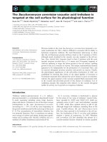
Fig. 1. Protein products of AR-JP genes: Proposed cross-talk of
pathways. Extracellular and intracellular cues activate universal cell-
signalling cascades including MAPK and phosphatidylinositol
3-kinase (PI3K) pathways that can target HtrA2, PINK1, Parkin and
DJ-1. Likely these PD-associated proteins are part of a complex
network including various signalling pathways. Although DJ-1
appears to act slightly more independently than PINK1, Parkin and
HtrA2, these PD-associated proteins seem to act in extremely com-
plex, multistepped and related pathways. The complexity and
cross-talk may be important in fine-tuning of cellular responses,
allowing points for interjection and feedback. There is mounting
evidence that these pathways may converge to influence protein
folding, protein stability and ultimately mitochondrial function
which appear to be central to the mechanism of neuronal cell death
in PD.
Autosomal recessive genes in Parkinson’s disease J. C. Fitzgerald and H. Plun-Favreau
5762 FEBS Journal 275 (2008) 5758–5766 ª 2008 The Authors Journal compilation ª 2008 FEBS
dissect biochemical pathways that lead to these diseases
and will be a major step forward in our understanding
of the pathogenesis of PD and ultimately to the
development of novel therapeutic approaches.
Acknowledgements
The authors wish to thank Professor Nicholas Wood
for his comments.
References
1 Kitada T, Asakawa S, Hattori N, Matsumine H,
Yamamura Y, Minoshima S, Yokochi M, Mizuno Y &
Shimizu N (1998) Mutations in the parkin gene cause
autosomal recessive juvenile parkinsonism. Nature 392,
605–608.
2Lu
¨
cking CB, Du
¨
rr A, Bonifati V, Vaughan J, De Mic-
hele G, Gasser T, Harhangi BS, Meco G, Dene
`
fle P,
Wood NW et al. (2000) Association between early-onset
Parkinson’s disease and mutations in the Parkin gene.
N Engl J Med 342, 1560–1567.
3 Schapira AH (2008) Mitochondria in the aetiology and
pathogenesis of Parkinson’s disease. Lancet Neurol 7,
97–109.
4 Hayashi S, Wakabayashi K, Ishikawa A, Nagai H, Sai-
to M, Maruyama M, Takahashi T, Ozawa T, Tsuji S &
Takahashi H (2000) An autopsy case of autosomal-
recessive juvenile parsinsonism with a homozygous
exon 4 deletion in the parkin gene. Mov Disord 15, 884–
888.
5 Farrer M, Chan P, Chen R, Tan L, Lincoln S, Hernan-
dez D, Forno L, Gwinn-Hardy K, Petrucelli L, Hussey
J et al. (2001) Lewy bodies and parkinsonism in families
with parkin mutations. Ann Neurol 50, 293–300.
6 Pramstaller PP, Schlossmacher MG, Jacques TS, Scara-
velli F, Eskelson C, Pepivani I, Hedrich K, Adel S,
Gonzales-McNeal M, Hilker R et al. (2005) Lewy body
Parkinson’s disease in a large pedigree with 77 parkin
mutation carriers. Ann Neurol 58, 411–422.
7 Shimura H, Hattori N, Kubo S, Mizuno Y, Asakawa
S, Minoshima S, Shimizu N, Iwai K, Chiba T, Tanaka
K et al. (2000) Familial Parkinson disease gene product,
Parkin, is a ubiquitin-protein ligase. Nat Genet 3, 302–
305.
8 Darios F, Corti O, Lu
¨
cking CB, Hampe C, Muriel MP,
Abbas N, Gu WJ, Hirsch EC, Rooney T, Ruberg M
et al. (2003) Parkin prevents mitochondrial swelling and
cytochrome c release in mitochondria-dependent cell
death. Hum Mol Genet 12, 517–526.
9 Kubo S, Kitami T, Noda S, Shimura H, Uchiyama Y,
Asakawa S, Minoshima S, Shimizu N, Mizuno Y &
Hattori N (2001) Parkin is associated with cellular
vesicles. J Neurochem 78, 42–54.
10 Wood-Kaczmar A, Gandhi S & Wood NW (2006)
Understanding the molecular causes of Parkinson’s
disease. Trends Mol Med 12, 521–528.
11 Doson MW & Guo M (2007) Pink1, Parkin, DJ-1 and
mitochondrial dysfunction in Parkinson’s disease. Curr
Opin Neurobiol 17, 331–337.
12 Valente EM, Abou-Sleiman PM, Caputo V, Muqit
MM, Harvey K, Gispert S, Ali Z, Del Turco D, Bentiv-
oglio AR, Healy DG et al. (2004) Hereditary early-
onset Parkinson’s disease caused by mutations in
PINK1. Science 304, 1120–1122.
13 Tan E & Skipper LM (2007) Pathogenic mutations in
Parkinson’s disease. Human Mut 28, 641–653.
14 Healey DG, Abou-Sleiman PM & Wood NW (2004)
PINK, PANK, or PARK? A clinicians’ guide to
familial parkinsonism Lancet Neurol
3, 652–662.
15 Gandhi S, Muqit MM, Stanyer L, Healy DG, Abou-Slei-
man PM, Hargreaves I, Heales S, Ganguly M, Parsons L,
Lees AJ et al. (2006) PINK1 protein in normal human
brain and Parkinson’s disease. Brain 129, 1720–1731.
16 Weihofen A, Ostaszewski B, Minami Y & Selkoe DJ
(2007) Pink1 Parkinson mutations, the Cdc37 ⁄ Hsp90
chaperones and Parkin all influence the maturation or
subcellular distribution of Pink1. Human Mol Genet 17 ,
602–616.
17 Haque EM, Thomas KJ, D’Souza C, Callaghan S, Kit-
ada T, Slack RS, Fraser P, Cookson MR, Tandon A &
Park DS (2008) Cytoplasmic Pink1 activity protects
neurons from dopaminergic neurotoxin MPTP. Proc
Natl Acad Sci USA 105, 1716–1721.
18 Hague S, Rogaeva E, Hernandez D, Gulick C, Single-
ton A, Hanson M, Johnson J, Weiser R, Gallardo M,
Ravina B et al. (2003) Early-onset Parkinson’s disease
caused by a compound heterozygous DJ-1 mutation.
Ann Neurol 54, 271–274.
19 Abou-Sleiman PM, Healy DG, Quinn N, Lees AJ &
Wood NW (2003) The role of pathogenic DJ-1 muta-
tions in Parkinson’s disease. Ann Neurol 54, 283–286.
20 Hedrich K, Djarmati A, Scha
¨
fer N, Hering R, Wellen-
brock C, Weiss PH, Hilker R, Vieregge P, Ozelius LJ,
Heutink P et al. (2004) DJ-1 (PARK7) mutations are
less frequent than Parkin (PARK2) mutations in early-
onset Parkinson disease. Neurology 62, 389–394.
21 Alves da Costa C (2007) DJ-1: a newcomer in Parkin-
son’s disease pathology. Curr Mol Med 7, 650–657.
22 Yoshida K, Sato Y, Yoshike M, Nozawa S, Ariga H &
Iwamoto T (2003) Immunocytochemical localisation of
DJ-1 in human male reproductive tissue. Mol Reprod
Dev 66, 391–397.
23 Le Naour F, Misek DE, Krause MC, Deneux L, Giord-
ano TJ, Scholl S & Hanash SM (2001) Proteomics-
based identification of RS ⁄ DJ-1 as a novel circulating
tumor antigen in breast cancer. Clin Cancer Res 7,
3328–3335.
J. C. Fitzgerald and H. Plun-Favreau Autosomal recessive genes in Parkinson’s disease
FEBS Journal 275 (2008) 5758–5766 ª 2008 The Authors Journal compilation ª 2008 FEBS 5763
24 Zhang L, Shimoji M, Thomas B, Moore DJ, Yu S,
Marupudi NI, Torp R, Torgner IA, Ottersen OP, Daw-
son TM et al. (2005) Mitochondrial localisation of the
Parkinson’s disease related protein DJ-1: implications
for pathogenesis. Hum Mol Genet 14, 2063–2073.
25 Bonifati V, Rizzu P, van Baren MJ, Schaap O, Breed-
veld GJ, Krieger E, Dekker MC, Squitieri F, Ibanez P,
Joosse M et al. (2003) Mutations in the DJ-1 gene asso-
ciated with autosomal recessive early-onset parkinson-
ism. Science 299, 256–259.
26 Canet-Avile
´
s RM, Wilson MA, Miller DW, Ahmad R,
McLendon C, Bandyopadhyay S, Baptista MJ, Ringe
D, Petsko GA & Cookson MR (2004) The Parkinson’s
disease protein DJ-1 is neuroprotective due to cysteine-
sulfinic acid-driven mitochondrial localization. Proc
Natl Acad Sci USA 101, 9103–9108.
27 Xu J, Zhong N, Wang H, Elias JE, Kim CY, Woldman
I, Pifl C, Gygi SP, Geula C & Yankner BA (2005) The
Parkinson’s disease-associated DJ-1 protein is a tran-
scriptional co-activator that protects against neuronal
apoptosis. Hum Mol Genet 14, 1231–1241.
28 Junn E, Taniguchi H, Jeong BS, Zhao X, Ichijo H &
Mouradian MM (2005) Interaction of DJ-1 with Daxx
inhibits apoptosis signal-regulating kinase 1 activity and
cell death. Proc Natl Acad Sci USA 102, 9691–9696.
29 Shendelman S, Jonason A, Martinat C, Leete T & Abe-
liovich A (2004) DJ-1 is a redox-dependent molecular
chaperone that inhibits alpha-synuclein aggregate for-
mation. PLoS Biol 2, e362.
30 Olzmann JA, Brown K, Wilkinson KD, Rees HD, Huai
Q, Ke H, Levey AI, Li L & Chin LS (2004) Familial
Parkinson’s disease-associated L166P mutation disrupts
DJ-1 protein folding and function. J Biol Chem 279,
8506–8515.
31 Lee SJ, Kim SJ, Kim IK, Ko J, Jeong CS, Kim GH,
Park C, Kang SO, Suh PG, Lee HS et al. (2003) Crystal
structures of human DJ-1 and Escherichia coli Hsp31,
which share an evolutionarily conserved domain. J Biol
Chem 278, 44552–44559.
32 Zhou W, Zhu M, Wilson MA, Petsko GA & Fink AL
(2006) The oxidation state of DJ-1 regulates its chaper-
one activity toward alpha-synuclein. J Mol Biol 356,
1036–1048.
33 Abeliovich A & Flint Beal M (2006) Parkinsonism
genes: culprits and clues. J Neurochem 99, 1062–1072.
34 Abou-Sleiman PM, Muqit MM & Wood NW (2006)
Expanding insights of mitochondrial dysfunction in Par-
kinson’s disease. Nat Rev Neurosci 7, 207–219.
35 De Girolamo LA, Billett EE & Hargreaves AJ (2000)
Effects of 1-methyl-4-phenyl-1,2,3,6-tetrahydropyridine
on differentiating mouse N2a neuroblastoma cells.
J Neurochem 75, 133–140.
36 Caneda-Ferron B, De Girolamo LA, Costa T, Beck
KE, Layfield R & Billett EE (2008) Assessment of the
direct and indirect effects of MPP
+
and dopamine on
the human proteasome: implications for Parkinson’s
disease aetiology. J Neurochem 105, 225–238.
37 Davis GC, Williams AC, Markey SP, Ebert MH, Caine
ED, Reichert CM & Kopin IJ (1979) Chronic parkin-
sonism secondary to intravenous injection of meperidine
analogues. Psychiat Res 1, 249–254.
38 Langston WJ, Ballard P, Tetrus JW & Irwin I (1983)
Chronic parkinsonism in humans due to a product of
meperidine-analog synthesis. Science 219, 979–980.
39 Casarejos MJ, Mene
´
ndez J, Solano RM, Rodrı
´
guez-
Navarro JA, Garcı
´
adeYe
´
benes J & Mena MA (2006)
Susceptibility to rotenone is increased in neurons from
Parkin null mice and is reduced by minocycline. J Neu-
rochem 97, 934–946.
40 Deng H, Jankovic J, Guo Y, Xie W & Le W (2005)
Small interfering RNA targeting the PINK1 induces
apoptosis in dopaminergic cells SH-SY5Y. Biochem Bio-
phys Res Commun 337, 1133–1138.
41 Gautier CA, Kitada T & Shen J (2008) Loss of PINK1
causes mitochondrial functional defects and increased
sensitivity to oxidative stress. Proc Natl Acad Sci USA
105, 11364–11369.
42 Plun-Favreau H & Hardy J (2008) Pink1 in mitochon-
drial function. Proc Natl Acad Sci USA 105, 11041–
11042.
43 Liu F, Nguyen JL, Hulleman JD, Li L & Rochet JC
(2008) Mechanisms of DJ-1 neuroprotection in a cellu-
lar model of Parkinson’s disease. J Neurochem 105,
2435–2453.
44 Meulener M, Whitworth AJ, Armstrong-Gold CE, Riz-
zu P, Heutink P, Wes PD, Pallanck LJ & Bonini NM
(2005) Drosophila DJ-1 mutants are selectively sensitive
to environmental toxins associated with Parkinson’s dis-
ease. Curr Biol 15, 1572–1577.
45 Clark IE, Dodson MW, Jiang C, Cao JH, Huh JR, Seol
JH, Yoo SJ, Hay BA & Guo M (2006) Drosophila
pink1 is required for mitochondrial function and inter-
acts genetically with Parkin. Nature 441, 1162–1166.
46 Park J, Lee SB, Lee S, Kim Y, Song S, Kim S, Bae E,
Kim J, Shong M, Kim JM et al. (2006) Mitochondrial
dysfunction in Drosophila PINK1 mutants is comple-
mented by Parkin. Nature 441, 1157–1161.
47 Yang Y, Gehrke S, Imai Y, Huang Z, Ouyang Y, Wang
JW, Yang L, Beal MF, Vogel H & Lu B (2006) Mito-
chondrial pathology and muscle and dopaminergic neu-
ron degeneration caused by inactivation of Drosophila
Pink1 is rescued by Parkin. Proc Natl Acad Sci USA
103, 10793–10798.
48 Poole AC, Thomas RE, Andrews LA, McBride HM,
Whitworth AJ & Pallanck LJ (2008) The PINK1 ⁄ Par-
kin pathway regulates mitochondrial morphology. Proc
Natl Acad Sci USA 105, 1638–1643.
49 Exner N, Treske B, Paquet D, Holmstro
¨
m K, Schiesling
C, Gispert S, Carballo-Carbajal I, Berg D, Hoepken
HH, Gasser T et al. (2007) Loss-of-function of human
Autosomal recessive genes in Parkinson’s disease J. C. Fitzgerald and H. Plun-Favreau
5764 FEBS Journal 275 (2008) 5758–5766 ª 2008 The Authors Journal compilation ª 2008 FEBS
PINK1 results in mitochondrial pathology and can be
rescued by Parkin. J Neurosci 27, 12413–12418.
50 Yang Y, Ouyang Y, Yang L, Beal MF, McQuibban
A, Vogel H & Lu B (2008) Pink1 regulates mitochon-
drial dynamics through interaction with the fis-
sion ⁄ fusion machinery. Proc Natl Acad Sci USA 105,
7070–7075.
51 Yang Y, Gehrke S, Haque ME, Imai Y, Kosek J, Yang
L, Beal MF, Nishimura I, Wakamatsu K, Ito S et al.
(2005) Inactivation of Drosophila DJ-1 leads to impair-
ments of oxidative stress response and phosphatidyl-
inositol 3-kinase ⁄ Akt signaling. Proc Natl Acad Sci
USA 102, 13670–13675.
52 Ekert PG & Vaux DL (2005) The mitochondrial death
squad: hardened killers or innocent bystanders? Curr
Opin Cell Biol 17, 626–630.
53 Muqit MM, Abou-Sleiman PM, Saurin AT, Harvey K,
Gandhi S, Deas E, Eaton S, Payne Smith MD, Venner
K, Matilla A et al. (2006) Altered cleavage and localiza-
tion of PINK1 to aggresomes in the presence of prote-
asomal stress. J Neurochem 98, 156–169.
54 Lin W & Kang UJ (2008) PINK1 characterization of
processing, stability, and subcellular localization. J Neu-
rochem 106, 464–474.
55 Plun-Favreau H, Klupsch K, Moisoi N, Gandhi S,
Kjaer S, Frith D, Harvey K, Deas E, Harvey RJ,
McDonald N et al. (2007) The mitochondrial protease
HtrA2 is regulated by Parkinson’s disease-associated
kinase PINK1. Nat Cell Biol 9, 1243–1252.
56 De Martino GN & Slaughter CA (1999) The protea-
some, a novel protease regulated by multiple mecha-
nisms. J Biol Chem 274, 22123–22126.
57 Tanaka K & Chiba T (1998) The proteasome: a pro-
tein-destroying machine. Genes Cells 3, 499–510.
58 Dawson TM & Dawson VL (2003) Molecular pathways
of neurodegeneration in Parkinson’s disease. Science
302, 819–822.
59 Leroy E, Boyer R, Auburger G, Leube B, Ulm G,
Mezey E, Harta G, Brownstein MJ, Jonnalagada S,
Chernova T et al. (1998) The ubiquitin pathway in
Parkinson’s disease. Nature 395, 451–452.
60 Yokota T, Sugawara K, Ito K, Takahashi R, Ariga H
& Mizusawa H (2003) Down regulation of DJ-1
enhances cell death by oxidative stress, ER stress, and
proteasome inhibition. Biochem Biophys Res Commun
312, 1342–1348.
61 Petrucelli L, O’Farrell C, Lockhart PJ, Baptista M,
Kehoe K, Vink L, Choi P, Wolozin B, Farrer M,
Hardy J et al. (2002) Parkin protects against the toxicity
associated with mutant a-synuclein: proteasome dys-
function selectively affects catecholaminergic neurons.
Neuron 36, 1007–1019.
62 Yang H, Zhou H, Li B, Niu G & Chen S (2007)
Downregulation of Parkin damages antioxidant
defenses and enhances proteasome inhibition-induced
toxicity in PC12 cells. J Neuroimmune Pharmacol 2
,
276–283.
63 Yang W, Chen L, Ding Y, Zhuang X & Kang UJ
(2007) Paraquat induces dopaminergic dysfunction and
proteasome impairment in DJ-1-deficient mice. Hum
Mol Genet 16, 2900–2910.
64 Miller DW, Ahmad R, Hague S, Baptista MJ, Canet-
Aviles R, McLendon C, Carter DM, Zhu PP, Stadler J,
Chandran J et al. (2003) L166P mutant DJ-1, causative
for recessive Parkinson’s disease, is degraded through
the ubiquitin-proteasome system. J Biol Chem 278,
36588–36595.
65 Moriwaki Y, Kim YJ, Ido Y, Misawa H, Kawashima
K, Endo S & Takahashi R (2008) L347P PINK1
mutant that fails to bind to Hsp90 ⁄ Cdc37 chaperones is
rapidly degraded in a proteasome-dependent manner.
Neurosci Res 61, 43–48.
66 Choi P, Ostrerova-Golts N, Sparkman D, Cochran E,
Lee JM & Wolozin B (2000) Parkin is metabolized by
the ubiquitin ⁄ proteasome system. NeuroReport 11,
2635–2638.
67 Hyun D, Lee M, Hattori N, Kubo S, Mizuno Y, Halli-
well B & Jenner P (2002) Effect of wild-type or mutant
Parkin on oxidative damage, nitric oxide, antioxidant
defenses, and the proteasome. J Biol Chem 277, 28572–
28577.
68 Pridgeon JW, Olzmann JA, Chin LS & Li L (2008)
PINK1 protects against oxidative stress by phosphory-
lating mitochondrial chaperone TRAP1. PLoS Biol 5,
1494–1503.
69 Young JC & Hartl FU (2003) A stress sensor for the
bacterial periplasm. Cell 113, 1–2.
70 Moore DJ, West AB, Dikeman DA, Dawson VL &
Dawson TM (2008) Parkin mediates the degradation-
independent ubiquitination of Hsp70. J Neurochem 105,
1806–1819.
71 Kahle PJ & Haass C (2004) How does Parkin ligate
ubiquitin to Parkinson’s disease? EMBO Rep 5, 681–
685.
72 Winklhofer KF, Henn IH, Kay-Jackson PC, Heller U
& Tatzelt J (2003) Inactivation of Parkin by oxidative
stress and C-terminal truncations: a protective role
of molecular chaperones. J Biol Chem 278, 47199–
47208.
73 Anderson JK (2004) Oxidative stress in neurodegenera-
tion: cause or consequence? Nat Rev Neurosci 10(Sup-
pl.), S18–S25.
74 Taira T, Saito Y, Niki T, Iguchi-Ariga SM, Takahashi
K & Ariga H (2004) DJ-1 has a role in antioxidative
stress to prevent cell death. EMBO Rep 5, 213–218.
75 Martinat C, Shendelman S, Jonason A, Leete T, Beal
MF, Yang L, Floss T & Abeliovich A (2004) Sensitivity
to oxidative stress in DJ-1-deficient dopamine neurons:
an ES-derived cell model of primary parkinsonism.
PLoS Biol 2, e327.
J. C. Fitzgerald and H. Plun-Favreau Autosomal recessive genes in Parkinson’s disease
FEBS Journal 275 (2008) 5758–5766 ª 2008 The Authors Journal compilation ª 2008 FEBS 5765
76 Moore DJ, Zhang L, Troncoso J, Lee MK, Hattori N,
Mizuno Y, Dawson TM & Dawson VL (2005) Associa-
tion of DJ-1 and Parkin mediated by pathogenic DJ-1
mutations and oxidative stress. Hum Mol Genet 14,
71–84.
77 Palacino JJ, Sagi D, Goldberg MS, Krauss S, Motz C,
Wacker M, Klose J & Shen J (2004) Mitochondrial dys-
function and oxidative damage in Parkin-deficient mice.
J Biol Chem 279, 18614–18622.
78 Pesah Y, Pham T, Burgess H, Middlebrooks B, Verstre-
ken P, Zhou Y, Harding M, Bellen H & Mardon G
(2004) Drosophila Parkin mutants have decreased mass
and cell size and increased sensitivity to oxygen radical
stress. Development 131, 2183–2194.
79 Wang D, Qian L, Xiong H, Liu J, Neckameyer WS,
Oldham S, Xia K, Wang J, Bodmer R & Zhang Z
(2006) Antioxidants protect PINK1-dependent dopami-
nergic neurons in Drosophila. Proc Natl Acad Sci USA
103, 13520–13525.
80 Beilina A, Van Der Brug M, Ahmad R, Kesavapany S,
Miller DW, Petsko GA & Cookson MR (2005) Muta-
tions in PTEN-induced putative kinase 1 associated
with recessive Parkinsonism have differential effects on
protein stability. Proc Natl Acad Sci USA 102, 5703–
5708.
81 Silvestri L, Caputo V, Bellacchio E, Atorino L, Dalla-
piccola B, Valente EM & Casari G (2005) Mitochon-
drial import and enzymatic activity of PINK1 mutants
associated to recessive parkinsonism. Hum Mol Genet
14, 3477–3492.
82 Unoki M & Nakamura Y (2001) Growth-suppressive
effects of BPOZ and EGR2, two genes involved in
the PTEN signaling pathway. Oncogene 20, 4457–
4465.
83 Kim RH, Peters M, Jang Y, Shi W, Pintilie M, Fletcher
GC, DeLuca C, Liepa J, Zhou L, Snow B et al. (2005)
DJ-1, a novel regulator of the tumor suppressor PTEN.
Cancer Cell 7 , 263–273.
84 Fallon L, Be
´
langer CM, Corera AT, Kontogiannea M,
Regan-Klapisz E, Moreau F, Voortman J, Haber M,
Rouleau G, Thorarinsdottir T et al. (2006) A regulated
interaction with the UIM protein Eps15 implicates Par-
kin in EGF receptor trafficking and PI(3)K-Akt signal-
ling. Nat Cell Biol 8, 834–842.
85 Yang L, Sun M, Sun XM, Cheng GZ, Nicosia SV &
Cheng JQ (2007) Akt attenuation of the serine protease
activity of HtrA2 ⁄ Omi through phosphorylation of
serine 212. J Biol Chem 282, 10981–10987.
86 Yamamoto A, Friedlein A, Imai Y, Takahashi R, Kah-
le PJ & Haass C (2005) Parkin phosphorylation and
modulation of its E3 ubiquitin ligase activity. J Biol
Chem 280, 3390–3399.
87 Avraham E, Rott R, Liani E, Szargel R & Engelender
S (2007) Phosphorylation of Parkin by the cyclin-depen-
dent kinase 5 at the linker region modulates its ubiqu-
itin-ligase activity and aggregation. J Biol Chem 282,
12842–12850.
88 Tang B, Xiong H, Sun P, Zhang Y, Wang D, Hu Z,
Zhu Z, Ma H, Pan Q, Xia JH et al. (2006) Association
of PINK1 and DJ-1 confers digenic inheritance of
early-onset Parkinson’s disease. Hum Mol Genet 15,
1816–1825.
89 Strauss KM, Martins LM, Plun-Favreau H, Marx FP,
Kautzmann S, Berg D, Gasser T, Wszolek Z, Mu
¨
ller T,
Bornemann A et al. (2005) Loss of function mutations
in the gene encoding Omi
⁄ HtrA2 in Parkinson’s disease.
Hum Mol Genet 14, 2099–2111.
90 Simo
´
n-Sa
´
nchez J & Singleton AB (2008) Sequencing
analysis of OMI ⁄ HTRA2 shows previously reported
pathogenic mutations in neurologically normal controls.
Hum Mol Genet 17, 1988–1993.
91 Bogaerts V, Nuytemans K, Reumers J, Pals P, Enge-
lborghs S, Pickut B, Corsmit E, Peeters K, Schymko-
witz J, De Deyn PP et al. (2008) Genetic variability in
the mitochondrial serine protease HTRA2 contributes
to risk for Parkinson disease. Hum Mutat 29, 832–840.
92 Anichtchik O, Diekmann H, Fleming A, Roach A,
Goldsmith P & Rubinsztein DC (2008) Loss of Pink1
function affects development and results in neurodegen-
eration in zebrafish. J Neurosci 28, 8199–8207.
93 Falkenburger BH & Schulz JB (2006) Limitations of
cellular models in Parkinson’s disease research. J Neural
Transm 70 (Suppl.), 261–268.
94 Hyun-Park I, Arora N, Huo H, Maherali N, Ahfeldt T,
Shimamura A, Lensch MW, Cowan C, Hochedlinger K
& Daley GQ (2008) Disease-specific induced pluripotent
stem cells. Cell 134, 1–10.
95 Nishikawa S, Goldstein RA & Nierras CR (2008) The
promise of human induced pluripotent stem cells
for research and therapy. Nat Rev Mol Cell Biol 9,
725–729.
96 Dimos JT, Rodolfa KT, Niakan KK, Weisenthal LM,
Mitsumoto H, Chung W, Croft GF, Saphier G, Leibel
R, Goland R et al. (2008) Induced pluripotent stem
cells generated from patients with ALS can be differen-
tiated into notor neurons. Science 321, 1218–1221.
Autosomal recessive genes in Parkinson’s disease J. C. Fitzgerald and H. Plun-Favreau
5766 FEBS Journal 275 (2008) 5758–5766 ª 2008 The Authors Journal compilation ª 2008 FEBS