Báo cáo khoa học: New insights into the functions and N-glycan structures of factor X activator from Russell’s viper venom pot
Bạn đang xem bản rút gọn của tài liệu. Xem và tải ngay bản đầy đủ của tài liệu tại đây (894.17 KB, 15 trang )
New insights into the functions and N-glycan structures of
factor X activator from Russell’s viper venom
Hong-Sen Chen
1
, Jin-Mei Chen
2
, Chia-Wei Lin
1
, Kay-Hooi Khoo
1,2
and Inn-Ho Tsai
1,2
1 Graduate Institute of Biochemical Sciences, National Taiwan University, Taiwan
2 Institute of Biological Chemistry, Academia Sinica, Taipei, Taiwan
Activators for zymogens of the blood coagulation cas-
cade are abundant in venoms of many Viperinae [1]
and some Elapidae [2,3]. The factor X activator from
the venom of Russell’s viper (Daboia russelli and
Daboia siamensis) (RVV-X) is a potent procoagulating
and lethal toxin [4]. Its action mechanism involves the
Ca
2+
-dependent hydrolysis of the peptide bond
between Arg51 and Ile52 of the heavy chain on
factor X, similar to the physiological activation by
factors IXa and VIIa [4,5]. In addition, RVV-X also
activates factor IX, but not prothrombin [6]. Given
these functional specificities, RVV-X has served as a
tool for thrombosis research and as a diagnostic
reagent [7].
RVV-X is a heterotrimeric glycoprotein composed
of one heavy chain (HC) and two distinct light chains
(LC1 and LC2) [8,9]. The heavy chain is a P-III metal-
loprotease [10], and both light chains belong to the
C-type lectin-like family. However, the light chain LC2
has yet to be fully sequenced [8]. Based on their
sequence similarity to other venom factor IX/X-bind-
ing proteins [8,11], both light chains of RVV-X have
Keywords
cDNA cloning; factor X activator; glycan
mass spectrometry; Lewis and sialyl-Lewis;
Russell’s viper venom
Correspondence
I. H. Tsai, Institute of Biological Chemistry,
Academia Sinica, PO Box 23-106, Taipei,
Taiwan
Fax: 886 22 3635038
Tel: 886 22 3620264
E-mail:
(Received 18 February 2008, revised 22
April 2008, accepted 5 June 2008)
doi:10.1111/j.1742-4658.2008.06540.x
The coagulation factor X activator from Russell’s viper venom (RVV-X) is
a heterotrimeric glycoprotein. In this study, its three subunits were cloned
and sequenced from the venom gland cDNAs of Daboia siamensis. The
deduced heavy chain sequence contained a C-terminal extension with four
additional residues to that published previously. Both light chains showed
77–81% identity to those of a homologous factor X activator from
Vipera lebetina venom. Far-western analyses revealed that RVV-X could
strongly bind protein S, in addition to factors X and IX. This might inacti-
vate protein S and potentiate the disseminated intravascular coagulation
syndrome elicited by Russell’s viper envenomation. The N-glycans released
from each subunit were profiled and sequenced by MALDI-MS and MS/
MS analyses of the permethyl derivatives. All the glycans, one on each
light chain and four on the heavy chain, showed a heterogeneous pattern,
with a combination of variable terminal fucosylation and sialylation on
multiantennary complex-type sugars. Amongst the notable features were
the presence of terminal Lewis and sialyl-Lewis epitopes, as confirmed by
western blotting analyses. As these glyco-epitopes have specific receptors in
the vascular system, they possibly contribute to the rapid homing of
RVV-X to the vascular system, as supported by the observation that slower
and fewer fibrinogen degradation products are released by desialylated
RVV-X than by native RVV-X.
Abbreviations
APTT, activated partial thromboplastin time; DIC, disseminated intravascular coagulation; FDP, fibrinogen degradation product; Gla,
c-carboxyglutamic acid; PNGase F, peptide N-glycosidase F; PVDF, poly(vinylidene difluoride); RVV-X, factor X activator from Russell’s viper
venom; SBHP, streptavidin-biotinylated horseradish peroxidase; TBST, Tris-buffered saline with Tween 20; VAP1, vascular apoptosis-inducing
protein 1; VLFXA, factor X activator from Vipera lebetina venom.
3944 FEBS Journal 275 (2008) 3944–3958 ª 2008 The Authors Journal compilation ª 2008 FEBS
been postulated to bind the c-carboxyglutamic acid
(Gla) domain of factor X and bring the heavy chain to
the Arg51 cleavage site of factor X [4]. This specula-
tion has been supported by a recent crystallographic
study of RVV-X at 2.9 A
˚
resolution [12]. In addition,
a homologous factor X activator from Vipera lebetina
venom (VLFXA) has been characterized, and its three
subunits have been cloned and fully sequenced [13,14].
Its heavy chain and light chain LC1 share high
sequence similarity (> 77%) to those of RVV-X.
The structures of the carbohydrate moieties of
RVV-X have been investigated previously. It was
found that RVV-X contains multiantennary complex-
type N-glycans, with bisecting GlcNAc and terminal
Neu5Aca2–3Gal sialylation. The glycan core structures
were additionally shown to be sufficient to maintain
the active conformation of RVV-X [9,15]. However,
details on the glycosylation and physiological signifi-
cance of these glycans remain to be explored. In this
study, we have cloned all the RVV-X subunits for the
first time and have solved their complete sequences.
The nucleotide sequences of HC, LC1 and LC2 have
been deposited in GenBank with accession numbers
DQ137799, AY734997 and AY734998, respectively.
The overall N-glycosylation profiles, as well as that of
the individual subunits and sites, were defined by
advanced mass spectrometry analyses. Unexpectedly,
terminal fucosylation contributing to Lewis (Le) and
sialyl-Lewis (SLe) epitopes was also identified, and
their functional implications were clarified by in vivo
studies.
Results and Discussion
Purification and characterization of RVV-X
RVV-X was purified from the crude venom of D. siam-
ensis (Flores Island, Indonesia) by two chromato-
graphic steps. The venom was separated into seven
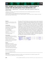
fractions using a Superdex G-75 column (Fig. 1A).
The first peak (indicated by a bar) exhibiting strong
procoagulating activity was further purified by anion
exchange chromatography (Fig. 1B). The yield of
RVV-X was approximately 3.4% (w/w) of the crude
venom, similar to that reported previously [4]. SDS-
PAGE of the purified protein revealed a single band at
93 kDa under nonreducing conditions, and three bands
of 62, 21 and 18 kDa under reducing conditions
(Fig. 1B, inset). The molecular mass of purified RVV-X
was also determined by an analytical ultracentrifuge as
92 972 ± 4356 Da (data not shown). After
electrophoresis and blotting, the protein band of LC2
was excised from the poly(vinylidene difluoride)
(PVDF) membrane. By automatic Edman sequencing,
its N-terminal sequence 1–25 was determined as
LDXPPDSSLYRYFXYRVFKEHKT (X denotes an
unidentified residue), which differs from that of
VLFXA LC2 by three residues at positions 10, 22 and
24 [14].
The stability of RVV-X under various conditions
was studied by activated partial thromboplastin time
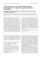
(APTT) coagulation assay. We first assigned a plot of
clotting time against dose of RVV-X that fitted well in
a power regression mode (Fig. 2A). On the basis of
this relationship, we determined the remaining activi-
ties after different treatments. The results showed that
RVV-X was stable in buffers of pH 6–10 and tempera-
tures below 37 °C (Fig. 2B,C), consistent with previous
studies showing that purified RVV-X was stable at
4 °Cin50mm Tris/H
3
PO
4
buffer, pH 6.0 for
2 months [16]. These properties were also similar to
those of the P-III metalloproteinase VAP1 (vascular
apoptosis-inducing protein 1) from Crotalus atrox
venom [17].
A
B
Fig. 1. Purification of RVV-X. (A) About 20 mg of D. siamensis
venom was dissolved in buffer and separated by Superdex G-75
gel filtration. The column was equilibrated and eluted with 100 m
M
ammonium acetate (pH 6.7). Fraction I (indicated by bar) possess-
ing coagulation activity was pooled and lyophilized. (B) Subsequent
purification of fraction I on a Mono Q column. The elution was
achieved by increasing (0–0.6
M) NaCl gradient in 50 mM Tris/HCl,
pH 8.0. The absorbance at 280 nm of the eluent was monitored
online. The inset shows the result of SDS-PAGE of purified RVV-X
under reducing (R) and nonreducing (NR) conditions.
H S. Chen et al. Daboia siamensis venom factor X activator
FEBS Journal 275 (2008) 3944–3958 ª 2008 The Authors Journal compilation ª 2008 FEBS 3945
Substrate specificities studied by far-western
analysis
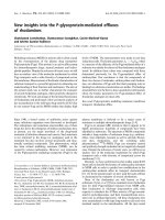
To investigate the binding specificity of RVV-X,
several human coagulation factors containing the Gla
domain were subjected to SDS-PAGE (Fig. 3A) and
then electroblotted onto a PVDF membrane. The blot
was incubated with biotinylated RVV-X, and binding
was detected with the streptavidin-biotinylated horse-
radish peroxidase (SBHP) system (Fig. 3B,C). In the
presence of a millimolar concentration of Ca
2+
ions,
RVV-X bound strongly to factors X and IX, whereas
its binding to prothrombin and protein C was hardly
detectable. When Ca
2+
ions were removed from the
solution, binding was no longer detectable (Fig. 3C),
confirming that exogenous Ca
2+
ions are essential for
substrate binding [18]. Furthermore, no signal could be
detected for factor X without the Gla domain (Fig. 3B,
lane 7).
Fig. 2. Effects of buffer pH and temperature on the coagulation
activity of RVV-X. (A) Relationship between the clotting time and
dose of RVV-X in APTT coagulation assay. Analysing the experimen-
tal data (0.1–10 ng) with power regression gives a correlation of
R
2
= 0.991 and a prediction equation of y = 16.624x
)0.2148
. (B) pH
stability profile. RVV-X (1 lgÆlL
)1
) was incubated at 4 ° C for 36 h in
buffers of different pH. (C) Thermal stability profile. RVV-X (1 lgÆlL
)1
in 100 mM Hepes, pH 8.0) was incubated at various temperatures for
1 h. The remaining activities of 5 ng of RVV-X after (B) and (C) treat-
ments were evaluated by the coagulation assay. The results are
expressed as the mean ± standard deviation (n = 3).
A
B
C
Fig. 3. Analysis of the binding of RVV-X to Gla-containing plasma
factors or proteins by far-western blotting. (A) Coagulation factors
were separated by SDS-PAGE and stained by Coomassie brilliant
blue G-250. Lane 1, 3 lg of factor X; lane 2, 0.3 lg of factor X; lane
3, 3 lg of factor IX; lane 4, 3 lg of prothrombin; lane 5, 3 lgof
protein C; lane 6, 3 lg of protein S; lane 7, 3 lg of Gla-domainless
factor X. (B) Instead of staining, the protein bands were blotted on
to a PVDF membrane after PAGE. The membrane was probed with
1.5 lgÆmL
)1
biotinylated RVV-X and detected with the SBHP sys-
tem in the presence of 5 m
M CaCl
2
. (C) Same as (B), except Ca
2+
ions were excluded. For lane 7, the arrow denotes residual factor X
present in the sample of Gla-domainless factor X.
Daboia siamensis venom factor X activator H S. Chen et al.
3946 FEBS Journal 275 (2008) 3944–3958 ª 2008 The Authors Journal compilation ª 2008 FEBS
Thus, the far-western results reflect the substrate
specificity of RVV-X [4,6], and its binding to sub-
strates involves their Gla domains [19]. Interestingly,
we found that protein S bound strongly to RVV-X
(Fig. 3B, lane 6). If RVV-X inactivates protein S
in vivo, it will interrupt the protein C pathway [20] and
stimulate the tissue factor pathway [21], both of which
may lead to an increase in the risk of coagulation
and disseminated intravascular coagulation (DIC)
syndrome.
Cloning and sequence alignment of RVV-X
subunits
PCR amplification and cloning of the light chains of
RVV-X were carried out using cDNA prepared from
venom glands of D. siamensis (Flores Island, Indone-
sia) as template. After RT-PCR, 20 clones encoding
C-type lectin-like proteins were sequenced. Of these, 10
clones were found to encode the LC2 and LC1 sub-
units. Others were found to encode other variants of
the C-lectin-like venom proteins. The amino acid
sequences of both subunits were deduced from the
nucleotide sequences, and were found to match the
N-terminal sequences of the corresponding proteins
[8]. The ORF of LC2 encodes a precursor of 158
amino acids, including a signal peptide of 23 residues
and mature protein of 135 residues. Its predicted mass
is 15 983 Da, its isoelectric point is 5.44 and it has
a potential N-glycosylation site at Asn59. The LC1
precursor contains 146 amino acids, including a signal
peptide of 23 residues, and the predicted sequence for
its mature protein matches that published previously
[8].
The amino acid sequences of LC1 and LC2, together
with those of other homologues of factor IX/X-bind-
ing lectin-like subunits, are aligned in Fig. 4. They
show the highest sequence identity (77–81%) to the
corresponding subunits of VLFXA [14]. Residues
Glu100 and Arg102 of LC2, presumably important for
interacting with the Gla domain of factor X [19], were
conserved in both LC2 subunits of RVV-X and
VLFXA. In addition to the conserved Cys residues
present in this lectin-like family, both LC2 subunits
contain an extra Cys at the extended C-terminus,
which probably forms an interchain disulfide bridge
with the heavy chain [14]. LC1 is covalently linked to
LC2 but not to the heavy chain.
The crystal structures of the factor IX/X-binding
lectin-like proteins from pit viper venom revealed that
each subunit contained one Ca
2+
-binding site and four
corresponding residues that coordinated Ca
2+
ions
[22]. It was shown later that only one subunit of fac-
tor IX/X-binding protein from Echis venom had a
Ca
2+
-binding site; the other non-Ca
2+
-binding subunit
was stabilized by C-terminal Lys/Arg residues [23]. We
found that the LC2 and LC1 sequences of RVV-X
(Fig. 4) lacked the Ca
2+
-binding acidic residues found
in the sequences of crotalid factor IX/X-binding
proteins; instead, they contained basic residues at these
A
B
Fig. 4. Sequence alignments of RVV-X light
chains with other factor IX/X-binding pro-
teins. Residues identical to those of LC2
and LC1 are denoted with dots; gaps are
marked with hyphens. Putative Ca
2+
-binding
sites and potential N-glycosylation sites are
shown in grey and underlined, respectively.
Accession numbers and venom species are
as follows: VLFXA LC2 (AY57811) and LC1
(AY339163), Macrovipera lebetina; ECLV IX/
X-bp a subunit (AAB36401) and b subunit
(AAB36402), Echis leucogaster; Acutus X-bp
A chain (1IODA) and B chain (1IODB), Dei-
nagkistrodon acutus; Habu IX/X-bp A chain
(P23806) and B chain (P23807), Habu X-BP
A chain (1J34A) and B chain (1J34B),
Protobothrops flavoviridis.
H S. Chen et al. Daboia siamensis venom factor X activator
FEBS Journal 275 (2008) 3944–3958 ª 2008 The Authors Journal compilation ª 2008 FEBS 3947
sites. This may reflect an evolutionary difference
between Viperinae and Crotalinae venoms in the struc-
ture of factor IX/X-binding protein families.
Using similar procedures, cDNA e ncoding the R VV-X
heavy chain (RVV-X HC) was cloned and sequenced.
Its ORF encodes a P-III precursor protein of 619
amino acids, including a 188-residue highly conserved
proenzyme domain followed by a mature protein of
431 residues (Fig. 5), consistent with its published pro-
tein sequence [8]. The proenzyme domain contains a
‘cysteine switch’ motif (PKMCGVT), which is possibly
required for its processing and activation. Notably, the
predicted RVV-X HC contains a C-terminal extension
of four additional residues (FSQI). Whether this
implies post-translational processing or geographical
variations amongst D. siamensis venoms is not clear. A
similar phenomenon has been reported for the deduced
protein sequence of HR1b, which has an additional
seven residues (TTVFSLI) at the C-terminus, and
proteolytic processing was suggested to have occurred
[24].
Figure 5 shows the alignment of the amino acid
sequences of RVV-X HC with those of other represen-
tative P-III enzymes. It shows highest similarity (82%)
to VLFXA HC, and lower similarity to other P-III
proteases, e.g. Ecarin (63%), Daborhagin (56%),
HR1b (54%) and VAP1 (53%). The proenzyme
domain, zinc-chelating motif, methionine turn and
three potential Ca
2+
-binding sites are all conserved
(Fig. 5). Notably, residue Cys562, which presumably
forms a disulfide bond with Cys135 of LC2, is located
within the highly variable region, which is important
for substrate recognition of the A disintegrin and
metalloproteinase (ADAM) family [25]. By this unique
linking to RVV-X HC, the light chains appear to con-
fer the substrate specificities of RVV-X [12]. Collec-
tively, the primary sequences of the three subunits of
RVV-X (Figs 4 and 5) suggest the possible presence of
three conformational Ca
2+
-binding sites in the heavy
chain and none in LC1 and LC2, in accordance with
the results of its crystallographic structure [12].
N-glycosylation profiles
The isolation of the individual heavy and light chains
in sufficient yield allowed a detailed structural charac-
terization of their respective N-glycosylation profiles to
be performed. Previous investigation based primarily
on lectin binding, sialidase treatment, glycosyl compo-
sition and linkage analyses has led to the conclusion
that the N-glycans of RVV-X are mostly of the com-
plex type, with bisecting GlcNAc and a2–3Neu5Ac
sialylation on a proportion of terminal b-Gal residues
as the most notable structural features [9]. More
specifically, it was estimated that about 5% of the total
N-glycans are of high mannose type, 65% are of bian-
tennary complex type and 30% are of tri-/tetra-anten-
nary complex type. On the basis of interactions with
immobilized erythroagglutinating phytohaemagglutinin
lectin, 50–60% of the total glycans are deduced to
carry a bisecting GlcNAc, consistent with the detection
of a substantial amount of 3,4,6-Man in a ratio of
2 : 1 relative to nonbisected 3,6-Man by methylation
analysis. Approximately 0.5–0.8 mol of terminal Fuc
was also detected per 3 mol of Man (1 mol of N-gly-
can), but the exact location was not defined as the
expected 4,6-linked GlcNAc residue, corresponding to
the reducing end GlcNAc in which core fucosylation is
normally attached, could not be identified. This overall
picture is mostly reproduced in our current analysis
based on MALDI-MS (Fig. 6) and advanced MS/MS
(Fig. 7) analyses of the permethylated N-glycans, but
with a few important new findings.
Overall, the salient structural characteristics of the
N-glycans released from the heavy and light chains are
similar. However, a major signal corresponding to the
high-mannose-type Man
5
GlcNAc
2
structure was only
found in the heavy chain. In addition, there is a rela-
tively higher abundance of the larger size, multianten-
nary glycans carried on the heavy chain, which gave a
much more heterogeneous and complex profile. As
listed in Table 1, the assigned compositions for the
major [M + Na]
+
molecular ion signals detected cor-
respond to the expected complex-type N-glycans with
up to five Hex-HexNAc units. The majority carry a
variable degree of Neu5Ac sialylation and an extra
HexNAc residue that is attributable to the bisecting
GlcNAc. Importantly, some of the larger structures
were found to contain more than one Fuc residue,
giving a first indication that not all fucosylation can be
ascribed to core a6-fucosylation. Core a3-fucosylation
was ruled out as these N-glycans were released by pep-
tide N-glycosidase F (PNGase F). It is thus likely that
some or all of the Fuc residues may be attached to the
terminal sequences.
As shown by MALDI-TOF/TOF MS/MS analyses
of representative Fuc-containing major N-glycans
(Fig. 7), the trimannosyl core structures are indeed
bisected by GlcNAc and are nonfucosylated. Fuc was
found to be attached to the 3-position of HexNAc of
the terminal Hex-HexNAc unit, giving rise to the Le
x
epitope and SLe
x
when additionally sialylated. The
characteristic D ions for Le
x
and SLe
x
were detected
at m/z 472 and 833, respectively, whereas the corre-
sponding ion indicative of Le
a
and SLe
a
at m/z
442 was either not found or was too minor to allow
Daboia siamensis venom factor X activator H S. Chen et al.
3948 FEBS Journal 275 (2008) 3944–3958 ª 2008 The Authors Journal compilation ª 2008 FEBS
unambiguous identification. Other terminal epitopes
include the nonsubstituted Hex-4HexNAc (Galb1–
4GlcNAcb1-, LacNAc), Neu5Aca2–3Hex-4HexNAc
and nonextended terminal HexNAc residues. The pres-
ence of bisecting GlcNAc was established from several
complementary ion series. First, the D ion formed at
the bisected 3,4,6-linked b-Man residue carried the
extra bisecting GlcNAc residue together with the
6-arm substituents. Second, a characteristic loss of
both the bisecting GlcNAc and the 3-arm substituents,
in concert with a
1,5
A-type ring cleavage at the b-Man
residue, yielded an ion at 321 mass units lower than
Fig. 5. Sequence alignments of RVV-X heavy chain with other P-III enzymes. Residues identical to those of RVV-X HC are denoted by dots,
and gaps are marked with hyphens. Putative Ca
2+
-binding sites and potential N-glycosylation sites are shown in grey or underlined, respec-
tively. Conserved cysteine switch, zinc-binding site, methionine turn and ECD motif are boxed. Accession numbers and venom species are
as follows: VLFXA HC (AAQ17467), Macrovipera lebetina; Ecarin (Q90495), Echis carinatus; Daborhagin (DQ137798), D. russelli; HR1b
(BAB92014), Protobothrops flavoviridis; VAP1 (BAB18307), Crotalus atrox.
H S. Chen et al. Daboia siamensis venom factor X activator
FEBS Journal 275 (2008) 3944–3958 ª 2008 The Authors Journal compilation ª 2008 FEBS 3949
the corresponding D ion. Third, the
0,4
A ion would
include the 6-arm substituents, but not the extra Glc-
NAc residue, if the latter bisects the b-Man residue at
the C4 position. Finally, an H ion would be formed
through concerted loss of the substituents on the
6-arm and the bisecting GlcNAc.
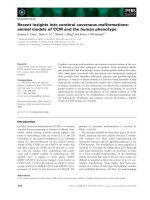
The identification of Le
x
and SLe
x
by MS/MS
sequencing was further corroborated by western blot
analyses (Fig. 8) using a panel of specific monoclonal
antibodies. Unexpectedly, the data indicated that, in
addition to Le
x
and SLe
x
, the heavy chain was also
stained positive with anti-SLe
a
serum. Although our
MS/MS data on the major Fuc-containing biantennary
N-glycans (Fig. 7) provided only convincing evidence
for the SLe
x
and Le
x
linkages, it is possible that a very
small amount of SLe
a
is also present amongst the iso-
mers, particularly on the multiantennary forms which
were of low abundance and not subjected to further
analysis. However, the monoclonal antibodies employed
failed to bind both light chains, although the MS data
clearly established the presence of at least Le
x
and SLe
x
on their N-glycans. It is possible that there is, overall, a
much higher abundance of the implicated epitopes
carried on the heavy chain, which contains five potential
N-glycosylation sites relative to one each on the two
light chains. The density of the presented epitopes would
be further amplified by a higher abundance of multian-
tennary structures on the heavy chain.
Glycopeptide analyses
To seek information on the potential N-glycosylation
site occupancies of the individual chains, tryptic
peptides from each of the purified HC, LC1 and LC2
chains were subjected to automated nano-LC-nESI-
MS/MS analyses, operated in a precursor ion discov-
ery mode to optimize for glycopeptide detection. For
the heavy chain, four distinct sets of glycopeptides
were detected, corresponding to glycoforms of tryptic
peptides carrying the N-glycosylated Asn28, Asn69,
Asn163 and Asn183 residues (data not shown). The
tryptic glycopeptide corresponding to the fifth poten-
tial site at Asn376 was not identified. The data are
therefore consistent with a previous report, which esti-
mated a total of four N-glycan chains carried on the
heavy chain, based on partial PNGase F digestion and
SDS-PAGE analysis [9,15]. There is apparently no
strict preference for any particular complex-type N-gly-
can structure to be localized on any of the four sites,
as most of the major structures found by MALDI-MS
mapping of the released N-glycans could be detected
amongst all four sets of glycopeptides observed.
A more definitive quantification of each individual
glycoform was not attempted as glycopeptides carrying
some of the larger multiantennary structures are rela-
tively minor and refractory to unambiguous identifica-
tion by direct online LC-MS/MS analysis. Interestingly
though, the single Man
5
GlcNAc
2
structure could only
be identified on Asn183.
For the light chains, tryptic glycopeptides carrying a
single N-glycosylation site could be identified. Notably,
the glycoform heterogeneity for LC1 was found to be
less complex than that of LC2 (data not shown).
Larger N-glycan structures extending up to (Hex-Hex-
NAc)
4
, with variable degrees of Fuc and Neu5Ac, were
found only on LC2 and not on LC1, despite earlier
A
B
Fig. 6. MALDI-MS profiling of the N-gly-
cans. N-glycans released from the heavy
chain (A) and LC1 (B) of RVV-X were perme-
thylated and profiled by MALDI-MS. The
N-glycans of LC1 and LC2 gave similar pro-
files, and only that of LC1 is shown here.
The molecular composition assignments of
the major signals detected are listed in
Table 1, several of which were further analy-
sed by MS/MS to deduce the terminal epi-
topes carried and their probable structures.
Daboia siamensis venom factor X activator H S. Chen et al.
3950 FEBS Journal 275 (2008) 3944–3958 ª 2008 The Authors Journal compilation ª 2008 FEBS
A
B
C
Fig. 7. MALDI-TOF/TOF MS/MS sequencing of Le
x
- and SLe
x
-containing N-glycans of RVV-X. The major N-glycans tentatively assigned as
carrying the Lewis and sialyl-Lewis epitopes of interest (Table 1) were further subjected to MALDI-TOF/TOF MS/MS analysis to derive link-
age-specific cleavage ions [40] for structural assignment. In general, the same molecular ion signals afforded by heavy and light chains gave
similar MS/MS spectra, indicative of similar structures. Representative MS/MS spectra for the sodiated parent ions at m/z 2490, 2647 and
2851 (Fig. 6) are shown in (A), (B) and (C), respectively. For clarity of presentation, only the most abundant linkage and/or sequence informa-
tive ions are schematically illustrated and annotated. The nomenclature for the ion series follows that proposed by Domon and Costello [42]
and Spina et al. [43], as adapted by Yu et al. [40]. Other nonannotated ions include: (1) a characteristic loss of 321 mass units from the D
ions formed at bisected b-Man; (2) oxonium ions for terminal HexNAc
+
(m/z 260), Neu5Ac
+
(m/z 376) and Hex-HexNAc
+
(m/z 464). In (A)
and (C), the presence of alternative isomers in which the nonfucosylated LacNAc is carried on the 6-arm is indicated by the D ion at m/
z 1125. Symbols used: r, Neu5Ac;
, Fuc; d, Hex (light-shaded for Gal and dark-shaded for Man, although these cannot be distinguished
by MS analysis); j, HexNAc (GlcNAc).
H S. Chen et al. Daboia siamensis venom factor X activator
FEBS Journal 275 (2008) 3944–3958 ª 2008 The Authors Journal compilation ª 2008 FEBS 3951
mapping of the released N-glycans indicating a rather
similar N-glycosylation profile for the two light chains.
It is possible that these larger N-glycan structures,
similar to those found on the heavy chain, are much
less abundant relative to the major biantennary ones,
and were not readily detectable without further glyco-
peptide purification and/or sample enrichment. The
data are consistent with previous findings, which indi-
cated that the mobility of LC2, but not of LC1, on
SDS-PAGE was shifted noticeably with sialidase treat-
ment [9]. This observation could be interpreted by the
fact that LC2 carries a more elaborate N-glycosylation,
with additional multisialylated and multiantennary
structures not found on LC1, albeit of relatively low
Table 1. Major RVV-X N-glycans detected by MS.
m/z
a
Composition
b
Deduced structure
c
1579.5 H
5
N
2
H
5
N
2
(high mannose)
2275.1 H
6
N
4
(HN)
1
-H
2
NC (hybrid)
N
2
,N
1
(HN)
1
or (HN)
2
/biantennary complex
1906.9 H
3
N
5
N
2
-NC
2111.0 H
4
N
5
N
1
(HN)
1
-NC
2286.1 F
1
H
4
N
5
F
1
N
1
(HN)
1
-NC
2647.2 NeuAc
1
F
1
H
4
N
5
NeuAc
1
F
1
N
1
(HN)
1
-NC
2070.1 H
5
N
4
(HN)
2
C
2245.1 F
1
H
5
N
4
F
1
(HN)
2
-C
2316.1 H
5
N
5
(HN)
2
-NC
2419.2 F
2
H
5
N
4
F
2
(HN)
2
-C
2490.3 F
1
H
5
N
5
F
1
(HN)
2
-NC
2677.3 NeuAc
1
H
5
N
5
NeuAc
1
(HN)
2
-NC
2851.4 NeuAc
1
F
1
H
5
N
5
NeuAc
1
F(HN)
2
-NC
3025.6 NeuAc
1
F
2
H
5
N
5
NeuAc
1
F
2
(HN)
2
-NC
3212.7 NeuAc
2
F
1
H
5
N
5
NeuAc
2
F(HN)
2
-NC
(HN)
3
/triantennary complex
2520.3 H
6
N
5
(HN)
3
-C
2765.4 H
6
N
6
(HN)
3
-NC
2939.5 F
1
H
6
N
6
F
1
(HN)
3
-NC
3126.7 NeuAc
1
H
6
N
6
NeuAc
1
(HN)
3
-NC
3300.8 NeuAc
1
F
1
H
6
N
6
NeuAc
1
F
1
(HN)
3
-NC
3474.8 NeuAc
1
F
2
H
6
N
6
NeuAc
1
F
2
(HN)
3
-NC
3661.9 NeuAc
2
F
1
H
6
N
6
NeuAc
2
F
1
(HN)
3
-NC
3835.9 NeuAc
2
F
2
H
6
N
6
NeuAc
2
F
2
(HN)
3
-NC
4198.1 NeuAc
3
F
2
H
6
N
6
NeuAc
3
F
2
(HN)
3
-NC
(HN)
4
/tetra-antennary complex
2969.5 H
7
N
6
(HN)
4
-C
3214.7 H
7
N
7
(HN)
4
-NC
3388.8 F
1
H
7
N
7
F
1
(HN)
4
-NC
3562.9 F
2
H
7
N
7
F
2
(HN)
4
-NC
3575.9 NeuAc
1
H
7
N
7
NeuAc
1
(HN)
4
-NC
3749.9 NeuAc
1
F
1
H
7
N
7
NeuAc
1
F
1
(HN)
4
-NC
3924.0 NeuAc
1
F
2
H
7
N
7
NeuAc
1
F
2
(HN)
4
-NC
3937.0 NeuAc
2
H
7
N
7
NeuAc
2
(HN)
4
-NC
4112.1 NeuAc
2
F
1
H
7
N
7
NeuAc
2
F
1
(HN)
4
-NC
4286.1 NeuAc
2
F
2
H
7
N
7
NeuAc
2
F
2
(HN)
4
-NC
4299.1 NeuAc
3
H
7
N
7
NeuAc
3
(HN)
4
-NC
4473.2 NeuAc
1
F
3
H
7
N
7
NeuAc
1
F
3
(HN)
4
-NC
4647.3 NeuAc
3
F
2
H
7
N
7
NeuAc
3
F
2
(HN)
4
-NC
(HN)
5
/penta-antennary complex
4026.0 NeuAc
1
F
2
H
8
N
8
NeuAc
1
F
2
(HN)
5
-NC
4374.2 NeuAc
1
F
2
H
8
N
8
NeuAc
1
F
2
(HN)
5
-NC
4561.3 NeuAc
2
F
1
H
8
N
8
NeuAc
2
F
1
(HN)
5
-NC
4736.4 NeuAc
2
F
2
H
8
N
8
NeuAc
2
F
2
(HN)
5
-NC
a
Only major peaks are labelled and tabulated. m/z value refers to the accu-
rate mass of the most abundant isotope peak.
b
Symbols used: F, Fuc; H,
Hex (Man or Gal); N, HexNAc (GlcNAc).
c
Deduced structures based on
the assumption that each of the N-glycans contains a trimannosyl core
Hex
3
HexNAc
2
, denoted as -C, which is mostly bisected (-NC) and not
fucosylated. MS/MS studies on selected peaks established that Fuc is
mostly on the HexNAc of the nonreducing terminal Hex-HexNAc or Lac-
NAc (Galb1–4GlcNAc) sequence, and that a HexNAc-HexNAc- or LacdiN-
Ac (GalNAcb1–4GlcNAc-) terminal sequence was not detected amongst
the major components. The LacNAc units are not fully sialylated and/or
fucosylated, and thus give rise to heterogeneity in the distribution of the
Le
x
and SLe
x
versus LacNAc and sialylated LacNAc terminal epitopes. The
assigned tri-, tetra- and penta-antennary structures have not been verified
by MS/MS, and may alternatively carry polyLacNAc sequences.
AB
CD
Fig. 8. Identification of Lewis epitopes on RVV-X using western
blotting analyses. In each gel, 7 lg of RVV-X and 5 lg of BSA were
loaded. Detections were performed with: (A) the Lewis x-specific
antibody SH1; (B) the sialyl-Lewis x-specific antibody KM3; (C) the
Lewis a-specific antibody CF4C4; and (D) the sialyl-Lewis a-specific
antibody B358. Different dosages of Lewis-glycan-conjugated BSAs
or human serum albumins were used as controls; the amounts
loaded on to the gels were 3 lg in (A), 0.5 lg in (B) and 1 lg in (C)
and (D).
Daboia siamensis venom factor X activator H S. Chen et al.
3952 FEBS Journal 275 (2008) 3944–3958 ª 2008 The Authors Journal compilation ª 2008 FEBS
abundance for each individual glycoform. In compari-
son, these larger structures occur at significantly higher
abundance on the heavy chain and, with contribution
from a total of four glycosylation sites, collectively
present a high density and multivalency of the impor-
tant terminal Le
x
and SLe
x
epitopes.
Functional significance of the glycans in venom
proteins
Previous studies have suggested that the trimannosyl
sugar cores are sufficient for the maintenance of the
conformation and in vitro enzymatic activity of RVV-X
[15], but have not addressed the in vivo contribution of
its glycans. We also added neuraminidase to remove
the terminal sialic acid residues from the glycans in
RVV-X, and the modified protein moved faster in the
electrophoresis gel, as expected (Fig. 9A). By APTT
assays, we f ound that the coagulating activity of RVV-X
was decreased slightly (by 5%) after sialidase treatment
(Fig. 9B). This is consistent with previous results,
which showed that RVV-X remained active after treat-
ment with various exoglycosidases [15].
Markedly elevated fibrinogen degradation product
(FDP) concentrations have been observed frequently in
the blood of patients affected by Russell’s viper bites,
indicating the activation of fibrinolysis and systemic
envenomation [26,27]. We thus compared the effects of
native and desialylated RVV-X on the plasma FDP
level in ICR mice using an immunochemical kit. As
shown in Fig. 9C, the serum FDP levels were elevated
within 1–8 h after intraperitoneal injection of a dose of
1.0 lgÆg
)1
of native RVV-X. In contrast, mice injected
with desialylated RVV-X showed a slower and
30–40% smaller FDP increment relative to those
injected with native RVV-X. As SLe
x
and SLe
a
epitopes present on RVV-X molecules (Figs 7 and 8)
can bind specifically to E- and P-selectins of activated
endothelial cells or platelets [28,29], removal of sialic
acid from RVV-X possibly abolishes or slows down its
homing and localization to the vascular system and
the generation of FDP.
We have also tested the lethal potency of RVV-X to
ICR mice by different routes of injection. The LD
50
value of intravenous injection (0.04 lgÆg
)1
mouse) was
about 50 times lower than that of intraperitoneal injec-
tion (2.0 lgÆg
)1
mouse), and intravenous injection
resulted in prominent systemic haemorrhage in mice.
These results emphasize the importance of the rapid
homing of RVV-X into microvessels to exert its effect.
The glycan structures of a number of venom glycopro-
teins have been characterized previously. The l-amino
acid oxidase of Malayan pitviper venom contains
bis-sialylated N-glycans, which possibly mediate bind-
ing to the cell surface and cause subsequent interna-
lization [30,31]. For cobra venom factor, the terminal
a-galactosyl residues of its N-glycans have been shown
to prevent its Le
x
-dependent uptake and clearance by
the liver [32,33]. Thus, it appears that sugars play
important roles in venom toxicology, not only by
increasing the solubility and stability of venom glyco-
proteins, but also by promoting their target recogni-
tion and specific binding in vivo.
Conclusions
By far-western analyses, we have shown that RVV-X
strongly binds protein S in addition to factors X and IX
under millimolar Ca
2+
ion concentrations. We have
A
C
B
Fig. 9. Effect of RVV-X desialylation on FDP induction. (A) SDS-
PAGE analysis of desialylated RVV-X. (B) Comparison of the in vitro
coagulation activities between native and desialylated RVV-X. (C)
Time course of induced FDP elevation. ICR mice were injected
(intraperitoneally) with either native or desialylated RVV-X at a dose
of 1.0 lgÆg
)1
body weight. The plasma FDP level in each sample
was determined after different times. The results are expressed as
the mean ± standard deviation (n = 3).
H S. Chen et al. Daboia siamensis venom factor X activator
FEBS Journal 275 (2008) 3944–3958 ª 2008 The Authors Journal compilation ª 2008 FEBS 3953
also cloned and solved the complete sequences of the
three subunits of RVV-X from D. siamensis venom. The
newly sequenced LC2 belongs to the A-chain subfamily
of venom C-lectin-like proteins and has one N-glycosyl-
ation site and an extra Cys135 residue linking to the
RVV-X heavy chain. Moreover, N-glycan profiling
revealed the presence of Le and SLe epitopes on
RVV-X, which have specific binding receptors on plate-
lets and endothelial cells. The important role of these
glycans in pharmacokinetics has been demonstrated by
the slower and smaller increment of FDP in vivo after
the injection of desialylated RVV-X rather than intact
RVV-X. As both RVV-X and RVV-V [34] are procoag-
ulating glycoproteins in the same venom, the common
glycosylation system in the endoplasmic reticulum Golgi
of venom glands presumably generates similar multiva-
lent glycoepitopes in these glycoproteins. It is probable
that these glycoepitopes may be responsible for the
cohoming of both venom enzymes to the vascular
system of the envenomated victims and for the activa-
tion of prothrombin synergistically.
Experimental procedures
Materials
Human coagulation factor X, Gla-domainless factor X,
prothrombin, protein C and protein S were purchased from
Haematologic Technologies Inc. (Essex, VT, USA). Fac-
tor IX was obtained from Baxter Healthcare Corp.
(Fremont, CA, USA). The anti-Le
x
(SH1) and anti-Le
a
(CF4C4) IgG were purchased from GlycoNex Inc. (Taipei,
Taiwan). The anti-SLe
x
(KM93) and anti-SLe
a
(B358) IgM
were obtained from Chemicon (Temacula, CA, USA) and
Biomeda (Foster City, CA, USA), respectively. For immu-
nochemical detection, a horseradish peroxidase-conjugated
goat anti-mouse IgG or IgM secondary serum was pur-
chased from Bethyl Laboratories Inc. (Montgomery, TX,
USA). Le
x
-BSA and SLe
x
-BSA were obtained from Calbio-
chem (Schwalbach, Germany); SLe
a
-human serum albumin
was purchased from GlycoTech Corp. (Gaithersburg, MD,
USA). To prepare Le
a
glycan epitope (used as a positive
control for anti-Le
a
specific IgG), 1 mgÆmL
)1
SLe
a
-human
serum albumin in 50 mm sodium acetate, pH 5.5 was trea-
ted with neuraminidase (Roche Diagnostics, Mannheim,
Germany) overnight to remove terminal sialic acids.
Purification of RVV-X
RVV-X was isolated from venom as described previously
[16] with minor modifications. About 20 mg of D. siamen-
sis limitus venom (Venom Supplies, Adelaide, Australia)
was dissolved in 200 lL of 0.1 m ammonium acetate
(pH 6.7) and loaded onto a Superdex G-75 column (10/300
GL; Pharmacia, Uppsala, Sweden) on an FPLC apparatus.
The column was eluted at a flow rate of 1.0 mLÆmin
)1
, and
fractions of 0.5 mL were collected. After assay for coagula-
tion activity, the active fractions were pooled, dialysed, and
lyophilized. The pooled fraction was further loaded onto a
Mono Q column (5/50 GL; Pharmacia) which had been
pre-equilibrated with 50 mm Tris/HCl buffer (pH 8.0), and
eluted with a two-step gradient of NaCl (0–0.6 m). Protein
concentrations were measured by the bicinchoninic acid
protein assay (Pierce Chemical Co., Rockford, IL, USA)
using BSA as a standard.
N-terminal sequencing of LC2
Purified RVV-X (10–20 lg per well) was subjected to SDS-
PAGE on a 1.0-mm-thick 12% gel under reducing condi-
tions. The protein bands were electroblotted to a PVDF
membrane. After staining with Amido Black (0.2% in 7%
acetic acid), the band corresponding to LC2 was excised
and sequenced using a gas-phase amino acid sequencer
Procise 492 (Applied Biosystems, Foster City, CA, USA).
Coagulation assay
APTT assays were carried out on an automatic coagulation
analyser (Hemostasis Analyzer KC-1; Sigma Diagnostics,
St Louis, MO, USA) according to the manufacturer’s pro-
tocol. Briefly, 50 lL of human plasma was incubated with
5 lL of sample at 37 °C for 1 min. Then, 50 lL of Alexin
Ò
(purified rabbit brain cephalin) was added and incubated
for 1.5 min. Finally, a 50 lL aliquot of CaCl
2
(20 mm) was
added to trigger coagulation, and the clotting time was
recorded automatically by the analyser.
Stability of RVV-X at different temperatures and
buffer pH values
Different doses of RVV-X (0.1, 0.5, 1, 5 and 10 ng) were first
tested by coagulation assay to establish a calibration curve
for data evaluation. To study its thermal stability, RVV-X
(1.0 lgÆlL
)1
) in 100 mm Hepes (pH 8.0) was incubated at –
20, 4, 25, 37, 50, 60, 70 and 80 °C for 60 min. In addition,
RVV-X (1.0 lgÆlL
)1
) was incubated at 4 °C for 36 h in
100 mm of various buffers, including sodium acetate
(pH 3–5), Hepes (pH 6–8) and glycine/NaOH (pH 9–11).
The remaining activity of 5 ng of RVV-X was determined by
measuring the clotting time on a coagulation analyser.
Biotinylation of RVV-X and far-western blotting
The BiotinTagÔ Micro-Biotinylation Kit (Sigma-Aldrich
Co., St Louis, MO, USA) was used; 0.6 mg of purified
RVV-X in 0.1 mL of 0.1 m phosphate buffer (pH 7.2) was
mixed with 10 mL of BAC-sulfoNHS solution (5 mgÆmL
)1
Daboia siamensis venom factor X activator H S. Chen et al.
3954 FEBS Journal 275 (2008) 3944–3958 ª 2008 The Authors Journal compilation ª 2008 FEBS
in 0.1 m phosphate buffer) and incubated with gentle
stirring for 30 min at room temperature. The biotinylated
protein was desalted by a Microspin G-50 column pre-
equilibrated with NaCl/P
i
, and stored at )20 °C until use.
Various human coagulation factors were subjected to
SDS-PAGE on an 8% gel under nonreducing conditions.
Protein bands in the gel were transferred to a PVDF mem-
brane, followed by incubation for 1 h in blocking solution
[1% BSA in Tris-buffered saline with Tween 20 (TBST:
20 mm Tris/HCl, pH 8.0, 150 mm NaCl and 0.1%
Tween 20)]. Subsequently, the membrane was incubated in
TBST with 1.5 lgÆmL
)1
biotinylated RVV-X for 1 h at
25 °C. After three 5 min washes with TBST, bound biotiny-
lated RVV-X was probed by the addition of a 1 : 1000-
diluted SBHP system in TBST for 1 h, and developed with
a solution containing 0.1 mgÆmL
)1
3,3¢-diaminobenzidine,
0.25% NiCl
2
and 0.05% H
2
O
2
in NaCl/Tris. For experi-
ments in the presence of Ca
2+
ions, 5 mm CaCl
2
was
included in the TBST solution in each step.
Cloning and sequencing
The venom gland mRNA and cDNA were prepared from
D. siamensis limitus, as described previously [35]. Two pairs
of primers corresponding to the conserved 5¢ signal peptide
and 3¢ noncoding region were designed based on the cDNA
sequences of snake venom C-type lectin proteins and
metalloproteases [36,37], and used to amplify specific cDNA
by PCR. For cloning of LC1and LC2, the sense primer
was 5¢-GGAA(C/G)GAAG(A/G)CCATGGGGCG-3¢ and
the antisense primer was 5¢-CTTC(C/T)TTGCTTCTC
CA(A/G)ACTTC-3¢. For cloning of the heavy chain, the
sense primer was 5¢-GCCAAAT(C/T)CAGCCTCCAAA
ATG-3¢ and the antisense primer was 5¢-CTGAGAGA
AGCCAGTGGTTGA-3¢. To clone its far 3¢ noncoding
region, the sense primer (a 20-mer designed from sequence
PRDQLQQ of the disintegrin domain) and antisense primer
(an 18-mer based on its far 3¢ end UTR) were used. The PCR
conditions were as follows: an initial denaturation at 94 °C
for 2 min, followed by 35 cycles of extension (72 °C, 1 min),
denaturation (94 °C, 1 min) and annealing (52 °C, 1 min),
and a terminal extension at 72 °C for 10 min. After PCR, the
products were cloned into the pGEM-T easy vector (Pro-
mega Corp., Madison, WI, USA) and transformed to Escher-
ichia coli strain JM 109. The white transformants were
screened and the positives were subjected to sequencing on a
DNA Sequencing System Model 373A and Taq-Dye-Deoxy
Terminator Cycle Sequencing Kit (Applied Biosystems).
Preparation of glycopeptides and release of
N-glycans for MS analysis
RVV-X in 50 mm ammonium bicarbonate (pH 8.4) was
first reduced with dithiothreitol at 37 °C for 1 h, and then
alkylated with iodoacetamide at room temperature for 1 h
in the dark, followed by the removal of excess reagents by
passing through a Sep-Pak C8 cartridge. For glycosylation
site analysis, the reduced alkylated sample was digested
with sequencing-grade modified trypsin (Promega Corp.),
and the resulting glycopeptide and peptide mixtures were
analysed directly by LC-MS/MS. For N-glycan analysis,
the sample was digested sequentially with trypsin (Sigma)
and chymotrypsin (Sigma) at 37 °C for 4 h each. After brief
boiling and cooling, the glycopeptide and peptide mixtures
were incubated with PNGase F (Roche Diagnostics) over-
night at 37 °C, and then passed through a C18 Sep-Pak
cartridge (Waters Co., Milford, MA, USA) in 5% acetic
acid, as described previously [38].
Desialylation and enzyme digestion for MS
analysis
Desialylation was performed by digestion with 50 mU of
Macrobdella decora a2,3 neuraminidase (Calbiochem) in
20 lLof50mm sodium acetate buffer, pH 6.0, at 37 °C
overnight. Further removal of b-Gal from desialylated
N-glycans was performed with b4-specific galactosidase of
Streptococcus pneumoniae (Calbiochem) in 100 lLof
50 mm sodium acetate buffer, pH 5.5, at 37 °C for 12 h.
MALDI-MS and MS/MS analysis
All glycans were permethylated using a modified NaOH/
dimethylsulfoxide method [38], originally described by
Ciucanu and Kerek [39], prior to MS analysis. For MALDI-
TOF MS glycan profiling, the permethyl derivatives in ace-
tonitrile were mixed 1 : 1 with 2,5-dihydroxybenzoic acid
matrix (10 mgÆmL
)1
in acetonitrile), spotted on to the target
plate, air dried and recrystallized on the plate with acetoni-
trile. Data acquisition was performed manually on a bench-
top MALDI LR system (Micromass, Manchester, UK)
operated in the reflectron mode. MALDI-MS/MS sequenc-
ing of the permethylated glycans was performed on both a
Q-TOF Ultima MALDI (Waters Micromass, Manchester,
UK) and 4700 Proteomics Analyzer (Applied Biosystems),
exactly as described previously [40].
LC-MS/MS analysis of glycopeptides
Online nanoLC-nanoESI-MS/MS analyses of the tryptic
peptides/glycopeptides from RVV-X were performed on a
Micromass Q-TOF Ultima API mass spectrometer fitted
with a nano-LC sprayer, a PepMap C18 m-precolumn car-
tridge (5 lm, 300 lm internal diameter · 5 mm; Dionex,
Sunnyvale, CA, USA) and an analytical C18 capillary col-
umn (15 cm · 75 lm internal diameter, packed with 5 lm
Zorbax 300 SB C18 particles; Micro-Tech Scientific, Vista,
CA, USA) at a flow rate of 300 nLÆmin
)1
using a 60 min
gradient of 5–80% acetonitrile in 0.1% formic acid. To
H S. Chen et al. Daboia siamensis venom factor X activator
FEBS Journal 275 (2008) 3944–3958 ª 2008 The Authors Journal compilation ª 2008 FEBS 3955
facilitate the identification of glycopeptides, automated
MS/MS data-dependent acquisition was operated under the
precursor ion discovery mode [41]. In brief, alternate low
(7 eV) and high (30 eV) collision energy LC-MS survey
scans were employed to trigger MS/MS acquisition on the
five most intense parent ions observed during the low-energy
survey scans, when glycan-specific oxonium ion fragments,
m/z 204.084 for HexNAc
+
and m/z 366.139 for HexHex-
NAc
+
, were detected at the corresponding high-energy
scans. MS/MS acquisition on false positives was limited to a
single scan if the monitored oxonium ions were not affor-
ded, so as to devote more analysis time to true positives.
Western blotting analyses of the glycan epitopes
Samples of 7 lg of RVV-X and 5 lg of BSA were analysed
by 8% SDS-PAGE under reducing conditions. Appropriate
amounts of Le
x/a
- and SLe
x/a
-conjugated BSAs and human
serum albumins were used as controls. After blotting onto
a PVDF membrane, immunoblotting was carried out using
anti-Le
x/a
and SLe
x/a
serum (1 : 1000 dilution) and horse-
radish peroxidase-conjugated second antibody (1 : 2000
dilution). Positive bands were detected using enhanced
chemiluminescent reagents (Pharmacia).
Desialylation and FDP measurement
To remove the terminal sialic acids, 120 lg of RVV-X was
treated with 25 mU of Vibrio cholerae a2,3 neuraminidase
(Roche) in 120 lLof50mm Hepes (pH 7.0) at 37 °C for
4 h. The modification was confirmed by analysis of the
product using SDS-PAGE.
The concentration of FDP was determined using the
NANOPIA P-FDP Kit (Daiichi Pure Chemicals Co.,
Tokyo, Japan). At different times after intraperitoneal
injection of RVV-X, ICR mouse blood was collected in
sodium citrate (9 : 1, v/v) and centrifuged at 1000 g at
room temperature for 10 min. The FDP concentration in
mouse plasma was measured following the manufacturer’s
procedure. Briefly, 8 lL of the plasma was incubated with
130 lL of P-FDP buffer at 37 °C for 5 min. After mixing
with 130 lL of Latex Reagent, the absorbance was mea-
sured immediately at 570 nm. The reaction was further
incubated at 37 °C for 5 min, and the absorbance was mea-
sured at 800 nm. The FDP concentration of each sample
was determined from a calibration curve, which was estab-
lished by differences between the absorbance at 570 and
800 nm versus the different concentrations of standard
FDP products (7.5, 14.3, 30, 60 and 120 lgÆmL
)1
).
Acknowledgements
We thank Ms Ying-Ming Wang for supplying venom
cDNA and Mr Sz-Wei Wu for the MALDI-TOF/TOF
MS/MS analyses of sugars. Mass spectrometry
data were acquired at the NRPGM Core Facilities
for Proteomics, Academia Sinica, supported by a
National Science Council grant (94-3112-B-001-009-Y).
This research was also supported by grants from
Academia Sinica and the National Science Council,
Taiwan.
References
1 Yamada D, Sekiya F & Morita T (1998) Prothrombin
and factor X activator activities in the venoms of
Viperidae snakes. Toxicon 35, 1581–1589.
2 Kini RM (2005) The intriguing world of prothrombin
activators from snake venom. Toxicon 45, 1133–1145.
3 Zhang Y, Xiong YL & Bon C (1995) An activator of
blood coagulation factor X from the venom of
Bungarus fasciatus . Toxicon 33, 1277–1288.
4 Morita T (1998) Proteases which activate factor X. In
Enzymes From Snake Venom (Bailey GS, ed.), pp. 179–
208. Alaken, Fort Collins, CO.
5 Jackson CM & Nemerson Y (1980) Blood coagulation.
Annu Rev Biochem 49, 765–811.
6 Lindquist PA, Fujikawa K & Davie EW (1978) Activa-
tion of bovine factor IX (Christmas factor) by fac-
tor XIa (activated plasma thromboplastin antecedent)
and a protease from Russell’s viper venom. J Biol Chem
253, 1902–1909.
7 Tans G & Rosing J (2001) Snake venom activators of
factor X: an overview. Haemostasis 31, 225–233.
8 Takeya H, Nishida S, Miyata T, Kawada S, Saisaka Y,
Morita T & Iwanaga S (1992) Coagulation factor X
activating enzyme from Russell’s viper venom (RVV-X).
A novel metalloproteinase with disintegrin (platelet
aggregation inhibitor)-like and C-type lectin domains.
J Biol Chem 267, 14109–14117.
9 Gowda DC, Jackson CM, Hensley P & Davidson EA
(1994) Factor X-activating glycoprotein of Russell’s
viper venom. Polypeptide composition and characteriza-
tion of the carbohydrate moieties. J Biol Chem 269,
10644–10650.
10 Fox JW & Serrano SM (2005) Structural considerations
of the snake venom metalloproteinases, key members
of the M12 reprolysin family of metalloproteinases.
Toxicon 45, 969–985.
11 Atoda H, Yoshida N, Ishikawa M & Morita T (1994)
Binding properties of the coagulation factor IX/factor
X-binding protein isolated from the venom of
Trimeresurus flavoviridis. Eur J Biochem 224, 703–708.
12 Takeda S, Igarashi T & Mori H (2007) Crystal structure
of RVV–X: an example of evolutionary gain of specific-
ity by ADAM proteinases. FEBS Lett 581, 5859–5864.
13 Siigur E, Tonismagi K, Trummal K, Samel M, Vija H,
Subbi J & Siigur J (2001) Factor X activator from
Daboia siamensis venom factor X activator H S. Chen et al.
3956 FEBS Journal 275 (2008) 3944–3958 ª 2008 The Authors Journal compilation ª 2008 FEBS
Vipera lebetina snake venom, molecular characterization
and substrate specificity. Biochim Biophys Acta 1568,
90–98.
14 Siigur E, Aaspollu A, Trummal K, Tonismagi K,
Tammiste I, Kalkkinen N & Siigur J (2004) Factor X
activator from Vipera lebetina venom is synthesized
from different genes. Biochim Biophys Acta 1702, 41–51.
15 Gowda DC, Jackson CM, Kurzban GP, McPhie P &
Davidson EA (1996) Core sugar residues of the
N-linked oligosaccharides of Russell’s viper venom
factor X-activator maintain functionally active
polypeptide structure. Biochemistry 35, 5833–5837.
16 Kisiel W, Hermodson MA & Davie EW (1976)
Factor X activating enzyme from Russell’s viper venom:
isolation and characterization. Biochemistry 15,
4901–4906.
17 Masuda S, Hayashi H & Araki S (1998) Two vascular
apoptosis-inducing proteins from snake venom are
members of the metalloprotease/disintegrin family.
Eur J Biochem 253, 36–41.
18 Skogen WF, Bushong DS, Johnson AE & Cox AC
(1983) The role of the Gla domain in the activation of
bovine coagulation factor X by the snake venom pro-
tein XCP. Biochem Biophys Res Commun 111 , 14–20.
19 Mizuno H, Fujimoto Z, Atoda H & Morita T (2001)
Crystal structure of an anticoagulant protein in complex
with the Gla domain of factor X. Proc Natl Acad Sci
USA 98, 7230–7234.
20 Dahlback B (1995) The protein C anticoagulant system:
inherited defects as basis for venous thrombosis.
Thromb Res 77, 1–43.
21 Hackeng TM, Sere KM, Tans G & Rosing J (2006)
Protein S stimulates inhibition of the tissue factor path-
way by tissue factor pathway inhibitor. Proc Natl Acad
Sci USA 103, 3106–3111.
22 Mizuno H, Fujimoto Z, Koizumi M, Kano H, Atoda H
& Morita T (1997) Structure of coagulation factors IX/
X-binding protein, a heterodimer of C–type lectin
domains. Nat Struct Biol 4, 438–441.
23 Atoda H, Kaneko H, Mizuno H & Morita T (2002)
Calcium-binding analysis and molecular modeling reveal
Echis coagulation factor IX/factor X-binding protein
has the Ca-binding properties and Ca ion-independent
folding of other C-type lectin-like proteins. FEBS Lett
531, 229–234.
24 Kishimoto M & Takahashi T (2002) Molecular cloning
of HR1a and HR1b, high molecular hemorrhagic fac-
tors, from Trimeresurus flavoviridis venom. Toxicon 40,
1369–1375.
25 Takeda S, Igarashi T, Mori H & Araki S (2006) Crystal
structures of VAP1 reveal ADAMs’ MDC domain
architecture and its unique C-shaped scaffold. EMBO J
25, 2388–2396.
26 Than-Than, Hutton RA, Myint-Lwin, Khin-Ei-Han,
Soe-Soe, Tin-Nu-Swe, Phillips RE & Warrell DA (1988)
Haemostatic disturbances in patients bitten by Russell’s
viper (Vipera russelli siamensis) in Burma. Br J Haema-
tol 69, 513–520.
27 Li QB, Yu QS, Huang GW, Tokeshi Y, Nakamura M,
Kinjoh K & Kosugi T (2000) Hemostatic disturbances
observed in patients with snakebite in south China.
Toxicon 38, 1355–1366.
28 Kannagi R, Izawa M, Koike T, Miyazaki K & Kimura N
(2004) Carbohydrate-mediated cell adhesion in
cancer metastasis and angiogenesis. Cancer Sci 95,
377–384.
29 Furie B & Furie BC (2004) Role of platelet P-selectin
and microparticle PSGL-1 in thrombus formation.
Trends Mol Med 10, 171–178.
30 Geyer A, Fitzpatrick TB, Pawelek PD, Kitzing K, Vrie-
link A, Ghisla S & Macheroux P (2001) Structure and
characterization of the glycan moiety of L-amino-acid
oxidase from the Malayan pit viper Calloselasma
rhodostoma. Eur J Biochem 268, 4044–4053.
31 Ande SR, Kommoju PR, Draxl S, Murkovic M,
Macheroux P, Ghisla S & Ferrando-May E (2006)
Mechanisms of cell death induction by l -amino acid
oxidase, a major component of ophidian venom.
Apoptosis 11, 1439–1451.
32 Gowda DC, Glushka J, Halbeek H, Thotakura RN,
Bredehorst R & Vogel CW (2001) N-linked
oligosaccharides of cobra venom factor contain novel
(1–3) galactosylated Le
x
structures. Glycobiology 11,
195–208.
33 Fu Q, Satyaswaroop PG & Gowda DC (1997) Tissue
targeting and plasma clearance of cobra venom factor
in mice. Biochem Biophys Res Commun 231, 316–320.
34 Tokunaga F, Nagasawa K, Tamura S, Miyata T, Iwa-
naga S & Kisiel W (1988) The factor V-activating
enzyme (RVV–V) from Russell’s viper venom. J Biol
Chem 263, 17471–17481.
35 Wang YM, Lu PJ, Ho CL & Tsai IH (1992) Character-
ization and molecular cloning of neurotoxic phospholip-
ases A
2
from Taiwan viper (Vipera russelli formosensis).
Eur J Biochem 209, 635–641.
36 Harrison RA, Oliver J, Hasson SS, Bharati K &
Theakston RD (2003) Novel sequences encoding venom
C–type lectins are conserved in phylogenetically and
geographically distinct Echis and Bitis viper species.
Gene 315, 95–102.
37 Tsai IH, Wang YM, Chiang TY, Chen YL & Huang
RJ (2000) Purification, cloning and sequence analyses
for pro-metalloprotease-disintegrin variants from
Deinagkistrodon acutus venom and subclassification of
the small venom metalloproteases. Eur J Biochem 267,
1359–1367.
38 Dell A, Reason AJ, Khoo KH, Panico M, McDowell
RA & Morris HR (1994) Mass spectrometry of carbo-
hydrate-containing biopolymers. Methods Enzymol 230,
108–132.
H S. Chen et al. Daboia siamensis venom factor X activator
FEBS Journal 275 (2008) 3944–3958 ª 2008 The Authors Journal compilation ª 2008 FEBS 3957
39 Ciucanu I & Kerek F (1984) A simple and rapid
method for the permethylation of carbohydrates.
Carbohydr Res 131, 209–217.
40 Yu SY, Wu S & Khoo KH (2006) Distinctive character-
istics of MALDI-Q/TOF and TOF/TOF tandem mass
spectrometry for sequencing of permethylated complex
type N-glycans. Glycoconj J 23, 355–369.
41 Ritchie MA, Gill AC, Deery MJ & Lilley K (2002)
Precursor ion scanning for detection and structural
characterization of heterogeneous glycopeptide
mixtures. J Am Soc Mass Spectrom 13, 1065–1077.
42 Domon B & Costello CE (1988) A systematic
nomenclature for carbohydrate fragmentations in
FAB-MS/MS spectra of glycoconjugates. Glycoconj J 5,
397–409.
43 Spina E, Sturiale L, Romeo D, Impallomeni G,
Garozzo D, Waidelich D & Glueckmann M
(2004) New fragmentation mechanisms in
matrix-assisted laser desorption/ionization
time-of-flight/time-of-flighttandem mass spectrometry
of carbohydrates. Rapid Commun Mass Spectrom 18 ,
392–398.
Daboia siamensis venom factor X activator H S. Chen et al.
3958 FEBS Journal 275 (2008) 3944–3958 ª 2008 The Authors Journal compilation ª 2008 FEBS