Báo cáo khoa học: Therapeutic approaches for prion and Alzheimer’s diseases pot
Bạn đang xem bản rút gọn của tài liệu. Xem và tải ngay bản đầy đủ của tài liệu tại đây (211.39 KB, 15 trang )
MINIREVIEW
Therapeutic approaches for prion and Alzheimer’s diseases
Thomas Wisniewski
1,2,3
and Einar M. Sigurdsson
2,3
1 Department of Neurology, New York University School of Medicine, NY, USA
2 Department of Pathology, New York University School of Medicine, NY, USA
3 Department of Psychiatry, New York University School of Medicine, NY, USA
Introduction
Alzheimer’s disease (AD) and prion disease belong to
a category of conformational disorders showing sub-
stantial overlap in pathologic mechanism [1–3]. The
basic pathomechanism in both disorders is related to a
conformational change of normally expressed proteins:
amyloid-b (Ab) in AD and the prion protein (PrP) in
Keywords
Alzheimer’s disease; metals; mucosal
vaccination; prion; vaccine
Correspondence
T. Wisniewski, New York University School
of Medicine, Departments of Neurology,
Psychiatry and Pathology, Millhauser
Laboratories, Room HN419, 560 First
Avenue, New York, NY 10016, USA
Fax: +1 212 263 7528
Tel: +1 212 263 7993
E-mail:
(Received 9 March 2007, revised 3 May
2007, accepted 4 May 2007)
doi:10.1111/j.1742-4658.2007.05919.x
Alzheimer’s and prion diseases belong to a category of conformational neu-
rodegenerative disorders [Prusiner SB (2001) N Eng J Med 344, 1516–1526;
Sadowski M & Wisniewski T (2007) Curr Pharm Des 13, 1943–1954;
Beekes M (2007) FEBS J 274, 575]. Treatments capable of arresting or at
least effectively modifying the course of disease do not yet exist for either
one of these diseases. Alzheimer’s disease is the major cause of dementia in
the elderly and has become an ever greater problem with the aging of
Western societies. Unlike Alzheimer’s disease, prion diseases are relatively
rare. Each year only approximately 300 people in the USA and approxi-
mately 100 people in the UK succumb to various forms of prion diseases
[Beekes M (2007) FEBS J 274, 575; Sigurdsson EM & Wisniewski T (2005)
Exp Rev Vaccines 4, 607–610]. Nevertheless, these disorders have received
great scientific and public interest due to the fact that they can be transmis-
sible among humans and in certain conditions from animals to humans.
The emergence of variant Creutzfeld–Jakob disease demonstrated the trans-
missibility of the bovine spongiform encephalopathy to humans [Beekes M
(2007) FEBS J 274, 575]. Therefore, the spread of bovine spongiform
encephalopathy across Europe and the recently identified cases in North
America have put a large human population at risk of prion infection. It is
estimated that at least several thousand Britons are asymptomatic carriers
of prion infections and may develop variant Creutzfeld–Jakob disease in
the future [Hilton DA (2006) J Pathol 208, 134–141]. This delayed emer-
gence of human cases following the near elimination of bovine spongiform
encephalopathy in the UK may occur because prion disease have a very
prolonged incubation period, ranging from months to decades, which
depends on the amount of inoculum, the route of infection and the genetic
predisposition of the infected subject [Hilton DA (2006) J Pathol 208, 134–
141]. Therefore, there is a great need for effective therapies for both Alzhei-
mer’s disease and prion diseases.
Abbreviations
ACT, a1-antichymotrypsin; AD, Alzheimer’s disease; Ab, amyloid-b; apoE, apolipoprotein E; BBB, blood–brain barrier; BSE, bovine
spongiform encephalopathy; CAA, congophilic amyloid angiopathy; CNS, central nervous system; CWD, chronic wasting disease;
DC, dendritic cell; GSSS, Gerstmann–Stra
¨
usler–Scheinker syndrome; PrP, prion protein; sAb , soluble Ab; sCJD, sporadic CJD;
Tg, transgenic; vCJD, variant Creutzfeld–Jakob disease.
3784 FEBS Journal 274 (2007) 3784–3798 ª 2007 The Authors Journal compilation ª 2007 FEBS
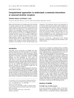
prion disease (Fig. 1) [4,5]. This occurs without an
alteration in the amino-acid sequence of the proteins.
Ab is a 40–43 amino acid peptide, which, in AD, self-
assembles into toxic oligomers and fibrils that accumu-
late in the brain, forming plaques and deposits in the
walls of meningocephalic vessels [6,7]. The same pep-
tide can be detected in most physiological fluids, such
as serum or cerebrospinal fluid, where it is called sol-
uble Ab (sAb) [7]. PrP
C
(C-cellular) is a 209 amino
acid, cell membrane anchored protein expressed at
highest levels by neurons and follicular dendritic cells
of the immune system. In the setting of prion disease,
this protein undergoes a transformation to toxic PrP
Sc
(Sc-scrapie) [8–10]. Fibrillar A b and PrP
Sc
have a high
b-sheet content which renders them insoluble, resistant
to proteolytic degradation and toxic to neurons. Neu-
rological symptoms in AD and prion disease are
directly related to loss of neurons and synaptic connec-
tions. Oligomeric and fibrillar A b can be directly
neurotoxic and ⁄ or can promote formation of neuro-
fibrillary tangles [7]. Both fibrillar Ab and PrP
Sc
are
capable of forming amyloid deposits. The presence of
amyloid deposits is necessary for making the diagnosis
of AD [11,12]. Abundant amyloid deposits composed
of PrP
Sc
(full length or fragments) are a neuropatho-
logical hallmark of variant Creutzfeld–Jakob disease
(vCJD), Gerstmann–Stra
¨
usler–Scheinker syndrome
(GSS), and kuru [13]. They are also present in 10% of
sporadic CJD (sCJD) cases [9].
A number of proteins may actively promote the con-
formational transformation of these disease specific pro-
teins and stabilize their abnormal structure. Examples
of such proteins in AD include apolipoprotein E (apoE),
especially its E
4
isoform [13,14], a1-antichymotrypsin
(ACT) [15] or C1q complement factor [16,17] (Fig. 1).
In their presence, the formation of Ab fibrils in a
solution of sAb is much more efficient [13,15]. These
‘pathological chaperone’ proteins have been found
histologically and biochemically in association with
fibrillar Ab deposits [18] but not in preamyloid aggre-
gates, which are not associated with neuronal loss [19].
Similarly, in prion disease, extensive data points toward
the existence of an unidentified protein X actively
involved in the conversion of PrP
C
into PrP
Sc
[20].
AD and prion diseases exist as sporadic and inher-
ited illnesses. In addition, prion disease can be trans-
mitted from one subject to another. In experimental
model settings, some evidence also exists for the infec-
tivity of AD [21,22]. An important event in the patho-
mechanism of AD is thought to be reaching a critical
concentration of sAb and ⁄ or chaperone proteins in the
brain, at which point the conformational change
occurs [23]. This leads to the formation of Ab aggre-
gates, initiating a neurodegenerative cascade. In
sporadic AD, this occurs due to an age-associated
overproduction of Ab, impaired clearance from the
brain, and ⁄ or influx into the central nervous system
(CNS) of sAb circulating in the serum [24]. Inherited
forms of AD are associated with various genetic
defects, resulting in overproduction of total sAb,or
more fibrillogenic Ab 1–42 species [25].
Sporadic prionoses like sCJD are thought to result
from the spontaneous conversion of PrP
C
into PrP
Sc
[26]. The mechanisms that stabilize PrP
C
structure are
largely unknown but, once PrP
Sc
assumes its patholo-
gical conformation, it can bind to PrP
C
and induce a
conformation change. This starts a self-perpetuating
vicious cycle allowing PrP
Sc
to replicate without DNA,
using the host cell’s PrP
C
as a template [9,26]. Most
inherited prionoses such as GSS or inherited forms of
CJD are the result of a point mutation in PrP
C
that
increases the propensity for it to assume an abnormal
conformation. Virtually all genetic defects implicated
in familial forms of AD and prionoses are inherited in
an autosomal dominant fashion. Unlike AD, prionoses
can be easily transmitted between subjects of the same
Protofibrils
Fibrils
Increased
Aggregated
Toxic
CONFORMATIONAL DISORDERS
PrP
C
PrP
Sc
Mainly
Random Coil
Monomers
Non-Toxic
Alzheimer’s Disease
A
β
Plaque
Neurofibrillary
Tangle
Prionoses
Neuronal loss
Spongiform changes
Pathological
Chaperones
Metals
Fig. 1. Conversion of sAb peptide or PrP
C
to their pathological
b-sheet conformers is a key step in the pathogenesis of AD and pri-
onoses, respectively. In AD, these b-sheet rich structures consist
of oligomers, protofibrils and fibrils that form plaques within the
brain parenchyma or deposit in the cerebrovasculature. A compar-
able entity in prion diseases consists of the proteinase K resistant
scrapie form of the prion protein (PrP
Sc
) that, in certain prion dis-
eases, fibrillizes and deposits as plaques within the brain. This pro-
cess is facilitated by various pathological chaperones as well as
several metals. The aim of most therapeutic interventions for these
conformational disorders is to reduce the amount of the substrate
(sAb, PrP
C
) and ⁄ or its availability for this structural alteration;
interfere with the conversion either directly or indirectly (via the
pathological chaperones or metals); and promote removal of the
disease-associated conformers.
T. Wisniewski and E. M Sigurdsson Therapy for prion disease and AD
FEBS Journal 274 (2007) 3784–3798 ª 2007 The Authors Journal compilation ª 2007 FEBS 3785
species. Transmissibility of prionoses between different
species is generally ineffective due to differences in the
PrP sequence. The phenomenon protecting one species
from acquiring a prion disease from another is called
‘the species barrier’. Therefore, scrapie (a prionosis
affecting sheep) is not transmissible to humans. The
species barrier does not provide absolute protection;
therefore, transmission of scrapie to cattle and trans-
mission of bovine spongiform encephalopathy (BSE)
from cattle to humans results in the emergence of
vCJD. In transmissible prionoses, exogenous PrP
Sc
present in the inoculum is responsible for the conform-
ational transformation of host PrP
C
. Upon entering an
organism, PrP
Sc
initially replicates within the lym-
phoreticular organs, including the spleen, lymph nodes
and tonsils, for months to years prior to neuroinvasion
and the onset of neurological symptoms. Therefore,
infected but asymptomatic individuals are a reservoir
of infectious material. This occurs because PrP
C
is
expressed by follicular dendritic cells and other lym-
phoid cells [27]. Accumulation of PrP
Sc
in the lympha-
tic organs of presymptomatic humans infected with
BSE has been demonstrated by immunohistochemistry
[28]. PrP
Sc
replication is possible because it does not
elicit an immune response [29]. This is related to the
inability of the immune system to distinguish between
PrP
C
and PrP
Sc
.
Vaccination approaches for AD
Vaccination was the first treatment approach demon-
strated to have genuine impact on disease process, at
least in animal models of AD. Vaccination of AD
transgenic (Tg) mice with Ab1–42 or Ab homologous
peptides coinjected with Freund’s adjuvant prevented
the formation of Ab deposition and, as a consequence,
eliminated the behavioral impairments that are related
to Ab deposition [30–35]. Similar effects on Ab load
and behavior have been demonstrated in AD Tg mice
by peripheral injections of anti-Ab monoclonal serum
indicating that the therapeutic effect of the vaccine
is based primarily on eliciting a humoral response
[36,37]. The striking biological effect of the vaccine in
preclinical testing and the apparent lack of side-effects
in AD Tg mice encouraged Elan ⁄ Wyeth to launch clin-
ical trials with a vaccine designated as AN1792 which
contained preaggregated Ab1–42 and QS21 as an adju-
vant. This type of vaccine design was aimed to induce
a strong cell-mediated immune response because QS21
is known to be a strong inducer of Th-1 lymphocytes
[38]. The initial safety testing of AN1792 in phase I of
the trial did not demonstrate any adverse effects. The
phase II of the trial was prematurely terminated when
6% of vaccinated patients manifested symptoms of
acute meningoencephalitis [38,39]. An autopsy per-
formed on one of the affected patients revealed an
extensive cytotoxic T-cell reaction surrounding some
cerebral vessels; however, analysis of the Ab load in
the brain cortex suggested that Ab clearance had
occurred [40]. It appeared that the immune reaction
triggered by AN1792 was a double-edge sword, where
the benefits of a humoral response against Ab were
overshadowed in some individuals by uncontrolled
cytotoxicity [41]. Not all patients who received
AN1792 responded with antibody production. The
majority mounted a humoral response and showed a
modest but statistically significant cognitive benefit
demonstrated as an improvement on some cognitive
testing scales compared to baseline and a slowed rate
of disease progression compared to patients who did
not form antibodies [42]. The follow-up data from the
‘Zurich’s cohort’, who are a subset of the Elan ⁄ Wyeth
trial followed by Dr Nitsch’s group [42,43], indicated
that the vaccination approach may be beneficial for
human AD patients but that the concept of the vaccine
has to be redesigned.
It appears that a humoral response elicited by the
vaccine has at least two mechanisms of action and both
of these are thought to be involved in amyloid clearance
[44,45]. Conformational selective anti-Ab serum may
target Ab deposits in the brain [43] leading to their
disassembly [46,47] and elicit Fc mediated phagocytosis
by microglia cells. The second mechanism by which
anti-Ab serum likely prevents Ab deposition is the cre-
ation of a ‘peripheral sink’ effect, where the removal of
excess sAb circulating in the blood stream leads to sAb
being drawn out from the brain [31,34, 47,48]. This per-
ipheral sink mechanism is likely to be the dominant
means of reducing Ab peptides in the brain.
The cause(s) for the toxicity in 6% of the Elan
trial patients are not entirely known; however, from
the available clinical and limited autopsy data, it is
thought that an excessive Th-1 cell-mediated response
within the brain was to blame [49]. The concept of a
redesigned AD vaccine puts emphasis on avoiding
this cell-mediated response in the following ways:
(a) avoiding stimulation of Th-1 lymphocytes so the
vaccine could potentially elicit a purely humoral res-
ponse; (b) using nontoxic and nonfibrillogenic Ab
homologous peptides, so that the immunogen can not
produce any direct toxicity; and (c) enhancing the
peripheral sink effect rather than central action.
Passive transfer of exogenous anti-Ab monoclonal
serum appears to be the easiest way to fulfill the goal of
providing anti-Ab serum without risk of uncontrolled
Th-1 mediated autoimmunity. AD Tg model mice
Therapy for prion disease and AD T. Wisniewski and E. M Sigurdsson
3786 FEBS Journal 274 (2007) 3784–3798 ª 2007 The Authors Journal compilation ª 2007 FEBS
treated this way had a significantly reduced Ab level and
demonstrated cognitive benefit [36,37]. The major draw-
backs of this approach are the high cost, limited half-life
of monoclonal antibodies (2–21 days depending on class
and isoform) and the potential for inducing serum sick-
ness with resultant complications such as renal failure
or lymphomas. Nevertheless, clinical trials for passive
immunization trials are underway. Alternative approa-
ches for passive immunization which are less likely to be
associated with toxicity, are use of Fv fragments or
mimetics of the active antibody binding site.
Another potential source of toxicity in association
with passive immunization is cerebral hemorrhage. The
mechanism of this hemorrhage is thought to be inflam-
mation in association with cerebral amyloid deposits
(congophilic amyloid angiopathy; CAA) that weakens
the blood vessel wall. Several reports have shown an
increase in microhemorrhages in different AD mouse
models following passive intraperitoneal immunization
with different monoclonal antibodies with high affinity
for Ab plaques and CAA [50–52]. The risk of micro-
hemorrhage following active immunization in animal
models has not been fully assessed. It has not been
a problem in our own active immunization studies
[34,35], but has been reported in one study [53].
Furthermore, the clinical trial data from the limited
number of autopsied cases suggests that vascular
amyloid was not being cleared and that hemorrhage
may have been increased [54–56]. In one of these
autopsies, numerous cortical bleeds, which are
typically rare in AD patients, were evident [55]. In
addition, the association of T lymphocytosis and
cuffing with the cerebral vessel Ab in these autopsies
suggests a potential role of CAA and an excessive
Th-1 response in the genesis of the inflammatory side-
effects [57]. This is an important issue because CAA is
present in virtually all AD cases, with approximately
20% of AD patients having ‘severe’ CAA [58].
Furthermore CAA is present in approximately 33% of
cognitively normal elderly, control populations [59–61].
Understanding the antigenic profile of Ab peptide,
allows engineering of modifications that favor a
humoral response and reduce the potential for a Th-1
mediated response. This approach has been termed
altered peptide ligands. Computer models have predic-
ted that Ab1–42 has one major antibody binding site
located on its N-terminus and two major T-cell epitopes
located at the central and C-terminal hydrophobic
regions encompassing residues 17–21 and 29–42,
respectively [62–64]. Therefore, their elimination or
modification provides a double gain by eliminating tox-
icity, as well as the potential for T-cell stimulation.
Sigurdsson et al. [34] immunized AD Tg mice with
K6Ab1–30[E
18
E
19
], a nontoxic Ab-homologous peptide,
where the first above mentioned T-cell epitope was
modified and the second removed. Polyamino acid
chains coupled to its N-terminus aimed to increase the
immunogenicity and solubility of the peptide. AD Tg
mice vaccinated with this peptide produced mainly an
IgM class antibodies and low or absent IgG titer. These
animals showed behavioral improvement and a partial
reduction of Ab deposits [34,35]. One of the advantages
of this design is that IgM, with a molecular mass of
900 kDa, does not penetrate the blood–brain barrier
(BBB) and therefore is unlikely to be associated with
any immune reaction in the brain. Like passive immun-
ization, this type of vaccine focuses its mechanism of
action on the peripheral sink. Furthermore, the IgM
response is reversible because it is T-cell independent;
hence memory T-cells that could maintain the immune
response are not generated. Therefore, this vaccine
method may potentially be safer than typical active
immunization.
Mucosal vaccination can be an alternative way to
achieve a primarily humoral response. This mechanism
is based on the presence of lymphocytes in the mucosa
of the nasal cavity and of the gastrointestinal tract. This
type of response produces primarily S-IgA antibodies
but, when the antigen is coadministrated with adjuvants
such as cholera toxin subunit B or heat-labile Escheri-
chia coli enterotoxin, significant IgG titer in the serum
may be achieved [65,66]. A marked reduction of Ab bur-
den in AD Tg mice immunized this way using Ab as an
antigen has been already demonstrated [66,67]. Interest-
ingly, this type of mucosal immunization has recently
been shown to be highly effective for prion infection
[68,69,70]. This promising approach requires further
exploration, especially using nonfibrillar and nontoxic
Ab homologous peptides as an antigen. Mucosal
immunization offers a great potential advantage in that
a more limited humoral immune response can be
obtained, with little or no cell-mediated immunity.
Inhibition of Ab fibrillization
Formation of Ab fibrils and deposition of Ab in the
brain parenchyma or in the brain’s vessels occurs in
the setting of increased local Ab peptide concentra-
tions [71]. Initially, conditions do not favor aggrega-
tion of fibrils; however, once a critical nucleus has
been formed, aggregation with fast kinetics is favored.
Any available monomer can then become entrapped in
an aggregate or fibril. Several compounds, such as
Congo red [71], anthracycline [73], rifampicin [74],
anionic sulphonates [75], or melatonin [76], can inter-
act with Ab and prevent its aggregation into fibrils
T. Wisniewski and E. M Sigurdsson Therapy for prion disease and AD
FEBS Journal 274 (2007) 3784–3798 ª 2007 The Authors Journal compilation ª 2007 FEBS 3787
in vitro, thereby reducing toxicity. It has been further
identified that certain nonfibrillogenic, Ab homologous
peptides can bind to Ab and break the formation of
b-sheet structure [77–80]. Therefore, these peptides
were termed b-sheet breakers. Several modifications
were used to extend serum half-life and increase BBB
permeability of these peptides. Permanne et al. [81],
using a BBB permeable five amino-acid long peptide
(iAb5), were able to demonstrate a reduction of Ab
load in AD Tg mice that received this peptide compared
with age-matched control group which received placebo.
Of interest, a similar concept of b-sheet breakers has
been shown to be applicable to prion disease [82].
Extensive evidence suggests that the most toxic forms
of Ab are oligomeric aggregates [83]. There is also evi-
dence implicating oligomeric aggregates in the medi-
ation of PrP
Sc
toxicity and infectivity [84,85]. Recently,
compounds and antibodies have been developed that
specifically target Ab oligomers [86–88]. Similar approa-
ches are being developed for prion oligomers.
Ab homologous peptides can aggregate and form
fibrils spontaneously in vitro; however, in vivo this pro-
cess appears more dependant on the presence of Ab
pathological chaperones. This group of proteins
promotes conformational transformation at certain
concentrations by increasing the b-sheet content of
these disease specific proteins and stabilizes their
abnormal structure [89,90]. Examples of such proteins
in AD include apoE, especially its E
4
isoform [18,91],
ACT [20] or C1q complement factor [21,22]. In
their presence, the formation of Ab fibrils in a solution
of sAb monomers becomes much more efficient
[18,20]. These ‘pathological chaperone’ proteins have
been found histologically and biochemically in associ-
ation with fibrillar Ab deposits [23,89,92,93] but not in
preamyloid aggregates that are not associated with
neuronal toxicity [24,94]. Inheritance of the apoE
4
isoform has been identified as the major identified
genetic risk factor for sporadic, late-onset AD [95] and
correlates with an earlier age of onset and greater Ab
deposition, in an allele-dose-dependent manner
[19,95,96]. In vitro, all apoE isoforms can propagate
the b-sheet content of Ab peptides promoting fibril
formation [92], with apoE
4
being the most efficient
[18]. The critical dependence of Ab deposition in
plaques on the presence of apoE has also been
confirmed in AD Tg APP
V717F
⁄ apoE
– ⁄ –
mice which
have a delayed onset of Ab deposition, a reduced Ab
load, and no fibrillar Ab deposits. Compared to
APP
V717F
⁄ apoE
+ ⁄ +
Tg mice, APP
V717F
⁄ apoE
+ ⁄ –
mice
demonstrate an intermediate level of pathology
[97–100]. Neutralization of the chaperoning effect of
apoE would therefore potentially have a mitigating
effect on Ab accumulation. ApoE hydrophobically
binds to the 12–28 amino acid sequence of Ab, form-
ing SDS insoluble complexes [101–103]. Ma et al. [104]
have demonstrated that a synthetic peptide homolog-
ous to 12–28 amino-acid sequence of Ab can be used
as a competitive inhibitor of the binding of full length
Ab to apoE, resulting in reduced fibril formation
in vitro and increased survival of cultured neurons.
The introduction of several modifications to Ab12–28
by replacing a valine for proline in position 18, making
this peptide nontoxic and nonfibrillogenic, as well as
end-protection by amidation and and acetylation of
the C- and N-termini, respectively, to increase serum
half-life, have allowed us to use this peptide therapeu-
tically in the APP
K670N ⁄ M671L
⁄ PS1
M146L
double Tg
mice model. Tg mice treated with Ab12–28P for
1 month demonstrated a 63.3% reduction in Ab load
in the cortex (P ¼ 0.0043) and a 59.5% (P ¼ 0.0087)
reduction in the hippocampus comparing to age-
matched control Tg mice that received placebo
[105,106]. The treated Tg mice also had a cognitive
benefit [105,106]. No antibodies against Ab were detec-
ted in sera of treated mice; therefore, the observed
therapeutic effect of Ab12–28P cannot be attributed to
an antibody clearance response. This experiment
demonstrates that compounds blocking the interaction
between Ab and its pathological chaperones may be
beneficial for treatment of Ab accumulation in AD
[14,105,106]. Whether similar approaches can be used
for prion disease remains to be determined.
Prion disease
Interest in prion disease has greatly increased subse-
quent to the emergence of BSE in England and the
resulting appearance of vCJD in human populations.
BSE arose from the feeding of cattle with prion con-
taminated meat and bone meal products, whereas
vCJD developed following entry of BSE into the
human food chain [107,108]. Since the original report
in 1995, a total of 201 probable or confirmed cases of
vCJD have been diagnosed, 165 in Great Britain, 21 in
France, four in Ireland, three in the USA, two in the
Netherlands and one each in Italy, Canada, Japan,
Saudi Arabia, Portugal and Spain. Most of the
patients from these countries resided in the UK during
a key exposure period of the UK population to the
BSE agent. It has proven difficult to predict the expec-
ted future numbers of vCJD. Mathematical analysis
has given a range from 1000 to approximately 136 000
individuals who will eventually develop the disease.
This broad range reflects a lack of knowledge regard-
ing the time of incubation and the number of patients
Therapy for prion disease and AD T. Wisniewski and E. M Sigurdsson
3788 FEBS Journal 274 (2007) 3784–3798 ª 2007 The Authors Journal compilation ª 2007 FEBS
who could be infected from a given dosage of BSE
agent. Because the vCJD agent is present at high levels
in the lymphatic tissue, screening for PrP
Sc
was per-
formed on sections from lymph nodes, tonsils, and
appendices archives in the UK. Three out of 12 674
randomly selected cases showed evidence of subclinical
infection, leading to a prediction that approximately
4000 vCJD further cases may occur in the UK [109].
However, there is much uncertainty about such a pre-
diction because it is not known whether all subclinical
infections will progress and also whether such screen-
ing of lymphoid tissue would capture all subclinical
cases. The initially predicted epidemic of vCJD does
not seem to be materializing because the number of
cases in the UK has declined from a peak of 28 in
2000 to five cases in 2006 [107]. A complicating factor
for estimating future numbers of vCJD is the docu-
mentation of several transfusion associated cases.
These occurred after incubation periods of 6–8 years.
One of these disease associated donations was made
more than 3 years before the donor became sympto-
matic, suggesting that vCJD can be transmitted from
silently infected individuals [110]. The estimated risk
for new cases of vCJD in other European countries
looks more optimistic. In the UK, 200 000 cases of
BSE were reported (it is estimated that four times this
number entered the food chain), compared to approxi-
mately 5600 BSE cases in other European countries
(with the highest numbers being 1590, 1030 and 986 in
Ireland, Portugal and Frances, respectively). This sug-
gests a significantly lower exposure of these popula-
tions to BSE prions. A few cases of BSE have also
been reported in other parts of the world, such as
Japan, the USA and Canada.
Of greater concern in North America is chronic wast-
ing disease (CWD). This disease is now endemic in
Colorado, Wyoming and Nebraska and continues to
spread to other parts in the USA, initially in the Mid-
west but now detected as far East as New York State
[111,112]. Most vulnerable to CWD infection are white
tailed deer and the disease is now found in areas with a
large population of these animals, which indicates that
its prevalence can be expected to increase substantially
in the future. The occurrence of CJD among three
young deer hunters from this same region raised the spe-
culation of transmission of the CWD to humans [113].
However, autopsy of these three subjects did not reveal
the extensive amyloidosis characteristic of vCJD and
CWD [114]. However like BSE, CWD is transmissible
to nonhuman primates and transgenic mice expressing
human PrP
C
[115,116]. Therefore, the possibility of such
transmission needs to be closely monitored. CWD is
similar to BSE in that the peripheral titers of the prion
agent are high. PrP
Sc
has been detected in both muscle
and saliva of CWD infected deer [117,118].
Vaccination as a therapeutic approach
for prionoses
The prion protein is a self-antigen; hence, prion infec-
tion is not known to elicit a classical immune response.
In fact, the immune system is involved in the peri-
pheral replication of the prion agent and its ultimate
access to the CNS [29,68]. This involvement is further
supported by the observation that immune suppression
with, for example, splenectomy or immunosuppressive
drugs, increases the incubation period. This interval,
during which time the prion agent replicates peripher-
ally, without producing any symptoms, is quite long,
lasting many months in experimental animals and up
to 56 years in documented human cases associated
with cannibalistic exposure to the prion agent [119].
Lymphatic organs such as the spleen, tonsils, lymph
nodes or gut associated lymphoid tissue contain high
concentrations of PrP
Sc
long before PrP
Sc
replication
starts in the brain [27,120,121]. Cells found to be par-
ticularly important for peripheral PrP
Sc
replication are
the follicular dendritic cells (DC) and the migratory
bone-marrow derived DC [121,122]. DC from infected
animals are capable of spreading the disease [122]. An
emerging therapeutic approach for prion infection is
immunomodulation [68,70,123].
Currently, there is no treatment that would arrest
and ⁄ or reverse progression of prion disease in non-
experimental settings, although many approaches have
been tried [124]. Partly due to the success in AD
models discussed above, similar experiments with
anti-PrP serum were initiated in prion infectivity cul-
ture models as well as active and passive immuniza-
tion studies in rodent models. Earlier in vivo studies
showed that infection with a slow strain of PrP
Sc
blocked expression of a more virulent fast strain of
PrP, mimicking vaccination with a live attenuated
organism [125]. In tissue culture studies, anti-PrP
serum and antigen binding fragments directed
against PrP were shown to inhibit prion replication
[126–128]. Although we first demonstrated that active
immunization with recombinant PrP delayed the onset
of prion disease in wild-type mice, the therapeutic
effect was relatively modest and, eventually, all the
mice succumbed to the disease [129]. This limited
therapeutic effect may be explained by the observa-
tion that antibodies generated against prokaryotic
PrP often do not have a high affinity towards PrP
C
[130], although, in our studies, the increase in the
incubation period correlated well with the antibody
T. Wisniewski and E. M Sigurdsson Therapy for prion disease and AD
FEBS Journal 274 (2007) 3784–3798 ª 2007 The Authors Journal compilation ª 2007 FEBS 3789
titers against PrP
C
. Our follow-up passive anti-PrP
immunization study confirmed the importance of the
humoral response, showing that anti-PrP serum is
able to prolong the incubation period [131]. Subse-
quently, other investigators, using a much higher anti-
body dosage, were able to completely prevent disease
onset in mice exposed to PrP
Sc
provided that passive
immunization was initiated within 1 month of expo-
sure [132]. This type of approach could be used
immediately following accidental exposure in humans
to prevent future infection. However, passive immun-
ization has not been found to be effective closer to
the clinically symptomatic stages of prion infection.
Also, passive immunization would be an approach
that is too costly for animal prion diseases.
In the development of immunotherapeutic approa-
ches targeting a self-antigen, designing a vaccine avoid-
ing auto-immune related toxicity is a major concern.
The emerging data from AD targeting immunization is
that toxicity is due to excessive cell-mediated immunity
within the CNS, whereas the therapeutic response is
linked to humoral immunity. In addition, toxicity
could be partially related to the immunogen and ⁄ or to
the adjuvant used; in the human AD vaccination trial,
fibrillar Ab1–42 was used as an immunogen. This pep-
tide is well characterized to be toxic. Hence, we have
been promoting the use of nonamyloidogenic deriva-
tives as immunogens for protein conformational disor-
ders, including AD and prion disease [31,34,38]. How
significant an issue direct toxicity of the immunogen
may be for prion vaccination remains unclear. Unlike
the Ab peptide used for vaccination in AD models,
direct application of recombinant PrP has not been
shown to be toxic. However, this issue has not been
investigated as thoroughly as in the Alzheimer’s field
and remains controversial. Several lines of evidence
suggest that intracellular accumulations of PrP
Sc
pro-
mote neurodegeneration [133].
A potential ideal means of using immunomodulation
to prevent prion infection is by mucosal immunization.
One important reason for this is that the gut is the
major route of entry for many prion diseases such as
CWD, BSE and vCJD. Furthermore, mucosal immun-
ization can be designed to induce primarily a humoral
immune response, avoiding the cell-mediated toxicity
that was seen in the human AD vaccine trial. In addi-
tion, mucosal vaccination has the advantage that it is
unlikely to induce significant immune response within
the brain. Although it has been shown that reduced
levels or absence of CNS PrP
C
by, for example, condi-
tional ablation by genetic manipulation of neuronal
PrP
C
[134] can prevent clinical prion infection, it is
likely that the immunological targeting of neuronal
PrP would be associated with inflammatory toxicity.
Recently, we have been developing prion vaccines that
target gut associated tissue, the main site of entry of
the prion agent. One of our approaches is to express
PrP in attenuated Salmonella strains as a live vector
for oral vaccination, which has resulted in prevention
or significant delay of prion disease in mice [69]. Live
attenuated strains of Salmonella enterica have been
used for many years as vaccines against salmonellosis
and as a delivery system for the construction of multi-
valent vaccines with a broad application in human and
veterinary medicine [135]. A main advantage for this
system is that the safety of human administration of
live attenuated Salmonella has been extensively con-
firmed in humans and animals [136,137]. Ruminants
and other veterinary species can be effectively immun-
ized by the oral route using attenuated Salmonella,to
induce humoral mucosal responses [138,139]. We are
currently exploring ways to increase the efficacy even
further. In these studies, the mucosal IgA anti-PrP titer
correlates well with the delay or prevention of prion
infection, further supporting the importance of the
humoral response for the therapeutic effect. Salmonella
target M-cells, antigen sampling cells in the intestines,
which may also be important for uptake of PrP
Sc
[27,68,121]. Hence, this approach is more targeted
than prior vaccination studies, likely explaining the
improved efficacy. By exploring other strains of attenu-
ated Salmonella, using different bacteria or oral
adjuvants, and ⁄ or by altering the expression levels or
sequence of the PrP antigen, it is likely that the
percentage of uninfected animals can be improved.
Our recent work utilizing this approach indicates that
complete protection to clinical prion infection via an
oral route is possible. Overall, this approach holds
great promise as an inexpensive prophylactic immuno-
therapy to prevent the spread of prion disease, partic-
ularly in animals at risk and perhaps eventually in
certain high risk human populations.
Metal chelation for prion and AD
Metal chelation is emerging as an important therapeu-
tic approach for AD, which is currently in clinical trial
[140,141]. This approach for AD is reviewed elsewhere
in this minireview series. Importantly, modulation of
metal levels, in particular copper, has been shown to
be important for the conversion of PrP
C
to PrP
Sc
,
highlighting another similarity between AD and prion
diseases [10]. Copper binding is thought to be part of
the normal function of PrP
C
[142–144]. The binding of
copper to PrP
C
gives the complex antioxidant activity
[145,146]; hence, it has been suggested that the reduced
Therapy for prion disease and AD T. Wisniewski and E. M Sigurdsson
3790 FEBS Journal 274 (2007) 3784–3798 ª 2007 The Authors Journal compilation ª 2007 FEBS
copper binding of PrP
Sc
with a consequent reduction
of antioxidant activity is part of the pathogenesis of
prion disease [147]. This hypothesis has been supported
by the finding that copper is reduced up to 50% in the
brains of sporadic CJD patients [148]. How copper
binding influences the PrP
C
to PrP
Sc
conversion is
complex [10,149]. We were the first to show that, sim-
ilar to studies in AD Tg models, metal chelation can
be used therapeutically [150] in prion infection. Our
studies indicated that penicillamine, a copper chelator,
prolongs the incubation period of scrapie in mice
[150]. Consistent with this observation, the presence of
copper has also been shown to stabilize the PrP
Sc
con-
formation using preformed fibrils [151–158], as well as
to induce aggregation of the prion peptide 106–126
[159]. Some tissue culture studies of prion infection
have also suggested that copper chelators are suitable
candidates for antiprion drugs [160]. However, there
are conflicting reports indicating that the interaction
between copper and PrP is likely to be quite complex.
For example, copper has been shown to inhibit the
in vitro conversion of recombinant PrP into amyloid
fibrils but, also in contrast, to enhance the protein-
ase K resistance of preformed fibrils [157]. These
findings indicate that copper may have a dual and
opposite effect on prion propagation. It may both
inhibit prion replication and prevent clearance of
potentially infectious forms of the prion protein.
Furthermore, copper treatment has also been shown to
inhibit PrP
Sc
amplification in reactions where brain
derived PrP
C
was used as a seed [161], as well as delay-
ing the onset of clinical disease in scrapie infected
hamsters [162]. In addition, it has been shown that
physiological levels of copper promote internalization
of PrP
C
[163]. The interaction between PrP
C
and cop-
per was found to be the overriding factor in stimula-
ting the internalization response with other metals
showing no effect. The decrease in detectable levels of
PrP
C
at the cell surface following copper treatment
was found to be the result of internalization rather
than loss into the surrounding environment [163].
Such internalization would limit the exposure of
PrP
C
to conversion from exogenous PrP
Sc
; however,
because cytoplasmic forms of PrP have been linked to
neurodegeneration [133], increased internalization
could also be deleterious in some settings. Copper has
also been shown to have immunomodulatory effects
[164] and, as discussed earlier, the immune system can
have profound effects on prion infection. Hence, it
appears that the deleterious or beneficial role of copper
in prion infection might vary depending on which
function predominates under the distinct experimental
conditions being used. Nevertheless, it is clear that a
greater understanding of the role of metal binding in
prion infection presents a therapeutic opportunity.
Conclusions
Immunization appears to be an effective therapeutic
method for prevention of Ab deposition and cognitive
decline in AD, provided that cell-mediated auto-
immune toxicity can be avoided. The second genera-
tion AD vaccines, which are under development, are
based on nontoxic and nonfibrillar Ab homologous
peptides that are modified to eliminate the potential
for inducing cellular immunity, and elicit primarily a
humoral response. Other related approaches include
direct administration of antibodies that target Ab.
These interventions would likely favor a peripheral
sink effect, clearing soluble Ab from the blood stream
and inducing efflux of Ab from the brain. Additional
potentially synergistic therapeutic approaches for AD
would include blocking the interaction of Ab with its
‘pathological chaperones’ such as apoE, as well as use
of b-sheet breaker compounds. Immunization approa-
ches could be used for sporadic AD, familial AD, and
AD associated with Down’s syndrome. The effective-
ness of treatment would depend on its initiation early
in the disease course. Therefore, such a treatment
needs to coincide with the development of a procedure
for the detection and monitoring of Ab deposits.
Both active and passive immunization appear to be
effective in prevention of prion infections in animal
models. Further studies are needed to develop specific
protocols applicable for human use. Active immuniza-
tion, using especially mucosal immunization could be
used to prevent spread of BSE through the oral
route, whereas passive immunization protocols would
be more appropriate for subjects accidentally infected
with prion contaminated material (e.g. blood transfu-
sion or organ transplant). Effective immunization for
prion infections works through prevention of entry of
PrP
Sc
via the gut and ⁄ or neutralization of PrP
Sc
replicating in the peripheral lymphoreticular system.
Metal chelation is another promising therapeutic
approach for AD, which is currently undergoing
clinical trials. Similar approaches are just emerging
for prion diseases. However, a greater understanding
of the role of copper and other metals in the PrP
C
to
PrP
Sc
conversion is needed before this therapeutic
strategy can be effectively harnessed for prion infection.
Acknowledgements
This manuscript is supported by NIH grants: AG15408,
AG20245, AG20197 and the Alzheimer’s Association.
T. Wisniewski and E. M Sigurdsson Therapy for prion disease and AD
FEBS Journal 274 (2007) 3784–3798 ª 2007 The Authors Journal compilation ª 2007 FEBS 3791
References
1 Prusiner SB (2001) Neurodegenerative diseases and pri-
ons. N Eng J Med 3444, 1516–1526.
2 Wisniewski T, Sigurdsson EM, Aucouturier P & Fran-
gione B (2001) Conformation as a therapeutic target in
the prionoses and other neurodegenerative conditions.
In Molecular and Cellular Pathology in Prion Disease
(Baker HF, ed.), pp. 223–236. Humana Press, Totowa,
NJ.
3 DeArmond SJ (1993) Alzheimer’s disease and Creutz-
feldt-Jakob disease: overlap of pathogenic mechanisms.
Curr Opin Neurol 6, 872–881.
4 Sadowski M & Wisniewski T (2004) Vaccines for con-
formational disorders. Exp Rev Vaccines 3, 89–100.
5 Sigurdsson EM (2006) Immunotherapy for conforma-
tional disorders. Curr Pharm Des 12, 2569–2585.
6 Small DH & Cappai R (2006) Alois Alzheimer and
Alzheimer’s disease: a centennial perspective. J Neuro-
chem 99, 708–710.
7 Walsh DM & Selkoe DJ (2007) Abeta oligomers ) a
decade of discovery. J Neurochem 101, 1172–1184.
8 DeArmond SJ & Prusiner SB (1995) Etiology and
pathogenesis of prion diseases. Am J Pathol 146,
785–811.
9 Sadowski M, Verma A & Wisniewski T, (2007) Prion
diseases. In Neurology in Clinical Practice, 5th edn.
Butterworth-Heinemann., Philadelphia, PA.
10 Caughey B & Baron GS (2006) Prions and their part-
ners in crime. Nature 443, 803–810.
11 Mirra SS, Gearing M & Nash F (1997) Neuropatholo-
gic assessment of Alzheimer’s disease. Neurology 49,
S14–S16.
12 Wisniewski HM, Bancher C, Barcikowska M, Wen GY
& Currie J (1989) Spectrum of morphological appear-
ance of amyloid deposits in AD. Acta Neuropathol 78,
337–347.
13 Wisniewski T, Castan
˜
o EM, Golabek AA, Vogel T &
Frangione B (1994) Acceleration of Alzheimer’s fibril
formation by apolipoprotein E in vitro. Am J Pathol
145, 1030–1035.
14 Sadowski M & Wisniewski T (2006) Apolipoproteins in
different amyloidoses. In Protein Misfolding, Aggrega-
tion and Conformational Diseases (Uversky VN, ed.).
Springer, New York, NY.
15 Ma J, Yee A, Brewer HB Jr, Das S & Potter H (1994)
Amyloid-associated proteins alpha 1-antichymotrypsin
and apolipoprotein E promote assembly of Alzheimer
beta-protein into filaments. Nature 372, 92–94.
16 Johnson LV, Leitner WP, Rivest AJ, Staples MK, Rad-
eke MJ & Anderson DH (2002) The Alzheimer’s A
beta-peptide is deposited at sites of complement activa-
tion in pathologic deposits associated with aging and
age-related macular degeneration. Proc Natl Acad Sci
USA 99, 11830–11835.
17 Boyett KW, DiCarlo G, Jantzen PT, Jackson J,
O’Leary C, Wilcock D, Morgan D & Gordon MN
(2003) Increased fibrillar beta-amyloid in response to
human C1q injections into hippocampus and cortex of
APP+PS1 transgenic mice. Neurochem Res 28, 83–93.
18 Wisniewski T, Lalowski M, Golabek AA, Vogel T &
Frangione B (1995) Is Alzheimer’s disease an apolipo-
protein E amyloidosis? Lancet 345, 956–958.
19 Wisniewski HM, Sadowski M, Jakubowska-Sadowska
K, Tarnawski M & Wegiel J (1998) Diffuse, lake-like
amyloid-beta deposits in the parvopyramidal layer of
the presubiculum in Alzheimer disease. J Neuropathol
Exp Neurol 57, 674–683.
20 Telling GC, Haga T, Torchia M, Tremblay P,
DeArmond SJ & Prusiner SB (1996) Interactions
between wild-type and mutant prion proteins modu-
late neurodegeneration transgenic mice. Genes Dev 10,
1736–1750.
21 Meyer-Luehmann M, Coomaraswamy J, Bolmont T,
Kaeser S, Schaefer C, Kilger E, Neuenschwander A,
Abramowski D, Frey P, Jaton AL et al. (2006) Exogen-
ous induction of cerebral beta-amyloidogenesis is gov-
erned by agent and host. Science 313, 1781–1784.
22 Walker LC, LeVine H III, Mattson MP & Jucker M
(2006) Inducible proteopathies. Trends Neurosci 29,
438–443.
23 Wisniewski T, Ghiso J & Frangione B (1994) Alzhei-
mer’s disease and soluble Ab. Neurobiol Aging 15,
143–152.
24 Shibata M, Yamada S, Kumar S, Calero M, Bading J,
Frangione B, Holtzman D, Miller CA, Strickland DK,
Ghiso J et al. (2000) Clearance of Alzheimer’s amyloid-
B 1–40 peptide from brain by LDL receptor-related
protein-1 at the blood–brain barrier. J Clin Invest 106,
1489–1499.
25 Hardy J (2006) A hundred years of Alzheimer’s disease
research. Neuron 52, 3–13.
26 Beekes M (2007) Prions and prion diseases. FEBS J
274, 575.
27 Beekes M & McBride PA (2007) The spread of prions
through the body in naturally acquired transmissible
spongiform encephalopathies. FEBS J 274, 588–605.
28 Hilton DA, Ghani AC, Conyers L, Edwards P,
McCardle L, Penney M, Ritchie D & Ironside JW
(2002) Accumulation of prion protein in tonsil and
appendix: review of tissue samples. Br Med J 325,
633–634.
29 Aucouturier P, Carp RI, Carnaud C & Wisniewski T
(2000) Prion diseases and the immune system. Clin
Immunol 96, 79–85.
30 Schenk D, Barbour R, Dunn W, Gordon G, Grajeda
H, Guido T, Hu K, Huang J, Johnson-Wood K, Khan
K et al. (1999) Immunization with amyloid-B attenu-
ates Alzheimer disease-like pathology in the PDAPP
mice. Nature 400, 173–177.
Therapy for prion disease and AD T. Wisniewski and E. M Sigurdsson
3792 FEBS Journal 274 (2007) 3784–3798 ª 2007 The Authors Journal compilation ª 2007 FEBS
31 Sigurdsson EM, Scholtzova H, Mehta P & Frangione
B & Wisniewski T (2001) Immunization with a
non-toxic ⁄ non-fibrillar amyloid-B homologous peptide
reduces Alzheimer’s disease associated pathology in
transgenic mice. Am J Pathol 159, 439–447.
32 Morgan D, Diamond DM, Gottschall PE, Ugen KE,
Dickey C, Hardy J, Duff K, Jantzen P, DiCarlo G,
Wilcock D et al. (2001) Ab peptide vaccination pre-
vents memory loss in an animal model of Alzheimer’s
disease. Nature 408, 982–985.
33 Janus C, Pearson J, McLaurin J, Mathews PM, Jiang
Y, Schmidt SD, Chishti MA, Horne P, Heslin D,
French J et al. (2000) Ab peptide immunization reduces
behavioural impairment and plaques in a model of
Alzheimer’s disease. Nature 408, 979–982.
34 Sigurdsson EM, Knudsen EL, Asuni A, Sage D, Goni
F, Quartermain D, Frangione B & Wisniewski T (2004)
An attenuated immune response is sufficient to enhance
cognition in an Alzheimer’s disease mouse model
immunized with amyloid-B derivatives. J Neurosci 24,
6277–6282.
35 Asuni A, Boutajangout A, Scholtzova H, Knudsen E,
Li Y, Quartermain D, Frangione B, Wisniewski T &
Sigurdsson EM (2006) A b derivative vaccination in
alum adjuvant prevents amyloid deposition and does
not cause brain microhemorrhages in Alzheimer’s
model mice. Eur J Neurosci 24, 2530–2542.
36 Bard F, Cannon C, Barbour R, Burke RL, Games D,
Grajeda H, Guido T, Hu K, Huang J, Johnson-Wood
K et al. (2000) Peripherally administered antibodies
against amyloid beta-peptide enter the central nervous
system and reduce pathology in a mouse model of
alzheimer disease. Nat Med 6, 916–919.
37 DeMattos RB, Bales KR, Cummins DJ, Dodart JC, Paul
SM & Holtzman DM (2001) Peripheral anti-A beta anti-
body alters CNS and plasma A beta clearance and decrea-
ses brain A beta burden in a mouse model of Alzheimer’s
disease. Proc Natl Acad Sci USA 98, 8850–8855.
38 Wisniewski T & Frangione B (2005) Immunological
and anti-chaperone therapeutic approaches for Alzhei-
mer’s disease. Brain Pathol 15, 72–77.
39 Wisniewski T (2005) Practice point commentary on
‘Clinical effects of Ab immunization (AN1792) in
patients with AD in an interupted trial. Nat Clin Pract
Neurol 1, 84–85.
40 Nicoll JAR, Wilkinson D, Holmes C, Steart P, Mark-
ham H & Weller RO (2003) Neuropathology of human
Alzheimer disease after immunization with amyloid-
beta peptide: a case report. Nat Med 9, 448–452.
41 Sadowski M & Wisniewski T (2007) Disease modifying
approaches for Alzheimer’s pathology. Curr Pharm Des
13, 1943–1954.
42 Hock C, Konietzko U, Straffer JR, Tracy J, Signorell A,
Muller-Tillmanns B, Lemke U, Henke K, Moritz E,
Garcia E et al. (2003) Antibodies against B-amyloid
slow cognitive decline in Alzheimer’s disease. Neuron
38, 547–554.
43 Hock C, Konietzko U, Paspassotiropoulos A, Wollmer A,
Streffer J, von Rotz RC, Davey G, Moritz E & Nitsch
RM (2002) Generation of antibodies specific for
B-amyloid by vaccination of patients with Alzheimer
disease.
Nat Med 8, 1270–1276.
44 Golde TE (2006) Disease modifying therapy for AD?
J Neurochem 99, 689–707.
45 Kennedy GJ, Golde TE, Tariot PN & Cummings JL
(2007) Amyloid-based interventions in Alzheimer’s dis-
ease. CNS Spectr 12, 1–14.
46 Bacskai BJ, Kajdasz ST, Christie RH, Carter C, Games
D, Seubert P, Schenk D & Hyman BT (2001) Imaging
of amyloid-B deposits in brains of living mice permits
direct observation of clearance of plaques with immu-
notherapy. Nat Med 7, 369–372.
47 Solomon B, Koppel R, Frankel D & Hanan-Aharon E
(1997) Disaggregation of Alzheimer beta-amyloid by
site-directed mAb. Proc Natl Acad Sci USA 94, 4109–
4112.
48 Sigurdsson EM, Wisniewski T & Frangione B (2002) A
safer vaccine for Alzheimer’s disease? Neurobiol Aging
23, 1001–1008.
49 Robinson SR, Bishop GM, Lee HG & Munch G
(2004) Lessons from the AN 1792 Alzheimer vaccine:
lest we forget., Neurobiol Aging 25, 609–615.
50 Pfeifer M, Boncristiano S, Bondolfi L, Stalder A, Del-
ler T, Staufenbiel M, Mathews PM & Jucker M (2002)
Cerebral hemorrhage after passive anti-Ab immuno-
therapy. Science 298, 1379–1380.
51 Wilcock DM, Rojiani A, Rosenthal A, Subbarao S,
Freeman MJ, Gordon MN & Morgan D (2004) Passive
immunization against Abeta in aged APP-transgenic
mice reverses cognitive deficits and depletes parenchymal
amyloid deposits in spite of increased vascular amyloid
and microhemorrhage. J Neuroinflammation 1, 24.
52 Racke MM, Boone LI, Hepburn DL, Parsadanian M,
Bryan MT, Ness DK, Piroozi KS, Jordan WH, Brown
DD, Hoffman WP et al. (2005) Exacerbation of cereb-
ral amyloid angiopathy-associated microhemorrhages
in amyloid precursor protein transgenic mice by immu-
notherapy is dependent on antibody recognition of
deposited forms of amyoid beta. J Neurosci 25, 629–
636.
53 Wilcock DM, Jantzen PT, Li Q, Morgan D & Gordon
MN (2007) Amyloid-beta vaccination, but not nitro-
nonsteroidal anti-inflammatory drug treatment, increa-
ses vascular amyloid and microhemorrhage while both
reduce parenchymal amyloid, Neuroscience 144, 950–
960.
54 Nicoll JA, Wilkinson D, Holmes C, Steart P, Markham
H & Weller RO (2005) Neuropathology of human Alz-
heimer disease after immunization with amyloid-beta
peptide: a case report. Nat Med 9, 448–452.
T. Wisniewski and E. M Sigurdsson Therapy for prion disease and AD
FEBS Journal 274 (2007) 3784–3798 ª 2007 The Authors Journal compilation ª 2007 FEBS 3793
55 Ferrer I, Boada RM, Sanchez Guerra ML, Rey MJ &
Costa-Jussa F (2004) Neuropathology and pathogenesis
of encephalitis following amyloid-beta immunization in
Alzheimer’s disease. Brain Pathol 14, 11–20.
56 Masliah E, Hansen L, Adame A, Crews L, Bard F, Lee
C, Seubert P, Games D, Kirby L & Schenk D (2005)
Ab vaccination effects on plaque pathology in the
absence of encephalitis in Alzheimer disease. Neurology
64, 129–131.
57 Gandy S & Walker L (2004) Toward modeling hem-
orrhagic and encephalitic complications of Alzheimer
amyloid-B vaccination in nonhuman primates. Curr
Opin Immunol 16, 607–615.
58 Jellinger KA (2002) Alzheimer disease and cerebro-
vascular pathology: an update. J Neural Transm 109,
813–836.
59 Jellinger KA & Attems J (2005) Prevalence and patho-
genic role of cerebrovascular lesions in Alzheimer dis-
ease. J Neurol Sci 229–230, 37–41.
60 Fernando MS & Ince PG (2004) Vascular pathologies
and cognition in a population-based cohort of elderly
people. J Neurol Sci 226, 13–17.
61 Zhang-Nunes SX, Maat-Schieman ML, Van Duinen
SG, Roos RA, Frosch MP & Greenberg SM (2006)
The cerebral beta-amyloid angiopathies: hereditary and
sporadic. Brain Pathol 16, 30–39.
62 Jameson BA & Wolf H (1988) The antigenic index: a
novel algorithm for predicting antigenic determinants.
Comput Appl Biosci 4, 181–186.
63 Singh H & Raghava GP (2001) ProPred: prediction of
HLA-DR binding sites. Bioinformatics 17, 1236–1237.
64 Singh H & Raghava GP (2003) ProPred1: prediction of
promiscuous MHC class-I binding sites. Bioinformatics
19, 1009–1014.
65 Lemere CA, Maron R, Selkoe DJ & Weiner HL (2001)
Nasal vaccination with beta-amyloid peptide for the
treatment of Alzheimer’s disease. DNA Cell Biol 20,
705–711.
66 Zhang J, Wu X, Qin C, Qi J, Ma S, Zhang H, Kong
Q, Chen D, Ba D & He W (2003) A novel recombinant
adeno-associated virus vaccine reduces behavioral
impairment and beta-amyloid plaques in a mouse
model of Alzheimer’s disease. Neurobiol Dis 14, 365–
379.
67 Weiner HL, Lemere CA, Maron R, Spooner ET, Gren-
fell TJ, Mori C, Issazadeh S, Hancock WW & Selkoe D
(2000) Nasal administration of amyloid-B peptide
decreases cerebral amyloid burden in a mouse model of
Alzheimer’s disease. Ann Neurol 48, 567–579.
68 Sigurdsson EM & Wisniewski T (2005) Promising
developments in prion immunotherapy. Exp Rev Vac-
cines 4, 607–610.
69 Goni F, Knudsen EL, Schreiber F, Scholtzova H, Pan-
kiewicz J, Carp RI, Meeker HC, Brown DR, Chabal-
goity JA, Sigurdsson EM et al. (2005) Mucosal
vaccination delays or prevents prion infection via an
oral route. Neuroscience 133, 413–421.
70 Wisniewski T, Chabalgoity JA & Goni F (2007) Is vac-
cination against transmissible spongiform encephalopa-
thy feasible? Rev Sci tech Off int Epiz 26(1), 243–251.
71 Barrow CJ, Yasuda A, Kenny PT & Zagorski MG
(1992) Solution conformations and aggregational prop-
erties of synthetic amyloid beta-peptides of Alzheimer’s
disease. Analysis of circular dichroism spectra. J Mol
Biol
225, 1075–1093.
72 Lorenzo A & Yankner BA (1994) Beta-amyloid neuro-
toxicity requires fibril formation and is inhibited by
congo red. Proc Natl Acad Sci USA 91, 12243–12247.
73 Merlini G, Ascari E , Amboldi N, Bellotti V , Arbustini E,
Perfetti V, Ferrari M, Zorzoli I, Marinone MG &
Garini P (1995) Interaction of the anthracycline
4¢-iodo-4¢-deoxydoxorubicin with amyloid fibrils:
inhibition of amyloidogenesis. Proc Natl Acad Sci USA
92, 2959–2963.
74 Tomiyama T, Shoji A, Kataoka K, Suwa Y, Asano S,
Kaneko H & Endo N (1996) Inhibition of amyloid B
protein aggregation and neurotoxicity by rifampicin )
its possible function as a hydroxyl radical scavenger.
J Biol Chem 271, 6839–6844.
75 Kisilevsky R, Lemieux LJ, Fraser PE, Kong X, Hultin
PG & Szarek WA (1995) Arresting amyloidosis in vivo
using small-molecule anionic sulphonates or sulphates:
implications for Alzheimer’s disease. Nat Med 1, 143–
148.
76 Pappolla M, Bozner P, Soto C, Shao H, Robakis NK,
Zagorski M, Frangione B & Ghiso J (1998) Inhibition
of Alzheimer beta-fibrillogenesis by melatonin. J Biol
Chem 273 , 7185–7188.
77 Hilbich C, Kisters-Woike B, Reed J, Masters CL &
Beyreuther K (1992) Substitutions of hydrophobic
amino acids reduce the amyloidogenicity of Alzheimer’s
disease bA4 peptides. J Mol Biol 228, 1–14.
78 Soto C, Kindy MS, Baumann M & Frangione B (1996)
Inhibition of Alzheimer’s amyloidosis by peptides that
prevent B-sheet conformation. Biochem Biophys Res
Commun 226 , 672–680.
79 Soto C, Sigurdsson EM, Morelli L, Kumar A, Castan
˜
o
EM & Frangione B (1998) B-sheet breaker peptides
inhibit fibrillogenesis in a rat brain model of amyloido-
sis: Implications for Alzheimer’s therapy. Nat Med 4,
822–826.
80 Sigurdsson EM, Permanne B, Soto C, Wisniewski T &
Frangione B (2000) In vivo reversal of amyloid B
lesions in rat brain. J Neuropath Exp Neurol 59, 11–17.
81 Permanne B, Adessi C, Saborio GP, Fraga S, Frossard
MJ, Van Dorpe J, Dewachter I, Banks WA, Van Leu-
ven F & Soto C (2002) Reduction of amyloid load and
cerebral damage in a transgenic mouse model of Alz-
heimer’s disease by treatment with a B-sheet breaker
peptide. FASEB J 16, 860–862.
Therapy for prion disease and AD T. Wisniewski and E. M Sigurdsson
3794 FEBS Journal 274 (2007) 3784–3798 ª 2007 The Authors Journal compilation ª 2007 FEBS
82 Soto C, Kascsak RJ, Saborio GP, Aucouturier P, Wis-
niewski T, Prelli F, Kascsak R, Mendez E, Harris DA,
Ironside J et al. (2000) Reversion of prion protein
conformational changes by synthetic B-sheet breaker
peptides. Lancet 355, 192–197.
83 Walsh DM & Selkoe DJ (2007) Abeta oligomers ) a
decade of discovery. J Neurochem 101, 1172–1184.
84 Demuro A, Mina E, Kayed R, Milton SC, Parker I &
Glabe CG (2005) Calcium dysregulation and membrane
disruption as a ubiquitous neurotoxic mechanism of
soluble amyloid oligomers. J Biol Chem 280, 17294–
17300.
85 Novitskaya V, Bocharova OV, Bronstein I & Baskakov
IV (2006) Amyloid fibrils of mammalian prion protein
are highly toxic to cultured cells and primary neurons.
J Biol Chem 281, 13828–13836.
86 Townsend M, Cleary JP, Mehta T, Hofmeister J,
Lesne S, O’Hare E, Walsh DM & Selkoe DJ (2006)
Orally available compound prevents deficits in memory
caused by the Alzheimer amyloid-beta oligomers. Ann
Neurol 60, 668–676.
87 Kayed R & Glabe CG (2006) Conformation-dependent
anti-amyloid oligomer antibodies. Methods Enzymol
413, 326–344.
88 Necula M, Kayed R, Milton S & Glabe CG (2007)
Small molecule inhibitors of aggregation indicate that
amyloid beta oligomerization and fibrillization path-
ways are independent and distinct. J Biol Chem 282,
10311–10324.
89 Wisniewski T & Frangione B (1992) Apolipoprotein E:
a pathological chaperone protein in patients with cereb-
ral and systemic amyloid. Neurosci Lett 135, 235–238.
90 Quinn J (2003) Vascular dementia. J Am Med Dir
Assoc 4, S155–S161.
91 Sanan DA, Weisgraber KH, Russell SJ, Mahley RW,
Huang D, Saunders A, Schmechel D, Wisniewski T,
Frangione B, Roses AD et al. (1994) Apolipoprotein E
associates with beta amyloid peptide of Alzheimer’s dis-
ease to form novel monofibrils. Isoform apoE4 associates
more efficiently than apoE3. J Clin Invest 94, 860–869.
92 Golabek AA, Soto C, Vogel T & Wisniewski T (1996)
The interaction between apolipoprotein E and Alzhei-
mer’s amyloid B-peptide is dependent on B-peptide
conformation. J Biol Chem 271, 10602–10606.
93 Permanne B, Perez C, Soto C, Frangione B & Wisniew-
ski T (1997) Detection of apolipoprotein E dimeric sol-
uble amyloid B complexes in Alzheimer’s disease brain
supernatants. Biochem Biophys Res Commun 240, 715–
720.
94 Kida E, Golabek AA, Wisniewski T & Wisniewski KE
(1994) Regional differences in apolipoprotein E immu-
noreactivity in diffuse plaques in Alzheimer’s disease
brain. Neurosci Lett 167, 73–76.
95 Schmechel DE, Saunders AM, Strittmatter WJ, Crain
BJ, Hulette CM, Joo SH, Pericak-Vance MA,
Goldgaber D & Roses AD (1993) Increased amyloid
beta-peptide deposition in cerebral cortex as a conse-
quence of apolipoprotein E genotype in late-onset
Alzheimer disease. Proc Natl Acad Sci USA 90, 9649–
9653.
96 Rebeck GW, Reiter JS, Strickland DK & Hyman BT
(1993) Apolipoprotein E in sporadic Alzheimer’s dis-
ease: allelic variation and receptor interactions. Neuron
11
, 575–580.
97 Bales KR, Verina T & Dodel RC, Du YS, Altstiel L,
Bender M, Hyslop P, Johnstone EM, Little SP, Cum-
mins DJ et al. (1997) Lack of apolipoprotein E dramat-
ically reduces amyloid B-peptide deposition. Nat Genet
17, 263–264.
98 Bales KR, Verina T & Cummins DJ, Du Y, Dodel RC,
Saura J, Fishman CE, DeLong CA, Piccardo P, Peteg-
nief V et al. (1999) Apolipoprotein E is essential for
amyloid deposition in the APPV717F transgenic mouse
model of Alzheimer’s disease. Proc Natl Acad Sci USA
96, 15233–15238.
99 Holtzman DM, Bales KR, Wu S, Bhat P, Parsadanian
M, Fagan AM, Chang LK, Sun Y & Paul SM (1999)
Expression of human apolipoprotein E reduces amy-
loid-beta deposition in a mouse model of Alzheimer’s
disease. J Clin Invest 103, R15–R21.
100 Holtzman DM, Bales KR, Tenkova T, Fagan AM,
Parsadanian M, Sartorius LJ, Mackey B, Olney J,
McKeel D, Wozniak D et al. (2000) Apolipoprotein E
isoform-dependent amyloid deposition and neuritic
degeneration in a mouse model of Alzheimer’s disease.
Proc Natl Acad Sci USA 97, 2892–2897.
101 Strittmatter WJ, Saunders AM, Schmechel D, Pericak-
Vance M, Enghild J, Salvesen GS & Roses AD (1993)
Apolipoprotein E: high-avidity binding to beta-amyloid
and increased frequency of type 4 allele in late-onset
familial Alzheimer disease. Proc Natl Acad Sci USA 90,
1977–1981.
102 Wisniewski T, Golabek AA, Matsubara E, Ghiso J &
Frangione B (1993) Apolipoprotein E: binding to sol-
uble Alzheimer’s beta-amyloid. Biochem Biophys Res
Commun 192, 359–365.
103 Naslund J, Thyberg J, Tjernberg LO, Wernstedt C,
Karlstrom AR, Bogdanovic N, Gandy SE, Lannfelt L,
Terenius L & Nordstedt C (1995) Characterization of
stable complexes involving apolipoprotein E and the
amyloid beta peptide in Alzheimer’s disease brain.
Neuron 15, 219–228.
104 Ma J, Brewer BH, Potter H & Brewer HB Jr (1996)
Alzheimer Ab neurotoxicity: promotion by antichymo-
trypsin, apoE4; inhibition by Ab-related peptides.
Neurobiol Aging 17, 773–780.
105 Sadowski M, Pankiewicz J, Scholtzova H, Ripellino
JA, Li Y, Schmidt SD, Mathews P, Fryer JD, Holtz-
man DM, Sigurdsson EM et al. (2004) Blocking the
apolipoprotein E ⁄B–amyloid interaction reduces
T. Wisniewski and E. M Sigurdsson Therapy for prion disease and AD
FEBS Journal 274 (2007) 3784–3798 ª 2007 The Authors Journal compilation ª 2007 FEBS 3795
B-amyloid toxicity and decreases B-amyloid load in
transgenic mice. Am J Pathol 165, 937–948.
106 Sadowski M, Pankiewicz J, Scholtzova H, Mehta P,
Prelli F & Quartermain D & Wisniewski T (2006)
Blocking the apolipoproteinE ⁄ amyloid B interaction
reduces the parenchymal and vascular amyloid-B
deposition and prevents memory deficit in AD trans-
genic mice. Proc Natl Acad Sci USA 103, 18787–
18792.
107 Manson JC, Cancellotti E, Hart P, Bishop MT & Bar-
ron RM (2006) The transmissible spongiform encephal-
opathies: emerging and declining epidemics. Biochem
Soc Trans 34, 1155–1158.
108 Butler R (2006) Prion diseases in humans: an update.
Br J Psychiatry 189, 295–296.
109 Hilton DA (2006) Pathogenesis and prevalence of vari-
ant Creutzfeldt-Jakob disease. J Pathol 208, 134–141.
110 Brown P, Brandel JP, Preese M & Sato T (2006) Iatro-
genic Creutzfeldt-Jakob disease: the waning of an era.
Neurology 67, 389–393.
111 Williams ES (2005) Chronic wasting disease. Vet Pathol
42, 530–549.
112 Aguzzi A & Sigurdson CJ (2004) Antiprion immuno-
therapy: to suppress or to stimulate? Nat Rev Immunol
4, 725–736.
113 Belay ED, Maddox RA, Williams ES, Miller MW,
Gambetti P & Schonberger LB (2004) Chronic wasting
disease and potential transmission to humans. Emerg
Infect Dis 10, 977–984.
114 Liberski PP, Guiroy DC, Williams ES, Walis A &
Budka H (2001) Deposition patterns of disease-associ-
ated prion protein in captive mule deer brains with
chronic wasting disease. Acta Neuropathol 102, 496–
500.
115 Marsh RF, Kincaid AE, Bessen RA & Bartz JC (2005)
Interspecies transmission of chronic wasting disease pri-
ons to squirrel monkeys (Saimiri sciureus). J Virol 79,
13794–13796.
116 Tamguney G, Giles K, Bouzamondo-Bernstein E, Bos-
que PJ, Miller MW, Safar J, DeArmond SJ & Prusiner
SB (2006) Transmission of elk and deer prions to trans-
genic mice. J Virol 80, 9104–9114.
117 Angers RC, Browning SR, Seward TS, Sigurdson CJ,
Miller MW, Hoover EA & Telling GC (2006) Prions in
skeletal muscles of deer with chronic wasting disease.
Science 311, 1117.
118 Mathiason CK, Powers JG, Dahmes SJ, Osborn DA,
Miller KV, Warren RJ, Mason GL, Hays SA, Hayes-
Klug J, Seelig DM et al. (2006) Infectious prions in the
saliva and blood of deer with chronic wasting disease.
Science 314, 133–136.
119 Collinge J, Whitfield J, McKintosh E, Beck J, Mead S,
Thomas DJ & Alpers MP (2006) Kuru in the 21st cen-
tury ) an acquired human prion disease with very long
incubation periods. Lancet 367, 2068–2074.
120 Brown KL, Ritchie DL, McBride PA & Bruce ME
(2000) Detection of PrP in extraneural tissues. Microsc
Res Tech 50, 40–45.
121 Mabbott NA & MacPherson GG (2006) Prions and
their lethal journey to the brain. Nat Rev Microbiol 4,
201–211.
122 Aucouturier P, Geissmann F, Damotte D, Saborio GP,
Meeker HC, Kascsak R, Kascsak R, Carp RI & Wis-
niewski T (2001) Infected dendritic cells are sufficient
for prion transmission to the CNS in mouse scrapie.
J Clin Invest 108, 703–708.
123 Sasson J, Sadowski M, Wisniewski T & Brown DR
(2005) Therapeutics and prion disease: can immuniza-
tion or drugs be effective? Mini Rev Med Chem 5,
361–366.
124 Trevitt CR & Collinge J (2006) A systematic review of
prion therapeutics in experimental models. Brain 129,
2241–2265.
125 Manuelidis L (1998) Vaccination with an attenuated
Creutzfeldt-Jakob disease strain prevents expression of a
virulent agent. Proc Natl Acad Sci USA 95, 2520–2525.
126 Enari M, Flechsig E & Weissmann C (2001) Scrapie
prion protein accumulation by scrapie-infected neuro-
blastoma cells abrogated by exposure to a prion pro-
tein antibody. Proc Natl Acad Sci USA 98, 9295–9299.
127 Peretz D, Williamson RA, Kaneko K, Vergara J, Lecl-
erc E, Schmitt-Ulms G, Mehlhorn IR, Legname G,
Wormald MR, Rudd PM et al. (2001) Antibodies inhi-
bit prion propagation and clear cell cultures of prion
infectivity. Nature 412, 739–743.
128 Pankiewicz J, Prelli F, Sy MS, Kascsak RJ, Kascsak
RB, Spinner DS, Carp RI, Meeker HC, Sadowski M &
Wisniewski T (2006) Clearance and prevention of prion
infection in cell culture by anti-PrP antibodies. Eur J
Neurosci 24, 2635–2647.
129 Sigurdsson EM, Brown DR, Daniels M, Kascsak RJ,
Kascsak R, Carp RI, Meeker HC, Frangione B & Wis-
niewski T (2002) Vaccination delays the onset of prion
disease in mice. Am J Pathol 161, 13–17.
130 Polymenidou M, Heppner FL, Pellicioli EC, Urich E,
Miele G, Braun N, Wopfner F, Schatzl HM, Becher B
& Aguzzi A (2004) Humoral immune response to
native eukaryotic prion protein correlates with anti-
prion protection. Proc Natl Acad Sci USA 101,
14670–14676.
131 Sigurdsson EM, Sy MS, Li R, Scholtzova H, Kascsak
RJ, Kascsak R, Carp RI, Meeker HC, Frangione B &
Wisniewski T (2003) Anti-PrP antibodies for prophy-
laxis following prion exposure in mice. Neurosci Lett
336, 185–187.
132 White AR, Enever P, Tayebl M, Mushens R, Linehan
J, Brandner S, Anstee D, Collinge J & Hawke S (2003)
Monoclonal antibodies inhibit prion replication and
delay the development of prion disease. Nature 422,
80–83.
Therapy for prion disease and AD T. Wisniewski and E. M Sigurdsson
3796 FEBS Journal 274 (2007) 3784–3798 ª 2007 The Authors Journal compilation ª 2007 FEBS
133 Tatzelt J & Schatzl HM (2007) Molecular basis of cer-
ebral neurodegeneration in prion diseases. FEBS J 274,
606–611.
134 Mallucci GR, White MD, Farmer M, Dickinson A,
Khatun H, Powell AD, Brandner S, Jefferys JG & Col-
linge J (2007) Targeting cellular prion protein reverses
early cognitive deficits and neurophysiological dysfunc-
tion in prion-infected mice. Neuron 53, 325–335.
135 Mastroeni P, Chabalgoity JA, Dunstan SJ, Maskell DJ
& Dougan G (2001) Salmonella: immune responses and
vaccines. Vet J 161, 132–164.
136 Tacket CO, Sztein MB, Wasserman SS, Losonsky G,
Kotloff KL, Wyant TL, Nataro JP, Edelman R,
Perry J, Bedford P et al. (2000) Phase 2 clinical trial of
attenuated Salmonella enterica serovar typhi oral live
vector vaccine CVD 908-htrA in US volunteers. Infect
Immun 68, 1196–1201.
137 Kirkpatrick BD, McKenzie R, O’Neill JP, Larsson CJ,
Bourgeois AL, Shimko J, Bentley M, Makin J, Chat-
field S, Hindle Z et al. (2006) Evaluation of Salmonella
enterica serovar typhi (Ty2 aroC-ssaV-) M01ZH09,
with a defined mutation in the Salmonella pathogenicity
island 2, as a live, oral typhoid vaccine in human vol-
unteers. Vaccine 24, 116–123.
138 Villarreal-Ramos B, Manser J, Collins RA, Dougan G,
Chatfield SN & Howard CJ (1998) Immune responses
in calves immunised orally or subcutaneously with a
live Salmonella typhimurium aro vaccine. Vaccine 16,
45–54.
139 Chabalgoity JA, Moreno M, Carol H, Dougan G &
Hormaeche CE (2000) A dog-adapted Salmonella
typhimurium strain as a basis for a live oral Echinococ-
cus granulosus vaccine. Vaccine 19, 460–469.
140 Doraiswamy PM & Finefrock AE (2004) Metals in our
minds: therapeutic implications for neurodegenerative
disorders. Lancet Neurol 3, 431–434.
141 Crouch PJ, Barnham KJ, Bush AI & White AR (2006)
Therapeutic treatments for Alzheimer’s disease based
on metal bioavailability. Drug News Perspect 19,
469–474.
142 Brown DR, Qin K, Herms JW, Madlung A, Manson J,
Strome R, Fraser PE, Kruck T, von Bohlen A, Schulz-
Schaeffer W et al. (1997) The cellular prion protein
binds copper in vivo. Nature 390, 684–687.
143 Brown DR (2002) Copper and prion diseases. Biochem
Soc Trans 30, 742–745.
144 Cheng F, Lindqvist J, Haigh CL, Brown DR & Mani
K (2006) Copper-dependent co-internalization of the
prion protein and glypican-1. J Neurochem 98,
1445–1457.
145 Brown DR, Nicholas RS & Canevari L (2002) Lack of
prion protein expression results in a neuronal pheno-
type sensitive to stress. J Neurosci Res 67, 211–224.
146 Brown DR, Wong B-S, Hafiz F, Clive C, Haswell SJ
& Jones IM (2001) Normal prion protein has an
activity like that of superoxide dismutase.
Biochem J
344, 1–5.
147 Brown DR (2001) Prion and prejudice: normal protein
and the synapse. Trends Neurosci 24, 85–90.
148 Wong BS, Chen SG, Colucci M, Xie Z, Pan T, Liu T,
Li R, Gambetti P, Sy MS & Brown DR (2001) Aber-
rant metal binding by prion protein in human prion
disease. J Neurochem 78, 1400–1408.
149 Varela-Nallar L, Gonzalez A & Inestrosa NC (2006)
Role of copper in prion diseases: deleterious or benefi-
cial? Curr Pharm Des 12, 2587–2595.
150 Sigurdsson EM, Brown DR, Alim MA, Scholtzova H,
Carp RI, Meeker HC, Prelli F, Frangione B &
Wisniewski T (2003) Copper chelation delays the onset
of prion disease. J Biol Chem 278, 46199–46202.
151 Wong BS, Venien-Bryan C, Williamson RA, Burton
DR, Gambetti P, Sy MS, Brown DR & Jones IM
(2000) Copper refolding of prion protein. Biochem
Biophys Res Commun 276, 1217–1224.
152 Wong B-S, Li R, Sasson J, Liu T, Pan T, Kang SC,
Wisniewski T, Brown DR & Sy MS (2003) Mapping
the antigenicity of copper-treated cellular prion protein
in the scrapie form. Cell Mol Life Sci 60, 1224–1234.
153 McKenzie D, Bartz J, Mirwald J, Olander D, Marsh R
& Aiken J (1998) Reversibility of scrapie inactivation is
enhanced by copper. J Biol Chem 273, 25545–25547.
154 Wong BS, Brown DR, Pan T, Whiteman M, Liu T,
Bu X, Li R, Gambetti P, Olesik J, Rubenstein R et al.
(2001) Oxidative impairment in scrapie-infected mice is
associated with brain metals perturbations and altered
antioxidant activities. J Neurochem 79, 689–698.
155 Wong E, Thackray AM & Bujdoso R (2004) Copper
induces increased beta-sheet content in the scrapie-sus-
ceptible ovine prion protein PrPVRQ compared with
the resistant allelic variant PrPARR. Biochem J 380,
273–282.
156 Nishina K & Jenks S & Supattapone S (2004) Ionic
strength and transition metals control PrPSc protease
resistance and conversion-inducing activity. J Biol
Chem 279, 40788–40794.
157 Bocharova OV, Breydo L, Salnikov VV & Baskakov
IV (2005) Copper(II) inhibits in vitro conversion of
prion protein into amyloid fibrils. Biochemistry 44,
6776–6787.
158 Kuczius T, Buschmann A, Zhang W, Karch H,
Becker K, Peters G & Groschup MH (2004) Cellular
prion protein acquires resistance to proteolytic degra-
dation following copper ion binding. Biol Chem 385,
739–747.
159 Jobling MF, Huang X, Stewart LR, Barnham KJ,
Curtain C, Volitakis I, Perugini M, White AR, Cherny
RA, Masters CL et al. (2001) Copper and zinc binding
modulates the aggregation and neurotoxic properties of
the prion peptide PrP106–126. Biochemistry 40, 8073–
8084.
T. Wisniewski and E. M Sigurdsson Therapy for prion disease and AD
FEBS Journal 274 (2007) 3784–3798 ª 2007 The Authors Journal compilation ª 2007 FEBS 3797
160 Fukuuchi T, Doh-ura K, Yoshihara S & Ohta S (2006)
Metal complexes with superoxide dismutase-like
activity as candidates for anti-prion drug. Bioorg Med
Chem Lett 16 5982–5987.
161 Orem NR, Geoghegan JC, Deleault NR, Kascsak R &
Supattapone S (2006) Copper (II) ions potently inhibit
purified PrPres amplification. J Neurochem 96, 1409–
1415.
162 Hijazi N, Shaked Y, Rosenmann H, Ben-Hur T &
Gabizon R (2003) Copper binding to PrPC may inhibit
prion disease propagation. Brain Res 993, 192–200.
163 Haigh CL, Edwards K & Brown DR (2005) Copper
binding is the governing determinant of prion protein
turnover. Mol Cell Neurosci 30, 186–196.
164 Percival SS (1998) Copper and immunity. Am J Clin
Nutr 67, 1064S–1068S.
Therapy for prion disease and AD T. Wisniewski and E. M Sigurdsson
3798 FEBS Journal 274 (2007) 3784–3798 ª 2007 The Authors Journal compilation ª 2007 FEBS