Báo cáo khoa học: The Rieske protein from Paracoccus denitrificans is inserted into the cytoplasmic membrane by the twin-arginine translocase doc
Bạn đang xem bản rút gọn của tài liệu. Xem và tải ngay bản đầy đủ của tài liệu tại đây (444.12 KB, 14 trang )
The Rieske protein from Paracoccus denitrificans is
inserted into the cytoplasmic membrane by the
twin-arginine translocase
Julie Bachmann
1
, Brigitte Bauer
1
, Klaus Zwicker
2
, Bernd Ludwig
1
and Oliver Anderka
1,
*
1 Institut fu
¨
r Biochemie, Johann Wolfgang Goethe-Universita
¨
t, Frankfurt, Germany
2 Zentrum der Biologischen Chemie, Institut fu
¨
r Molekulare Bioenergetik, Universita
¨
ts-Klinikum, Frankfurt, Germany
Mitochondrial respiratory complex III ⁄ cytochrome bc
1
is among the best-characterized membrane proteins,
with structures elucidated from several species [1–4].
These structures revealed the organization of three cat-
alytic subunits (SU) in the homodimeric complex; these
are cytochrome b, cytochrome c
1
, and the Rieske iron–
sulfur protein (ISP). Cytochrome b forms eight trans-
membrane (TM) helices that bind two hemes and is
largely contained within the membrane bilayer. Both
cytochrome c
1
and the ISP are single-spanning TM
proteins with globular hydrophilic domains located in
the periplasmic space; these ecto-domains carry the
heme and [2Fe)2S] cofactors, respectively. As the first
cytochrome bc
1
structures were characterized, two fea-
tures of the ISP came as a major surprise: (a) the ISP
intertwines between the monomeric halves of the
enzyme, such that the N-terminal TM helix of a given
ISP is anchored within one monomer, whereas the
Keywords
cytochrome bc
1
complex; membrane
targeting; Paracoccus denitrificans;
Rieske iron–sulfur protein; twin-arginine
translocation
Correspondence
O. Anderka, Institut fu
¨
r Biochemie,
Johann Wolfgang Goethe-Universita
¨
t,
D-60438 Frankfurt, Germany
Fax: +49 69 3058 1901
Tel: +49 69 3051 2418
E-mail: oliver.anderka@sanofi-aventis.com
*Present address
Sanofi-aventis, TD Metabolism, Frankfurt,
Germany
(Received 16 June 2006, revised 23 August
2006, accepted 24 August 2006)
doi:10.1111/j.1742-4658.2006.05480.x
The Rieske [2Fe)2S] protein (ISP) is an essential subunit of cyto-
chrome bc
1
complexes in mitochondrial and bacterial respiratory chains.
Based on the presence of two consecutive arginines, it was argued that the
ISP of Paracoccus denitrificans, a Gram-negative soil bacterium, is inserted
into the cytoplasmic membrane via the twin-arginine translocation (Tat)
pathway. Here, we provide experimental evidence that membrane integra-
tion of the bacterial ISP indeed relies on the Tat translocon. We show that
targeting of the ISP depends on the twin-arginine motif. A strict require-
ment is established particularly for the second arginine residue (R16); con-
servative replacement of the first arginine (R15K) still permits substantial
ISP transport. Comparative sequence analysis reveals characteristics com-
mon to Tat signal peptides in several bacterial ISPs; however, there are
distinctive features relating to the fact that the presumed ISP Tat signal
simultaneously serves as a membrane anchor. These differences include an
elevated hydrophobicity of the h-region compared with generic Tat signals
and the absence of an otherwise well-conserved ‘+5’-consensus motif lysine
residue. Substitution of the +5 lysine (Y20K) compromises ISP export
and ⁄ or cytochrome bc
1
stability to some extent and points to a specific role
for this deviation from the canonical Tat motif. EPR spectroscopy confirms
cytosolic insertion of the [2Fe)2S] cofactor. Mutation of an essential cofac-
tor binding residue (C152S) decreases the ISP membrane levels, possibly
indicating that cofactor insertion is a prerequisite for efficient translocation
along the Tat pathway.
Abbreviations
EPR, electron paramagnetic resonance spectroscopy; ISF, Rieske iron–sulfur protein soluble fragment; ISP, Rieske iron–sulfur protein;
SU, subunit; Tat, twin-arginine translocation; TM, transmembrane.
FEBS Journal 273 (2006) 4817–4830 ª 2006 The Authors Journal compilation ª 2006 FEBS 4817
periplasmic domain structurally and functionally inter-
acts with the other monomer; (b) the periplasmic
domain seems to undergo large-scale motion in order
to shuffle electrons between cytochromes b and c
1
.Up
to eight accessory subunits surround the catalytic core
of the enzyme; they are probably required for assembly
and ⁄ or stability of the complex, but their precise func-
tion is largely unknown.
Cytochrome bc
1
complexes from bacterial respirat-
ory chains, e.g. from Paracoccus denitificans, are made
up of only the catalytically essential subunits, and
show high sequence identity towards their mitochond-
rial counterparts [5,6]. There is considerable interest in
studying these minimal complexes as model systems;
they are readily amenable to genetic manipulation and
therefore allow unsolved issues of mechanism or bio-
genesis to be tackled. However, structures of such
‘minimal’ bc
1
complexes cannot currently be solved to
high resolution. In the case of the related b
6
f complex
of oxygenic photosynthesis, a structure of prokaryotic
origin has recently been characterized [7].
Despite its relatively simple composition, there is
currently little information about biogenesis and
assembly of the prokaryotic bc
1
complex. It is not
known how cytochrome b as the central and largest
subunit is inserted into the membrane. The cyto-
chrome c
1
precursor is translocated along the Sec
translocon; its heme cofactor is exported to the peri-
plasm and attached to the apo-protein by the c-type
cytochrome maturation machinery [8,9]. A twin-argin-
ine-dependent translocation (Tat) was first proposed
for the Rieske iron–sulfur protein by Berks [10], based
on the occurrence of a specific consensus motif in its
N-terminal region. Since then, considerable informa-
tion has been obtained about the Tat system [11–13].
Its hallmarks are: (a) the occurrence of and export
dependence on a S ⁄ T-R-R-x-F-L-K consensus motif
within a tripartite signal peptide; (b) proton-motive
force-dependent and ATP-independent transport; (c)
insertion of cofactors and ⁄ or assembly of different
subunits at a cytosolic stage; and (d) export in a fully
folded conformation, which is probably the most
remarkable feature. Components of the Tat trans-
locon are TM proteins TatA, B, and C. TatBC seems
to form the initial receptor [14]. Electron microscopy
reveals that multiple copies of TatA form ring-like
structures which are thought to represent the translo-
cation pore [15]. Recently, specific chaperones have
been identified which seem to exert ‘proof-reading’ or
‘quality control’ on the Tat translocon substrates
[16,17]. In thylakoids, a ‘DpH pathway’ has been des-
cribed that is homologous to the bacterial Tat pathway
[18].
Currently known Tat substrates are almost exclu-
sively soluble periplasmic proteins; to date only five
Escherichia coli proteins containing a C-terminal mem-
brane anchor have been shown to be transported along
the Tat pathway. In contrast, the Rieske ISP is N-ter-
minally anchored, which is novel and unique for a
putative Tat substrate: The N-terminus would serve a
dual role of export signal and membrane anchor. In
the thylakoid system, it has been already shown that
the ISP is transported via the DpH ⁄ Tat pathway
[19,20]. Interestingly, the chloroplast ISP displays a
KR motif, which is only the second known example
of natural deviation from the otherwise invariant
RR motif [21].
We examined membrane translocation of the ISP
from P. denitrificans. Experimental evidence is provi-
ded that the bacterial ISP is indeed a substrate of the
Tat translocon, and transport depends on the presence
of the Tat consensus motif. However, as in the case of
the thylakoid ISP and in contrast to the majority of
other Tat substrates, transport of the P. denitrificans
ISP shows more relaxed requirements regarding the
conserved RR motif. Furthermore, bioinformatic ana-
lysis and site-directed mutagenesis reveal distinctive
features of a Tat signal that simultaneously serves as a
membrane anchor.
Results
Bacterial Rieske proteins contain signal
sequences that deviate from the canonical Tat
consensus
In an early review on double-arginine signal sequences,
the ISP of P. denitrificans was listed as a potential sub-
strate for what was later named the Tat pathway [10].
To substantiate this assignment, the P. denitrificans
ISP was initially analysed using bioinformatic tools. Its
primary sequence was aligned to ISP sequences from
other proteobacteria. Sequences were selected accord-
ing to a phylogenetic study on Rieske proteins [22]. All
chosen ISPs are subunits of respiratory cytochrome bc
1
complexes. The main part of the sequence representing
the cluster-binding periplasmic domain was omitted
from the comparison to avoid biasing the alignment
towards this highly conserved protein region, which
might mask the similarities of interest within the
N-terminal part.
The alignment shown in Fig. 1 reveals that all selec-
ted sequences contain the indicative twin-arginine.
Comparison with the canonical Tat consensus (S ⁄ T)-
R-R-x-F-L-K [10] shows good agreement in the other
positions of the motif, with the remarkable exception
Rieske protein from Paracoccus denitrificans J. Bachmann et al.
4818 FEBS Journal 273 (2006) 4817–4830 ª 2006 The Authors Journal compilation ª 2006 FEBS
of the C-terminal lysine residue, which is not found in
any of the ISP sequences examined. On average, a
lysine residue appears in this position in > 60% of
general Tat signal sequences [10]. This position is num-
bered ‘+5’, relative to the first invariant arginine; it
corresponds to Y20 in the P. denitrificans sequence
and is discussed in more detail later. Upstream of the
consensus motif, a mean of 11 residues is found, con-
sistent with the frequently observed extended n-region
of Tat signal sequences relative to Sec signals [23]. The
upstream sequences do not exhibit sequence conserva-
tion; in contrast, it has been observed that signal pep-
tides for proteins binding a given cofactor (e.g. the
[Ni–Fe] hydrogenase small subunits) often show
marked sequence conservation within this region [11].
For the n-region of cofactor-containing Tat substrates,
an a-helical structure was proposed [12]. Using jpred
(see Experimental procedures), a corresponding secon-
dary structure could not be predicted for bacterial
Rieske proteins (data not shown). The h-region was
defined according to a set of rules given by Cristobal
et al. [23]; it consists of 19 residues, in good agreement
with a length of 15–20 residues found within known
Tat signals. It is in remarkable contrast to established
Tat substrate proteins that a number of conserved resi-
dues can be found within the ISP h-region (for discus-
sion, see below). The c-region of the putative ISP Tat
signal predominantly displays an initial proline residue
which has been described for other Tat signals as a
helix breaker following the a-helical h-region [23].
However, Tat signal peptides characteristically contain
basic amino acids within the h-region that serve as a
‘Sec-avoidance signal’; these basic residues are not
observed in the analysed ISP sequences. All Rieske
sequences lack the AxA cleavage site at the end of the
c-region for obvious reasons, as this part of the ISP
serves as a membrane anchor. The c-region overlaps
with the flexible hinge-region observed in the crystal
structures of the mitochondrial enzyme which allows
for movement of the Rieske ecto-domain within the
cytochrome bc
1
complex [1,4,24]. Taken together, the
N-terminal domain of Rieske proteins displays several
hallmarks of Tat signal peptides, such as the invariant
twin-arginine and the tripartite structure. However, it
also deviates in important aspects, missing for example
the consensus lysine or a ‘Sec-avoidance’ signal.
The N-terminal part of Rieske proteins serves as a
membrane anchor, whereas the majority of known Tat
substrates are exported to the periplasm where their
export signals are cleaved. In order to examine this
difference in detail, the corresponding h-regions were
compared. Kyte–Doolittle analysis was performed with
a sequence window size of 19, appropriate for detect-
ing potential TM helices [25]. ISP sequences were selec-
ted according to the sequence alignment in Fig. 1. For
known Tat substrates, used as a comparison group,
three to five sequences were taken from five different
classes each: [NiFe] hydrogenase small subunits,
MauM family ferredoxins, NapA periplasmic nitrate
reductases, NosZ nitrous oxide reductases, and TorA
Trimethylamine-N-oxide reductases (for details, see
Experimental procedures). The resulting Kyte–Doolit-
tle data were aligned relative to the Tat consensus
motif. An averaged hydropathy index was calculated
for the ISP sequences and the comparison group
(Fig. 2). Both curves display a positive score in the
h-region which corresponds to relative hydrophobicity.
The data show that the ISP group is distinctly more
hydrophobic than the comparison group; this differ-
ence is statistically significant (P<0.001, two-tailed
Fig. 1. Bacterial ISP sequences contain the twin-arginine consensus specific of the Tat translocation pathway. Sequence alignment of Rieske
proteins that are subunits of proteobacterial cytochrome bc
1
complexes. The C-terminal portion of the sequences representing the cluster-
binding hydrophilic domain is removed to avoid the alignment being biased towards this highly conserved protein region. Sequences were
retrieved from the SwissProt server and the alignment performed with
CLUSTAL X, as detailed in the Experimental procedures. Star symbols
denote invariant residues, colons highly conserved and dots conserved positions. Limits of the h-region were determined following rules
given previously [23]. The start of the so-called hinge region is indicated [4,57]. (Lower) Canonical Tat consensus motif.
J. Bachmann et al. Rieske protein from Paracoccus denitrificans
FEBS Journal 273 (2006) 4817–4830 ª 2006 The Authors Journal compilation ª 2006 FEBS 4819
Mann–Whitney U-test with pooled data for the corres-
ponding h-regions). However, the ISP group h-region
shows relatively weak hydrophobicity with hydropathy
values < 1.5 compared with TM helices of multispan-
ning membrane proteins which typically reach hydro-
pathy values of > 1.8 in the Kyte–Doolittle analysis,
using a window size of 19 [25]. This was confirmed
with a small selection of single-spanning membrane
proteins from P. denitrificans; here, hydropathy values
for the TM helices ranged between 2 and 3 (data not
shown). It was observed that the h-region of Sec signal
peptides is significantly more hydrophobic than the
h-region of common Tat peptides [23]; this also holds
true when the ISP signal sequence is compared with
Sec signal peptides. For a set of 20 predicted Sec sub-
strates from P. denitrificans, a mean hydropathy value
of 1.8 (± 0.1 SEM) was obtained for the h-region;
the set of ISP h-regions showed a mean hydropathy
value of only 1.0 (± 0.1 SEM) in the Kyte–Doolittle
analysis (details not shown; here, a window size of 9
was applied to both data sets).
To obtain comparative information from a different
method, TM helix prediction was performed using the
program tmap [26]. As an input, multiple sequence
alignments were used that were generated with clu-
stal x [27]. The algorithm predicts a TM helix for the
ISP group only, not for the five different classes of Tat
substrates mentioned above. Most remarkably, predic-
tion of the ISP TM helix was absolutely dependent on
the natural deviation from the canonical consensus
motif described above: When a ‘+5’ lysine residue of
the Tat consensus was introduced in silico (e.g. a
Y20K mutation in the P. denitrificans sequence, see
also below), TM prediction failed in all examined ISP
sequences. In conclusion, slightly higher mean hydro-
phobicity compared with average Tat signal peptides
and the exchange of the canonical ‘+5’ lysine residue
against a more hydrophobic amino acid (isoleucine,
phenylalanine, tyrosine) provide a clear discrimination
and an initial evidence for the ISP signal sequence to
simultaneously serve as a membrane anchor.
Finally, to predict whether the Tat translocation
machinery is operating in P. denitrificans, the draft
version of the genome was inspected (Joint Genome
Institute Microbial Sequencing Program). Three genes
annotated as TatA, TatB, and TatC homologues
could be found on contig 67; TatB and TatC are
adjacent genes and might form a transcriptional unit,
whereas the TatA homologue is found in a separate
locus.
Specific mutations demonstrate membrane
insertion of the P. denitrificans Rieske protein via
the Tat pathway
In order to analyse membrane insertion of the ISP in
P. denitrificans, a number of mutants was generated.
Individually and in combination, the invariant arginine
residues of the consensus motif were conservatively
exchanged for lysine. In addition, a Y20K mutation
introduces the ‘+5’ lysine residue that is ‘missing’ in
the ISP sequences. As export via the Tat pathway clas-
sically requires previous cofactor-insertion in the cyto-
plasm, a mutation C152S was introduced that
conservatively replaces one of the cluster-binding lig-
ands. Site-directed mutagenesis and cloning procedures
were performed as described in the Experimental pro-
cedures, and mutations were confirmed by sequencing.
Wild-type and ISP mutants of the complete fbc operon
coding for the three-subunit cytochrome bc
1
complex
under control of its native promotor were cloned into
a broad host-range vector and introduced into a
P. denitrificans Dfbc::Km strain [28] via conjugation.
Expression of the ISP subunit was probed by western
blotting of whole-cell samples (not shown).
For subcellular fractionation of P. denitrificans cells,
a protocol originally developed for E. coli was adapted
(Experimental procedures). To check the effectiveness
of the process, three markers characteristic for each
subcellular fraction were assayed (Table 1). Redox-
difference spectra were recorded and the amount of
soluble c-type cytochromes determined, these are
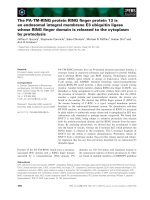
Fig. 2. The signal sequence h-region in general Tat substrates is
significantly less hydrophobic compared with Rieske proteins.
Kyte–Doolittle plot comparing the hydropathy of ISPs and general
Tat substrate proteins. Values fall within a range of +4 to )4, with
hydrophilic residues having a negative score. Each data point
represents an averaged hydropathy value derived from analysis of
multiple sequences, as detailed in the Experimental procedures.
A relative sequence numbering is given, with position 0 represent-
ing the first invariant arginine residue of the consensus motif.
Boundaries of the h-region are indicated as defined in Fig. 1.
Rieske protein from Paracoccus denitrificans J. Bachmann et al.
4820 FEBS Journal 273 (2006) 4817–4830 ª 2006 The Authors Journal compilation ª 2006 FEBS
normally found only in the periplasm, but not in the
cytosolic fraction [29]. Enzymatic activities of cytosolic
malate dehydrogenase and membrane-bound cyto-
chrome c oxidase were the two other markers that
allowed any cross-contamination to be assessed. The
periplasm was isolated efficiently, as demonstrated by
the high relative yield of c-type cytochromes given in
Table 1. Within the periplasmic fraction, no malate
dehydrogenase activity was detectable; this confirms
that practically no cell lysis occurred during extraction
of the periplasm. The enzymatic activites of malate
dehydrogenase and cytochrome c oxidase show that
there is little cross-contamination between the cytosolic
and membrane fractions. However, good separation,
as demonstrated here, could be achieved only after
repeated ultracentrifugation. Small differences between
the total activity in the nonfractionated cell lysate and
the sum of the individual fractions can be easily
explained by either loss of material or protein degrada-
tion during the procedure. Taken together, the sub-
cellular fractionation method applied here resulted in
essentially quantitative separation with cross-contamin-
ation of a few per cent at most.
Localization of the ISP variants was analysed by
western blotting of subcellular fractions derived from
small-scale cultures of P. denitrificans in the exponen-
tial growth phase (50 mL, D
600
1.5). The result of
this experiment is given in Fig. 3. The deletion strain
with the wild-type protein expressed in trans showed a
dominant ISP signal in the membrane fraction; how-
ever, substantial amounts of the protein could be
found in the cytoplasm. It should be mentioned that
the Rieske protein typically separates into two bands
on SDS ⁄ PAGE; the lower band can be seen here only
in the fractions with elevated ISP amounts. Exchange
of the first invariant arginine (R15K) leads to a com-
parable distribution, but the total ISP level is clearly
diminished. In contrast, both the R16K mutation and
the R15K⁄ R16K double mutation result in almost
complete loss of the signal in the membrane fraction.
The bands for the Y20K mutant resemble the wild-
type, with slightly decreased levels. A much weaker sig-
nal was obtained for the C152S mutant, which should
abolish cofactor binding. This is in line with earlier
observations by Davidson et al. [30]; they prepared an
equivalent mutation in the closely related Rhodobacter
capsulatus complex and observed a strongly decreased
membrane level of the apo-ISP that was between one
and two orders of magnitude less than the wild-type
overproducer. Interestingly, a decrease or loss of the
membrane-bound form in the mutant strains was not
accompanied by ISP accumulation in the cytosol,
pointing to rapid degradation of ISP that cannot be
targeted to the membrane.
Table 1. P. denitrificans cells are efficiently fractionated. Cell cultures at exponential growth (D
600
)1.5) were fractionated as described in the
Experimental procedures. For each cell fraction, a marker protein was assayed. Periplasmically located c-type cytochromes were quantified
using redox difference spectra. Activities of the cytosolic malate dehydrogenase and the membrane-integral cytochrome c oxidase were
assayed as described in the Experimental procedures, and the total activities of the cell fractions were compared. Three separate fractiona-
tions gave consistent results; values given here are from a single representative experiment. ND, values not determined.
Marker
Cell fraction (%)
Cell lysate Periplasm Cytoplasm Membrane
c-type cytochromes ND 95 5 nd
Malate dehydrogenase activity 100 < 1 92 3
Cytochrome c oxidase activity 100 nd 1 95
Fig. 3. Specific mutations strongly inhibit membrane insertion of the P. denitrificans Rieske protein. Detection of ISP by western blotting in
cell fractions from different P. denitrificans strains. Strain variants are indicated above the corresponding lanes, as follows: wt, P. denitrifi-
cans bc
1
deletion strain MK6 expressing the wild-type fbc operon from plasmid pAN42; R15K, R16K, R15 ⁄ R16K, Y20K, and C152S, MK6
strain bearing pAN42 derivatives with the respective mutation(s) in the fbcF gene. Total amounts of 15 lg protein were loaded in each lane.
The applied cell fractions are indicated: C, cytoplasm; M, membrane; periplasmic samples did not show any ISP signal and therefore are
omitted. The Rieske protein typically separates into two bands on SDS ⁄ PAGE, as indicated by the two arrows; the lower band shows up
only with higher protein amounts.
J. Bachmann et al. Rieske protein from Paracoccus denitrificans
FEBS Journal 273 (2006) 4817–4830 ª 2006 The Authors Journal compilation ª 2006 FEBS 4821
Cytochrome bc
1
enzymatic activity was assayed to
obtain a more quantitative measure of decreased ISP
amounts in the mutant membranes. To this end, we
assume that mutations in the signal sequence do not
exert an effect on the intrinsic activity of the enzyme;
this seems plausible as the structures of mitochondrial
homologues show that the N-terminal part of the ISP
is far from the catalytic centres and is separated by the
membrane dielectric in the native environment [3,4].
Obviously, this argument does not apply for the
C152S mutant. Specific QH
2
:cytochrome c oxidoreduc-
tase activities of membrane samples are given in
Table 2. The data show that mutant membranes R16K
and R15K ⁄ R16K were essentially inactive, whereas the
R15K and the Y20K mutant membranes contained
considerable amounts of fully assembled and active
enzyme. As expected, the C152S mutant was fully inac-
tive. For the Y20K mutant, an interesting observation
was made when the membranes were subjected to
sodium carbonate treatment prior to activity measure-
ments. Although this treatment had only a minor
effect on wild-type membranes, pretreated Y20K mem-
branes showed severe instability when recording
steady-state activities spectroscopically: traces with ini-
tially normal slopes became a flat line a few seconds
after the addition of substrate (data not shown).
In conclusion, enzymatic data from Table 2 and the
western blot results given in Fig. 3 provide a consistent
picture with strong evidence for a Tat-dependent trans-
location of the P. denitrificans ISP. Conservative
exchanges of the twin-arginine motif for a lysine pair
blocked membrane insertion; the single mutations on
the arginine pair showed differential effects, with a
more important role for the second arginine residue.
As anticipated by the above sequence analysis, ‘restor-
ation’ of the canonical Tat consensus with the Y20K
mutant has a negative impact on membrane insertion;
somewhat surprisingly, this effect is rather mild, and
to a considerable extent the mutant ISP is still func-
tionally incorporated into the cytoplasmic membrane.
In addition, the effect of sodium carbonate treatment
points to a structurally destabilizing effect of this
mutation. Finally, removal of cofactor binding capa-
city with the C152S mutant strongly impairs mem-
brane insertion; this result might be explained by the
postulated ‘cofactor-proof-reading’ operating in the
Tat translocation scheme [11,31]. However, certain
amounts of the apo-ISP are still found in the mem-
brane; there is obviously no strict requirement for
cofactor insertion prior to transport. Furthermore, a
potential drawback of our data is that the effects of
site-specific mutants are deduced only from the steady-
state distribution of the ISP; no kinetic data for mem-
brane translocation were obtained. Particularly in case
of the C152S cofactor insertion mutant and the Y20K
mutation in the ‘+5’ position, it is conceivable that
secondary effects such as proteolytic degradation due
to improper folding or an assembly defect of the
bc
1
complex with subsequent proteolysis might also
account for the reduced amounts of ISP in the mem-
brane. Further experiments will be needed to fully
exclude such side effects.
The [2Fe)2S] cluster of the Rieske protein is
inserted in the cytoplasm
As cytosolic cofactor insertion is one of the hallmarks
of Tat translocation, the cofactor loading status of
the ISP in P. denitrificans cytosolic fractions was
examined using electron paramagnetic resonance
(EPR). Cells from a 0.5 L culture in the exponential
growth phase were harvested; cytosolic fractions of
the complemented wild-type, the export mutant
R15K ⁄ R16K, and the cofactor binding mutant C152S
were isolated and concentrated by ultrafiltration.
Membranes from the complemented wild-type and the
C152S mutant were used as positive and negative
controls for the presence of the Rieske [2Fe )2S] clus-
ter EPR signal. A reference spectrum was recorded
with a purified sample of the Rieske protein fragment
(ISF). EPR samples were reduced with 5 mm sodium
ascorbate (Fig. 4).
Spectrum I shows the reference spectrum of the
purified ISF; the complemented wild-type in spec-
trum II gave a clear Rieske signal in the membrane
fraction, with shifted peak positions relative to
Table 2. Cytochrome bc
1
activity reflects the decreased ISP con-
tent in mutant membranes. Membranes were isolated from the
parental fbc::Km deletion strain that was complemented with a
wild-type copy of the fbc operon in trans or mutants thereof. Val-
ues given are the average of three to five measurements. To elim-
inate unspecific activity effects, activity data were corrected for the
slope measured when the enzyme was inhibited with 10 l
M anti-
mycin A. Relative activity refers to the wild-type complemented
strain.
Strain
Specific activity of
membrane fraction
(mUÆmg
)1
)
Relative activity
(%)
fbc::Km deletion strain 3 < 1
Complemented wild-type 4018 100
R15K 1397 35
R16K 67 2
R15K ⁄ R16K 29 1
Y20K 929 23
C152S 12 < 1
Rieske protein from Paracoccus denitrificans J. Bachmann et al.
4822 FEBS Journal 273 (2006) 4817–4830 ª 2006 The Authors Journal compilation ª 2006 FEBS
spectrum I. This shift most probably arises from
h-bond interactions of a histidine cluster ligand with
the quinone substrate bound to the membrane integral
cytochrome bc
1
complex [32]. No [2Fe)2S] cluster sig-
nal was visible in the cytosolic and membrane fractions
of the C152S mutant, demonstrating: (a) the inability
of the mutant to insert a cofactor, and (b) the specific
origin of the signal in the other samples. The cytosolic
fraction of the complemented wild-type clearly showed
the EPR signature of the Rieske cluster (spectrum V),
which provides strong evidence for the cytosolic assem-
bly of the holoprotein. Likewise, the cytosol of the
R15K ⁄ R16K double mutant contained the Rieske clus-
ter, albeit at lower concentration compared with the
wild-type cytosol (spectrum VI). In order to demon-
strate the presence of the cluster in this strain more
convincingly, the cytosolic fraction obtained from a
2.5 L culture was further enriched. It was applied to a
Q Sepharose column, and the 150–250 mm NaCl salt
gradient eluate was pooled and concentrated; the ISP
is known to elute at 200 mm NaCl [33]. The
enriched cytosolic fraction clearly shows the indicative
Rieske spectrum (VII). The existence of the Rieske
cluster in the cytosol of the R15K ⁄ R16K which is
incompetent of membrane insertion (Fig. 3) clearly
rules out the possibility that the signal could arise
from membrane remnants in the cytosolic fraction.
Furthermore, no Rieske cluster signal was observed in
membrane samples of the R15K ⁄ R16K double mutant
(not shown). Thus, the EPR results clearly demon-
strate cytosolic cofactor insertion which is a typical
feature of Tat substrate proteins.
Discussion
The aim of this study was to obtain experimental evi-
dence of whether the Rieske [2Fe)2S] protein subunit
of bacterial cytochrome bc
1
complexes is targeted to
the cytoplasmic membrane by means of twin-arginine-
dependent translocation. Studying the ISP from
P. denitrificans with sequence analysis tools, site-direc-
ted mutagenesis, and EPR spectroscopy, we found
the key requirements of Tat translocation fulfilled:
the characteristic features of the signal sequence, the
export dependence on the conserved arginine pair,
Fig. 4. The [2Fe)2S] cluster is inserted into the Rieske apoprotein
in the cytoplasm. EPR spectra of: I, purified Rieske protein frag-
ment (ISF); II, membranes of complemented wild-type strain; III,
membranes of C152S mutant; IV, cytosol of C152 mutant; V, cyto-
sol of complemented wild-type; VI, cytosol of R15K ⁄ R16K double
mutant; VII, IEX chromatography-enriched cytosol fraction of
R15K ⁄ R16K double mutant (150–250 m
M NaCl eluate). Samples
were reduced with 5 m
M sodium ascorbate. For representation pur-
poses, spectra are scaled differently on the y-axis: Spectra II–VII
are magnified by a scaling factor of 500 relative to spectrum I. This
scaling factor includes differences in sample concentration, spectral
accumulation, and graphical scaling. Hence, spectral intensities do
not reflect the concentration ratio between membrane and cytosol
fractions. Peaks in the g
x
and g
y
region are indicated by vertical
lines; the g
z
region is omitted due to overlaps with EPR signals
from other proteins. The positions of g
x
signals in samples I and VII
(g
x1
) are shifted to higher magnetic field because of the occurrence
of ligand-free iron–sulfur protein. Conditions for EPR spectroscopy
are given in the Experimental procedures.
J. Bachmann et al. Rieske protein from Paracoccus denitrificans
FEBS Journal 273 (2006) 4817–4830 ª 2006 The Authors Journal compilation ª 2006 FEBS 4823
and cytosolic cofactor insertion as a prerequisite for
membrane targeting.
To our knowledge, the only experimental evidence
for Tat dependence of the bacterial ISP to date is the
finding that a DtatBC deletion mutant of R. leguminos-
arum lacks a functional bc
1
complex [34]. In contrast,
insertion of the chloroplast ISP into the thylakoidal
membrane via the Tat ⁄ DpH-pathway is well documen-
ted [19,20]. This protein was the first Tat substrate
shown to be an integral membrane polypeptide with a
signal sequence that is not cleaved after translocation.
Another interesting feature is the lack of the ‘invariant’
twin-arginine; instead, a KR sequence is found in the
corresponding position. In contrast, cyanobacteria as
supposed ancestors of chlorplasts contain ISPs with a
perfect twin-arginine motif. It was argued that the RR
to KR transition has a functional role in slowing
import of the now nucleus-encoded ISP to allow for
proper cofactor insertion in the stroma [19]. Associ-
ation of the ISP with stromal chaperonin Cpn60
and ⁄ or Hsp70 was observed [19,35]. Furthermore, evi-
dence was found for interplay with components of the
Sec system and it was hypothesized that the ISP is an
‘intermediate’ substrate in the evolution of the chloro-
plast export pathways [19].
The second conserved arginine residue plays a
critical role in ISP translocation
The relative amounts of ISP, detected by immunologi-
cal means in the membrane fractions of the variant
strains R15K, R16K, and R15K ⁄ R16K, show that
both arginine residues are important for membrane
targeting. However, the second arginine appears the
most critical, and even a conservative mutation in this
position leads to an essentially complete block,
whereas replacement of the first arginine allows sub-
stantial membrane insertion. This is a remarkable find-
ing in the light of the naturally occurring KR motif
in plant ISPs. This observation raises the question
whether the Tat translocon is especially ‘permissive’
towards the Rieske protein, allowing for variation at
the first arginine position, or whether the stricter role
of the second arginine is a general feature of Tat sub-
strates. Originally, an absolute requirement for both
arginines was stated [11,36,37]. A gain-of-function
mutant screen with a Tat-targeted GFP reporter con-
struct, however, indicated that both positions tolerate
variation, with the second position even being more
flexible. Similarly, the E. coli multi-copper oxidase
superfamily homologue SufI allowed single conserva-
tive substitutions at both positions. In contrast to this,
several authors report a critical role only for the
second arginine [21,38–40]. Furthermore, apart from
the plant ISPs, another example of natural ‘KR’ vari-
ation of the Tat motif is known, interestingly, also in
case of a protein carrying an iron–sulfur cofactor [21].
Taken together, these examples show that in some sig-
nal peptides at least, conservative variation especially
in the first position of the twin-arginine is possible,
and this idea is corroborated by this study.
The ISP Tat signal serves as a membrane anchor
Sequence analysis of the N-terminal ISP portion is in
good overall agreement with the structure of general
Tat signal peptides. However, it shows some clear dis-
tinctions that may account for its dual role as a Tat
signal and as a membrane anchor. The ISP h-region
exhibits a significantly higher hydrophobicity than
average Tat signal peptides (Fig. 2). Likewise, it is well
established that Sec signal peptides show higher hydro-
phobicity than typical Tat signals. An engineered
increase in hydrophobicity in a Tat signal peptide even
leads to a (nonphysiological and functionally inexpedi-
ent) re-routing of a precursor protein to the Sec trans-
locon [23]. We wondered if the ISP h-region has a
similar degree of hydrophobicity as the corresponding
portion of Sec signal peptides. However, when we
compared the h-region hydropathy values of the ISPs
and a set of Sec substrates, we found the ISP h-region
to be considerably less hydrophobic (not shown).
Therefore, a clear ranking of hydropathy values
becomes apparent, with Sec h-regions as the most
hydrophobic, followed by ISP signal h-regions, which
again are distinctly more hydrophobic than generic
Tat h-regions. Together with the fact that we do not
find a basic ‘Sec-avoidance’ signal [41] in the c-region,
the elevated hydrophobicity of the h-region raises the
intriguing question whether the bacterial ISP may also
interact with Sec components. Evidence for such inter-
play exists in case of the chloroplast ISP, where it was
suggested that soluble components involved in Sec-
targeting also deliver the Rieske protein to the Tat
translocon [19]. It will be interesting to see if future
experiments show a similar linkage in case of the
bacterial ISP.
As another critical determinant for membrane
anchorage, the moderately hydrophobic residue at the
‘+5’ position was identified in silico, which was found
in place of the consensus lysine in the ISP Tat motifs.
Genetic substitution by the canonical lysine residue
leads to slightly decreased ISP levels in the cytoplasmic
membrane. This could be interpreted in line with the
observation made by Stanley et al. [42] that a ‘+5’
lysine slows export of Tat substrates. Alternatively,
Rieske protein from Paracoccus denitrificans J. Bachmann et al.
4824 FEBS Journal 273 (2006) 4817–4830 ª 2006 The Authors Journal compilation ª 2006 FEBS
our results may be explained by secondary effects of
the Y20K mutation which might destabilize the bc
1
complex and lead to proteolysis. At any rate, the
hypothesis of these authors that this slowing has the
physiological role of allowing for proper cofactor
insertion is not substantiated here; the ISP is a cofac-
tor-containing protein but lacks the ‘+5’ lysine in its
native sequence. Probably, retardation is needed for
other cofactor classes or in the case of heterodimeric
proteins, where the ‘hitch-hiking’ subunit is granted
time to associate with the subunit containing the Tat
signal peptide [12].
Activity measurements with Y20K mutant mem-
branes pretreated with carbonate show apparent rapid
loss of cytochrome bc
1
activity; this can be tentatively
interpreted as a less stable insertion of the ISP into the
hydrophobic core of the enzyme complex. It has been
a frequent observation that the Rieske subunit appears
only poorly associated with the bc
1
complex, is easily
lost during purification of the bc
1
complex, and can be
extracted by high detergent concentrations or chao-
tropic salt treatment [43]; before the crystal structure
information emerged, it was still a matter of debate
whether the ISP is a true integral membrane protein
[44,45]. This weak association may be explained by the
h-region hydrophobicity as given in Fig. 2, which,
albeit being higher as for the Tat substrate average, is
still rather low compared with TM helices of other
membrane-anchored proteins. Possibly, the lacking
‘+5’ lysine, on the one hand, and the comparatively
low hydrophobicity, on the other hand, represent a
compromise between the conflicting requirements of
TM helix formation and acceptance as a substrate by
the Tat translocon.
Recently, the crystal structure of a cytochrome bc
1
homologue, the cytochrome b
6
f complex from the
cyanobacterium Mastigocladus laminosus was solved
[7]. As discussed by Berks et al. [13], this enzyme also
contains an ISP subunit with a putative Tat signal.
Here, an asparagine residue is in the ‘+5’-position;
sequence alignments of ISP subunits from bacterial b
6
f
complexes show that for this subgroup asparagine is
the most frequent amino acid in this position (data not
shown), whereas the canonical lysine residue cannot be
found, as in the case of bacterial bc
1
complexes. In the
enzyme structure, no major interactions were found
for the ‘+5’-Asn side chain. However, whereas the
invariant Arg residues are located at the membrane–
water interface, the ‘+5’-residue lies well within the
TM region of the enzyme. It is a reasonable assump-
tion that a Lys residue cannot be accommodated in
this hydropohobic environment and is therefore absent
from the Tat motif of bacterial ISPs.
Potential steps during ISP biogenesis
The observation that cofactor-containing periplasmic
proteins carry a conserved twin-arginine motif led to
discovery of the Tat translocation pathway [10]. Cyto-
solic cofactor insertion is a key feature of Tat sub-
strates and was experimentally confirmed by a number
of studies for the bacterial and the homologous thylak-
oid system, as reviewed by Berks et al. [11]. Our EPR
data demonstrate the presence of holo-ISP in cytosolic
fractions, thereby confirming that cluster assembly
takes place in the cytosol. Impaired membrane inser-
tion in the C152S mutant may indicate that cofactor
insertion is a prerequisite for efficient export of the
ISP. It was convincingly shown by different authors
that a lack of the cofactor prevents export of Tat sub-
strates [31,36]. However, because the kinetics of mem-
brane insertion were not examined experimentally, the
possibility exists that the apo-ISP is targeted to the
membrane perfectly normally and only secondary pro-
teolysis depletes the membrane fraction. It is an inter-
esting observation that the cluster content of the
R15K ⁄ R16K mutant cytosol was much lower than in
the complemented wild-type (Fig. 4); for an export-
deficient strain, rather an accumulation of the signal
might be expected. It is therefore tempting to speculate
on a potential interplay of the cluster insertion
machinery and the Tat translocation process. With a
closer look at the spectra in Fig. 4, it is remarkable to
see that the chromatographically enriched cytosol frac-
tion of R15K ⁄ R16K (spectrum VII) shows the same
g-value positions as the purified reference protein (I),
whereas cytosolic fractions taken directly for EPR
measurements (spectra V + VI) resemble spectrum II
from intact bc
1
complex where the [2Fe)2S] cluster
histidine ligand is involved in hydrogen bonding inter-
actions. Interaction with a quinone molecule in the
cytosol appears quite unlikely; therefore, this observa-
tion may give a hint to a putative binding parter of
the ISP in the cytosol, probably playing some chaper-
one role. The chromatographic purification step may
have removed this binding partner. Further experi-
ments are needed to examine this aspect in detail.
A potential binding partner could be involved in cluster
assembly; likely candidates are components of the Nif
or Isc machinery responsible for iron–sulfur cluster
assembly in bacteria and mitochondria [46,47]. Prelim-
inary data from our laboratory indicate that overex-
pression of the isc or nif operon can indeed promote
[2Fe)2S] cluster assembly to the P. denitrificans ISP in
the heterologous host E. coli [48]. In a BLAST search
on the draft version of the P. denitrificans genome,
putative genes homologous to those of the isc operon
J. Bachmann et al. Rieske protein from Paracoccus denitrificans
FEBS Journal 273 (2006) 4817–4830 ª 2006 The Authors Journal compilation ª 2006 FEBS 4825
from E. coli and the nif operon from A. vinelandii were
identified (data not shown).
Alternatively, ‘proof-reading’ chaperones may inter-
act with the ISP in the cytosol. Recent evidence
points to such specific chaperones acting on Tat sub-
strates and preventing premature translocation [16,49].
However, such specific proof-reading chaperones were
typically acknowledged as accessory genes in the
operon context of the respective Tat substrate protein
[40]. No such ORF of yet unknown function is pre-
sent in the fbc operon coding for the cytochrome bc
1
complex of P. denitrificans. Also, chaperone binding is
assumed to be associated with conserved sequence ele-
ments in the signal peptide n-region [12]. We did not
find such sequence conservation in case of bacterial
ISPs; however, comparison of h-regions shows signifi-
cant similarities among the different ISPs and might
therefore be specifically recognized by a putative
chaperone.
An additional level of control for export competence
is designated ‘quality control’ [17]. Here, the folding
status of the protein is examined, presumably by the
Tat translocon itself. From our data it is not clear
whether cofactor ‘proof-reading’ or general ‘quality-
control’ keeps the apo-ISP from being efficiently trans-
located and inserted into the membrane; a third
explanation for the lower membrane levels, as men-
tioned above, is that the apo-ISP is normally targeted
but subsequently degraded by periplasmic proteases.
From the crystal structure of the homologous bovine
soluble Rieske protein fragment (ISF), it seems plaus-
ible that the apo-protein may adopt its almost terminal
tertiary structure, as the cluster is bound only by a
minor subdomain on top of the b-sandwich fold [50].
Furthermore, a CD spectrum of the heterologously
expressed and refolded apo-ISF shows secondary
structure features similar to the native holo-protein.
However, native PAGE indicates a partially mobile or
disordered structure for the refolded apo-ISF [48]. In
addition, the Rieske protein carries a cystine bridge,
and it was shown that various disulfide-containing pro-
teins may be exported by the Tat pathway only under
conditions in which a mutant strain provides an oxid-
izing cytosolic environment [17]. However, it is reason-
able to assume that disulfide bonds are formed in the
periplasm, catalysed by homologues of the DsbA ⁄ B
machinery. Therefore, we argue that the cystine bridge
is not essential for an export-competent structure of
the ISP. In summary, it seems that the apo-ISP can
adopt its tertiary structure to a large extent and might
well be accepted by the Tat translocon; however, disor-
dered elements (the cluster binding subdomain) may
hamper this process.
Genetic inactivation of Tat machinery components
in P. denitrificans will be an interesting goal for future
experiments. Respiration is obligatory for this bac-
terium [29]; however, a Tat-inactivated strain should
by viable under oxic conditions, given the bioenergetic
flexibility of P. denitrificans. The expected defect of
the cytochrome bc
1
complex can be bypassed by the
ba
3
quinol oxidase. If such a mutant can be obtained,
it will certainly provide valuable information about
the assembly of redox proteins in this important
model system for the study of respiratory chains.
Another interesting outlook is the identification of a
putative cytosolic binding partner of the ISP, e.g.
by using chemical cross-linking approaches and MS,
giving interesting insights into the assembly of Rieske
proteins.
Experimental procedures
Bioinformatic tools
Protein sequences were obtained from Swiss-Prot pro-
tein database ( (a) bacterial
Rieske proteins: R. rubrum P23136, R. capsulatus P08500,
R. sphaeroides Q02762, R viridis P81380, B. japonicum
P51130, P. denitrificans P05417; (b) [NiFe] hydrogenase
small subunits: A. chroococcum P18190, A. hydrogenophilus
P33375, B. japonicum P12635, R. capsulatus P15283; (c)
MauM ferredoxins: M. extorquens Q49130, M. flagellatum
Q50423, M. methylotrophus Q50235, P. denitrificans
Q51659; (d) NapA periplasmic nitrate reductases: A. eutro-
phus P39185, D. desulfuricans P81186, R. sphaeroides
Q53176, P. pantotrophus Q56350; (e) NosZ nitrous oxide
reductases: A. eutrophus Q59105, P. aeruginosa Q01710,
P. denitrificans Q51705, P. stutzeri P19573, R. meliloti
Q59746; (f) TorA trimethylamine-N-oxide reductases:
E. coli P33225, R. capsulatus Q52675, R. sphaeroides
Q57366. Multiple sequence alignments were performed
using clustal x v. 1.81 [27]. For secondary structure
prediction based on multiple alignments, the web server
JPRED [51] () was used.
Kyte–Doolittle hydropathy plots [25] were generated
using an online tool from the ExPASy molecular bio-
logy server ( />the window size was set to 19 residues for comparison
of ISP sequences and the comparison group of Tat-
translocated proteins. Differences in hydropathy were
statistically assessed with a two-tailed Mann–Whitney
U-test ( />To estimate the hydropathy of TM helices, a limited
collection of P. denitrificans TM proteins was examined:
cytochrome c
552
, cytochrome c
1
, cytochrome b and cyto-
chrome c oxidase SU II. A set of 20 predicted Sec-
exported proteins was obtained from the SPDb server
Rieske protein from Paracoccus denitrificans J. Bachmann et al.
4826 FEBS Journal 273 (2006) 4817–4830 ª 2006 The Authors Journal compilation ª 2006 FEBS
( here, due to the
shorter n-region of the Sec substrates, a window size of 9
was applied in the comparative Kyte–Doolittle hydropathy
analysis. For TM prediction based on multiple alignments,
the program tmap [26] ( />tmap/) was used. Tat gene homologues in P. denitrificans
were found in the draft genome annotation (Joint Genome
Institute Microbial Sequencing Program, http://genome.
jgi-psf.org/draft_microbes/parde/parde.home.html).
Bacterial strains and growth conditions
Export of the Rieske protein variants was studied in P. den-
itrificans strain MK6 (fbc::Km
R
), a derivative of Pd1222
with the bc
1
-coding fbc operon replaced by a kanamycin
resistance gene. E. coli strain JM109 was used for standard
cloning procedures and E. coli DH5a RP4-4 served as a
helper strain in the conjugative transfer of plasmids to
P. denitrificans via triparental mating. E. coli strains were
grown aerobically in Luria–Bertani medium; P. denitrificans
was cultivated aerobically in succinate medium [52]. Antibi-
otics were used at the following final concentrations: ampi-
cillin, 50 lgÆmL
)1
; kanamycin, 25 lgÆmL
)1
; streptomycin,
25 lgÆmL
)1
; rifampicin, 80 lgÆmL
)1
.
Mutagenesis and cloning procedures
For site-directed mutagenesis, the QuickChange mutagen-
esis system (Stratagene, La Jolla, CA) was employed.
Sequences of mutagenic primers were as follows, with the
mutated positions in bold: R15K, 5¢-GATCACGGCGCC
ACGAAGAGGGACTTCCTCTAC-3¢; R16K, 5¢-CACGG
CGCCACCCGGAAGGACTTCCTCTAC-3¢; R15K ⁄ R16K,
5¢-GATCACGGCGCCACCAAGAAGGACTTCCTCTAC
TACG-3¢; Y20K, 5¢-GGAGGGACTTCCTGAAGTAC
GCGACGGCCGGTG-3¢; C152S, 5¢-GGCGGCTGGTTC
AGCCCGTGCCATGG-3¢. For the mutagenesis reactions,
a SacI ⁄ NcoI cassette of the fbcF coding sequence was
cloned into pSL1180 (GE Healthcare, Chalfont St Giles,
UK). All introduced mutations were checked by sequencing
of the full insert. For expression of the cytochrome bc
1
var-
iants, the mutagenized SacI ⁄ NcoI fbcF fragments were
introduced into pAN42, a derivative of broad host range
vector pRI2 [53], which carries the complete fbc operon
under the control of its native promotor.
Subcellular fractionation
To analyse Tat-dependent translocation, subcellular local-
ization of the Rieske protein variants was determined. Sub-
cellular fractionation of P. denitrificans cells was performed
analogous to a protocol originally designed for E. coli (pET
System Manual, Merck Biosciences, Darmstadt, Germany).
P. denitrificans strain MK6 expressing the fbc operon
in trans was cultivated in 50 mL succinate medium and har-
vested during exponential growth at D
600
1.5. Cells were
resuspended in 30 mL buffer containing 30 mm Tris pH 8,
500 mm sucrose and 1 mm EDTA. After 10 min incubation
at room temperature, intact cells were harvested at 6000 g
and 4 °C; the following steps were performed at 4 °C
throughout. Cells were osmotically shocked by resuspension
in 20 mL 5 mm MgSO
4
buffer containing 100 lm Pefabloc
SC (Roche Applied Science, Mannheim, Germany). After
20 min incubation, spheroplasts were separated from the
periplasmic supernatant by centrifugation at 10 000 g.
Spheroplasts were resuspended in 50 m m KP
i
pH 7, 10 mm
EDTA, 100 lm Pefabloc SC, and 0.1 mgÆmL
)1
lysozyme
and lysed by sonication. Intact cells and cellular debris was
removed by centrifugation (10 min, 10 000 g). The mem-
brane fraction was separated from the cytoplasm by two
successive ultracentrifugation steps (1 h, 125 000 g). The
membrane pellet was homogenized in 50 lL20mm KP
i
pH 8; protein concentration of all samples was determined
following a modified Lowry method [54]. Western blotting
was performed using a polyclonal antibody against the
P. denitrificans Rieske protein and an anti-(rabbit IgG)
alkaline phosphatase conjugate as secondary antibody that
was detected using enzyme substrates nitrobluetetrazolium
and 5-bromo-4-chloro-3-indolylphosphate.
Enzymatic assays and spectroscopy
All enzymatic measurements were performed at ambient
temperature on a Hitachi U-3000 spectrophotometer (Hita-
chi, Tokyo, Japan).
Malate dehydrogenase assay
Malate dehydrogenase was used as a marker for the cyto-
plasmic fraction. Activity was measured by determining
the absorbance change at 340 nm due to oxidation of
NADH. The reaction mixture contained 50 mm Tris
pH 7.4, 50 mm NaCl, 300 lm NADH, and 300 lm oxalo-
acetate.
Cytochrome c oxidase activity
Cytochrome c oxidase activity as a marker for the mem-
brane fraction was monitored by following the oxidation of
20 lm horse heart ferrocytochrome c (Sigma, St. Louis,
MO) at 550 nm; buffer conditions were 20 mm KP
i
pH 8,
20 mm KCl, 1 mm EDTA and 0.02% lauryl maltoside.
Cytochrome bc
1
activity
The reduction of 25 lm horse heart ferricytochrome c by
80 lm n-decyl-ubihydroquinone (Sigma; chemically prere-
duced) [55] was followed at 550 nm; the buffer contained
J. Bachmann et al. Rieske protein from Paracoccus denitrificans
FEBS Journal 273 (2006) 4817–4830 ª 2006 The Authors Journal compilation ª 2006 FEBS 4827
50 mm Mops pH 7.5, 100 mm NaCl, 1 mm EDTA, 1 mm
KCN, and 0.04% lauryl maltoside. In order to assess the
stability of the cytochrome bc
1
complex of the Y20K
mutant strain, membranes from the Y20K mutant and the
wild-type (as control) were subjected to a sodium carbonate
treatment prior to the enzymatic assay: membrane aliquots
in 20 mm KPi pH 8 (protein concentration 20 mgÆmL
)1
)
were diluted 1 : 10 in 0.1 m sodium carbonate pH 11.5 and
incubated for 30 min on ice; it was checked that the phos-
phate buffer of the membrane samples did not interfere
with the desired alkaline pH. For the following enzymatic
measurement, the pretreated sample was typically diluted
1 : 200 in assay buffer.
Optical spectroscopy
c-Type cytochromes served as a periplasmic marker, as they
exhibit characteristic redox difference spectra in the visible
range. For the oxidized spectra, 1 mm ferricyanide was added
to the samples; for reduction of the c-type cytochromes,
samples were treated with 10 mm sodium ascorbate.
EPR spectroscopy
X-band EPR spectra at 9.46 GHz were recorded at 16 K
on a Bruker ESP 300E spectrometer equipped with a
liquid helium continuous flow cryostat, ESR 900 from
Oxford Instruments (Eynsham, UK). Microwave power
was 1 mW and modulation amplitude was 10 G. Samples
were reduced with 5 mm sodium ascorbate, shock-frozen
in cold isopentane ⁄ methylcyclohexane ( 81 K) and stored
in liquid nitrogen until measurement. For samples with
low Rieske cluster content, spectra were accumulated up
to 10 times. In order to enhance the Rieske cluster signal,
the cytosolic fraction of the R15K ⁄ R16K double mutant
was chromatographically enriched in one set of EPR
measurements: The cytosol from a 2.5 L culture harvested
at exponential growth was applied to a 40 mL Q-Seph-
arose column that was equilibrated with 50 mm KP
i
; chro-
matography was performed on a FPLC system
(Pharmacia). Proteins were eluted with a 0–300 mm NaCl
gradient; the eluate from 150 to 250 mm salt was pooled
and concentrated by ultrafiltration (cut-off 5 kDa) to a
final protein concentration of 200 mgÆmL
)1
. To obtain
a reference Rieske [2Fe-2S] cluster spectrum, a soluble
fragment of the Rieske iron–sulfur-protein was produced
by limited proteolysis of the purified P. denitrificans cyto-
chrome bc
1
complex, followed by chromatographic purifi-
cation steps, as detailed elsewhere [56].
Acknowledgements
We are grateful to Andrea Herrmann for excellent
technical help and to Uli Brandt for provision of EPR
facilities. This work was supported by Deutsche For-
schungsgemeinschaft (SFB 472).
References
1 Zhang Z, Huang L, Shulmeister VM, Chi YI, Kim KK,
Hung LW, Crofts AR, Berry EA & Kim SH (1998)
Electron transfer by domain movement in cytochrome
bc
1
. Nature 392 , 677–684.
2 Xia D, Yu CA, Kim H, Xia JZ, Kachurin AM, Zhang
L, Yu L & Deisenhofer J (1997) Crystal structure of the
cytochrome bc
1
complex from bovine heart mitochon-
dria. Science 277, 60–66.
3 Iwata S, Lee JW, Okada K, Lee JK, Iwata M,
Rasmussen B, Link TA, Ramaswamy S & Jap BK
(1998) Complete structure of the 11-subunit bovine
mitochondrial cytochrome bc
1
complex. Science 281,
64–71.
4 Hunte C, Koepke J, Lange C, Rossmanith T & Michel
H (2000) Structure at 2.3 A
˚
resolution of the cyto-
chrome bc
1
complex from the yeast Saccharomyces cere-
visiae co-crystallized with an antibody Fv fragment.
Struct Fold Design 8, 669–684.
5 Kurowski B & Ludwig B (1987) The genes of the Para-
coccus denitrificans bc
1
complex. Nucleotide sequence
and homologies between bacterial and mitochondrial
subunits. J Biol Chem 262, 13805–13811.
6 Yang XH & Trumpower BL (1986) Purification of a
three-subunit ubiquinol–cytochrome c oxidoreductase
complex from Paracoccus denitrificans. J Biol Chem 261,
12282–12289.
7 Kurisu G, Zhang H, Smith JL & Cramer WA (2003)
Structure of the cytochrome b
6
f complex of oxygenic
photosynthesis: tuning the cavity. Science 302, 1009–
1014.
8 Tho
¨
ny-Meyer L (2002) Cytochrome c maturation: a
complex pathway for a simple task? Biochem Soc Trans
30, 633–638.
9 Tho
¨
ny-Meyer L (1997) Biogenesis of respiratory
cytochromes in bacteria. Microbiol Mol Biol Rev 61,
337–376.
10 Berks BC (1996) A common export pathway for pro-
teins binding complex redox cofactors? Mol Microbiol
22, 393–404.
11 Berks BC, Sargent F & Palmer T (2000) The Tat
protein export pathway. Mol Microbiol 35, 260–274.
12 Palmer T, Sargent F & Berks BC (2005) Export of
complex cofactor-containing proteins by the bacterial
Tat pathway. Trends Microbiol 13, 175–180.
13 Berks BC, Palmer T & Sargent F (2005) Protein target-
ing by the bacterial twin-arginine translocation (Tat)
pathway. Curr Opin Microbiol 8, 174–181.
14 Alami M, Luke I, Deitermann S, Eisner G, Koch HG,
Brunner J & Mu
¨
ller M (2003) Differential interactions
Rieske protein from Paracoccus denitrificans J. Bachmann et al.
4828 FEBS Journal 273 (2006) 4817–4830 ª 2006 The Authors Journal compilation ª 2006 FEBS
between a twin-arginine signal peptide and its translo-
case in Escherichia coli. Mol Cell 12, 937–946.
15 Sargent F, Gohlke U, De Leeuw E, Stanley NR, Palmer
T, Saibil HR & Berks BC (2001) Purified components
of the Escherichia coli Tat protein transport system
form a double-layered ring structure. Eur J Biochem
268, 3361–3367.
16 Jack RL, Buchanan G, Dubini A, Hatzixanthis K,
Palmer T & Sargent F (2004) Coordinating assembly
and export of complex bacterial proteins. EMBO J 23,
3962–3972.
17 DeLisa MP, Tullman D & Georgiou G (2003) Folding
quality control in the export of proteins by the bacterial
twin-arginine translocation pathway. Proc Natl Acad
Sci USA 100 , 6115–6120.
18 Settles AM, Yonetani A, Baron A, Bush DR, Cline K
& Martienssen R (1997) Sec-independent protein trans-
location by the maize Hcf106 protein. Science 278,
1467–1470.
19 Molik S, Karnauchov I, Weidlich C, Herrmann RG &
Klosgen RB (2001) The Rieske Fe ⁄ S protein of the
cytochrome b
6
⁄ f complex in chloroplasts: missing link in
the evolution of protein transport pathways in chloro-
plasts? J Biol Chem 276, 42761–42766.
20 Finazzi G, Chasen C, Wollman FA & de Vitry C (2003)
Thylakoid targeting of Tat passenger proteins shows no
delta pH dependence in vivo. EMBO J 22, 807–815.
21 Hinsley AP, Stanley NR, Palmer T & Berks BC (2001)
A naturally occurring bacterial Tat signal peptide lack-
ing one of the ‘invariant’ arginine residues of the con-
sensus targeting motif. FEBS Lett 497, 45–49.
22 Schmidt CL & Shaw L (2001) A comprehensive phylo-
genetic analysis of Rieske and Rieske-type iron–sulfur
proteins. J Bioenerg Biomembr 33, 9–26.
23 Cristobal S, de Gier JW, Nielsen H & von Heijne G
(1999) Competition between Sec- and TAT-dependent
protein translocation in Escherichia coli. EMBO J 18,
2982–2990.
24 Kim H, Xia D, Yu CA, Xia JZ, Kachurin AM, Zhang
L, Yu L & Deisenhofer J (1998) Inhibitor binding
changes domain mobility in the iron–sulfur protein of
the mitochondrial bc
1
complex from bovine heart. Proc
Natl Acad Sci USA 95, 8026–8033.
25 Kyte J & Doolittle RF (1982) A simple method for dis-
playing the hydropathic character of a protein. J Mol
Biol 157, 105–132.
26 Persson B & Argos P (1997) Prediction of membrane
protein topology utilizing multiple sequence alignments.
J Protein Chem 16, 453–457.
27 Thompson JD, Higgins DG & Gibson TJ (1994) CLUS-
TAL W: improving the sensitivity of progressive multi-
ple sequence alignment through sequence weighting,
position-specific gap penalties and weight matrix choice.
Nucleic Acids Res 22, 4673–4680.
28 Korn M (1994) Doppeldeletion der Cytochrom c
Oxidase und Reduktase (cta und fbc Operon) in
Paracoccus denitrificans (Diploma thesis). Johann
Wolfgang Goethe-Universita
¨
t, Frankfurt am Main.
29 Baker SC, Ferguson SJ, Ludwig B, Page MD, Richter
OMH & van Spanning RJM (1998) Molecular genetics
of the genus Paracoccus: metabolically versatile bacteria
with bioenergetic flexibility. Microbiol Mol Biol Rev 62,
1046–1078.
30 Davidson E, Ohnishi T, Atta-Asafo-Adjei E & Daldal F
(1992) Potential ligands to the [2Fe)2S] Rieske cluster
of the cytochrome bc
1
complex of Rhodobacter capsula-
tus probed by site-directed mutagenesis. Biochemistry
31, 3342–3351.
31 Santini CL, Ize B, Chanal A, Muller M, Giordano G &
Wu LF (1998) A novel sec-independent periplasmic pro-
tein translocation pathway in Escherichia coli. EMBO J
17, 101–112.
32 Samoilova RI, Kolling D, Uzawa T, Iwasaki T, Crofts
AR & Dikanov SA (2001) The interaction of the Rieske
iron–sulfur protein with occupants of the Qo-site of the
bc
1
complex, probed by 1D and 2D electron spin echo
envelope modulation. J Biol Chem 277, 4605–4608.
33 de Vries S & Cherepanov A (1998) Spectroscopic inves-
tigations on the water-soluble fragment of the Rieske
[2Fe)2S] protein from Paracoccus denitrificans. Inorgan
Chim Acta 275–276, 493–499.
34 Meloni S, Rey L, Sidler S, Imperial J, Ruiz-Argueso T
& Palacios JM (2003) The twin-arginine translocation
(Tat) system is essential for Rhizobium–legume symbi-
osis. Mol Microbiol 48, 1195–1207.
35 Maduen
˜
o F, Napier JA & Gray JC (1993) Newly
Imported Rieske iron–sulfur protein associates with
both Cpn60 and Hsp70 in the chloroplast stroma. Plant
Cell 5, 1865–1876.
36 Halbig D, Wiegert T, Blaudeck N, Freudl R & Sprenger
GA (1999) The efficient export of NADP-containing glu-
cose–fructose oxidoreductase to the periplasm of Zymo-
monas mobilis depends both on an intact twin-arginine
motif in the signal peptide and on the generation of a
structural export signal induced by cofactor binding. Eur
J Biochem 263, 543–551.
37 Chaddock AM, Mant A, Karnauchov I, Brink S,
Herrmann RG, Klosgen RB & Robinson C (1995) A new
type of signal peptide: central role of a twin-arginine
motif in transfer signals for the delta pH-dependent thyla-
koidal protein translocase. EMBO J 14, 2715–2722.
38 Niviere V, Wong SL & Voordouw G (1992) Site-direc-
ted mutagenesis of the hydrogenase signal peptide con-
sensus box prevents export of a beta-lactamase fusion
protein. J Gen Microbiol 138, 2173–2183.
39 Buchanan G, Sargent F, Berks BC & Palmer T (2001)
A genetic screen for suppressors of Escherichia coli Tat
signal peptide mutations establishes a critical role for
J. Bachmann et al. Rieske protein from Paracoccus denitrificans
FEBS Journal 273 (2006) 4817–4830 ª 2006 The Authors Journal compilation ª 2006 FEBS 4829
the second arginine within the twin-arginine motif. Arch
Microbiol 177, 107–112.
40 Palmer T & Berks BC (2003) Moving folded proteins
across the bacterial cell membrane. Microbiology 149,
547–556.
41 Bogsch E, Brink S & Robinson C (1997) Pathway speci-
ficity for a delta pH-dependent precursor thylakoid
lumen protein is governed by a ‘Sec-avoidance’ motif in
the transfer peptide and a ‘Sec-incompatible’ mature
protein. EMBO J 16, 3851–3859.
42 Stanley NR, Palmer T & Berks BC (2000) The twin
arginine consensus motif of tat signal peptides is
involved in Sec-independent protein targeting in Escheri-
chia coli. J Biol Chem 275, 11591–11596.
43 Engel WD, Michalski C & von Jagow G (1983) Recon-
stitution of the ubiquinol : cytochrome c reductase from
a bc
1
subcomplex and the ‘Rieske’ iron–sulfur protein
isolated by a new method. Eur J Biochem 132, 395–407.
44 Breyton C, de Vitry C & Popot JL (1994) Membrane
association of cytochrome b
6
f subunits. The Rieske
iron–sulfur protein from Chlamydomonas reinhardtii is
an extrinsic protein. J Biol Chem 269, 7597–7602.
45 Gonzalez-Halphen D, Vazquez-Acevedo M & Garcia-
Ponce B (1991) On the interaction of mitochondrial
complex III with the Rieske iron–sulfur protein (subunit
V). J Biol Chem 266, 3870–3876.
46 Gerber J & Lill R (2002) Biogenesis of iron–sulfur pro-
teins in eukaryotes: components, mechanism and
pathology. Mitochondrion 2, 71–86.
47 Frazzon J & Dean DR (2003) Formation of iron–sulfur
clusters in bacteria: an emerging field in bioinorganic
chemistry. Curr Opin Chem Biol 7, 166–173.
48 Anderka O (2005) Strukturelle und funktionelle Unter-
suchungen am Cytochrom bc
1
-Komplex von Paracoccus
denitrificans. PhD thesis, Johann Wolfgang Goethe-
Universita
¨
t, Frankfurt am Main.
49 Hatzixanthis K, Clarke TA, Oubrie A, Richardson DJ,
Turner RJ & Sargent F (2005) Signal peptide–chaperone
interactions on the twin-arginine protein transport path-
way. Proc Natl Acad Sci USA 102, 8460–8465.
50 Iwata S, Saynovits M, Link TA & Michel H (1996)
Structure of a water soluble fragment of the ‘Rieske’
iron–sulfur protein of the bovine heart mitochondrial
cytochrome bc
1
complex determined by MAD phasing
at 1.5 A
˚
resolution. Structure 4, 567–579.
51 Cuff JA, Clamp ME, Siddiqui AS, Finlay M &
Barton GJ (1998) JPred: a consensus secondary
structure prediction server. Bioinformatics 14, 892–893.
52 Ludwig B (1986) Cytochrome c oxidase from
Paracoccus denitrificans. Methods Enzymol 126,
153–159.
53 Pfitzner U, Odenwald A, Ostermann T, Weingard L,
Ludwig B & Richter OM (1998) Cytochrome c oxidase
(heme aa
3
) from Paracoccus denitrificans: analysis of
mutations in putative proton channels of subunit I.
J Bioenerg Biomembr 30, 89–97.
54 Markwell MA, Haas SM, Bieber LL & Tolbert NE
(1978) A modification of the Lowry procedure to
simplify protein determination in membrane and
lipoprotein samples. Anal Biochem 87, 206–210.
55 Crane FL & Barr R (1971) Determination of ubiqui-
nones. Methods Enzymol 18, 137–165.
56 Ritter M, Anderka O, Ludwig B, Ma
¨
ntele W & Hellwig
P (2003) Electrochemical and FTIR spectroscopic
characterization of the cytochrome bc
1
complex from
Paracoccus denitrificans: evidence for protonation
reactions coupled to quinone binding. Biochemistry 42,
12391–12399.
57 Darrouzet E, Valkova-Valchanova M & Daldal F
(2000) Probing the role of the Fe–S subunit hinge region
during Q(o) site catalysis in Rhodobacter capsulatus bc
1
complex. Biochemistry 39, 15475–15483.
Rieske protein from Paracoccus denitrificans J. Bachmann et al.
4830 FEBS Journal 273 (2006) 4817–4830 ª 2006 The Authors Journal compilation ª 2006 FEBS