Báo cáo khoa học: Activation of activating transcription factor 2 by p38 MAP kinase during apoptosis induced by human amylin in cultured pancreatic b-cells ppt
Bạn đang xem bản rút gọn của tài liệu. Xem và tải ngay bản đầy đủ của tài liệu tại đây (809.82 KB, 13 trang )
Activation of activating transcription factor 2 by p38 MAP
kinase during apoptosis induced by human amylin in
cultured pancreatic b-cells
Shaoping Zhang
1
, Hong Liu
1
, Junxi Liu
1
, Cynthia A. Tse
1
, Michael Dragunow
2
and
Garth J. S. Cooper
1
1 The School of Biological Sciences, Faculty of Science, University of Auckland, New Zealand
2 Department of Pharmacology and Clinical Pharmacology, Faculty of Medical and Health Sciences, University of Auckland, New Zealand
Progressive b-cell loss and defective insulin production
and secretion accompanied by the presence of islet
amyloid deposits are characteristic pathological fea-
tures of type 2 diabetes mellitus (T2DM) [1–3]. Cur-
rent studies have indicated that amyloid formation
may contribute to the development of hyperglycemia
by causing islet dysfunction [4,5]. The major protein
component of islet amyloid has been identified as a 37
amino acid peptide, called amylin (also known as islet
amyloid polypeptide) [6–8]. Human amylin (hA) can
self-assemble to form b-sheet-containing aggregates
that are cytotoxic to b-cells, as observed in vitro and
Keywords
activating transcription factor 2; amylin;
b-cell apoptosis; p38 kinase; type-2 diabetes
Correspondence
G. J. S. Cooper, School of Biological
Sciences, University of Auckland, Level 4,
3A Symonds Street, Private Bag 92019,
Auckland, New Zealand
Fax: +64 93737045
Tel: +64 93737599 ext. 87239
E-mail:
(Received 3 May 2006, accepted 16 June
2006)
doi:10.1111/j.1742-4658.2006.05386.x
Amylin-mediated islet b-cell death is implicated in diabetogenesis. We pre-
viously reported that fibrillogenic human amylin (hA) evokes b-cell apopto-
sis through linked activation of Jun N-terminal kinase 1 (JNK 1) and a
caspase cascade. Here we show that p38 kinase [p38 mitogen-activated pro-
tein (MAP) kinase] became activated by hA treatment of cultured b-cells
whereas extracellular signal-regulated kinase (ERK) did not; by contrast,
nonfibrillogenic rat amylin (rA) altered neither. Pretreatment with the p38
kinase-inhibitor SB203580 decreased hA-induced apoptosis and caspase-3
activation by 30%; as did combined SB203580 and JNK inhibitor I, by
about 70%; and the combination of SB203580, the JNK inhibitor I and a
caspase-8 inhibitor, by 100%. These findings demonstrate the requirement
for concurrent activation of the p38 kinase, JNK and caspase-8 pathways.
We further showed that hA elicits time-dependent activation of activating
transcription factor 2 (ATF-2), which was largely suppressed by SB203580,
indicating that this activation is catalyzed mainly by p38 kinase. Further-
more, hA-induced apoptosis was suppressed by specific antisense ATF-2,
and increased phospho-ATF-2 (p-ATF-2) was associated with increased
CRE (cAMP-response element) DNA binding and CRE-mediated tran-
scriptional activity, as well as enhancement of c-jun promoter activation.
We also detected changes in the phosphorylation status and composition of
the CRE complex that may play important roles in regulation of distinct
downstream target genes. These studies establish p38 MAP kinase-mediated
activation of ATF-2 as a significant mechanism in hA-evoked b-cell death,
which may serve as a target for pharmaceutical intervention and effective
suppression of b-cell failure in type-2 diabetes.
Abbreviations
AP-1, activator protein-1; AS-jnk1, antisense jnk1; ATF-2, activating transcription factor 2; CAT, chloramphenicol acetyltransferase; CRE,
cAMP-response element; ERK, extracellular signal-regulated kinase; GFP, green fluorescent protein; GST, glutathione S-transferase; hA,
human amylin; JNK, Jun N-terminal kinase; MAPK, mitogen-activated protein kinase; p38 kinase, p38 MAP kinase; p-ATF-2, phosphorylated
activating transcription factor 2; rA, rat amylin; T2DM, type 2 diabetes mellitus.
FEBS Journal 273 (2006) 3779–3791 ª 2006 The Authors Journal compilation ª 2006 FEBS 3779
in vivo [9–13]. In contrast, rat amylin (rA), whose
sequence varies from the human at six residues, does
not aggregate and exhibits random conformations in
physiological solutions [9,14]. Extracellular application
of fibrillogenic hA, but not nonfibrillogenic rA, induces
apoptosis in cultured human and rat b-cells [10,15,16].
In addition, onset of diabetes, associated with islet
amyloid formation and decreased b-cell mass, has been
demonstrated in transgenic mice expressing hA in their
b-cells [13,17]. Formation of amylin aggregates in the
pancreatic islets may thus play an important role in
triggering islet b-cell death and dysfunction in T2DM.
Since all humans produce amylin with the propensity
to self-assemble, but most do not lose b-cell mass and
develop diabetes, the mechanism by which amylin
aggregates and causes cytotoxicity is attracting increas-
ing attention as a potential molecular target for phar-
macological intervention.
Several putative molecular mechanisms by which hA
might lead to or cause b-cell toxicity have been identi-
fied. One envisages that hA evokes b-cell toxicity
through apoptosis (programmed cell death), wherein
contact of protein aggregates with b-cell membranes is
necessary for death induction [10,11,16]. Another pos-
sibility is increased cellular pro-oxidant responses and
low density lipoprotein uptake evoked by aggregate–
cell interactions [18]. Small or intermediate-sized hA
aggregates ⁄ oligomers, rather than monomers or large,
mature amylin fibrils, have been associated with b-cell
membrane leakage, instability and apoptosis [19,20].
More recent studies suggest that Ca
2+
signaling dis-
ruptions may be the common mechanism for oligomer-
mediated cytotoxicity in many amyloidogenic diseases
including T2DM [21]. Thiol reducing agents can pre-
vent hA-induced b-cell cytotoxicity [22]. In addition, it
is now known that hA-induced b-cell apoptosis entails
alterations in RNA and protein synthesis from genes
such as p53, p21
WAF1 ⁄ CIP1
and c-jun [10,15,16]. We
previously showed that hA elicited b-cell apoptosis via
stimulated expression and activation of c-Jun accom-
panied by increased activator protein-1 (AP-1) DNA
binding and c-Jun transcriptional activation [15]. We
also found that fibrillogenic amylin evoked b-cell
apoptosis through linked activation of a caspase
cascade and Jun N-terminal kinase (JNK) 1 [23].
However, non-b-sheet forming ⁄ nonfibrillogenic amylin
variants such as rA or triprolyl-hA elicited neither
apoptosis nor caspase ⁄ JNK activation [23]. Together,
these findings support the hypothesis that small hA
aggregates or oligomers interact with b-cell membranes
in a specific, conformation-dependent manner that in
turn activates specific intracellular signal transduction
pathways that elicit apoptosis.
The intracellular signaling pathways mediating
hA-induced b-cell apoptosis are incompletely under-
stood. hA-evoked cytotoxicity has been associated with
activation of JNK and p38 mitogen activating protein
(MAP) kinase (p38 kinase) followed by caspase-3 acti-
vation [24]. Mitogen-activated protein kinases (MAPKs)
are a group of protein serine ⁄ threonine kinases that
play central roles in cellular responses to various extra-
cellular stimuli [25–27]. In general, the extracellular sig-
nal-regulated kinase (ERK) pathway is required for cell
proliferation and differentiation [27,28]. Conversely,
the JNK and p38 kinase pathways are preferentially
activated by genotoxic agents and cytokines, and tend
to mediate the stress response, growth arrest and apop-
totic pathways [26,29,30]. However, activation of
ERK1 ⁄ 2 was reported to contribute to cytokine-evoked
apoptosis in primary rat pancreatic b-cells [31]. Stress-
activated JNK and p38 kinase were also reportedly
associated with cell proliferation, anticytotoxicity and
antiapoptotic activity [32–34]. Thus, these MAPK path-
ways fulfill complex physiological roles in mediation of
distinct cellular responses in different cell lineages.
MAPK may either contribute to or prevent cell death,
depending on the duration of activation and the bal-
ance of activity between the MAP kinase, ERK, and
the stress-activated kinases, JNK and p38 [26].
Activation of MAPK-mediated signaling pathways
could result in phosphorylation of several protein tar-
gets, including activating transcription factor 2 (ATF-
2). This protein regulates gene expression by binding
either to cAMP-response element (CRE) DNA response
elements as a homodimer, or to both AP-1 and CRE
sequences as a heterodimer, which it can form with
other members of the ATF family or with Jun ⁄ Fos fam-
ily members [35,36]. The most common of these is the
ATF-2 ⁄ c-Jun heterodimer that recognizes both AP-1
and CRE sites in the promoter regions of its target
genes. The c-jun gene is a major ATF-2 target, and both
c-Jun and ATF-2 are influential regulators of its expres-
sion [37]. ATF-2, together with c-Jun, has been implica-
ted in a wide variety of biological processes, for
example, neuronal apoptosis [38]. ATF-2 activity is
regulated by phosphorylation of Thr69 and Thr71 resi-
dues in its NH
2
-terminal region [39], and either JNK or
p38 kinase can catalyze these phosphorylation events
in vitro and in vivo [27,40,41]. We have previously shown
that human amylin elicits c-Jun activation in islet b-cells,
through activation of the JNK pathway [15,23]. In the
current study, we planned to determine whether either
of the other two MAP kinases, ERK and p38 kinase,
and activation of their target transcription factors, such
as ATF-2, could modulate this apoptotic pathway.
If they do, how might they co-operate to control
ATF-2 activation mediates hA-evoked apoptosis S. Zhang et al.
3780 FEBS Journal 273 (2006) 3779–3791 ª 2006 The Authors Journal compilation ª 2006 FEBS
CRE-mediated transcriptional activity of downstream
genes? The results from the current study were expected
to contribute to a better molecular understanding of
nuclear events that occur in b-cells in response to hA
treatment.
Results
Increased p38 kinase activity, but not ERK
activity, in hA-induced b-cell apoptosis
Given the important role played by MAPK in the
regulation of transcription factor activities and gene
expression, we sought here to investigate whether hA
treatment might increase the activity of ERK1 ⁄ 2or
p38 kinase, and whether such activation by phosphory-
lation could contribute to subsequent hA-evoked b-cell
death. Two insulinoma b-cell lines, rat RINm5F and
human CM, were cultured and exposed to hA for var-
ious periods. The hA solutions employed were pre-
pared in water as previously described [16]. We have
analyzed equivalent preparations and shown them to
contain polymorphic fibrillar structures, composed pre-
dominantly of protofibrils, and also minute soluble
oligomeric aggregates [14,42]. We believe that the latter
are likely to directly elicit hA-mediated cytotoxicity.
The amylin concentrations and time-points used in
these experiments were based on previous studies in
which hA-elicited activation of the caspase cascade and
the JNK pathway were characterized [23]. The calcula-
ted EC
50
-value for the concentration dependence of hA
cytotoxicity was determined to be 10 lm [43]. Studies
of time dependence of cell killing by 10 lm hA indica-
ted that cell death reached half-maximal after 24 h.
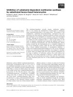
Figure 1 shows that hA induced time-dependent activa-
tion of p38 kinase, which was detectable from 1 h,
peaked at 4 h, then declined by 8 h and had returned
to the 1-h level by 16 h after treatment. In contrast,
hA did not activate ERK1 ⁄ 2 over the 24-h time course
studied (data not shown). Thus, hA treatment elicited
activation of p38 kinase, but not pERK1 ⁄ 2, in both
RINm5F and CM b-cells. Furthermore, nonfibrillogen-
ic rA activated neither ERK1 ⁄ 2 nor p38 kinase activ-
ity, as shown in Fig. 1, indicating that sequence
differences between hA and rA and the fibrillogenic
potential of the human peptide are required for hA-
induced p38 kinase activation. In contrast, the same
effects are not observed when b-cells are exposed to
solutions containing large mature hA fibrils, prepared
by dissolution in NaCl ⁄ P
i
and 7-day incubation prior
(data not shown). This finding is consistent with the
current view that the early aggregates rather than the
mature fibrils, are the primary toxic species [20,44].
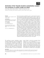
Similar results regarding hA-elicited p38 kinase acti-
vation were obtained in studies wherein an immunocom-
plex kinase assay was employed (shown in Fig. 2A).
Here, p38 kinase immunoprecipitated from 4-h hA-
treated cells catalyzed phosphorylation of glutathione
S-transferase (GST)-ATF-2, whereas phosphorylation
of GST-Elk-1 mediated by ERK1 ⁄ 2 immunoprecipita-
tion did not increase with 4-h treatment and did not
differ from that in untreated controls. These results are
consistent with the observations obtained from direct
western blot analysis described above. In addition, a
dose-dependence experiment showed that p38 kinase
activity increased with hA concentrations (Fig. 2B),
demonstrating a dose-responsive effect of hA on activa-
tion of p38 kinase.
In parallel experiments, we studied the effects of inhi-
bition of ERK1 ⁄ 2 and p38 kinase on hA-induced
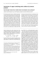
caspase-3 activation and apoptosis. Figure 3 shows that
pretreatment of RINm5F or CM cells with the selective
p38 kinase-inhibitor SB203580 for 1 h prior to hA expo-
sure significantly inhibited apoptosis by 32% (Fig. 3A)
and caspase-3 activation by 30% (Fig. 3B). In contrast,
no pretreatment with either SB202474 (negative inhib-
itor-control) or PD98059 (inhibitor of the kinase
upstream from ERK1 ⁄ 2) elicited increased apoptosis or
caspase-3 activation when compared with non-pretreat-
ed controls. Thus, activation of p38 kinase, but not the
ERK pathway, contributes to the molecular mechanism
through which hA induces b-cell apoptosis. The inhibi-
tory effect of SB203580 was incomplete, however, indi-
cating that p38-kinase activation is not the only
mechanism by which hA induces b-cell apoptosis. We
detected a further reduction in caspase-3 activation and
apoptosis in cells pretreated with combined SB203580
and JNK inhibitor I ( 70% reduction in total), and full
suppression with the combination of SB203580, JNK
inhibitor I and caspase-8 inhibitor (Fig. 3A,B). In addi-
tion, treatment of cells with hA and JNK inhibitor I can
ratio
1.0
2.9
4.8
3.3
1.6
0.6
-p-p38
CM
0h
1h
2h 4h
8h 16h
24h 1h
4h
8h
24h
hA
rA
-p-p38
ratio
1.0
2.1
3.7
1.9
1.2 0.5
RINm5F
Fig. 1. Western blot analysis of p-p38 kinase protein in RINm5F
and CM cells. Total cell extracts were prepared from cells treated
with 10 l
M hA or rA at the indicated time-points and subjected to
western blot analysis using anti-p-p38 kinase IgG. Fold induction of
p-p38 kinase (shown as a ratios) was calculated based on levels at
1 h, which were set at one. All results shown are the average of
three independent experiments.
S. Zhang et al. ATF-2 activation mediates hA-evoked apoptosis
FEBS Journal 273 (2006) 3779–3791 ª 2006 The Authors Journal compilation ª 2006 FEBS 3781
significantly but not fully suppress caspase-3 activity
and apoptosis, whereas the inhibitors themselves did
not prevent apoptosis in the absence of hA (data not
shown). These findings support a mechanism in which
multiple apoptotic pathways, including those mediated
via JNK, p38 and initiator caspase-8, cooperate to
mediate hA-evoked b-cell apoptosis.
Increased phosphorylation of ATF-2 in response to
hA treatment is catalyzed mainly by p38 kinase
We examined protein expression and phosphorylation
of ATF-2 by p38 kinase during hA-evoked b-cell
0
1
2
3
4
5
6
7
8
0 5 10 20 40 0 5 10 20 40
Relative phosphorylation
p38 immunoprecipitated
hA
rA
*
*
*
*
*
*
μΜ
CM RINm5F
0
0.5
1
1.5
2
2.5
3
3.5
4
4.5
co hA rA co hA rA
Relative phosphorylation
CM
RINm5F
*
*
GST-ATF-2 (p38 immunoprecipitated)
GST-Elk-1 (ERK immunoprecipitated)
A
B
Fig. 2. Immunocomplex kinase assay for hA-induced ERK and p38
kinase activity. (A) Total cell extracts were prepared from RINm5F
and CM cells untreated (co) or treated with 10 l
M of hA or rA for
4 h. Whole cell kinase activity assay was performed using ERK and
p38 kinase immunoprecipitated with c-
32
P-ATP and GST-Elk-1 (for
ERK assay) or GST-ATF-2 (for p38 kinase assay) as substrates. The
phosphorylation reactions were visualized by autoradiography after
SDS ⁄ PAGE, quantified by PhosphorImager and presented relative
to untreated control. The results are mean ± SEM of three inde-
pendent experiments, each performed in duplicate. *P < 0.01 ver-
sus respective controls. (B) RINm5F and CM cells were untreated
(co) or treated with various concentrations of hA or rA for 4 h as
indicated. p38 kinase activity was measured as described above
using p38 kinase immunoprecipitated with c-
32
P-ATP and GST-
ATF-2 as substrates.
Fig. 3. Effects of MAPK inhibitors on hA-induced apoptosis and acti-
vation of caspase-3. Cultured RINm5F and CM cells were pre-incuba-
ted with specific MAPK inhibitor alone (SB203580, JNK inhibitor I or
PD98059), or combinations of inhibitors (SB203580 + JNK inhibitor I)
or (SB203580 + JNK inhibitor I + caspase-8 inhibitor) or inhibitor-neg-
ative control (SB202474) for 1 h before exposure to hA. (A) Apoptosis
was assessed after 24-h exposure using a quantitative cell death
detection ELISA. Results shown represent enrichment of nucleo-
somes (fragmented DNA). (B) Caspase-3 activity was determined
after 16-h hA-exposure using synthetic fluorogenic oligopeptide sub-
strate z-DEVD-AFC. The fluorescence was measured at excitation
k ¼ 400 nm and emission ¼ 540 nm. All data were presented relat-
ive to the untreated control (co) and calculated as mean ± SEM
of four independent experiments, each performed in duplicate.
†P<0.01 versus control; * P < 0.01 versus hA-treated cells.
ATF-2 activation mediates hA-evoked apoptosis S. Zhang et al.
3782 FEBS Journal 273 (2006) 3779–3791 ª 2006 The Authors Journal compilation ª 2006 FEBS
apoptosis. Figure 4 shows the result of a representative
western blot analysis using specific antibodies for
ATF-2 and phosphorylated activating transcription
factor 2 (p-ATF-2). Human amylin-induced apoptosis
in CM cells was accompanied by time-dependent
increases in phosphorylation (activation) of ATF-2
(Fig. 4A). Phosphorylated ATF-2 had reached max-
imal levels by 4 h after initiation of hA treatment
(four- to five-fold increase), which coincided with the
time at which the level of p-p38 kinase had increased.
Augmented phosphorylation of ATF-2 was also detec-
ted in RINm5F cells after hA treatment (data not
shown). However, no increment in p-ATF-2 level was
detected in cells treated with either vehicle alone or
noncytotoxic rA in either RINm5F or CM cells,
indicating that increased activation of ATF-2 is corre-
lated with induction of apoptosis and the ability of hA
to form b-sheet-containing aggregates. In contrast, we
found that levels of nonphosphorylated ATF-2 were
unaffected by hA treatment throughout the 24-h study
(Fig. 4B). In addition, hA-induced apoptosis was
suppressed by specific antisense ATF-2, demonstrating
the important role played by ATF-2 in cell death
(Fig. 4C). Effects of hA on ATF-2 mRNA expression
were also measured using quantitative RT-PCR, which
showed that tissue ATF-2 mRNA content remained
unchanged throughout this period (data not shown).
Thus, hA treatment had no measurable effect on ATF-2
mRNA or protein expression, and hA-stimulation of
ATF-2 activity was not attributable to enhanced tissue
ATF-2 content.
ATF-2 is a transcription factor whose activation can
be catalyzed by either p38 or JNK, or by both
[27,41]. To further clarify the role of the MAPKs in
hA-evoked activation of ATF-2, we used selective
MAPK inhibitors to ascertain the major upstream
kinase that activates ATF-2. Figure 5A shows that
- pATF-2
ratios
1.0 0.19 0.81 0.79
1.1
oc
Ah
0
8530
2
B
S
+A
h
Ibihni
K
N
J
+Ah
1
knj-
S
A
+Ah
9
5
089
DP+Ah
A
- p-c-JunSer63
ratios
1.0
0.42 0.38 0.45
0.92
B
- c-Jun
1.0 0.37 0.40 0.36 0.96
ratios
- ATF-2
C
D
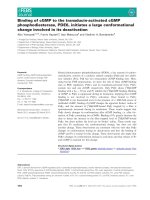
Fig. 5. Effects of inhibition of MAPK on hA-evoked expression and
activation of ATF-2 and c-Jun. (A) CM cells were pre-incubated with
specific MAPK inhibitors (SB203580, PD98059 or JNK inhibitor I)
for 1 h or transfected with AS-jnk1 for 24 h before exposure to hA.
Total cell extracts were prepared and subjected to western blot
analysis using anti-p-ATF-2 IgG. (B) The same western blot mem-
brane as in (A) was stripped and re-probed with anti-p-c-Jun IgG.
(C) Cell treatments were performed as in (A) and western blot ana-
lyzed using anti-ATF-2 IgG. (D) Cell treatments were performed as
in (A) and western blot analyzed using anti-c-Jun IgG. All changes
of protein levels were calculated based on those in corresponding
hA-treated cells, which were set at one. Results shown are the
average of three independent experiments.
Fig. 4. Activation of ATF-2 is required for human amylin-induced
b-cell apoptosis. (A,B) Representative western blot analysis of
time-dependent activation and expression of ATF-2. Total cell
extracts were prepared from CM cells untreated (co) or treated
with 10 l
M hA or rA at the indicated time-points. Western blots
were performed using anti-p-ATF-2 IgG (A) or ATF-2 IgG (B) and
specific protein bands were visualized using ECL chemilumines-
cence reagent. Fold induction of p-ATF-2 (shown as ratios) was cal-
culated based on levels at 1 h, which were set at one. Results
shown are the average of three independent experiments. (C)
RINm5F and CM cells were transfected with antisense ATF-2 (AS-
ATF-2) or sense ATF-2 (S-ATF-2) for 24 h before exposure to hA.
Apoptosis was assessed after 24-h exposure using a quantitative
cell death detection ELISA. All data were presented relative to the
untreated control (co) and calculated as mean ± SEM of four inde-
pendent experiments, each performed in duplicate. †P<0.01
versus control; *P < 0.01 versus hA-treated cells.
S. Zhang et al. ATF-2 activation mediates hA-evoked apoptosis
FEBS Journal 273 (2006) 3779–3791 ª 2006 The Authors Journal compilation ª 2006 FEBS 3783
pretreatment of CM cells with SB203580 caused a
large decline (averaging about 80%) in hA-induced
ATF-2 phosphorylation compared with non-pretreat-
ment controls. Pretreatment of CM cells with JNK
inhibitor I or transfection with antisense jnk1 (AS-
jnk1) caused lesser inhibitory effects (20% decre-
ments), and PD98059 failed to decrease hA-induced
ATF-2 phosphorylation at all, indicating that ATF-2
phosphorylation is catalyzed mainly by p38 kinase
and, to a lesser extent, by JNK1. Similar effects were
observed following pretreatment with MAPK inhibi-
tors of RINm5F cells, wherein we also found that
SB203580 could largely inhibit hA-induced ATF-2
phosphorylation (data not shown). Therefore, p38 kin-
ase rather than JNK is the primary upstream kinase
for ATF-2 in hA-induced b-cell apoptosis.
It is known that, unlike JNK, p38 kinase does not
directly phosphorylate c-Jun [27]. However, we set out
to investigate whether p38 activation has any ultimate
indirect downstream effect on hA-induced activation of
c-Jun. The same western blot membrane, which had
been used for analysis of p-ATF-2 above, was stripped
and re-probed with the p-c-JunSer63-specific antibody.
The results shown in Fig. 5B demonstrate that suppres-
sion of p38 kinase activation by SB203580, as well as
suppression of JNK1 activation by JNK inhibitor I
and AS-jnk1, caused equal inhibition of c-Jun phos-
phorylation. ATF-2 and c-Jun protein levels were ana-
lyzed by western blot, as shown in Fig. 5C,D. ATF-2
protein level was unchanged and c-Jun protein levels
were equivalently decreased by pretreatment with
SB203580, JNK inhibitor I, or AS-jnk1. These data
indicate that the decreased c-Jun phosphorylation
evoked by SB203580 may be due to decreased c-jun
transcription. Furthermore, we found that inhibition of
c-jun expression was more pronounced by simultaneous
treatment with both SB203580 and JNK inhibitor I,
indicating that p38 kinase and JNK1 act co-operatively
to control c-Jun expression and activation. In addition,
pretreatment of PD98059 did not decrease activity of
either p-ATF-2 or p-c-JunSer63 (Fig. 5A,B), further
indicating that hA-elicited activation of c-Jun and
ATF-2 are independent of the ERK pathway.
Activation of ATF-2 is associated with increased
CRE-DNA binding activity
To determine whether increased ATF-2 activation fol-
lowing hA treatment is associated with a change in the
DNA binding activity at the CRE site, nuclear proteins
were extracted from 8-h hA-treated and untreated
RINm5F and CM cells and subjected to electropho-
retic mobility shift assay (Fig. 6A). Two shifted bands
corresponding to two different forms of CRE DNA-
binding complexes were detected. ATF-2-CRE DNA
binding activity was markedly induced following 8-h
hA treatment, as shown by increased intensities of
both shifted bands in hA-treated cells. In addition,
the increased CRE binding activity was suppressed
by JNK inhibitor I and SB 203580, implying that
hA-evoked CRE binding is mediated by both the JNK
and the p38 kinase pathways.
Additionally, supershift assays (antibody pre-incuba-
tions) were performed to determine which types of
CRE-binding protein complexes were induced upon
hA treatment. We detected the appearance of two
supershifted bands, corresponding to the CRE-anti-
body supershifted complexes, in hA-treated cells
following pre-incubation of antibody against p-c-Jun-
Ser63, indicating that both of these shifted CRE com-
plexes contain p-c-JunSer63 (Fig. 6B). We also found
that pre-incubation with antibodies against c-Jun,
ATF-2 or p-ATF-2 enable competition of these anti-
bodies on binding of labeled CRE to protein com-
plexes (shown by weakening in the two shifted bands).
However, pre-incubation of specific blocking peptide
with these antibodies before incubation with nuclear
extract, did not weaken the shift bands (data not
shown). Thus c-Jun and p-c-JunSer63, ATF-2 and
p-ATF-2 are all part of the two CRE-binding com-
plexes in hA-treated cells. Interestingly, the antibodies
for p-c-JunSer63 and p-ATF-2 were more efficient
than the antibodies for unphosphorylated ATF-2 and
c-Jun in supershifting or competing with the CRE
complexes, suggesting that these complexes are mainly
composed of the active forms of c-Jun and ATF-2. In
contrast, only antibodies for unphosphorylated ATF-2
and c-Jun competed with binding of labeled DNA to
the CRE complex from untreated control cells, indica-
ting that the unphosphorylated forms of ATF-2 and
c-Jun are the major components of the complexes
associated with CRE DNA sequences in untreated con-
trol cells. Taken together, our results demonstrate
changes in protein composition and phosphorylation
state of the CRE-binding complexes, with the emer-
gence of functionally significant p-ATF-2 and p-c-Jun-
Ser63 in hA-treated apoptotic cells.
Activation of ATF-2 increases transcriptional
transactivation potential of ATF-2
The correlation of ATF-2 activation with CRE-
mediated transcriptional activity after hA treatment
was studied using a CRE-driven luciferase reporter
construct (pCRE-luc). CM cells were transiently trans-
fected with pCRE-luc and luciferase activity was
ATF-2 activation mediates hA-evoked apoptosis S. Zhang et al.
3784 FEBS Journal 273 (2006) 3779–3791 ª 2006 The Authors Journal compilation ª 2006 FEBS
measured to determine the effects of amylin on
modulation of CRE-mediated transcriptional activity.
Results demonstrate that treatment of transfected cells
with hA caused increased transactivation activity in
comparison with untreated control samples, as meas-
ured by increased production of relative light units of
luciferase activity (Fig. 7). Luciferase activity reached
maximum induction at 8 h (about four-fold increase),
which coincided with the observed elevation in CRE -
binding activity. In contrast, noncytotoxic rA, which
does not elicit ATF-2 activation, had no effect on
CRE-mediated transcriptional activation. Thus, hA
activates CRE-driven gene transcription and the
increased transactivation potential of ATF-2 is correla-
ted with hA’s fibrillogenic and cytotoxic properties.
Also shown in Fig. 7 is evidence that suppression of
p38 kinase by SB203580 inhibits ATF-2-mediated tran-
scriptional activation. Together, these data demon-
strate a role for the p38 kinase-mediated signal
transduction pathway in transcriptional responses
mediated by ATF-2 in hA-treated b-cells. The cooper-
ative effect of JNK and p38 kinase on CRE-mediated
transcriptional activation was also demonstrated by
inhibition of luciferase expression using JNK inhib-
itor I, as well as its simultaneous use with SB203580.
We showed that suppression of CRE-luciferase activity
was more pronounced by combined inhibition of JNK
and p38 kinase (Fig. 7). Control treatment of trans-
fected cells with inhibitors alone had no effect on lucif-
erase activity (data not shown).
To determine whether hA-stimulated activation of
ATF-2 activates ATF-2-dependent transcription of
c-jun, a time course experiment was performed wherein
CM cells were transfected with a c-jun promoter-
chloramphenicol acetyltransferase (CAT) reporter
construct. CAT activity was measured at various time-
points in transfected-cells pretreated with SB203580,
which selectively inhibits phosphorylation of ATF-2
but not of c-Jun, and hA stimulated ATF-2-mediated
c-jun expression (Fig. 8). The maximum induction of
CAT activity was about three- to four-fold above con-
trol values following 8 h of exposure, whereas in
contrast, CAT activity remained consistently low in
rA-treated b-cells. The time at which the CAT activity
p
oc
hA
ogilocificeps+Ah
ogilocificeps-non+Ah
Ib
ihniKN
J+Ah
08
5302BS+Ah
oc
Ah
ogilocific
e
ps+Ah
ogilocificeps-non+A
h
Ib
i
hniKNJ+Ah
0
8
5
3
02BS
+Ah
RINm5F
CM
_
_
CRE-
complexes
_
_
A
nu
J
-c-i
t
na+oc
2
-FT
A
-itna+oc
nuJ-c-p-itna+oc
2-FTA-p-itna+oc
nuJ-c-itna+Ah
2-FTA-itna+Ah
nuJ-
c
-p-it
n
a+A
h
2-FT
A-p-itna+Ah
p
_
_
CRE-
complexes
_
_
p-c-Jun-CRE-
complexes
oc
Ah
_
_
_
_
B
Fig. 6. Representative electrophoretic mobility shift assays of CRE-DNA binding activated by hA. (A) The binding reactions were performed
using nuclear extracts prepared from RINm5F and CM cells that had been treated with hA or vehicle control (co) for 8 h. Nuclear extracts
were also prepared from cells that had been pre-incubated with SB203580 or JNK inhibitor I (JNK inhib I) before treatment with hA. For the
assay of CRE binding specificity, nuclear extracts were incubated for 1 h with unlabelled specific or nonspecific oligonucleotides, respect-
ively, before addition of labeled CRE probe. P denotes reaction containing only labeled CRE probe without nuclear extract. (B) Supershift
experiments were carried out by incubation of nuclear extracts with different antibodies for 1 h, before addition of labeled CRE probe. The
binding reaction samples were then analyzed as described above.
S. Zhang et al. ATF-2 activation mediates hA-evoked apoptosis
FEBS Journal 273 (2006) 3779–3791 ª 2006 The Authors Journal compilation ª 2006 FEBS 3785
reached maximal coincided with the observed elevation
in CRE binding and CRE-mediated transcriptional
activity. SB203580, a suppressor of p38 kinase-medi-
ated ATF-2 activation, inhibited c-jun promoter trans-
activation. Thus, activated ATF-2 and p38 kinase play
critical roles in stimulation of c-jun transcription dur-
ing hA-evoked b-cell apoptosis. In addition, control
treatment with inhibitors alone had no effect on CAT
activity (data not shown); hA-induced c-jun promoter
activation was partially suppressed by JNK inhibitor I
and more completely suppressed by combined pretreat-
ment with SB203580 and JNK inhibitor I (Fig. 8),
further indicating that both the JNK- and p38 kinase-
mediated pathways are necessary for hA-induced
transactivation of c-jun gene expression.
Discussion
We have previously shown that hA elicits islet b-cell
apoptosis through activation of c-Jun and the JNK
pathway [15,23]. We have also shown that activated
JNK1 interacts with a caspase cascade in controlling
this apoptotic process [23]. The objective of the current
studies was to clarify possible roles of ERK and p38
kinase and their downstream target ATF-2, in hA-elici-
ted b-cell apoptosis using the same b-cell lines, rat
RINm5F and human CM that we previously employed
in our studies of JNK activation. The CM line was
originally established from ascitic cells taken from a
human subject with a malignant insulinoma [45]. CM
cells express genes typical of the islet b-cell lineage,
such as insulin and certain of the GLUT genes,
respond to glucose stimulation and posses a functional
glucose-signaling pathway, thus representing a good
model for studies of b-cell function and signaling [46].
We show here that, in addition to the JNK pathway,
the p38 kinase pathway is also required for hA-evoked
b-cell apoptosis, whereas no role for the ERK pathway
was apparent. p38 kinase activation is related to the
presence of fibrillogenic hA, and hA-induced activation
of the p38 kinase pathway in b-cells is consistent with
the general role of the p38 kinase pathway in cellular
regulation of antiproliferation and apoptosis. However,
this pathway is only partially p38-dependent and tar-
geting multiple pathways, including caspase-8, JNK
and p38 kinase, is required for complete suppressed of
hA-induced b-cell apoptosis. Our results are supported
by the report that hA, at nanomolar concentrations,
Fig. 7. Analysis of CRE driven luciferase activity induced by hA
treatment. CM cells were transfected with a CRE-driven luciferase
reporter construct (pCRE-luc) for 24 h before exposure to hA, rA or
vehicle control (co) for various times as indicated. Transfected CM
cells were also pre-incubated with SB203580, JNK inhibitor I or
with combination of SB203580 and JNK inhibitor I before exposure
to hA. Cell lysates were then prepared and analyzed using a lucif-
erase reporter gene assay system. Resulting values, shown as
relative light units, are mean ± SEM of four independent experi-
ments, each performed in duplicate. †P<0.01 versus control;
*P < 0.01 versus hA-treated cells.
Fig. 8. Analysis of ATF-2 dependent c-jun promoter activation in hA
treated CM cells. Cells were transfected with a c-jun promoter-CAT
reporting construct for 24 h before exposure to hA, rA or vehicle
control (co) for various times as indicated. Transfected CM cells
were also pre-incubated with SB203580, JNK inhibitor I or with
combination of SB203580 and JNK inhibitor I before exposure to
hA. Cell lysates were then prepared and analyzed using a CAT Elisa
kit. Results were presented as relative CAT activities based on
untreated control levels, which were set at one. All values are
mean ± SEM of four independent experiments, each performed in
duplicate. †P<0.01 versus control; *P < 0.01 versus hA-treated
cells.
ATF-2 activation mediates hA-evoked apoptosis S. Zhang et al.
3786 FEBS Journal 273 (2006) 3779–3791 ª 2006 The Authors Journal compilation ª 2006 FEBS
induced strong and sustained phosphorylation of JNK
and p38 kinase in RINm5F cells [24]. Consistent with
our current results, these data also indicate that ERK
activation does not play a role in hA-induced RINm5F
cell apoptosis, although therein an early ERK activa-
tion was detected at which the effect was not concom-
itant with JNK and ⁄ or p38 activation [24].
We have shown here that hA elicits distinct and spe-
cific effects on phosphorylation of ATF-2 by p38 kin-
ase-mediated signaling pathways, although a lesser
effect of JNK was also detected. Alterations in ATF-2
phosphorylation correspond closely with the previously
observed pattern of changes in the levels of phosphor-
ylated c-JunSer63 [15]. p-ATF-2 has been identified
previously as part of the AP-1 complex that regulates
AP-1-mediated transcriptional activation evoked by
hA in apoptotic b-cells [15]. Collective results from
previous and current studies indicate that ATF-2 could
form homodimers with itself or heterodimers with
c-Jun to bind to the specific AP-1 and CRE consensus
sites in the promoter regions of target genes, including
those of c-jun. Inhibition of p38 kinase by SB203580,
which decreases ATF-2 phosphorylation, could sup-
press induction of CRE binding and c-jun promoter
activation in response to hA, consistent with the ability
of activated-ATF-2 to transactivate the expression of
target genes. Although p38 kinase does not directly
phosphorylate c-Jun, we detected a decrease in c-Jun
activation as a result of pretreatment of b-cells with
SB203580. This is likely because the suppression in
p38 kinase activity caused by SB203580 can cause
decreased p-ATF-2, which in turn lessens its binding
to and transcriptional activation of the c-jun promoter.
The resulting decrease in c-jun expression would cause
diminished amounts of c-Jun protein to be available
for JNK1-mediated phosphorylation. In addition, our
studies with JNK and p38 kinase inhibitors showed
that effects of direct and indirect inhibition of c-Jun
phosphorylation were similar, indicating that there is
no major competition between JNK and p38 kinase on
direct and indirect activation of c-Jun. Furthermore,
JNK is responsible for increasing the activity of c-Jun
during hA-evoked b-cell apoptosis, as shown by our
previous study [15]. However, although both JNK and
p38 kinase elicited phosphorylation of ATF-2, our cur-
rent data show that p38 is more important for the acti-
vation of ATF-2 evoked by hA in both our b-cell
systems. Thus, these parallel pathways may well con-
verge at AP-1 and CRE sites, mediating hA-induced
induction of expression of their target genes, including
c-jun as demonstrated herein.
The composition of the ATF-2-associated transcrip-
tion factor complexes may differ between various
physiological and pathological states, so that even clo-
sely related members of the same protein family may
contribute to quite distinct biological phenomena. We
have demonstrated changes in the protein composition
and phosphorylation state of the CRE complex during
hA-induced b-cell apoptosis. The two shifted bands,
corresponding to hA-induced CRE-binding complexes
detected here, may represent different dimers formed
from some of the identified components, including
c-Jun, p-c-Jun, ATF-2 and p-ATF-2. This supports
our idea that variation in CRE-complex composition
and phosphorylation between hA-treated and
untreated cells, can result in formation of different
dimers that may have distinguishable CRE-binding
specificity and activity. Moreover, changes in the
composition or phosphorylation state of CRE com-
plexes can modulate their transcriptional activity and
thereby alter target-gene specificity, leading to apopto-
sis in hA-treated b-cell systems.
Apoptosis is an important form of b-cell death in
diabetes. Formation of islet amyloid, rather than the
presence of islet amyloid per se, was related to
increased b-cell apoptosis in a mouse model of T2DM
[5]. We expect that our current investigations into the
molecular mechanisms relating the structure of amylin
aggregates ⁄ oligomers to their function and the associ-
ated b-cell apoptosis will ultimately lead to a better
understanding of the causes of b-cell failure and islet
dysfunction in T2DM. These insights may allow the
development of new approaches to preserve islet b-cell
survival in vivo. Moreover, the current findings may
also be relevant to other forms of amyloid-associated
cell death, such as occur in Alzheimer’s disease and
the prion encephalopathies.
Experimental procedures
Cell culture treatments
For amylin treatment, peptide solutions were prepared by
dissolving hA (Lot 524836; Bachem, Torrance, CA, USA) or
rA (Lot ZM275; Bachem) in water and incubation at room
temperature for 10 min, as previously described [15,16]. Rat
and human insulinoma cell lines, RINm5F and CM, were
cultured and treated with hA or rA as previously described
[15,16]. Both cell lines were originally derived from trans-
formed b-cells, and retain numerous differentiated features
of their cell lineage (e.g. insulin synthesis and secretion).
For MAPK-inhibitor treatment, a selective p38 kinase
inhibitor (SB203580), a selective ERK inhibitor (PD 98059)
or negative inhibitor control (SB 202474) (Calbiochem, La
Jolla, CA, USA) were prepared by dissolution in dimethyl
sulfoxide. The inhibitors were then added to RINm5F or
S. Zhang et al. ATF-2 activation mediates hA-evoked apoptosis
FEBS Journal 273 (2006) 3779–3791 ª 2006 The Authors Journal compilation ª 2006 FEBS 3787
CM cell cultures, to final concentrations of 10 lm or
100 lm, respectively, 1 h before exposure to hA. Alternat-
ively, JNK inhibitor I or JNK inhibitor I-negative control
peptides (Calbiochem) were dissolved in water and applied
to cell cultures to final concentrations of 1 lm, 1 h before
hA addition. The doses selected have been tested and treat-
ments with inhibitors alone shown to have no effect on
b-cell proliferation and viability.
Quantitative cell death detection ELISA
RINm5F and CM cells were cultured in 96-well plates in
the presence or absence of specific MAPK inhibitors for
1 h before exposure to hA for 24 h as described above. For
ATF-2 antisense and sense oligonucleotide transfection,
cells were incubated with 0.2 lm phosphothiorate-modified
antisense and sense ATF-2 (antisense: CACATGTAACTT
GAATTTCAT and sense: ATGAAATTCAAGTTACAT
GTG) using lipofectin reagent as previous described for
transfection of antisense c-jun [15]. Cells were then exposed
to hA and apoptotic cell death measured using a cell death-
detection ELISA system (Roche Applied Science, Man-
nheim, Germany) as previously described [15,23].
Caspase-3 activity assay
RINm5F and CM cells were cultured on 24-well plates in
the presence or absence of specific MAPK inhibitors for
1 h before exposure to synthetic hA for 16 h, as described
above. Cells were then lyzed and caspase-3 activity assays
performed as previously described [23]. One hundred micro-
grams of each cell extract was incubated in reaction buffer
containing 40 ngÆlL
)1
of the specific fluorogenic caspase-3
substrate, Ac-DEVD-AFC (Bio-Rad Hercules, CA, USA),
in the presence or absence of a caspase-3 inhibitor
(z-DEVD-FMK), in a black 96-well plate at 37 °C for 3–
4 h. Caspase activity was determined by measuring the
AFC released using a fluorescence MP reader (Spectra Max
Gemini XS; Molecular Devices: excitation at 400 nm; emis-
sion at 540 nm).
Western blot analysis
RINm5F and CM cells were untreated, or treated with
MAPK inhibitors as indicated, before exposure to hA or
rA. CM cells were also transfected with AS-jnk1 as previ-
ously described, before exposure to hA [23]. Total cell
lysates were then prepared and protein concentrations
determined as previously described [15,23]. Twenty-five
micrograms of each whole-cell extract were separated by
12% SDS ⁄ PAGE, and Ponceau S staining was performed
to confirm the equal loading. Western blots were performed
using either rabbit anti-p-p38 kinase (Cell Signaling, Bev-
erly, MA, USA), rabbit anti-p-ERK1 ⁄ 2 (Cell Signaling),
rabbit anti-ATF-2 (Santa Cruz Biotechnology, Santa Cruz,
CA, USA), rabbit anti-p-ATF-2 (Cell Signaling), mouse
anti-p-c-JunSer63 (Santa Cruz Biotechnology) or rabbit
anti-c-Jun (Oncogene Science, San Diego, CA, USA).
Specific signals were detected using a horseradish peroxi-
dase-conjugated secondary anti-rabbit or anti-mouse IgG
(Jackson Immuno Research, Soham, UK) and an enhanced
ECL reagent according to the manufacturer’s instructions
(Roche Applied Science). Intensities of the reactive bands
were determined by scanning autoradiography on an ima-
ging densitometer (ScanMaker, Microtek).
Immunocomplex kinase assay
RINm5F and CM cells were cultured in six-well plates and
exposed to hA as described above. Cells were lyzed and kin-
ase activities of p38 and ERK1 ⁄ 2 determined by in vitro im-
munocomplex kinase assay, as described [47]. Briefly, 100 lg
of protein from each cell extract were incubated with 2 lgof
an antibody for ERK1 ⁄ 2 or p38 kinase for 2 h at 4 °C,
respectively, in the presence of protein A–Sepharose (Amer-
sham Biosciences, Uppsala, Sweden). The immunocomplexes
were then collected by centrifugation and resuspended in
30 lL of kinase reaction buffer (20 mm Hepes, pH 7.5,
20 mm b-glycerophosphate, 10 mm p-nitrophenol phosphate,
5mm MgCl
2
,1mm 2-mercaptoethanol and 50 lm Na
3
VO
4
),
containing 10 lCi of c-
32
P-ATP (Amersham Biosciences).
Following incubation for 30 min at 30 °C with 5 lg of GST-
ATF-2 (for p38 kinase assay) or GST-Elk-1 (for ERK1 ⁄ 2
assay), the reactions were terminated by addition of
SDS ⁄ PAGE sample buffer and heating for 5 min at 95 °C.
The samples were then analyzed using 12% SDS ⁄ PAGE.
The protein bands (phosphorylated substrates) were ana-
lyzed using a phosphorImager (FLA 2040 Fuji, Japan).
Electrophoretic mobility shift assay
Cells were grown in T25 tissue culture flasks and either left
untreated or treated with MAPK inhibitors before exposure
to hA, as described above. Cells were then harvested for
preparation of nuclear extracts, as described [15]. The dou-
ble-stranded DNA-binding probe for the CRE complex was
5¢-end labeled with c-
32
P-ATP using T4 polynucleotide kin-
ase (Invitrogen, Carlsbad, CA, USA). The top strand con-
sensus sequence for complex binding was: 5¢-TCGATT
GGC
TGACGTCAGAGAGAG-3, where the CRE binding
site is underlined. CRE binding reaction and electrophoretic
mobility shift assays were carried out as previously des-
cribed [15]. For competition experiments, unlabelled oligo-
nucleotides, either specific (containing the CRE sequence),
or nonspecific [containing the SP1 binding site (5¢-AT
TCGATCGGGGCGGGGCGAGC-3¢) in 200-fold excess],
were added to the reaction before addition of the labeled
probe. For supershift experiments, specific antibodies (anti-
ATF-2 activation mediates hA-evoked apoptosis S. Zhang et al.
3788 FEBS Journal 273 (2006) 3779–3791 ª 2006 The Authors Journal compilation ª 2006 FEBS
ATF-2, anti-p-ATF2, anti-c-Jun, anti-p-JunSer63) were
mixed with nuclear protein extract for 1 h prior to the addi-
tion of the labeled probe. A total of 15 lL of the binding
reaction mixture was then electrophoresed on 5% nondena-
turing polyacrylamide gels and the DNA-protein binding
signals were visualized by phosphor-imaging (FLA 2040,
Fuji, Japan).
Construction of c-jun promoter-reporting
construct
Total genomic DNA from human CM cells was isolated
using DNAzol reagent (Invitrogen) as previously described
[16]. A 636 nt DNA fragment for the c-jun promoter was
generated by PCR using primers: 5¢-CCCAAAACCACTG
GCCTGGTTC-3¢ and 5¢-CACAGGCGCTAGATCTGGG
CAG-3¢. This fragment was then cloned into a promoter
activity assay vector pOPI3CAT (Stratagene, La Jolla, CA,
USA) between BstX I and Bgl II restriction sites using
standard molecular cloning techniques [48]. The cloned
c-jun promoter sequences were verified by DNA sequencing
(DNA sequencing service, Centre for Genomics and Proteo-
mics, the University of Auckland, New Zealand).
Luciferase reporter gene assay
CM cells were plated (12-well plates at a density of 5 · 10
4
cellsÆwell
)1
) one day before transfection. A total of 1.5 lg
of either luciferase-reporter gene construct (pCRE-Luc;
Stratagene) or vector DNA was transfected into cells using
Fugene 6 reagent (Roche) according to the manufacturer’s
protocol. Transfection efficiency was checked by cotransfec-
tion with 0.5 lg of pEGFP plasmid DNA (Clontech,
Mountain View, CA, USA) and green fluorescent protein
(GFP) expression measured (excitation at 395 nm; emission
at 510 nm). Cells were pretreated with inhibitors and
exposed to hA or rA for 1, 4, 8, 16 and 24 h, beginning
24 h after transfection. Luciferase activity assays were per-
formed using a highly sensitive luciferase reporter gene
assay system (Roche) as previously described. Luciferase
activity was determined by measuring light emission at
562 nm using a luminescence MP reader (SpectraMAX
Gemini XS; Molecular Devices), and the results were nor-
malized relative to the levels of GFP expression.
CAT activity assay
A total of 1.0 lg of either c-jun promoter-CAT reporter
construct or vector DNA was transfected into cells using
Fugene 6 reagent (Roche) according to the manufacturer’s
protocol. Transfection efficiency was checked by cotransfec-
tion with 0.5 lg of pEGFP plasmid DNA (Clontech) as
described above. CM cells were subsequently pretreated
with inhibitors and exposed to hA or rA for 1, 4, 8, 16 and
24 h, 24 h after transfection. Cells were then harvested and
CAT activity assays performed using a CAT Elisa kit
(Roche). Briefly, cell extracts were prepared using the lysis
buffer provided and 200 lg of each sample were incubated
in the anti-CAT-coated MP modules (covered with foil) for
1 h at 37 °C, followed by washing and addition of 200 lL
of anti-CAT-DIG working solution. The MP modules were
further incubated for 1 h at 37 °C and re-rinsed. Two hun-
dred microliters of anti-digoxigenin-peroxidase (anti-DIG-
POD) was then added to each well and incubated a further
1 h at 37 °C. After washing, 200 lL of POD substrate 2,2¢-
azino-di-[3-ethylbenzthiazoline sulfonate (6)]diammonium
salt (ABTS) with substrate enhancer was added to each well
and incubated with shaking for about 30 min at room tem-
perature to enable photometric reaction. Absorbance was
measured at k ¼ 405 nm (MP reader; SPECTRA MAX
340, Molecular Devices). Results were normalized relative
to the levels of GFP expression as described above.
Statistical analysis
All results are presented as mean ± sem. Differences
between experimental groups were analyzed by paired Stu-
dent’s t-tests or, in the case of multiple comparisons, by
anova followed by Dunnett’s or Tukey’s post hoc multiple
comparisons tests (to analyze more than two conditions) as
appropriate. Statistical significance was determined at
P < 0.05.
Acknowledgements
We wish to thank P. Pozzilli (Department of Diabetes
and Metabolism, St Bartholomew’s Hospital, London,
UK) for kindly providing the CM cells and H. K. Oie
(NIH, Bethesda, MD, USA) for kindly providing the
RINm5F cells. We thank X. Li for her enthusiastic
assistance with statistical analyses. This work was sup-
ported by the Endocore Research Trust, the Maurice
and Phyllis Paykel Trust, and the New Zealand Lot-
tery Grants Board, and by Programme Grants from
the New Zealand Health Research Council to GC and
MD, and from the Foundation for Research, Science
and Technology (NZ) to GC.
References
1 Butler AE, Janson J, Bonner-Weir S, Ritzel RA, RR &
Butler PC (2003a) Beta cell deficit and increased beta-
cell apoptosis in human with type 2 diabetes. Diabetes
52, 102–110.
2 Kahn SE (2003) The relative contributions of insulin
resistance and beta-cell dysfunction to the pathophysiol-
ogy of type 2 diabetes. Diabetologia 46, 3–19.
S. Zhang et al. ATF-2 activation mediates hA-evoked apoptosis
FEBS Journal 273 (2006) 3779–3791 ª 2006 The Authors Journal compilation ª 2006 FEBS 3789
3 Cooper GJS (1994) Amylin compared with calcitonin
gene-related peptide: structure, biology, and relevance
to metabolic disease. Endocrine Rev 15, 163–201.
4 Marzban L, Park K & Verchere CB (2003) Islet amyloid
polypeptide and type 2 diabetes. Exp Gerontol 38, 347–
351.
5 Butler AE, Janson J, Soeller WC & Butler PC (2003b)
Increased beta-cell apoptosis prevents adaptive increase
in beta-cell mass in mouse model of type 2 diabetes –
evidence for role of islet amyloid formation rather than
direct action of amyloid. Diabetes 52, 2304–2314.
6 Cooper GJS, Willis AC, Clark A, Turner RC, Sim RB
& Reid KBM (1987) Purification and characterization
of a peptide from amyloid-rich pancreases of type 2
diabetic patients. Proc Natl Acad Sci USA 84, 8628–
8632.
7 Lukinius A, Wilander E, Westermark GT, Engstro
¨
mU
& Westermark P (1989) Co-localization of islet amyloid
polypeptide and insulin in the B cell secretory granules
of the human pancreatic islets. Diabetologia 32, 240–
244.
8 Westermark P, Wernstedt C, Wilander E, Hayden DW,
O’Brien TD & Johnson KH (1987) Amyloid fibrils in
human insulinoma and islets of Langerhans of the dia-
betic cat are derived from a neuropeptide-like protein
also present in normal islet cells. Proc Natl Acad Sci
USA 84, 3881–3885.
9 Goldsbury CS, Cooper GJS, Goldie KN, Mu
¨
ller SA,
Saafi EL, Gruijters WTM, Misur MP, Engel A, Aebi U
& Kistler J (1997) Polymorphic fibrillar assemblies of
human amylin. J Struct Biol 119, 17–27.
10 Lorenzo A, Razzaboni B, Weir GC & Yankner BA
(1994) Pancreatic islet cell toxicity of amylin associated
with type-2 diabetes mellitus. Nature 368, 756–760.
11 Saafi EL, Konarkowska B, Zhang S, Kistler J & Cooper
GJS (2001) Ultrastructural evidence that apoptosis is
the mechanism by which human amylin evokes death in
RINm5F pancreatic islet b-cells. Cell Biol Intl 25, 339–
350.
12 Kapurniotu A (2001) Amyloidogenicity and cytotoxicity
of islet amyloid polypeptide. Biopolymers 60, 438–459.
13 Janson J, Soeller WC, Roche PC, Nelson RT, Torchia
AJ, Kreutter DK & Butler PC (1996) Spontaneous dia-
betes mellitus in transgenic mice expressing human islet
amyloid polypeptide. Proc Natl Acad Sci USA 93,
7283–7288.
14 Goldsbury C, Kistler J, Aebi U, Arvinte T & Cooper
GJS (1999) Watching amyloid fibrils grow by time-lapse
atomic force microscopy. J Mol Biol 285, 33–39.
15 Zhang SP, Liu JX, MacGibbon G, Dragunow M &
Cooper GJS (2002) Increased expression and activation
of c-Jun contributes to human amylin-induced apoptosis
in pancreatic islet beta-cells. J Mol Biol 324, 271–285.
16 Zhang SP, Liu JX, Saafi EL & Cooper GJS (1999)
Induction of apoptosis by human amylin in RINm5F
islet beta-cells is associated with enhanced expression of
p53 and p21 (WAF1 ⁄ CIP1). FEBS Lett 455, 315–320.
17 Verchere CB, D’Alessio DA, Palmiter RD, Weir GC,
Bonner-Weir S, Baskin DG & Kahn SE (1996) Islet
amyloid formation associated with hyperglycemia in
transgenic mice with pancreatic beta cell expression of
human islet amyloid polypeptide. Proc Natl Acad Sci
USA 93, 3492–3496.
18 Janciauskiene S & Ahren B (2000) Fibrillar islet amy-
loid polypeptide differentially affects oxidative mechan-
isms and lipoprotein uptake in correlation with
cytotoxicity in two insulin-producing cell lines. Biochem
Biophys Res Commun 267
, 619–625.
19 Sparr E, Engel MFM, Sakharov DV, Sprong M, Jacobs
J, de Kruijff B, Hoppener JWM & Killian JA (2004)
Islet amyloid polypeptide-induced membrane leakage
involves uptake of lipids by forming amyloid fibers.
FEBS Lett 577, 117–120.
20 Janson J, Ashley RH, Harrison D, McIntyre S & Butler
PC (1999) The mechanism of islet amyloid polypeptide
toxicity is membrane disruption by intermediate-sized
toxic amyloid particles. Diabetes 48, 491–498.
21 Demuro A, Mina E, Kayed R, Milton SC, Parker I &
Glabe CG (2005) Calcium dysregulation and membrane
disruption as a ubiquitous neurotoxic mechanism of
soluble amyloid oligomers. J Biol Chem 280, 17294–
17300.
22 Konarkowska B, Aitken JF, Kistler J, Zhang S &
Cooper GJS (2005) Thiol reducing compounds prevent
human amylin-evoked cytotoxicity. FEBS J 272, 4949–
4959.
23 Zhang SP, Liu JX, Dragunow M & Cooper GJS (2003)
Fibrillogenic amylin evokes islet beta-cell apoptosis
through linked activation of a caspase cascade and
JNK1. J Biol Chem 278, 52810–52819.
24 Rumora L, Hadzija M, Barisic K, Maysinger D & Gru-
biic TZ (2002) Amylin-induced cytotoxicity is associated
with activation of caspase-3 and MAP kinases. Biol
Chem 383, 1751–1758.
25 Cowan KJ & Storey KB (2003) Mitogen-activated pro-
tein kinases: new signaling pathways functioning in cel-
lular responses to environmental stress. J Exp Biol 206 ,
1107–1115.
26 Xia Z, Dickens M, Raingeaud J, Davis RJ & Greenberg
ME (1995) Opposing effects of ERK and JNK-p38
MAP kinases on apoptosis. Science 270, 1326–1331.
27 Davis RJ (1993) The mitogen-activated protein kinase
signal transduction pathway. J Biol Chem 268, 14553–
14556.
28 Hill CS & Treisman R (1995) Transcriptional regulation
by extracellular signals: mechanisms and specificity. Cell
80, 199–211.
29 Ichijo H, Nishida E, Irie K, Dijke PT, Saitoh M,
Moriguchi T, Takagi M, Matsumoto K, Miyazono K &
Gotoh Y (1997) Induction of apoptosis by ASK1, a
ATF-2 activation mediates hA-evoked apoptosis S. Zhang et al.
3790 FEBS Journal 273 (2006) 3779–3791 ª 2006 The Authors Journal compilation ª 2006 FEBS
mammalian MAPKKK that activates SAPK ⁄ JNK and
p38 signaling pathways. Science 275, 90–94.
30 Saldeen J, Lee JC & Welsh N (2001) Role of p38 mito-
gen-activated protein kinase (p38 MAPK) in cytokine-
induced rat islet cell apoptosis. Biochem Pharmacol 61,
1561–1569.
31 Pavlovic D, Andersen NA, Mandrup-Poulsen T &
Eizirik DL (2000) Activation of extracellular signal-
regulated kinase (ERK) 1 ⁄ 2 contributes to cytokine-
induced apoptosis in purified rat pancreatic beta-cells.
Eur Cytokine Network 11, 267–274.
32 Nemoto S, Xiang J, Huang S & Lin A (1998) Induction
of apoptosis by SB202190 through inhibition of p38b
mitogen-activated protein kinase. J Biol Chem 273,
16415–16420.
33 Nishina H, Fischer KD, Radvanyi L, Shahinian A,
Hakem R, Rubie EA, Bernstein A, Mak TW, Woodgett
JR & Penninger JM (1997) stress-signalling kinase Sek1
protects thymocytes from apoptosis mediated by CD95
and CD3. Nature 385, 350–353.
34 Wisdom R, Johnson R, S & Moore C (1999) c-Jun reg-
ulates cell cycle progression and apoptosis by distinct
mechanisms. EMBO J 18, 188–197.
35 Maekawa T, Sakura H, Kanei-Ishii C, Sudo T, Yoshim-
ura T, Fujisawa J, Yoshida M & Ishii S (1989) Leucine
zipper structure of the protein CRE-BP1 binding to the
cyclic AMP response element in brain. EMBO J 8,
2023–2028.
36 Hai T & Curran T (1991) Cross-family dimerization of
transcription factor Fos ⁄ Jun and ATF ⁄ CREB alters
DNA binding specificity. Proc Natl Acad Sci USA 88,
3720–3724.
37 van Dam H, Wilhelm D, Herr I, Steffen A, Herrlich P
& Angel P (1995) ATF-2 is preferentially activated by
stress-activated protein kinases to mediate c-jun induc-
tion in response to genotoxic agents. EMBO J 14,
1798–1811.
38 Walton M, Woodgate AM, Sirimanne E, Gluckman P
& Dragunow M (1998) ATF-2 phosphorylation
in apoptotic neuronal death. Mol Brain Res 63, 198–
204.
39 Livingstone C, Patel G & Jones N (1995) ATF-2 con-
tains a phosphorylation-dependent transcriptional acti-
vation domain. EMBO J 14, 1785–1797.
40 Zayzafoon M, Botolin S & McCabe LR (2002) p38 and
activating transcription factor-2 involvement in osteo-
blast osmotic response to elevated extracellular glucose.
J Biol Chem 277, 37212–37218.
41 Davis RJ (1999) Signal transduction by the c-Jun N-ter-
minal kinase. Biochem Soc Symp 64, 1–12.
42 Goldsbury C, Goldie K, Pellaud J, Seelig J, Frey P,
Muller SA, Kistler J, Cooper GJS & Aebi U (2000)
Amyloid fibril formation from full-length and fragments
of amylin. J Struct Biol 130, 352–362.
43 Bai JZ, Saafi EL, Zhang S & Cooper GJS (1999) Role
of calcium in apoptosis evoked by human amylin in
pancreatic islet b-cells. Biochem J 343, 53–61.
44 Hull RL, Westermark GT, Westermark P & Kahn SE
(2004) Islet amyloid: a critical entity in the pathogenesis
of type 2 diabetes. J Clin Endocrinol Metab 89, 3629–
3643.
45 Gueli N, Toto A, Palmieri G, Carmenini G, Delpino A
& Ferrini U (1987) In vitro growth of a cell line origi-
nated from a human insulinoma. J Exp Clin Cancer Res
6, 281–285.
46 Baroni MG, Cavallo MG, Mark M, Monetini L, Stoeh-
rer B & Pozzilli P (1999) Beta-cell gene expression and
functional characterisation of the human insulinoma cell
line CM. J Endocrinol 161, 59–68.
47 Stadheim TA, Saluta GR & Kucera GL (2000) Role of
c-Jun N-terminal kinase ⁄ p38 stress signaling in 1-b-d-
arabinofuranosylcytosine-induced apoptosis. Biochem
Pharmacol 59, 407–418.
48 Sambrook J, Fritsch EF & Maniatis T (1989) Molecular
Cloning: a Laboratory Manual, 2nd edn. Cold Spring
Harbour Laboratory, Cold Spring Harbor, NY.
S. Zhang et al. ATF-2 activation mediates hA-evoked apoptosis
FEBS Journal 273 (2006) 3779–3791 ª 2006 The Authors Journal compilation ª 2006 FEBS 3791