Báo cáo khoa học: Iron regulatory protein-independent regulation of ferritin synthesis by nitrogen monoxide pot
Bạn đang xem bản rút gọn của tài liệu. Xem và tải ngay bản đầy đủ của tài liệu tại đây (813.62 KB, 9 trang )
Iron regulatory protein-independent regulation of ferritin
synthesis by nitrogen monoxide
Marc Mikhael
1,2
, Sangwon F. Kim
2
, Matthias Schranzhofer
3
, Shan S. Lin
1,4
, Alex D. Sheftel
1,2
,
Ernst W. Mullner
3
and Prem Ponka
1,2
1 Department of Physiology, McGill University, Montreal, Canada
2 Lady Davis Institute for Medical Research, Jewish General Hospital, Montreal, Canada
3 Department of Medical Biochemistry, Division of Molecular Biology, Max F. Perutz Laboratories, Medical University of Vienna, Austria
4 Division of Experimental Medicine, McGill University, Montreal, Canada
Iron (Fe) is essential for life, functioning as a metal
cofactor for many proteins containing either heme
or nonheme iron [1–3]. Hemoproteins have crucial
biological functions, such as oxygen binding, oxygen
metabolism, and electron transfer. Many nonheme
iron-containing proteins catalyze key reactions involved
in energy metabolism and DNA synthesis. However,
the chemical properties of iron which are exploited
for a remarkable range of biological functions have
created problems for living organisms. In excess, cel-
lular ‘free’ iron catalyzes the Haber–Weiss reaction
that can lead to the production of cytotoxic oxygen
radicals [4,5]. The safe storage and sequestration of
iron is therefore an absolute necessity within the cell
Keywords
ferritin; iron; iron regulatory proteins;
nitrogen monoxide; NO
Correspondence
1
P. Ponka, Lady Davis Institute, McGill
University, 3755 Cote Ste-Catherine Road,
Montreal, Quebec, H3T 1E2, Canada
Fax: +1 514 340 7502
Tel: +1 514 340 8260
E-mail:
(Received 2 June 2006, revised 20 June
2006, accepted 22 June 2006)
doi:10.1111/j.1742-4658.2006.05390.x
The discovery of iron-responsive elements (IREs), along with the identifica-
tion of iron regulatory proteins (IRP1, IRP2), has provided a molecular
basis for our current understanding of the remarkable post-transcriptional
regulation of intracellular iron homeostasis. In iron-depleted conditions,
IRPs bind to IREs present in the 5¢-UTR of ferritin mRNA and the
3¢-UTR of transferrin receptor (TfR) mRNA. Such binding blocks the
translation of ferritin, the iron storage protein, and stabilizes TfR mRNA,
whereas the opposite scenario develops when iron in the intracellular tran-
sit pool is plentiful. Nitrogen monoxide (commonly designated nitric oxide;
NO), a gaseous molecule involved in numerous functions, is known to
affect cellular iron metabolism via the IRP ⁄ IRE system. We previously
demonstrated that the oxidized form of NO, NO
+
, causes IRP2 degrada-
tion that is associated with an increase in ferritin synthesis [Kim, S &
Ponka, P (2002) Proc Natl Acad Sci USA 99, 12214–12219]. Here we report
that sodium nitroprusside (SNP), an NO
+
donor, causes a dramatic and
rapid increase in ferritin synthesis that initially occurs without changes in
the RNA-binding activities of IRPs. Moreover, we demonstrate that the
translational efficiency of ferritin mRNA is significantly higher in cells trea-
ted with SNP compared with those incubated with ferric ammonium cit-
rate, an iron donor. Importantly, we also provide definitive evidence that
the iron moiety of SNP is not responsible for such changes. These results
indicate that SNP-mediated increase in ferritin synthesis is, in part, due to
an IRP-independent and NO
+
-dependent post-transcriptional, regulatory
mechanism.
Abbreviations
DFO, desferoxamine; FAC, ferric ammonium citrate; Ft, ferritin; hDFO, high molecular mass version of DFO; IFN, interferon; IRE, iron-
responsive element; IRP, iron regulatory protein; LPS, lipopolysaccharide; NO, nitric oxide; PIH, pyridoxal isonicotinoyl hydrazone;
SIH, salicylaldehyde isonicotinoyl hydrazone; SNP, sodium nitroprusside; TfR, transferrin receptor.
3828 FEBS Journal 273 (2006) 3828–3836 ª 2006 The Authors Journal compilation ª 2006 FEBS
[3,6,7]. Hence, virtually all organisms can synthesize
the icosikaitetrameric protein, ferritin, which can safely
house thousands of iron at oms in a shell-like structure.
Ferritin is a 430–460 kDa protein made up of 24
subunits of heavy (H; 21 kDa) and light (L; 19 kDa)
ferritin chains [3,8]. While both H- and L-ferritin are
involved in incorporating iron, H-ferritin is several
times more efficient than L-ferritin. This difference
appears to be due to a ferroxidase center associated
with the H-ferritin subunit that promotes the oxida-
tion of ferrous iron [9]. By contrast, the L-subunit
has a higher capacity than the H-subunit to induce
iron-core nucleation [10,11], suggesting that both
ferritin chains cooperate in the overall uptake and
storage of iron.
The regulation of ferritin synthesis is largely accom-
plished via an elegant regulatory system that tightly
controls intracellular iron levels. The structurally sim-
ilar iron regulatory proteins 1 and 2 (IRP1 and 2)
function as iron sensors [4–7]. In iron-depleted condi-
tions, IRPs are active and consequently bind specific
nucleotide sequences, iron-responsive elements (IRE),
located in the 5¢-UTR of ferritin mRNA and the
3¢-UTR of transferrin receptor (TfR) mRNA. Such
binding leads to translational repression of ferritin
mRNA and stabilization of the TfR message. Con-
versely, under iron-replete conditions, IRP binding
decreases, leading to TfR mRNA destabilization while
ferritin mRNA is efficiently translated. IRP1 assumes
cytosolic aconitase activity in such iron-replete condi-
tions, whereas IRP2 is targeted for degradation via the
ubiquitin–proteasome pathway [1,2,12,13].
It is well established that IRP-binding activities are
also modulated by noniron stimuli such as hydrogen
peroxide, hypoxia, phosphorylation, and nitric oxide
(NO) [14–21]. NO, in particular, has emerged as an
extraordinary signaling molecule [22,23] whose targets
differ depending on its redox state [24]. The reduced
form of NO, the NO radical (NO
•
), transduces signals
primarily via direct interactions with the iron of heme
moieties in guanylyl cyclase [25–27]; NO
•
also binds to
iron in the iron–sulfur clusters of IRP1 [19,28] and
mitochondrial aconitase [29,30]. Numerous laborator-
ies have shown that NO
•
increases the RNA-binding
activities of IRP1 in many cell types [14,15,18,
20,28,31]. In contrast, oxidized NO, the nitrosonium
ion (NO
+
), reacts with thiol groups of cysteine resi-
dues, typically resulting in a reversible signaling mech-
anism known as S-nitrosylation [24,32]. A multitude of
proteins have been identified as targets of S-nitrosyla-
tion [23,33,34] including IRP2, whose S-nitrosylation
leads to its ubiquitination and subsequent proteosomal
degradation [35].
We have previously shown that macrophages acti-
vated by lipopolysaccharide (LPS) and interferon-c
(IFNc), a condition known to induce NO synthesis
[36], exhibit NO-dependent IRP2 degradation accom-
panied by an increase in ferritin synthesis [20,21,37].
Moreover, sodium nitroprusside (SNP), a NO
+
donor,
was also found to cause IRP2 degradation followed by
a dramatic induction of ferritin synthesis [20,35,37].
Recently, Bourdon et al. [38] proposed that SNP, a
compound containing complexed iron, contributes to
IRP2 degradation by supplying iron to cells. In this
study, we show that the iron component of SNP is not
involved in the stimulation of ferritin synthesis. More-
over, we have discovered that, in RAW
3
264.7 cells (a
macrophage cell line), SNP stimulates ferritin synthe-
sis, at least in part, by a mechanism that does not
require IRP2 degradation. We also report a similar
phenomenon in INFc ⁄ LPS-treated macrophages.
Results
NO
+
-mediated induction of ferritin synthesis
precedes changes in IRP/IRE binding
We have previously shown that treatment of
RAW 264.7 cells with the NO
+
donor, SNP, causes
the degradation of IRP2 associated with an increase of
ferritin synthesis [20,37]. Here, we examined the kinet-
ics of ferritin synthesis and changes in RNA-binding
activities of IRPs in response to SNP exposure for var-
ious time intervals. First, RAW 264.7 cells were trea-
ted with 100 lm SNP for 15–180 min, after which the
cells were thoroughly washed and incubated with [
35
S]-
methionine for 1 h. Figure 1A shows that exposure of
cells to SNP for a time interval as short as 30 min led
to a significant increase in ferritin synthesis levels.
Interestingly, IRP2 degradation was noticeable only
at 2 h following incubation of RAW 264.7 cells with
SNP, whereas the RNA-binding activity of IRP1
remained largely unaffected (Fig. 1B). Surprisingly,
the rapid induction of ferritin synthesis by SNP
in RAW 264.7 cells occurred much earlier than any
decrease of IRP activities could be detected (Fig. 1A,B;
0–60 min). This strongly suggests that SNP-mediated
induction of ferritin synthesis is, at least in part, inde-
pendent of IRP ⁄ IRE regulation.
IFNc
⁄
LPS-mediated ferritin synthesis occurs
without changes in IRP activity
We have also previously shown that a combination of
IFNc and LPS is able to increase ferritin synthesis
in macrophages in an IRP2-dependent manner via the
M. Mikhael et al. IRP-independent effects of NO
+
on Ft synthesis
FEBS Journal 273 (2006) 3828–3836 ª 2006 The Authors Journal compilation ª 2006 FEBS 3829
production of NO by inducible nitric oxide synthase
[21,37]. Because SNP is able to mediate ferritin synthe-
sis before IRP activities are changed, we hypothesized
that endogenously produced NO is also able to repro-
duce such a phenomenon. Indeed, when we treated
RAW 264.7 macrophages with both IFNc and LPS
for as little as 1 h, we observed a more than twofold
induction of ferritin synthesis (Fig. 2A). As expected,
the increase in ferritin synthesis was accompanied
by NO production (nitrite concentrations, Fig. 2A).
Importantly, IRP levels did not change during the first
two hours of IFNc ⁄ LPS treatment (Fig. 2B), suggest-
ing that, like SNP-derived NO, endogenously produced
NO is able to mediate changes in ferritin synthesis
prior to the modulation of IRP activities.
SNP enhances ferritin synthesis even in the
absence of IRP activity
The above experiments indicate that SNP may increase
ferritin synthesis via an IRE ⁄ IRP-independent mechan-
ism. To find further support for this conclusion we
pretreated RAW 264.7 cells with an iron donor, ferric
ammonium citrate (FAC; 50 lgÆmL
)1
) for 18 h,
washed and then incubated them with or without SNP
for an additional 3 h. As expected, pretreatment of
RAW 264.7 cells with FAC for 18 h led to abolish-
ment of IRP-binding activities (Fig. 3A, lane 2) with
a concomitant increase in ferritin synthesis (Fig. 3B,
lane 2). The addition of SNP to FAC-pretreated cells
augmented ferritin synthesis by more than twofold
(Fig. 3B, lanes 2 versus 3) despite similar levels of
IRP-binding activity in both conditions (Fig. 3A, lanes
2 versus 3). This indicates that SNP is able to augment
ferritin synthesis beyond the levels capable solely by
the classical IRE ⁄ IRP system.
The bioavailability of SNP iron is negligible
SNP, which contains iron [Na
2
Fe(CN)
5
NO], is a well-
established NO
+
donor [24,39] that reacts with thiol
groups leading to S-nitrosylation of target proteins
[32,40]. We have previously shown that SNP causes
S-nitrosylation of Cys178 in IRP2, which, in turn, trig-
gers the ubiquitination and degradation of the protein
[35]. It has, however, been suggested that the ability of
SNP to both stimulate IRP2 degradation and induce
ferritin synthesis is accomplished through its iron moi-
ety [38]. Hence, we examined whether SNP releases
A
B
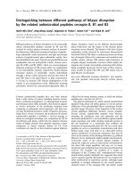
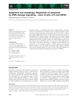
Fig. 2. Effects of IFNc ⁄ LPS on ferritin (Ft) synthesis (A) and IRP-
binding activities (B) in RAW 264.7 cells. (A) Cells were incubated
in the presence of IFNc (100 UÆmL
)1
) and LPS (5 lgÆmL
)1
) for the
indicated time intervals and were then washed and pulse labeled
(1 h) with [
35
S]-methionine and harvested. Ferritin was immunopre-
cipitated by using anti-ferritin IgG and analyzed by SDS ⁄ PAGE fol-
lowed by autoradiography. DA, densitometric analysis, in arbitrary
units. (B) Cells were treated with IFNc ⁄ LPS as in (A) and the pro-
tein extracts assayed for IRE-binding activities using gel-retardation
assays [20]. Nitrate was assayed by using the Greiss reagent as
described by Green et al. [52].
35
S H+L
1.0 1.4 3.2 7.1 15.7 24.8 (D.A.)
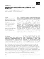
100μ
M SNP
0 15 30 60 120 180 (min)
IRP 1
IRP 2
+β-ME
A
B
100μM SNP
0 15 30 60 120 180 (min)
IRP 1
IRP 2
Fig. 1. Effects of SNP on ferritin synthesis (A) and IRP-binding
activities (B) in RAW 264.7 cells. (A) Cells were incubated in the
presence of SNP (100 l
M) for the indicated time intervals and were
then washed and pulse labeled (1 h) with [
35
S]-methionine and har-
vested. Ferritin was immunoprecipitated by using anti-ferritin IgG
and analyzed by SDS ⁄ PAGE followed by autoradiography. DA, den-
sitometric analysis, in arbitrary units. (B) Cells were treated with
SNP as in (A) and the protein extracts assayed for IRE-binding activ-
ities using gel-retardation assays [20], performed in the absence or
presence of 2% b-mercaptoethanol (b-ME), a condition that reveals
total RNA binding activity of IRP1 [51].
IRP-independent effects of NO
+
on Ft synthesis M. Mikhael et al.
3830 FEBS Journal 273 (2006) 3828–3836 ª 2006 The Authors Journal compilation ª 2006 FEBS
iron which could be responsible for the SNP-mediated
induction of ferritin synthesis. To do so, equimolar
amounts of either ferric citrate or SNP were incubated
with desferoxamine (DFO) in tissue culture medium at
different time intervals during which the absorption
spectra were recorded (Fig. 4A). Iron-laden chelators
exhibit a characteristic absorption pattern (Fig. 4A).
The amount of iron transferred from SNP to the
chelator gradually increased but was extremely slow
(Fig. 4B). Importantly, there was no detectable loss
of iron from SNP in 3 h as no Fe–DFO complexes
were observed at this time (Fig. 4B). Identical results
were obtained using other chelators such as pyrid-
oxal isonicotinoyl hydrazone (PIH), salicylaldehyde
isonicotinoyl hydrazone (SIH) and a high molecular
mass version of DFO (hDFO; data not shown). These
results were corroborated by the experimental out-
come that IRP1 levels are not decreased after the
treatment of RAW 264.7 cells with SNP for 3 h
(Fig. 1A).
Further support for our conclusion that SNP is not
a source of chelatable iron comes from our earlier
observation that DFO, which is commonly used to
intercept intracellular iron, was unable to attenuate
SNP-induced degradation of IRP2 [20]. Here we
exploited hDFO, which is unable to penetrate cell
membranes, to show that hDFO was unable to prevent
SNP-mediated increases in ferritin synthesis (Fig. 5A),
indicating that SNP does not donate iron to the cell
culture medium. Moreover, neither the SNP-like com-
pound, potassium ferricyanide, nor cyanide and nitrate
compounds were able to increase ferritin synthesis
(Fig. 5B) further indicating that it is NO
+
that is
responsible for SNP-mediated induction of ferritin
synthesis.
NO
+
enhances the efficiency of ferritin mRNA
translation
To elucidate the mechanism by which NO
+
derived
from SNP induces ferritin synthesis independent of
the IRP ⁄ IRE system, we examined the levels of ferritin
mRNA associated with polysomes in untreated
RAW 264.7 cells or those treated with either FAC
(50 lgÆmL
)1
,18h)
4
or SNP (100 lm, 3 h). Figure 6
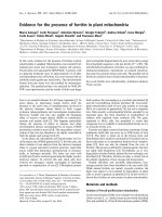
Fig. 4. SNP releases minimal amounts of iron during incubation
with DFO. SNP (100 l
M) or ferric citrate (FC) (100 lM) were incuba-
ted (37 °C) with or without DFO (100 l
M) for various time intervals
following which the Fe–DFO complexes were detected using spec-
trophotometric analysis at wavelengths 350–550 nm. (A) Represen-
tative spectrophotometric profiles of FC, DFO and DFO + FC at
time 0 h. (B) Relative levels of Fe–DFO formation for media with
FC, DFO + FC and DFO + SNP. Absorbance measurements were
taken at 410 nm; the peak that corresponds to Fe–DFO complexes
as observed in (A).
9
Fig. 3. Effect of SNP on IRP-binding activities (A) and ferritin syn-
thesis (B) in control or FAC-pretreated cells. RAW 264.7 cells were
incubated with either control medium or FAC (50 lgÆmL
)1
) for 18 h,
washed with cold NaCl ⁄ P
i
and incubated with either control med-
ium or with SNP (100 l
M) for 3 h. (A) Gel-retardation analysis of
protein (10 lg) extracted from RAW 264.7 cells after different treat-
ments. (B) RAW 264.7 cells, treated as in (A), were pulse labeled
(2 h) with [
35
S]-methionine, and [
35
S]-ferritin was immunoprecipitat-
ed (by using anti-ferritin IgG) and analyzed by SDS ⁄ PAGE followed
by autoradiography.
M. Mikhael et al. IRP-independent effects of NO
+
on Ft synthesis
FEBS Journal 273 (2006) 3828–3836 ª 2006 The Authors Journal compilation ª 2006 FEBS 3831
shows that in cells incubated with FAC, 10% of the
ferritin message can be found in a polysome-bound
form. However, SNP treatment yielded a significantly
elevated fraction of ferritin mRNA associated with
polysomes (50%), indicating that NO
+
increases the
efficiency of ferritin translation significantly above the
levels that can be achieved with iron.
Discussion
It is well known that the inflammatory signals cause
macrophages to produce NO [36,41]. We have pre-
viously shown that IFNc ⁄ LPS-mediated activation of
murine macrophages caused NO-dependent IRP2 de-
gradation [21], and that such changes led to an increase
in ferritin synthesis [20,37]. Moreover, preventing the
degradation of IRP2 by proteasomal inhibitors also
blocked the ferritin synthesis increase [37], indicating
that inflammatory signals in murine macrophages can
activate ferritin synthesis via the degradation of IRP2.
Our laboratory reported that SNP, a NO
+
donor, was
also able to trigger IRP2 degradation followed by an
increase in ferritin synthesis [37]. Such NO-dependent
IRP2 degradation was caused by the S-nitrosylation of
Cys178 which led to ubiquitination of the protein fol-
lowed by its degradation in the proteosome [35]. These
results suggest that NO-mediated IRP2 degradation is
largely responsible for the increase in ferritin synthesis
in both SNP and IFNc ⁄ LPS-treated macrophages.
In this report, we show that SNP enhances ferritin
synthesis not only by the mechanism involving IRP2
degradation, but also by an IRP ⁄ IRE-independent pro-
cess. We show that treatment of RAW 264.7 cells with
SNP increases ferritin synthesis much faster than IRP
activity decreases (Fig. 1). In addition, we also show
that IFNc ⁄ LPS treatment for as little as 1 h is able
to produce a similar phenomenon, whereby ferritin
5
levels increase more than twofold without any signifi-
cant change in IRP ⁄ IRE-binding activities (Fig. 2).
Fig. 5. hDFO does not block the induction of ferritin synthesis in
SNP-treated RAW 264.7 cells (A); effects of various control com-
pounds on ferritin synthesis are also shown (B). Cells were incuba-
ted with SNP or various other reagents [hDFO, K
3
Fe(CN)
6
, KCN,
NaCN, NaNO
3
] for 3 h, following which they were washed and
then pulse-labeled (1 h) with [
35
S]-methionine and harvested. Fer-
ritin was immunoprecipitated by using anti-ferritin IgG and analyzed
by SDS ⁄ PAGE followed by autoradiography.
Fig. 6. Polysome profiles of mRNAs isolated
by sucrose gradient fractionation. RNA was
extracted from RAW 264.7 cells treated
with either FAC (50 lgÆmL
)1
) for 18 h or
SNP (100 l
M) for 3 h and blotted onto nylon
membranes. The filters were hybridized
sequentially with [
32
P]dCTP[aP]-labeled
probes specific for H-ferritin. 18S and 28S
rRNA profiles from a representative
polysome gradient are shown as control for
RNA integrity (loading).
IRP-independent effects of NO
+
on Ft synthesis M. Mikhael et al.
3832 FEBS Journal 273 (2006) 3828–3836 ª 2006 The Authors Journal compilation ª 2006 FEBS
Importantly, IFNc ⁄ LPS treatment was also accom-
panied by an increase in NO production (Fig. 2A).
Moreover, SNP is able to enhance ferritin synthesis
above the levels seen following the pretreatment of cells
with the iron donor, FAC. This occurs despite the fact
that similar levels of IRP-binding activity are detectable
in samples treated with FAC alone and those exposed
to both FAC and SNP together (Fig. 3, lane 2 versus 3).
These results suggest the existence of a yet unidentified
regulatory mechanism of ferritin translation that can
operate independently of the IRE ⁄ IRP system.
It has been proposed that the active effector compo-
nent of SNP is iron [38], even though SNP has been
extensively used as a NO donor by many laboratories
[24,26,27,42–44]. Bourdon et al. [38] claimed that SNP
is capable of donating iron to cells even though there is
no chemical evidence for iron release from SNP [39,45].
Indeed, we have shown that iron transfer from SNP to
DFO and other chelators is negligible under our experi-
mental conditions in which SNP causes an increase of
ferritin synthesis. Moreover, we showed that ferricya-
nide, an iron complex similar to SNP, did not affect
IRP2 levels [20] or ferritin synthesis (Fig. 5B).
Bourdon et al. also reported that the iron chelator
DFO was able to prevent both SNP-mediated IRP2
degradation and the induction of ferritin synthesis; the
authors concluding that it is SNP-derived iron, rather
than NO, which is responsible for such changes [38].
However, we previously reported [20] that neither
DFO nor EDTA (a cell-impermeable iron chelator)
added together with SNP were able to attenuate SNP-
mediated IRP2 degradation, indicating that SNP-
derived iron was not responsible for IRP2 degradation.
This conclusion is also supported by our finding that
IRP1 levels remain unchanged during 10 h of treat-
ment of RAW 264.7 cells with SNP [20]. The discrep-
ancy between our results and those of Bourdon et al.
[38] may be because we examined an acute response to
SNP (3–10 h), whereas Bourdon et al. incubated cells
with SNP or SNP and DFO for 18 h. It is known that
SNP has a short half-life (0.5–1 h) [20,46] and the
effect of DFO is rather slow due to its poor membrane
permeability [47]. Therefore, it can be expected that in
the study by Bourdon et al. DFO did not actually
block the effect of SNP per se but rather decreased
intracellular iron levels when the effect of SNP expired,
and an increase in IRP2 levels, that suppressed ferritin
synthesis, resumed.
In order for mRNA to be translated into protein,
the message has to become associated with ribosomes,
forming polysomes. IRP binding to the IRE on the
5¢-UTR of ferritin mRNA prevents translation of the
protein. In this report we demonstrate that treatment
of RAW 264.7 cells with iron (50 lgÆmL
)1
FAC, 18 h)
and the resulting decrease in IRP activity will cause
10% of the total ferritin message to become poly-
some associated (Fig. 6). Importantly, SNP treatment
of the cells for only 3 h redistributed as much as 50%
of the ferritin mRNA from the polysome-free form to
the polysome-bound form. These data, along with the
fact that we were unable to detect any transcriptional
changes in ferritin expression by SNP treatment (data
not shown), are congruent with our observations that
translational upregulation of ferritin synthesis is rap-
idly and dramatically achieved to levels greater than
those attainable by iron loading when RAW 264.7 cells
are exposed to the nitrosonium ion donor. To the best
of our knowledge, this is the first report showing that
NO can regulate ferritin synthesis in a manner that is,
at least in part, independent of the IRP ⁄ IRE system.
In conclusion, we have previously shown that chem-
ically produced NO
+
, which causes S-nitrosylation of
the thiol groups of proteins, decreased the RNA-bind-
ing activity of IRP2 followed by IRP2 degradation
and an increase in ferritin synthesis [6,20,35,37]. We
have also provided strong evidence that the iron com-
ponent of SNP is not responsible for IRP2 degrada-
tion. We showed that: (a) the effect of SNP on IRP2
degradation was not prevented by EDTA or DFO
[20]; (b) SNP did not decrease the RNA-binding activ-
ity of IRP1, which would be expected if iron was liber-
ated [20]; and (c) SNP stimulated iron incorporation
into ferritin [37], which would likely decrease iron lev-
els in the labile iron pool
6
. In this study, we have defin-
itively demonstrated that the effect of SNP is not due
to its integrated iron moiety and that NO
+
from SNP
is responsible for its effect on ferritin synthesis. More-
over, acute regulation of ferritin synthesis by NO
+
is
accomplished by a rapid mobilization of polysome-free
ferritin mRNA that occurs much more efficiently than
in iron-treated cells. It is likely that S-nitrosylation
of a protein(s) involved in the activation of ferritin
translation is the mechanism underlying our findings,
therefore further research is needed to delineate the
players involved in NO
+
-mediated, IRP2-independent
stimulation of ferritin mRNA translation.
Experimental procedures
Chemicals
Dulbecco’s modified Eagle’s medium (DMEM) was
obtained from Wisent Inc. (Saint-Jean-Baptiste de Rouville,
Canada); fetal bovine serum, penicillin, streptomycin, and
glutamine were from Invitrogen Corp. (Carlsbad, CA).
SNP, FAC, and LPS were from Sigma (St. Lous, MO); and
M. Mikhael et al. IRP-independent effects of NO
+
on Ft synthesis
FEBS Journal 273 (2006) 3828–3836 ª 2006 The Authors Journal compilation ª 2006 FEBS 3833
[
35
S]-methionine was from Perkin–Elmer (Boston, MA);
[
32
P]-UTP was from Amersham Biosciences (Little Chalfont,
UK) The iron chelators PIH and SIH were synthesized as
described previously [39]; DFO was obtained from Pharma
Science (Montreal, Canada); hDFO was obtained from Bio-
medical Frontiers Inc. (Minneapolis, MN). IFNc was
obtained from Roche (Indianapolis, IN). All other chemi-
cals were obtained from Sigma, unless specified otherwise.
Cells
RAW 264.7 murine macrophages were obtained from
American Type Culture Collection. Cells were grown in
60 cm
2
plastic culture dishes (Falcon, Franklin Lakes, NJ)
in a humidified atmosphere of 95% air and 5% CO
2
at 37 °C
in DMEM containing 10% fetal bovine serum, extra l-gluta-
mine (300 lgÆmL
)1
), sodium pyruvate (110 lgÆmL
)1
), peni-
cillin (100 unitsÆmL
)1
), and streptomycin (100 l g ÆmL
)1
).
Gel-retardation assay
The gel-retardation assay used to measure the interaction
between IRPs and IREs was carried out as described previ-
ously [20]. Briefly, 6 · 10
6
cells were washed with ice-cold
NaCl ⁄ P
i
and lyzed at 4 °Cin80lL of lysis(+) buffer
(10 mm Hepes, pH 7.5, 3 mm MgCl
2
,40mm NaCl, 5% gly-
cerol, 1 mm dithiothreitol, and 0.2% Nonidet P-40). After
lysis, the samples were centrifuged for 5 min at 10 000 g to
remove the nuclei. Samples of cytoplasmic extract were dilu-
ted with two volume of lysis(–) buffer (without 0.2% Noni-
det P-40) to a protein concentration of 1 lgÆ l L
-1
, and 10 lg
aliquots were analyzed for IRP binding by incubating them
with an excess amount of
32
P-labeled pSRT-fer RNA tran-
script, which contains one IRE [49]. This RNA was tran-
scribed in vitro from linearized plasmid template using T7
RNA polymerase in the presence of [
32
P]-UTP. To form
RNA–protein complexes, cytoplasmic extracts were incuba-
ted for 10 min at room temperature with excess amount of
labeled RNA. Heparin (5 mgÆmL
)1
) was added for another
10 min to prevent nonspecific binding. RNA–protein com-
plexes were analyzed in 6% nondenaturing polyacrylamide
gels. In parallel, duplicate samples were treated with 2%
b-mercaptoethanol before the addition of the RNA probe.
Metabolic labeling and immunoprecipitation
Cells were labeled for 1 h with (100 lCiÆmL
)1
)[
35
S]-methi-
onine in methionine-free RPMI media, washed three times
with cold NaCl ⁄ P
i
, after which they were lyzed with RIPA
buffer (50 mm Tris ⁄ HCl, 150 mm NaCl, 1% Nonidet P-40,
0.5% sodium deoxycholate, 0.1% SDS) for 30 min at 4 °C.
Anti-ferritin IgG obtained from Roche (Indianapolis, IN)
was added to the lysates and incubated overnight at 4 °C,
then 60 lL of protein A–Sepharose was added for 3 h at
4 °C to precipitate the immune complexes. The beads were
washed three times with cold RIPA buffer and then boiled
with SDS loading dye. Immunoprecipitated protein was
resolved by using 12.5% SDS ⁄ PAGE. The gel was dried
and analyzed by autoradiography.
Analysis of ferritin mRNA association with
polysomes
Sucrose-gradient fractionation was performed essentially as
described [50]. Extracts from resting and activated cells were
prepared by lysis at 4 °C in extraction buffer (10 mm
Tris ⁄ HCl, pH 8.0, 140 mm NaCl, 1.5 mm MgCl
2
, 0.5%
Nonidet P-40 and 500 UÆmL
)1
RNAsin), and nuclei were
removed by centrifugation (12 000 g ,10s,4°C). The super-
natant was supplemented with 20 mm dithiothreitol,
150 lgÆmL
)1
cycloheximide, 1.5 mgÆmL
)1
heparin and 1 mm
phenylmethylsulfonyl fluoride and centrifuged (12 000 g,
5 min, 4 °C) to eliminate mitochondria. The supernatant
was layered onto a 10 mL linear sucrose gradient (15–40%
sucrose w ⁄ v supplemented with 10 mm Tris ⁄ HCl, pH 7.5,
140 mm NaCl, 1.5 mm MgCl
2
,10mm dithiothreitol,
100 lgÆmL
)1
cycloheximide, and 0.5 mgÆmL
)1
heparin) and
centrifuged in a SW41Ti rotor (Beckman, Palo Alto, CA)
(178 305 g, 120 min, 4 °C)
7
without brake. Fractions
(550 lL) were collected and digested with 150 l g Æ mL
)1
pro-
teinase K in 1% SDS and 10 mm EDTA (30 min, 37 °C).
RNAs were then recovered by phenol ⁄ chloroform ⁄ isoamyl
alcohol extraction, followed by ethanol precipitation. RNAs
were analyzed by electrophoresis on denaturing 1.2% for-
maldehyde agarose gels and subsequent northern blotting.
After RNA transfer to nylon membranes (GeneScreen,
NEN, Boston, MA) and UV cross-linking, the distribution
of 18S and 28S rRNAs was visualized by methylene blue
staining of the filters [35]. The membranes were sequentially
hybridized with various [
32
P]dCTP[aP]-labeled random-
primed ferritin cDNA probes or antisense [
32
P]UTP[aP]-
labeled RNA probes. After washing and autoradiography,
signals were quantified by PhosphorImaging (Molecular
Dynamics, Sunnyvale, CA).
Iron transfer from SNP to iron chelators
Equimolar amounts of either SNP or ferric citrate (100 lm)
were incubated in a humidified atmosphere of 95% air and
5% CO
2
at 37 °C in DMEM containing 10% fetal bovine
serum, extra l-glutamine (300 lgÆmL
)1
), sodium pyruvate
(110 lgÆmL
)1
), penicillin (100 UÆmL
)1
), and streptomycin
(100 lgÆmL
)1
), with or without iron chelators for various
time intervals. Experiments were done using DFO, hDFO,
PIH and SIH as iron chelators.
Statistics
Experiments were repeated at least three times and the
representative data are presented.
IRP-independent effects of NO
+
on Ft synthesis M. Mikhael et al.
3834 FEBS Journal 273 (2006) 3828–3836 ª 2006 The Authors Journal compilation ª 2006 FEBS
Acknowledgements
This work was supported by a grant (to PP), a fellow-
ship (to SFK), and a scholarship (to ADS) from the
Canadian Institutes of Health Research (CIHR) and the
‘Fonds zur Fo
¨
rderung der Wissenschaftlichen Forschung’
(FWF), Austria, grant SFB F-28 (to EWM) and the
Hertzfelder Family Foundation (to EWM). We thank
Biomedical Frontiers for their generous gift of hDFO.
References
1 Eisenstein RS (2000) Iron regulatory proteins and the
molecular control of mammalian iron metabolism. Annu
Rev Nutr 20, 627–662.
2 Klausner RD, Rouault TA & Harford JB (1993) Regu-
lating the fate of mRNA: the control of cellular iron
metabolism. Cell 72, 19–28.
3 Munro HN & Linder MC (1978) Ferritin: structure,
biosynthesis, and role in iron metabolism. Physiol Rev
58, 317–396.
4 McCord JM (1998) Iron, free radicals, and oxidative
injury. Semin Hematol 35, 5–12.
5 Eaton JW & Qian M (2002) Molecular bases of cellular
iron toxicity. Free Radical Biol Med 32, 833–840.
6 Ponka P, Beaumont C & Richardson DR (1998) Func-
tion and regulation of transferrin and ferritin. Semin
Hematol 35, 35–54.
7 Arosio P & Levi S (2002) Ferritin, iron homeostasis,
and oxidative damage. Free Radical Biol Med 33, 457–
463.
8 Harrison PM & Arosio P (1996) The ferritins: molecular
properties, iron storage function and cellular regulation.
Biochim Biophys Acta 1275, 161–203.
9 Levi S, Luzzago A, Cesareni G, Cozzi A, Franceschin-
elli F, Albertini A & Arosio P (1988) Mechanism of fer-
ritin iron uptake: activity of the H-chain and deletion
mapping of the ferro-oxidase site. A study of iron
uptake and ferro-oxidase activity of human liver, recom-
binant H-chain ferritins, and of two H-chain deletion
mutants. J Biol Chem 263, 18086–18092.
10 Levi S, Santambrogio P, Cozzi A, Rovida E, Corsi B,
Tamborini E, Spada S, Albertini A & Arosio P (1994)
The role of the 1-chain in ferritin iron incorporation.
Studies of homo- and heteropolymers. J Mol Biol 238,
649–654.
11 Santambrogio P, Levi S, Arosio P, Palagi L, Vecchio G,
Lawson DM, Yewdall SJ, Artymiuk PJ, Harrison PM
& Jappelli R (1992) Evidence that a salt bridge in the
light chain contributes to the physical stability differ-
ence between heavy and light human ferritins. J Biol
Chem 267, 14077–14083.
12 Hentze MW, Muckenthaler MU & Andrews NC (2004)
Balancing acts: molecular control of mammalian iron
metabolism. Cell 117, 285–297.
13 Richardson DR & Ponka P (1997) The molecular
mechanisms of the metabolism and transport of iron in
normal and neoplastic cells. Biochim Biophys Acta 1331,
1–40.
14 Pantopoulos K & Hentze MW (1995) Nitric oxide sig-
naling to iron-regulatory protein: direct control of ferri-
tin mRNA translation and transferrin receptor mRNA
stability in transfected fibroblasts. Proc Natl Acad Sci
USA 92, 1267–1271.
15 Weiss G, Goossen B, Doppler W, Fuchs D, Pantopou-
los K, Werner-Felmayer G, Wachter H & Hentze MW
(1993) Translational regulation via iron-responsive ele-
ments by the nitric oxide ⁄ NO-synthase pathway. EMBO
J 12, 3651–3657.
16 Hanson ES & Leibold EA (1998) Regulation of iron
regulatory protein 1 during hypoxia and hypoxia ⁄ reoxy-
genation. J Biol Chem 273, 7588–7593.
17 Eisenstein RS, Tuazon PT, Schalinske KL, Anderson
SA & Traugh JA (1993) Iron-responsive element-bind-
ing protein. Phosphorylation by protein kinase C. J Biol
Chem 268, 27363–27370.
18 Drapier JC, Hirling H, Wietzerbin J, Kaldy P & Kuhn
LC (1993) Biosynthesis of nitric oxide activates iron reg-
ulatory factor in macrophages. EMBO J 12, 3643–3649.
19 Hentze MW & Kuhn LC (1996) Molecular control of
vertebrate iron metabolism: mRNA-based regulatory
circuits operated by iron, nitric oxide, and oxidative
stress. Proc Natl Acad Sci USA 93, 8175–8182.
20 Kim S & Ponka P (1999) Control of transferrin receptor
expression via nitric oxide-mediated modulation of iron-
regulatory protein 2. J Biol Chem 274, 33035–33042.
21 Kim S & Ponka P (2000) Effects of interferon-gamma
and lipopolysaccharide on macrophage iron metabolism
are mediated by nitric oxide-induced degradation of
iron regulatory protein 2. J Biol Chem 275, 6220–6226.
22 Bredt DS & Snyder SH (1994) Nitric oxide: a physiolo-
gic messenger molecule. Annu Rev Biochem 63, 175–195.
23 Foster MW, McMahon TJ & Stamler JS (2003)
S-Nitrosylation in health and disease. Trends Mol Med
9, 160–168.
24 Stamler JS, Singel DJ & Loscalzo J (1992) Biochemistry
of nitric oxide and its redox-activated forms. Science
258, 1898–1902.
25 Ignarro LJ (1994) Regulation of cytosolic guanylyl
cyclase by porphyrins and metalloporphyrins. Adv
Pharmacol 26, 35–65.
26 Ignarro LJ (1996) Physiology and pathophysiology of
nitric oxide. Kidney Int Suppl 55, S2–S5.
27 Ignarro LJ, Barry BK, Gruetter DY, Edwards JC,
Ohlstein EH, Gruetter CA & Baricos WH (1980)
Guanylate cyclase activation of nitroprusside and
nitrosoguanidine is related to formation of S-nitro-
sothiol intermediates. Biochem Biophys Res Commun
94, 93–100.
M. Mikhael et al. IRP-independent effects of NO
+
on Ft synthesis
FEBS Journal 273 (2006) 3828–3836 ª 2006 The Authors Journal compilation ª 2006 FEBS 3835
28 Kennedy MC, Mende-Mueller L, Blondin GA & Beinert
H (1992) Purification and characterization of cytosolic
aconitase from beef liver and its relationship to the
iron-responsive element binding protein. Proc Natl Acad
Sci USA 89, 11730–11734.
29 Kennedy MC, Antholine WE & Beinert H (1997) An
EPR investigation of the products of the reaction of
cytosolic and mitochondrial aconitases with nitric oxide.
J Biol Chem 272, 20340–20347.
30 Gardner PR, Costantino G, Szabo C & Salzman AL
(1997) Nitric oxide sensitivity of the aconitases. J Biol
Chem 272, 25071–25076.
31 Richardson DR, Neumannova V, Nagy E & Ponka P
(1995) The effect of redox-related species of nitrogen
monoxide on transferrin and iron uptake and cellular
proliferation of erythroleukemia (K562) cells. Blood 86,
3211–3219.
32 Stamler JS (1994) Redox signaling: nitrosylation and
related target interactions of nitric oxide. Cell 78, 931–
936.
33 Jaffrey SR, Erdjument-Bromage H, Ferris CD, Tempst
P & Snyder SH (2001) Protein S-nitrosylation: a physio-
logical signal for neuronal nitric oxide. Nat Cell Biol 3,
193–197.
34 Mannick JB, Hausladen A, Liu L, Hess DT, Zeng M,
Miao QX, Kane LS, Gow AJ & Stamler JS (1999)
Fas-induced caspase denitrosylation. Science 284, 651–
654.
35 Kim S, Wing SS & Ponka P (2004) S-Nitrosylation of
IRP2 regulates its stability via the ubiquitin–proteasome
pathway. Mol Cell Biol 24, 330–337.
36 MacMicking J, Xie QW & Nathan C (1997) Nitric
oxide and macrophage function. Annu Rev Immunol 15,
323–350.
37 Kim S & Ponka P (2002) Nitrogen monoxide-mediated
control of ferritin synthesis: implications for macro-
phage iron homeostasis. Proc Natl Acad Sci USA 99,
12214–12219.
38 Bourdon E, Kang DK, Ghosh MC, Drake SK, Wey J,
Levine RL & Rouault TA (2003) The role of endogen-
ous heme synthesis and degradation domain cysteines in
cellular iron-dependent degradation of IRP2. Blood
Cells Mol Dis 31, 247–255.
39 Wang PG, Xian M, Tang X, Wu X, Wen Z, Cai T &
Janczuk AJ (2002) Nitric oxide donors: chemical activities
and biological applications. Chem Rev 102, 1091–1134.
40 Beltran B, Orsi A, Clementi E & Moncada S (2000)
Oxidative stress and S-nitrosylation of proteins in cells.
Br J Pharmacol 129, 953–960.
41 Bosca L, Zeini M, Traves PG & Hortelano S (2005)
Nitric oxide and cell viability in inflammatory cells: a
role for NO in macrophage function and fate. Toxicol-
ogy 208, 249–258.
42 Gruetter CA, Barry BK, McNamara DB, Gruetter DY,
Kadowitz PJ & Ignarro L (1979) Relaxation of bovine
coronary artery and activation of coronary arterial
guanylate cyclase by nitric oxide, nitroprusside and a
carcinogenic nitrosoamine. J Cyclic Nucleotide Res 5,
211–224.
43 Ignarro LJ, Lippton H, Edwards JC, Baricos WH,
Hyman AL, Kadowitz PJ & Gruetter CA (1981)
Mechanism of vascular smooth muscle relaxation by
organic nitrates, nitrites, nitroprusside and nitric
oxide: evidence for the involvement of S-nitrosothiols
as active intermediates. J Pharmacol Exp Ther 218
,
739–749.
44 Hara MR, Agrawal N, Kim SF, Cascio MB, Fujimuro
M, Ozeki Y, Takahashi M, Cheah JH, Tankou SK,
Hester LD et al. (2005) S-Nitrosylated GAPDH initiates
apoptotic cell death by nuclear translocation following
Siah1 binding. Nat Cell Biol 7, 665–674.
45 Butler AR & Megson IL (2002) Non-heme iron nitro-
syls in biology. Chem Rev 102, 1155–1166.
46 Kaul P, Singh I & Turner RB (1999) Effect of nitric
oxide on rhinovirus replication and virus-induced inter-
leukin-8 elaboration. Am J Respir Crit Care Med 159,
1193–1198.
47 Richardson D, Ponka P & Baker E (1994) The effect of
the iron (III) chelator, desferrioxamine, on iron and
transferrin uptake by the human malignant melanoma
cell. Cancer Res 54, 685–689.
48 Ponka P, Borova J, Neuwirt J, Fuchs O & Necas E
(1979) A study of intracellular iron metabolism
using pyridoxal isonicotinoyl hydrazone and other
synthetic chelating agents. Biochim Biophys Acta 586,
278–297.
49 Mullner EW, Neupert B & Kuhn LC (1989) A specific
mRNA binding factor regulates the iron-dependent sta-
bility of cytoplasmic transferrin receptor mRNA. Cell
58, 373–382.
50 Mikulits W, Pradet-Balade B, Habermann B, Beug H,
Garcia-Sanz JA & Mullner EW (2000) Isolation of
translationally controlled mRNAs by differential screen-
ing. FASEB J 14 , 1641–1652.
51 Hentze MW, Rouault TA, Harford JB & Klausner RD
(1989) Oxidation-reduction and the molecular mechan-
ism of a regulatory RNA–protein interaction. Science
244, 357–359.
52 Green LC, Wagner DA, Glogowski J, Skipper PL,
Wishnok JS & Tannenbaum SR (1982) Analysis of
nitrate, nitrite, and [
15
N] nitrate in biological fluids.
8
Anal Biochem 126, 131–138.
IRP-independent effects of NO
+
on Ft synthesis M. Mikhael et al.
3836 FEBS Journal 273 (2006) 3828–3836 ª 2006 The Authors Journal compilation ª 2006 FEBS