Báo cáo khoa học: Selectins and glycosyltransferases in leukocyte rolling in vivo pdf
Bạn đang xem bản rút gọn của tài liệu. Xem và tải ngay bản đầy đủ của tài liệu tại đây (707.52 KB, 13 trang )
MINIREVIEW
Selectins and glycosyltransferases in leukocyte rolling
in vivo
Markus Sperandio
University Children’s Hospital Heidelberg, Division of Neonatal Medicine, University of Heidelberg, Germany
Leukocyte recruitment is a crucial immunological pro-
cess that enables leukocytes to leave the intravascular
compartment and transmigrate into tissue where they
fulfill their task as immune cells [1,2]. Recruitment of
leukocytes proceeds along a cascade of events, begin-
ning with the capture of free flowing leukocytes to the
vessel wall. This is followed by leukocyte rolling along
the endothelium. Capture and rolling are mediated by
a group of glycoproteins, called selectins, which bind
to carbohydrate determinants on selectin ligands.
During rolling, leukocytes are intimately engaged with
the endothelium, which gives endothelial bound chemo-
kines the opportunity to bind to their respective
chemokine receptors on leukocytes. This triggers the
activation of integrins, leading to firm leukocyte adhe-
sion to the endothelium and transmigration into tissue
[3]. A detailed online illustration of the leukocyte
recruitment cascade can be found at http://www.
bme.virginia.edu/ley/
Leukocyte rolling
Leukocyte rolling is mediated by selectins and is consid-
ered an important step for the successful recruitment of
Keywords
glycosylation; glycosyltransferases;
leukocyte rolling; selectin ligand; selectin
Correspondence
M. Sperandio, Children’s Hospital, Division
of Neonatal Medicine, University of
Heidelberg, INF 150, 69120 Heidelberg,
Germany
Fax: +49 622156 4208
Tel: +49 622156 1759
E-mail: markus.sperandio@med.
uni-heidelberg.de
(Received 15 May 2006, accepted 3 July
2006)
doi:10.1111/j.1742-4658.2006.05437.x
Leukocyte rolling is an important step for the successful recruitment of leu-
kocytes into tissue and occurs predominantly in inflamed microvessels and
in high endothelial venules of secondary lymphoid organs. Leukocyte roll-
ing is mediated by a group of C-type lectins, termed selectins. Three differ-
ent selectins have been identified – P-, E- and L-selectin – which recognize
and bind to crucial carbohydrate determinants on selectin ligands. Among
selectin ligands, P-selectin glycoprotein ligand-1 is the main inflammatory
selectin ligand, showing binding to all three selectins under in vivo condi-
tions. Functional relevant selectin ligands expressed on high endothelial
venules of lymphoid tissue are less clearly defined at the protein level.
However, high endothelial venule-expressed selectin ligands were instru-
mental in uncovering the crucial role of post-translational modifications for
selectin ligand activity. Several glycosyltransferases, such as core 2 b1,6-
N-acetylglucosaminyltransferase-I, b1,4-galactosyltransferases, a1,3-fucosyl-
transferases and a2,3-sialyltransferases have been described to participate
in the synthesis of core 2 decorated O-glycan structures carrying the tetra-
saccharide sialyl Lewis X, a carbohydrate determinant on selectin ligands
with binding activity to all three selectins. In addition, modifications, such
as carbohydrate or tyrosine sulfation, were also found to contribute to the
synthesis of functional selectin ligands.
Abbreviations
CHO, Chinese hamster ovary; CLA, cutaneous lymphocyte-associated antigen; core 2 GlcNAcT, core 2 b1,6 N-acetylglucosaminyltransferase;
FucT, a1,3-fucosyltransferase; HEC, high endothelial cell; PNAd, peripheral node addressin; ppGalNAcT, polypeptide galactosaminyl-
transferase; PSGL-1, P-selectin glycoprotein ligand-1; sLe
x
, sialyl Lewis X; ST3Gal, a2,3 sialyltransferase; TNF-a, tumor necrosis factor-a;
TPST, tyrosylprotein sulfotransferase.
FEBS Journal 273 (2006) 4377–4389 ª 2006 The Author Journal compilation ª 2006 FEBS 4377
leukocytes into tissue. Three different selectins are
known: P-, E- and L-selectin. They are all members of a
family of glycoproteins called C-type lectins [4]. Accord-
ingly, the characteristic feature of selectins is their abil-
ity to recognize and bind to specific carbohydrate
determinants on selectin ligands in a calcium-dependent
manner [5]. Binding takes place under dynamic condi-
tions, where continuous shear forces, exerted by the
flowing blood, act on leukocytes rolling along the endo-
thelium at rolling velocities 100–1000-fold slower than
the mean blood flow velocity. To achieve controlled and
stable leukocyte rolling under these conditions, selectin
binding to selectin ligands needs to comply with the fol-
lowing three requirements (a) rapid bond formation at
the leading edge, (b) high tensile strength during bind-
ing and (c) fast dissociation rates. Interestingly, recent
reports have revealed that selectin binding duration
(bond lifetime) adjusts to increasing forces by decreas-
ing off-rates [i.e. the bond locks more tightly when
blood flow is increased (catch bonds)]. This enables leu-
kocytes to roll even at high shear rates [6,7]. However,
after reaching a certain shear rate threshold, the bond
properties change towards a slip bond behaviour, which
eventually leads to breakage of the bond [8]. These
properties contribute significantly to the creation of
an effective breaking system that recruits free flow-
ing leukocytes to the endothelial wall and prepares
them (during rolling) for subsequent adhesion and
transmigration.
Functionally, leukocyte rolling serves two main pur-
poses. First, leukocyte rolling participates in the
successful recruitment of neutrophils, monocytes, eos-
inophils, some effector T cells and dendritic cells to
sites of acute and chronic inflammation. This requires
the up-regulation of P- and E-selectin and of endothel-
ial L-selectin ligands on inflamed endothelium. In rest-
ing vascular endothelial cells, P-selectin is stored in
secretory granules, called Weibel-Palade bodies. In
addition, P-selectin is found in a-granules of platelets.
Upon stimulation with pro-inflammatory mediators,
including histamine, tumor necrosis factor- a (TNF-a),
lipopolysaccharide, thrombin, complement C5a and
calcium ionophores, P-selectin can be rapidly mobil-
ized to the cell surface [9]. P-selectin is the
predominant leukocyte rolling receptor on acutely
inflamed endothelial cells in vivo. This has been dem-
onstrated by intravital microscopy studies of inflamed
mouse cremaster muscle venules from P-selectin defici-
ent mice, where leukocyte rolling was almost com-
pletely absent shortly after exteriorization of the
cremaster muscle [10]. Except for skin microvessels,
E-selectin is not constitutively expressed on resting vas-
cular endothelium. Expression has to be stimulated
with TNF-a, lipopolysaccharide, interleukin-1, or other
pro-inflammatory mediators involving transcriptional
mechanisms [11]. Kraiss and colleagues recently
showed that fluid flow reduces E-selectin expression by
inhibiting E-selectin translation [12]. In collaboration
with P-selectin, E-selectin shares distinct, as well as
overlapping, functions as rolling receptor [13]. In addi-
tion, E-selectin co-operates with the chemokine recep-
tor CXCR-2 in mediating the transition from slow
rolling to firm leukocyte arrest [14].
During inflammation, E- and P-selectin bind to
selectin ligands expressed on rolling leukocytes
(Table 1). In vivo studies using mice deficient in P-se-
lectin glycoprotein ligand-1 (PSGL-1) have shown that
PSGL-1 is the predominant, if not the only, P-selectin
ligand during inflammation [15,16]. PSGL-1, a ho-
modimeric sialomucin expressed on most leukocytes,
also functions as an important capture ligand for
E-selectin, while the characteristically slow E-selectin
mediated rolling velocity, as well as the E-selectin
dependent transition from slow rolling to firm arrest,
is not dependent on PSGL-1 [14,16].
Besides PSGL-1, many other E- as well as P-selectin
ligands have been identified under in vitro conditions
(Table 1), but most of these selectin ligand candidates
failed to demonstrate relevance under in vivo condi-
tions. Recently, CD44 and CD43 have been proposed
to be functionally relevant E-selectin ligands. Katayam-
a and colleagues showed that immunopurified CD44
from peripheral blood polymorphonuclear cells binds
to E-selectin [20]. Tunicamycin and O-sialoglycoprotein
endopeptidase treatment of myeloid cells revealed that
N-linked, but not O-linked, glycans on CD44 con-
tribute to the observed binding of CD44 to E-selectin,
suggesting that distinct N-glycan-modified CD44
glycoforms exist for binding to E-selectin [20]. To test,
in greater detail, the in vivo relevance of CD44 as an
E-selectin ligand, additional intravital microscopy
experiments were conducted in TNF-a stimulated
cremaster muscle venules where E- and P-selectin medi-
ated leukocyte rolling occurs. Similarly to a1,3-fucosyl-
transferase (FucT)-IV deficient mice [27], CD44
– ⁄ –
mice exhibited a significant increase in rolling velocity
without affecting the number of rolling leukocytes [20].
This provides indirect evidence that CD44 may be an
E-selectin ligand in vivo. Using E-selectin transfected
Chinese hamster ovary (CHO) cells and recombinant
murine CD43 immobilized on the surface of glass capil-
laries, Matsumoto et al. demonstrated that CD43
supports rolling of E-selectin transfected CHO cells,
but not of control CHO cells [25]. In another report,
the core 2 decorated glycoform of CD43 isolated
from cutaneous lymphocyte-associated antigen (CLA)+
Leukocyte rolling and glycosyltransferases M. Sperandio
4378 FEBS Journal 273 (2006) 4377–4389 ª 2006 The Author Journal compilation ª 2006 FEBS
human T cells supported rolling via E-selectin, but not
via P-selectin. Interestingly, the same study identified
that the CLA epitope recognized by mAb high endo-
thelial cell (HEC) A-452 is not restricted to PSGL-1
but also found on the core 2 modified glycoform of
CD43 from CLA+ human T cells [28]. Both studies on
CD43 clearly demonstrate that CD43 interacts with
E-selectin under static and dynamic conditions in vitro.
However, the role of CD43 as a relevant E-selectin lig-
and in vivo remains to be determined.
Table 1. Relevant selectin ligands for leukocyte rolling under in vivo conditions. ESL-1, E-selectin ligand-1; GlyCAM, glycosylation-dependent
cell adhesion molecule; HEV, high endothelial venule; MAdCAM-1, mucosal addressin cell adhesion molecule-1; PNAd, peripheral node
addressin; PSGL-1, P-selectin glycoprotein ligand-1.
Selectin ligand Expression Function
During inflammation
PSGL-1 Most leukocytes,
chronically inflamed
endothelium in a
spontaneous model
of chronic ileitis
Predominant inflammatory selectin ligand in vivo
Mediates P-selectin-dependent rolling [17]
Probably the only relevant P-selectin ligand during inflammation [15,16]
Mediates
L-selectin dependent secondary and primary
tethering events in inflamed venules [18]
Endothelial expressed PSGL-1 mediates
L-selectin dependent
recruitment of T cells into chronically inflamed ileum [19]
Capture ligand for E-selectin [16]
No influence on slow E-selectin-dependent rolling velocity or
E-selectin-mediated arrest [14,16]
CD44 Expressed on leukocytes,
erythrocytes and in
the brain
Strong indirect evidence that CD44 functions as E-selectin ligand
during inflammation from in vivo studies in CD44-deficient mice [20]
Binding to E-selectin via specific N-glycan decorated
glycoform of CD44 [20]
Mediates
L-selectin-dependent rolling in a flow chamber assay [21]
During lymphocyte homing
MAdCAM-1 Constitutive expression in
Peyer’s patch HEV and in
intestinal lamina propria
vessels; induced expression
in chronically inflamed venules
Mediates
L-selectin-dependent leukocyte rolling in Peyer’s
patch HEV [22]
The only relevant
L-selectin homing ligand in vivo identified at present
PNAd (GlyCAM-1,
CD34, podocalyxin
and endomucin)
Constitutive expression in HEV
of peripheral lymph nodes;
induced expression in
chronically inflamed venules
No functional evidence that single members of the group are relevant
L-selectin ligands in vivo
Normal lymphocyte homing in GlyCAM-1
– ⁄ –
and CD34
– ⁄ –
mice [23,24]
Probably overlapping
L-selectin ligand function by all members of
the PNAd group
Other selectin ligands
identified under in vitro
conditions with no proven
relevance for leukocyte
rolling in vivo
CD24 Different tumor cells,
neutrophils, B lymphocytes,
immature thymocytes,
erythrocytes
Mediates tumor metastasis in different mouse models
Mediates P-selectin-dependent tumor cell rolling in vitro
CD43 Expressed on most
hematopoietic cells
Mediates E-selectin-dependent rolling in vitro [25]
ESL-1 Low expression on neutrophil
surface, but abundantly
expressed in the Golgi
apparatus
Supports leukocyte rolling in vitro
Heparin derivatives Ubiquitously expressed Contribution to leukocyte rolling in vivo unknown
Versican Renal tubular cells Binds to
L-selectin in vitro
Nucleolin Weakly expressed on
leukocyte surface
Binds to
L-selectin in static in vitro assays [26]
M. Sperandio Leukocyte rolling and glycosyltransferases
FEBS Journal 273 (2006) 4377–4389 ª 2006 The Author Journal compilation ª 2006 FEBS 4379
L-selectin mediated rolling, observed during acute
inflammation in vivo, is independent of endothelial
L-selectin ligands but dependent on PSGL-1. This has
been shown in PSGL-1 deficient mice, where L-selectin
dependent leukocyte rolling was completely absent in
two models of acute inflammation, suggesting that
PSGL-1 is the main (if not the only) inflammatory
L-selectin ligand [18]. Using the same in vivo models in
control mice, it was noted that L-selectin dependent
rolling occurred mostly via interactions between free
flowing and adherent leukocytes (secondary tethering)
and, to a lesser degree, between free flowing leukocytes
and leukocyte fragments deposited on the inflamed
endothelium (primary tethering) [18]. In contrast to
acute inflammation, endothelial L-selectin ligand acti-
vity has been reported during chronic inflammatory
states in several disease models, including multiple
sclerosis and rheumatoid arthritis. The induction of
endothelial L-selectin ligand activity is frequently
accompanied by the development of inflammatory
infiltrates that exhibit lymphoid organ characteristics,
suggesting that the molecular structure of these endo-
thelial L-selectin ligands are similar to those L-selectin
ligands constitutively expressed on high endothelial
venules (HEVs) of secondary lymphoid organs [29,30].
However, a recent study identified PSGL-1 expression
on chronically inflamed microvessels of the small intes-
tine and on mesenteric lymph node HEV in a sponta-
neous model of chronic ileitis [19]. Additional
intravital microscopy studies revealed that blockade of
PSGL-1, using the monoclonal mAb, 4RA10, led to a
significant reduction in rolling leukocytes on inflamed
serosal venules of the terminal ileum, suggesting a
crucial role of PSGL-1 in leukocyte recruitment to
inflamed small intestine in chronic ileitis [19]. These
results may stimulate follow-up studies to evaluate
PSGL-1 as a potential target for the treatment of
human chronic inflammatory bowel disease.
Apart from its function as a rolling receptor,
L-selectin also influences leukocyte adhesion and trans-
migration during inflammation (reviewed in [31]).
In vitro studies revealed that the cross-linking of
L-selectin on neutrophils induces Mac-1 up-regulation
followed by an increase in firm adhesion and
transmigration under shear flow [32,33]. In addition,
Hickey and colleagues investigated leukocyte recruit-
ment in response to chemokines and chemotactic fac-
tors in the mouse cremaster muscle [34]. The authors
found that superfusion of keratinocyte-derived chemo-
kine or platelet-activating factor over the cremaster
muscle of L-selectin-deficient mice did not alter leuko-
cyte rolling or adhesion, but led to a significant
decrease in the number of emigrated leukocytes when
compared with control mice. Furthermore, the authors
demonstrated that directed leukocyte migration
towards a keratinocyte-derived chemokine-containing
gel within the cremaster muscle tissue was significantly
impaired in L-selectin deficient mice [34].
Besides the important role of leukocyte rolling dur-
ing inflammation, the second major purpose of leuko-
cyte rolling involves the successful exit of T- and B
lymphocytes from HEV into the parenchyma of secon-
dary lymphoid organs. Leukocyte rolling on HEV is
almost exclusively mediated by L-selectin and an essen-
tial step for the effective transmigration of lympho-
cytes into secondary lymphoid organs [35]. L-selectin is
expressed on the microtips of most leukocytes, inclu-
ding all myeloid cells, naı
¨
ve T- and B cells, as well as
some activated T cells and memory T cells. Therefore,
leukocyte rolling on HEV is not restricted to lympho-
cytes but also involves other leukocyte populations.
This explains the observation that more than 50% of
leukocytes passing through HEVs of secondary lym-
phoid organs are rolling [36]. It is obvious that most
rolling leukocytes will eventually detach from the sur-
face of HEV and return into free flow because they
lack the proper signals from specific chemokines neces-
sary to trigger the activation of integrins, which leads
to firm leukocyte arrest. Successful leukocyte adhesion
and consecutive transmigration is only possible for
those lymphocytes expressing the appropriate chemo-
kine receptors, which then interact with their respective
chemokines immobilized on the surface of high endo-
thelial cells [37].
In HEVs, L-selectin interacts with HEV-expressed
L-selectin ligands, which have been mainly defined as a
group of heterogeneous glycoproteins recognized by
mAb MECA-79 and termed peripheral node addressins
(PNAd) [38]. The PNAd group includes glycosylation-
dependent cell adhesion molecule-1, CD34, sgp200,
HEV-expressed podocalyxin, and a recently identified
glycoprotein called endomucin (Table 1) [39]. To fur-
ther investigate the contribution of the different PNAd
members for selectin ligand function in vivo, lympho-
cyte homing was investigated in glycosylation-depend-
ent cell adhesion molecule-1
– ⁄ –
mice that demonstrated
normal lymphocyte trafficking [23]. Similarly, CD34
– ⁄ –
mice had no defect in lymphocyte trafficking [24] sug-
gesting that L-selectin ligand activity on HEV is not
dependent on a single member of the PNAd family,
but comprises a redundant system where the loss of
one member is compensated by the presence of the
others. In addition, it indicates that other regulatory
mechanisms, such as post-translational modifications,
contribute to cell-specific and activation-specific
expression of functional selectin ligands in vivo.
Leukocyte rolling and glycosyltransferases M. Sperandio
4380 FEBS Journal 273 (2006) 4377–4389 ª 2006 The Author Journal compilation ª 2006 FEBS
Post-translational glycosylation of
selectin ligands
Selectin ligands belong to a growing number of glyco-
proteins where protein function is closely linked to
its proper post-translational glycosylation. Posttransla-
tional glycosylation is mainly performed in the Golgi
apparatus, involving a group of Golgi resident enzymes
termed glycosyltransferases. Glycosyltransferases are
type II transmembrane proteins that specifically trans-
fer activated sugar nucleotide donors, including UDP-
N-acetylgalactosamine, UDP-N-acetylglucosamine,
UDP-galactose, GDP-fucose, and CMP-sialic acid to
glycoconjugate acceptors [40]. In general, each glycosyl-
transferase recognizes only one type of sugar nucleo-
tide. Furthermore, transfer of the sugar nucleotide is
restricted to specific acceptor molecules and glycosidic
bonds formed. Additional factors, such as the expres-
sion level of specific glycosyltransferases and the loca-
tion of glycosyltransferases along the different Golgi
compartments, add to the complex machinery necessary
for the synthesis of specific carbohydrate determinants
on glycoproteins. Characterization of the carbohydrate
epitopes crucial for selectin ligand activity revealed that
selectins are low affinity receptors to a2,3-sialylated
and a1,3-fucosylated core 2 decorated O-glycans carry-
ing the sialyl Lewis X (sLe
x
) motif as capping group
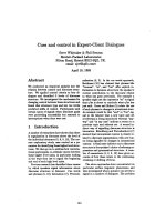
(Fig. 1) [41]. Several glycosyltransferases, including
core 2 b1,6-N-acetylglucosaminyltransferase [42,43],
b1,4-galactosyltransferases (Gal-T)-I and -IV [44,45],
FucT-VII and -IV [27,46], and a2,3-sialyltransferase
(ST3Gal)-IV [47] have been identified to participate
directly in the synthesis of functional selectin ligands
in vivo (Table 2). In addition, several other modifica-
tions have been described to contribute to selectin
ligand function (Table 2). Two enzymes catalyzing
carbohydrate sulfation [N-acetylglucosamine 6-O-sulfo-
transferase (GlcNAc6ST)-1 and -2] were found to be
involved in the generation of 6-sulfo sLe
x
which is
important for l-selectin ligand activity on HEV (Fig. 2)
[48,49]. Furthermore, sulfation of tyrosine residues at
the N-terminus of PSGL-1 has been reported to signifi-
cantly influence binding of selectins to PSGL-1 (Fig. 1)
[50].
Figure 1 gives an overview on the biosynthetic path-
way of core 2 modified O-glycans terminated with
sLe
x
. O-glycan biosynthesis is initiated with the addi-
tion of galactosamine to serine or threonine residues at
the protein backbone [61]. This step is catalysed by
UDP-GalNAc:polypeptide GalNAcT (ppGalNAcT).
Twenty-four different ppGalNAcT have been described
in humans [51]. No data are available on the role of
ppGalNAcT on selectin ligand activity. However, in
view of the abundance of different isoenzymes it seems
likely that a high degree of redundancy exists which
may be an indication that ppGalNAcT is not rate-
limiting in the synthesis of functional selectin ligands.
After the addition of galactose to GalNAc in b1,3
linkage, which gives rise to the core 1 extension, core 2
b1,6 N-acetylglucosaminyltransferase (core 2 Glc-
NAcT-I) initiates the core 2 extension by adding Glc-
NAc to GalNAc in b1,6 linkage. This is followed by
the alternate action of b1,4-galactosyltransferase (b1,4-
GalT) and b1,3-GlcNAcT, which elongate the core 2
branch by forming a polylactosamine chain of various
length. During elongation, a1,3-fucosylation of Glc-
NAc residues by FucT-IV may occur within the poly-
lactosamine chain. Elongation of core 2 branches is
terminated by the addition of sialic acid, in a2,3 link-
age, to galactose (Fig. 1). This is followed by the addi-
tion of fucose to the penultimate GlcNAc, in a1,3
linkage, resulting in the formation of sLe
x
at the end
of core 2 decorated O-glycans (Fig. 1). In the following
section, the contribution of glycosyltransferases
involved in the synthesis of functional selectin ligands
in vivo are discussed.
Core 2 GlcNAcT-I
Core 2 GlcNAcT-I is the key branching enzyme in the
synthesis of core 2 decorated O-glycans. Core 2 Glc-
NAcT-I catalyzes the addition of N-acetylglucosamine
to N-acetylgalactosamine in b1,6 linkage, which initi-
ates the core 2 extension (Fig. 1). Direct evidence that
core 2 GlcNAcT-I is important for leukocyte rolling
in vivo comes from mice deficient in core 2 GlcNAcT-I,
Fig. 1. Biosynthetic pathway for the synthesis of core 2 decorated
O-glycans carrying the sialyl Lewis X (sLe
x
) determinant. During
inflammation, the main inflammatory selectin ligand, P-selectin gly-
coprotein ligand-1 (PSGL-1), interacts with P- and L-selectin under
in vivo conditions via core 2 decorated sLe
x
, in co-operation with
nearby sulfated tyrosines located at the N-terminus of PSGL-1.
M. Sperandio Leukocyte rolling and glycosyltransferases
FEBS Journal 273 (2006) 4377–4389 ª 2006 The Author Journal compilation ª 2006 FEBS 4381
Table 2. Enzymes involved in the post-translational modification of selectin ligands. CDG, congenital deficiency of glycosylation; CHST-2, car-
bohydrate sulfotransferase 2; core 2 GlcNAcT, core 2 b1,6 N-acetylglucosaminyltransferase; FucT, a1,3 fucosyltransferase; GalT, galactosyl-
transferase; GlcNAc6ST, N-acetylglucosamine 6-O-sulfotransferase; GST, Gal ⁄ GalNAc ⁄ GlcNAc 6-O-sulfotransferase; HEC, high endothelial
cell; HEV, high endothelial venule; LSST, L-selectin sulfotransferase; ppGalNAcT, polypeptide galactosaminyltransferase; PSGL-1, P-selectin
glycoprotein ligand-1; ST3Gal, a2,3 sialyltransferase; TPST, tyrosylprotein sulfotransferase.
Suspected ⁄ identified leukocyte rolling defect Reference
Glycosyltransferases
ppGalNAcT Influence on leukocyte rolling unknown at present
Probably overlapping function of different ppGalNAcT
in the initiation of O-glycan biosynthesis
[51]
Core 1 b1,3-GalT Initiates the core 1 extension
MECA-79 recognizes GlcNAc-6-O-sulfate on core 1 branch
[52]
ST3Gal-I Indirect influence on leukocyte rolling
Sialylates core 1 extensions
Competes with core 2 GlcNAcT-I for substrate
[53]
Core2 b1,6-GlcNAcT-I P- and
L-selectin-dependent rolling strongly reduced in core 2
GlcNAcT-I
– ⁄ –
during inflammation in vivo
Regulates capture ligand for E-selectin during inflammation
No influence on E-selectin-dependent slow rolling velocity
Lymphocyte homing to Peyer’s patches unaffected in core 2
GlcNAcT-I
– ⁄ –
Reduced lymphocyte homing to peripheral lymph nodes of
core 2 GlcNAcT-I
– ⁄ –
Reduced lymphocyte rolling on HEV of peripheral lymph nodes
in core 2 GlcNAcT-I
– ⁄ –
Increased rolling velocity on HEV of peripheral lymph nodes in
core 2 GlcNAcT-I
– ⁄ –
[36,42,43,54]
FucT-VII P- and E-selectin ligand-dependent rolling dramatically reduced in
FucT-VII
– ⁄ –
during inflammation in vivo
L-selectin-dependent rolling almost completely absent in peripheral
lymph node HEV of FucT-VII
– ⁄ –
[46]
FucT-IV Influences slow E-selectin-dependent rolling velocity
P- and L-selectin ligand function unaffected in FucT-IV
– ⁄ –
[27]
ST3Gal-IV Influences slow E-selectin-dependent rolling velocity
P-selectin-dependent rolling unaffected in ST3Gal-IV
– ⁄ –
[47]
b1,4GalT-I Influence on leukocyte rolling unknown at present
Binding of soluble P-selectin to b1,4GalT-I
– ⁄ –
neutrophils impaired
Normal lymphocyte homing to peripheral lymph nodes
Deficiency of b1,4GalT-I described in humans (CDG IId)
[44,55]
b1,4GalT-IV Influence on leukocyte rolling unknown at present
Acts specifically on core 2 linked GlcNAc 6-O-sulfate
Participates in the synthesis of 6-sulfo sialyl Lewis
x
[45]
Sulfotransferases
GlcNAc6ST-1
(also called GST-2 or CHST-2)
Moderate reduction in lymphocyte homing to peripheral lymph nodes
Modest increase in rolling velocity of B- and T cells on HEV of
peripheral lymph nodes
Overlapping and distinct function with GlcNAcT6ST-2 on
L-selectin
ligand activity in HEV of lymphoid tissue
Contributes to abluminal MECA-79 staining in HEV
[48,56]
GlcNAc6ST-2
(also called HEC-GlcNAc6ST,
GST-3, LSST and CHST-4)
Marked reduction in lymphocyte homing to peripheral lymph nodes
Number of rolling cells on HEV not affected in GlcNAc6ST-2
– ⁄ –
Significant increase in rolling velocity in HEV of GlcNAc6ST-2
– ⁄ –
Reduced leukocyte adhesion in HEV of GlcNAc6ST-2
– ⁄ –
Highly restricted expression on HEV of lymphoid tissue and
lymphoid-like aggregates of chronically inflamed tissue
Not expressed on Peyer’s patch HEV
Overlapping and distinct function with GlcNAcT6ST-1 on L-selectin
ligand activity in HEV of lymphoid tissue
crucial for MECA-79 reactivity on the luminal side of HEV
[57–59]
Leukocyte rolling and glycosyltransferases M. Sperandio
4382 FEBS Journal 273 (2006) 4377–4389 ª 2006 The Author Journal compilation ª 2006 FEBS
which have been generated recently [42]. Intravital
microscopy studies, conducted in untreated and TNF-a
pretreated cremaster muscle venules of core 2 Glc-
NAcT-I deficient mice, revealed a dramatic reduction
in P- and L-selectin mediated rolling, and a less pro-
nounced reduction in E-selectin dependent rolling
[36,43]. In contrast, leukocyte rolling was unchanged
in Peyer’s patch HEV, where rolling is predominantly
mediated by L-selectin and, to a lesser degree, by a
4
b
7
-
integrin and P-selectin [36], suggesting that core 2
GlcNAcT-I is dispensable for L-selectin ligand func-
tion on HEV. This was confirmed, in part, by Yeh and
colleagues who identified 6-sulfo sLe
x
on core 1 exten-
ded O-glycans of core 2 GlcNAcT-I deficient mice [52].
Core 1 decorated 6-sulfo sLe
x
serve, in collaboration
with core 2 decorated 6-sulfo sLe
x
, as L-selectin lig-
ands on HEV [62]. However, subsequent studies of
lymphocyte trafficking to peripheral lymph nodes of
core 2 GlcNAcT-I
– ⁄ –
mice revealed a defect in B-cell
(and less pronounced in T-cell) homing, which consis-
ted of reduced B- and T-cell rolling on peripheral
lymph node HEV accompanied by increased rolling
velocities [54]. The difference in B- and T-cell homing
observed in core 2 GlcNAcT-I
– ⁄ –
mice was mainly
attributed to a lower expression of L-selectin on B
cells, which led to a further, functionally relevant,
decrease in L-selectin mediated interactions [54].
b1,4-GalT
To date, seven b1,4-GalT have been identified [63].
Two of them – b1,4-GalT-I and b1,4-GalT-IV – have
been implicated in the synthesis of functional selectin
ligands. b1,4-GalT-I catalyzes the addition of UDP-
galactose to terminal N-acetylgalactosamine and acts
in concert with b1,3-N-acetyl-glucosaminyltransferase
to synthesize polylactosamine extensions of core 2 dec-
orated O-glycans. In addition, b1,4-GalT-I also partici-
pates in the generation of sLe
x
. Using b1,4-GalT-I
deficient mice, Asano and colleagues investigated the
contribution of b1,4-GalT-I on selectin ligand activity.
They found that binding of soluble P-selectin to neu-
trophils and monocytes of b1,4-GalT-I
– ⁄ –
mice was
significantly impaired [44], suggesting a role of b1,4-
GalT-I in P-selectin mediated rolling in vivo. Although
not formally investigated, a putative P-selectin depend-
ent rolling defect in b1,4-GalT-I deficient mice would
be sufficient to explain the observed increase in leuko-
cyte and neutrophil counts, as well as the significant
reduction of recruited neutrophils into zymosan treated
earlobes [44]. Lymphocyte homing to peripheral lymph
nodes, which requires L-selectin ligand activity on
HEVs, was not affected in the absence of b1,4-GalT-I,
suggesting that b1,4-GalT-I does not contribute to
the biosynthesis of HEV-expressed L-selectin ligands
in vivo [44]. Recently, the first patient, a 16-month-old
boy, with a deficiency in b1,4-GalT-I, has been des-
cribed and was designated as having congenital defici-
ency of glycosylation IId [55]. The little boy suffers
from mental retardation, Dandy-Walker malformation
with hydrocephalus, myopathy and blood clotting
problems [55].
Among the seven b1,4-GalTs, b1,4-GalT-IV is the
only b1,4-GalT that specifically acts on core 2 linked
6-sulfo GlcNAc, which is further processed to 6-sulfo
sLe
x
[45], a carbohydrate determinant found on
L-selectin ligands in HEVs of secondary lymphoid
organs and crucial for binding to L-selectin. Co-
expression profiles of b1,4-GalT-IV and 6-sulfo sLe
x
revealed no correlation in expression, suggesting that
b1,4-GalT-IV is not rate limiting for the synthesis of
6-sulfo sLe
x
[45].
Fucosyltransferases
Transfer of the monosaccaride fucose to core 2
decorated O-glycans is dependent on two a1,3-fucosyl-
transferases, namely FucT-VII and FucT-IV [41].
Expression of a1,3-fucosyltransferases (similarly to
other glycosyltransferases) is primarily regulated at the
transcriptional level. Both FucT-VII and FucT-IV, are
expressed in leukocytes. FucT-VII has also been identi-
fied in murine high endothelial cells of secondary lym-
phoid organs, suggesting a role of FucT-VII in the
synthesis of functional L-selectin ligands on HEV [64].
Direct evidence for a role of FucT-VII and FucT-IV in
selectin ligand function in vivo comes from intravital
microscopy studies conducted in mice deficient in
FucT-VII [46] and FucT-IV [27]. FucT-VII
– ⁄ –
mice,
which have a significantly increased leukocyte count,
Table 2. (Continued).
Suspected ⁄ identified leukocyte rolling defect Reference
TPST-1 and -2 Catalyze sulfation of crucial tyrosines at the N-terminus of PSGL-1
Important for P- and
L-selectin ligand function
Contribution of different TPSTs on leukocyte rolling unknown
TPST-1
– ⁄ –
and TPST-2
– ⁄ –
with no reported defect in PSGL-1 function
[60]
M. Sperandio Leukocyte rolling and glycosyltransferases
FEBS Journal 273 (2006) 4377–4389 ª 2006 The Author Journal compilation ª 2006 FEBS 4383
demonstrate an almost complete absence of leukocyte
rolling in inflamed venules of the ear and the cremaster
muscle, suggesting a dramatic reduction in E- and
P-selectin ligand function in FucT-VII
– ⁄ –
mice. Leuko-
cyte rolling in lymph node HEV of FucT-VII
– ⁄ –
mice
was also dramatically impaired and accompanied by
small hypocellular lymph nodes and a severe defect in
lymphocyte homing to secondary lymphoid organs
[46]. FucT-IV
– ⁄ –
mice appear healthy and show leuko-
cyte counts within the normal range. Analysis of leu-
kocyte rolling in inflamed venules of the ear revealed a
similar number of rolling leukocytes when compared
Fig. 2. L-selectin ligand activity on high endothelial venules (HEV) of secondary lymphoid organs is predominantly mediated by 6-sulfo sialyl
Lewis X (sLe
x
), which can be found as a capping group on core 2 extensions, core 1 extensions or on biantennary (core 2 and core 1) exten-
sions.
Leukocyte rolling and glycosyltransferases M. Sperandio
4384 FEBS Journal 273 (2006) 4377–4389 ª 2006 The Author Journal compilation ª 2006 FEBS
with control mice. However, leukocyte rolling veloci-
ties were significantly increased, suggesting that FucT-
IV contributes to E-selectin dependent rolling, distinct
from FucT-VII [27].
Sialyltransferases
Sialylation was the first post-translational glycosylation
reported to be crucial for functional L-selectin ligands
on HEV [65]. Subsequent studies identified the tetrasac-
charide sLe
x
on selectin ligands to show binding affinity
to all three selectins. Sialylation of Le
x
is catalyzed by
a2,3-sialyltransferases. From the six different a2,3-sial-
yltransferases (ST3GalI-VI) described to date, ST3Gal-
IV, ST3Gal-VI and, to a lesser degree, ST3Gal-III,
transfer sialic acid residues to terminal galactose resi-
dues of type II oligosaccharides on core 2 decorated
O-glycans [66]. Recently, mice deficient in ST3Gal-IV
have been generated [67]. In vivo studies investigating
P- and E-selectin mediated leukocyte rolling in inflamed
cremaster muscle venules of ST3Gal-IV
– ⁄ –
mice
revealed no defect in P-selectin dependent rolling [47].
However, E-selectin dependent leukocyte rolling velo-
city was significantly increased, with no defect in
E-selectin mediated leukocyte capture, suggesting that
ST3Gal-IV regulates E-selectin dependent rolling velo-
city while it does not affect the efficiency of E-selectin
to attract free flowing leukocytes to inflamed endothe-
lium [47]. These results imply that PSGL-1, which
mediates P-selectin dependent rolling and functions as
a capture ligand for E-selectin, is not strictly dependent
on ST3Gal-IV, but may also be sialylated by another
a2,3-sialyltransferase, probably ST3Gal-VI.
Although ST3Gal-I is not directly involved in the
synthesis of selectin ligands, it is worth mentioning
that ST3Gal-I may exhibit indirect influence on selec-
tin ligand function, and hence leukocyte rolling, by
competing with core 2 GlcNAcT-I for the same
substrate. ST3Gal-I specifically catalyzes the sialyla-
tion of core 1 extensions (NeuAca2,3Galb1,3GalNAc-
Ser ⁄ Thr) [68]. In ST3Gal-I deficient mice, the expres-
sion of Galb1,3GalNAc-Ser ⁄ Thr is significantly
increased [53]. This is accompanied by strong up-regu-
lation of core 2 decorated O-glycans, which may lead
to enhanced binding of selectins to selectin ligands
[53].
Carbohydrate sulfotransferases
GlcNAc-6-O-sulfation of HEV-expressed L-selectin lig-
ands is an important post-translational modification,
leading to enhanced binding of L-selectin to its ligands
under in vitro and in vivo conditions [30]. Five different
GlcNAc-6-O-sulfotransferases (GlcNAc6ST1-5) exist.
Two of them – GlcNAc6ST-1 and GlcNAc6ST-2 –
contribute to the elaboration of 6-sulfo sLe
x
(Fig. 2),
the most important sulfate modification of functional
L-selectin ligands. GlcNAc6ST-1, also known as
Gal ⁄ GalNAc ⁄ GlcNAc 6-O-sulfotransferase-2 or carbo-
hydrate sulfotransferase-2, is broadly expressed and
demonstrates some overlapping, as well as distinct,
functions with GlcNAc6ST-2 [48,49]. Mice deficient in
GlcNAc6ST-1 show a moderate reduction in lympho-
cyte homing to peripheral lymph nodes, mesenteric
lymph nodes and Peyer’s patches [56]. Intravital micro-
scopy studies revealed no defect in lymphocyte rolling
flux in HEV of peripheral lymph nodes. However, roll-
ing velocities of B- and T cells were modestly increased
[48]. Expression of GlcNAc6ST-2 (also known as
HEC-GlcNAc6ST, Gal⁄ GalNAc ⁄ GlcNAc 6-O-sulfo-
transferase-3, L-selectin sulfotransferase, and carbohy-
drate sulfotransferase-4) is highly restricted to HEVs
of lymphoid tissue and lymphoid-like aggregates in
chronically inflamed tissue [30,59]. In contrast to Glc-
NAc6ST-1, GlcNAc6ST-2 is not expressed on Peyer’s
patch HEV: this may indicate a distinct role of Glc-
NAc6ST-1 in the synthesis of functional selectin lig-
ands on Peyer’s patch HEV. GlcNAc6ST-2 leads
predominantly to GlcNAc-6-O-sulfation of extended
core 1 structures (Fig. 2), which is recognized by mAb
MECA-79 [52]. Accordingly, absence of GlcNAc6ST-2
dramatically reduced the binding of MECA-79 to
HEV. Interestingly, MECA-79 staining in Glc-
NAc6ST-2
– ⁄ –
mice was only reduced at the luminal
site. Abluminal staining was found to be mainly
dependent on GlcNAc6ST-1 [56]. Functional assays
revealed that lymphocyte homing was reduced by 50%
in GlcNAc6ST-2 deficient mice, whereas leukocyte roll-
ing flux on HEV was not affected in GlcNAc6ST-2
– ⁄ –
mice. However, rolling velocities were significantly
increased and accompanied by a marked reduction in
leukocyte adhesion [69]. To further investigate the con-
tribution of sulfation on L-selectin ligand activity, mice
deficient in GlcNAc6ST-1 and -2 have been generated
recently [48,49]. These mice showed a dramatic reduc-
tion in lymphocyte homing to peripheral lymph nodes.
MECA-79 staining, as a reporter for PNAd activity,
was completely absent. Intravital analysis revealed that
leukocyte rolling flux was significantly, but not com-
pletely, reduced. In addition, rolling velocity was
substantially increased. Residual leukocyte rolling
observed in the double knockout mouse was com-
pletely abolished by the addition of the L-selectin
blocking mAb, MEL-14, suggesting that sulfation-
independent L-selectin ligands (probably decorated by
sLe
x
) exist.
M. Sperandio Leukocyte rolling and glycosyltransferases
FEBS Journal 273 (2006) 4377–4389 ª 2006 The Author Journal compilation ª 2006 FEBS 4385
Tyrosylprotein sulfotransferases
In mice and humans, two tyrosylprotein sulfotrans-
ferases (TPST-1 and -2) have been identified to medi-
ate tryrosine O-sulfation [60]. Tyrosine O-sulfation is
an important post-translational modification of critical
tyrosine residues at the N-terminus of PSGL-1, leading
to enhanced binding of P- and L-selectin to PSGL-1
[50]. Functional studies revealed that both tyrosyl-
protein sulfotransferases contribute equally to the sulf-
ation of peptides modelled on the N-terminus of
PSGL-1 [70], suggesting a role for both enzymes in the
synthesis of functional PSGL-1. However, investiga-
tions in TPST-1
– ⁄ –
or TPST-2
– ⁄ –
mice have not repor-
ted any decrease in binding activity of P- or L-selectin
to PSGL-1, suggesting that either enzyme is able to
compensate for the loss of the other [71,72].
Conclusion
Leukocyte rolling is an important step in the recruit-
ment of leukocytes into tissue and has been considered
to be a rather nonspecific process, allowing leukocytes
to obtain intimate contact with the vascular wall. Dur-
ing rolling, leukocytes have the opportunity to screen
the endothelial surface for specific trigger signals, which
brings about a decision for extravasation into tissue.
Recent advancements in the elucidation of post-transla-
tional modifications relevant for selectin ligand func-
tion in vivo challenge this view and indicate that subtle
differences in the post-translational glycosylation ⁄ sulfa-
tion of endothelium- or leukocyte-expressed selectin lig-
ands might constitute an important early determinant
for the successful recruitment of leukocytes.
References
1 Springer TA (1995) Traffic signals on endothelium for
lymphocyte recirculation and leukocyte emigration.
Annu Rev Physiol 57, 827–872.
2 Butcher EC (1991) Leukocyte-endothelial cell recogni-
tion – Three (or more) steps to specificity and diversity.
Cell 67, 1033–1036.
3 Hamann A & Engelhardt B (2005) Leukocyte Traffick-
ing. Wiley-VCH, Weinheim, Germany.
4 Zelensky AN & Gready JE (2005) The C-type lectin-like
domain superfamily. FEBS J 272, 6179–6217.
5 Vestweber D & Blanks JE (1999) Mechanisms that
regulate the function of the selectins and their ligands.
Physiol Rev 79, 181–213.
6 Marshall BT, Long M, Piper JW, Yago T, McEver
RP & Zhu C (2003) Direct observation of catch bonds
involving cell-adhesion molecules. Nature 423, 190–
193.
7 Yago T, Wu J, Wey CD, Klopocki AG, Zhu C &
McEver RP (2004) Catch bonds govern adhesion
through L-selectin at threshold shear. J Cell Biol 166,
913–923.
8 Smith ML, Smith MJ, Lawrence MB & Ley K (2002)
Viscosity-independent velocity of neutrophils rolling on
p-selectin in vitro or in vivo. Microcirculation 9, 523–
536.
9 Sperandio M & Ley K (2005) The physiology and
pathophysiology of P-selectin. Mod Asp Immunobiol 15,
24–26.
10 Ley K, Bullard DC, Arbones ML, Bosse R, Vestweber
D, Tedder TF & Beaudet AL (1995) Sequential contri-
bution of L- and P-selectin to leukocyte rolling in vivo.
J Exp Med 181, 669–675.
11 Bevilacqua MP, Stengelin S, Gimbrone MA Jr & Seed B
(1989) Endothelial leukocyte adhesion molecule-1: An
inducible receptor for neutrophils related to complement
regulatory proteins and lectins. Science 243, 1160–1165.
12 Kraiss LW, Alto NM, Dixon DA, McIntyre TM,
Weyrich AS & Zimmerman GA (2003) Fluid flow regu-
lates E-selectin protein levels in human endothelial cells
by inhibiting translation. J Vasc Surg 37, 161–168.
13 Ley K (2001) Pathways and bottlenecks in the web of
inflammatory adhesion molecules and chemoattractants.
Immunol Rev 24, 87–95.
14 Smith ML, Olson TS & Ley K (2004) CXCR2- and
E-selectin-induced neutrophil arrest during inflammation
in vivo. J Exp Med 200, 935–939.
15 Yang J, Hirata T, Croce K, Merrill-Skoloff G,
Tchernychev B, Williams E, Flaumenhaft R, Furie B &
Furie BC (1999) Targeted gene disruption demonstrates
that PSGL-1 is required for P-Selectin mediated but not
E-Selectin mediated neutrophil rolling and migration.
J Exp Med 190, 1769–1782.
16 Xia L, Sperandio M, Yago T, McDaniel JM, Cummings
RD, Pearson-White S, Ley K & McEver RP (2002)
P-selectin glycoprotein ligand-1-deficient mice have
impaired leukocyte tethering to E-selectin under flow.
J Clin Invest 109, 939–950.
17 Mayadas TN, Johnson RC, Rayburn H, Hynes RO &
Wagner DD (1993) Leukocyte rolling and extravasation
are severely compromised in P selectin-deficient mice.
Cell 74, 541–554.
18 Sperandio M, Smith ML, Forlow SB, Olson TS, Xia L,
McEver RP & Ley K (2003) P-selectin glycoprotein
ligand-1 mediates L-selectin-dependent leukocyte rolling
in venules. J Exp Med 197, 1355–1363.
19 Rivera-Nieves J, Burcin TL, Olson TS, Morris MA,
McDuffie M, Cominelli F & Ley K (2006) Critical role
of endothelial P-selectin glycoprotein ligand 1 in chronic
murine ileitis. J Exp Med 203, 907–917.
20 Katayama Y, Hidalgo A, Chang J, Peired A & Frenette
PS (2005) CD44 is a physiological E-selectin ligand on
neutrophils. J Exp Med 201
, 1183–1189.
Leukocyte rolling and glycosyltransferases M. Sperandio
4386 FEBS Journal 273 (2006) 4377–4389 ª 2006 The Author Journal compilation ª 2006 FEBS
21 Dimitroff CJ, Lee JY, Fuhlbrigge RC & Sackstein R
(2000) A distinct glycoform of CD44 is an L-selectin
ligand on human hematopoietic cells. Proc Natl Acad
Sci USA 97, 13841–13846.
22 Bargatze RF, Jutila MA & Butcher EC (1995) Distinct
roles of L-selectin and integrins a
4
b
7
and LFA-1 in lym-
phocyte homing to Peyer’s patch-HEV in situ: the
multistep model confirmed and refined. Immunity 3,
99–108.
23 Watson SR (1997) Glycoprotein ligands for l-selectin.
In The Selectins (Vestweber D, ed.), pp. 179–193. Har-
wood Academic Publishers, Amsterdam.
24 Suzuki A, Andrew DP, Gonzalo J-A, Fukumoto M,
Spellberg J, Hashiyama M, Suda T, Takimoto H, Ger-
win N, Webb J et al. (1996) CD34 deficient mice have
reduced eosinophil accumulation after allergen exposure
and reveal a novel crossreactive 90 kD protein. Blood
87, 3550–3562.
25 Matsumoto M, Atarashi K, Umemoto E, Furukawa Y,
Shigeta A, Miyasaka M & Hirata T (2005) CD43 func-
tions as a ligand for E-Selectin on activated T cells.
J Immunol 175, 8042–8050.
26 Harms G, Kraft R, Grelle G, Volz B, Dernedde J &
Tauber R (2001) Identification of nucleolin as a new
L-selectin ligand. Biochem J 360, 531–538.
27 Weninger W, Ulfman LH, Cheng G, Souchkova N,
Quackenbush EJ, Lowe JB & von Andrian UH (2000)
Specialized contributions by a (1,3)-fucosyltransferase-
IV and FucT-VII during leukocyte rolling in dermal
microvessels. Immunity 12, 665–676.
28 Fuhlbrigge RC, King SL, Sackstein R & Kupper TS
(2005) CD43 is a ligand for E-selectin on CLA+ human
T cells. Blood 107, 1421–1426.
29 Bistrup A, Tsay D, Shenoy P, Singer MS, Bangia N,
Luther SA, Cyster JG, Ruddle NH & Rosen SD (2004)
Detection of a sulfotransferase (HEC-GlcNAc6ST) in
high endothelial venules of lymph nodes and in high
endothelial venule-like vessels within ectopic lymphoid
aggregates: relationship to the MECA-79 epitope. Am J
Pathol 164, 1635–1644.
30 Rosen SD (2004) Ligands for L-selectin: homing,
inflammation, and beyond. Annu Rev Immunol 22,
129–156.
31 Khan AI & Kubes P (2003) L-selectin: an emerging
player in chemokine function. Microcirculation 10,
351–358.
32 Crockett-Torabi E, Sulenbarger B, Smith CW & Fan-
tone JC (1995) Activation of human neutrophils
through L-selectin and Mac-1 molecules. J Immunol
154, 2291–2302.
33 Simon SI, Burns AR, Taylor AD, Gopalan PK, Lynam
EB, Sklar LA & Smith CW (1995) L-selectin (CD62L)
cross-linking signals neutrophil adhesive functions via
the Mac-1 (CD11b ⁄ CD18) beta 2-integrin. J Immunol
155, 1502–1514.
34 Hickey MJ, Forster M, Mitchell D, Kaur J, De Caigny
C & Kubes P (2000) L-selectin facilitates emigration
and extravascular locomotion of leukocytes during acute
inflammatory responses in vivo. J Immunol 165, 7164–
7170.
35 Warnock RA, Askari S, Butcher EC & von Andrian UH
(1998) Molecular mechanisms of lymphocyte homing to
peripheral lymph nodes. J Exp Med 187, 205–216.
36 Sperandio M, Forlow SB, Thatte J, Ellies LG, Marth
JD & Ley K (2001) Differential requirements for core2
glucosaminyltransferase for endothelial L-selectin ligand
function in vivo. J Immunol 167, 2268–2274.
37 Olson TS & Ley K (2002) Chemokines and chemokine
receptors in leukocyte trafficking. Am J Physiol Regul
Integr Comp Physiol 283, R7–R28.
38 Streeter PR, Rouse BTN & Butcher EC (1988) Immu-
nohistologic and functional characterization of a vascu-
lar addressin involved in lymphocyte homing into
peripheral lymph nodes. J Cell Biol
107, 1853–1862.
39 Samulowitz U, Kuhn A, Brachtendorf G, Nawroth R,
Braun A, Bankfalvi A, Bocker W & Vestweber D
(2002) Human endomucin: distribution pattern, expres-
sion on high endothelial venules, and decoration with
the MECA-79 epitope. Am J Pathol 160, 1669–1681.
40 Lowe JB & Varki A (1999) Glycosyltransferases. In
Essentials of Glycobiology (Varki A, Cummings RD,
Esko J, Freeze HH, Hart G & Marth JD, eds), pp. 253–
266. Cold Spring Harbor Laboratory Press, Cold Spring
Harbor, New York.
41 Lowe JB (2002) Glycosylation in the control of selectin
counter-receptor structure and function. Immunol Rev
186, 19–36.
42 Ellies LG, Tsuboi S, Petryniak B, Lowe JB, Fukuda
M & Marth JD (1998) Core 2 oligosaccharide bio-
synthesis distinguishes between selectin ligands essential
for leukocyte homing and inflammation. Immunity 9,
881–890.
43 Sperandio M, Thatte A, Foy D, Ellies LG, Marth JD &
Ley K (2001) Severe impairment of leukocyte rolling in
venules of core 2 glucosaminyltransferase-deficient mice.
Blood 97, 3812–3819.
44 Asano M, Nakae S, Kotani N, Shirafuji N, Nambu A,
Hashimoto N, Kawashima H, Hirose M, Miyasaka M,
Takasaki S et al. (2003) Impaired selectin-ligand biosyn-
thesis and reduced inflammatory responses in beta-1,4-
galactosyltransferase-I-deficient mice. Blood 102, 1678–
1685.
45 Seko A, Dohmae N, Takio K & Yamashita K (2003)
Beta 1,4-galactosyltransferase (beta 4GalT)-IV is specific
for GlcNAc 6-O-sulfate. Beta 4GalT-IV acts on keratan
sulfate-related glycans and a precursor glycan of 6-sulfo-
sialyl-Lewis X. J Biol Chem 278, 9150–9158.
46 Maly P, Thall AD, Petryniak B, Rogers CE, Smith PL,
Marks RM, Kelly RJ, Gersten KM, Cheng G, Saunders
TL et al. (1996) The a (1,3) fucosyltransferase Fuc-TVII
M. Sperandio Leukocyte rolling and glycosyltransferases
FEBS Journal 273 (2006) 4377–4389 ª 2006 The Author Journal compilation ª 2006 FEBS 4387
controls leukocyte trafficking through an essential role
in L-, E-, and P-selectin ligand biosynthesis. Cell 86,
643–653.
47 Ellies LG, Sperandio M, Underhill GH, Yousef J,
Smith M, Priatel JJ, Kansas GS, Ley K & Marth J
(2002) Sialyltransferase specifity in selectin ligand for-
mation. Blood 100, 3618–3625.
48 Uchimura K, Gauguet JM, Singer MS, Tsay D,
Kannagi R, Muramatsu T, von Andrian UH & Rosen
SD (2005) A major class of L-selectin ligands is elimi-
nated in mice deficient in two sulfotransferases
expressed in high endothelial venules. Nat Immunol 6,
1105–1113.
49 Kawashima H, Petryniak B, Hiraoka N, Mitoma J,
Huckaby V, Nakayama J, Uchimura K, Kadomatsu K,
Muramatsu T, Lowe JB et al. (2005) N-acetylglucosa-
mine-6-O-sulfotransferases 1 and 2 cooperatively control
lymphocyte homing through L-selectin ligand biosynthe-
sis in high endothelial venules. Nat Immunol 6, 1096–
1104.
50 Ramachandran V, Nollert MU, Qiu H, Liu WJ, Cum-
mings RD, Zhu C & McEver RP (1999) Tyrosine repla-
cement in P-selectin glycoprotein ligand-1 affects
distinct kinetic and mechanical properties of bonds with
P- and L-selectin. Proc Natl Acad Sci USA 96, 13771–
13776.
51 Ten Hagen KG, Fritz TA & Tabak LA (2003) All in
the family: the UDP-GalNAc: polypeptide N-acetylga-
lactosaminyltransferases. Glycobiology 13, 1R–16R.
52 Yeh J, Hiraoka N, Petryniak B, Nakayama J, Ellies LG,
Rabuka D, Hindsgaul O, Marth JD, Lowe JB &
Fukuda M (2001) Novel sulfated lymphocyte homing
receptors and their control by a core1 extension
beta1,3-N-Acetylglucosaminyltransferase. Cell 105, 957–
969.
53 Priatel JJ, Chui D, Hiraoka N, Simmons CJ, Richard-
son KB, Page DM, Fukuda M, Varki NM & Marth JD
(2000) The ST3Gal-I sialyltransferase controls CD8+
T lymphocyte homeostasis by modulating O-glycan bio-
synthesis. Immunity 12, 273–283.
54 Gauguet JM, Rosen SD, Marth JD & von Andrian UH
(2004) Core 2 branching beta1,6-N-acetylglucosaminyl-
transferase and high endothelial cell N-acetylglucosa-
mine-6-sulfotransferase exert differential control over
B- and T-lymphocyte homing to peripheral lymph
nodes. Blood 104, 4104–4112.
55 Hansske B, Thiel C, Lubke T, Hasilik M, Honing S,
Peters V, Heidemann PH, Hoffmann GF, Berger EG,
von Figura K et al. (2002) Deficiency of UDP-galactose:
N-acetylglucosamine beta-1,4-galactosyltransferase I
causes the congenital disorder of glycosylation type IId.
J Clin Invest 109, 725–733.
56 Uchimura K, Kadomatsu K, El Fasakhany FM, Singer
MS, Izawa M, Kannagi R, Takeda N, Rosen SD &
Muramatsu T (2004) N-acetylglucosamine 6-O-sulfo-
transferase-1 regulates expression of 1-selectin ligands
and lymphocyte homing. J Biol Chem 279, 35001–
35008.
57 Hiraoka N, Petryniak B, Nakayama J, Tsuboi S,
Suzuki M, Yeh JC, Izawa D, Tanaka T, Miyasaka M,
Lowe JB et al. (1999) A novel, high endothelial venule-
specific sulfotransferase expresses 6-sulfo sialyl lewis (x),
an L-selectin ligand displayed by CD34. Immunity 11,
79–89.
58 Bistrup A, Bhakta S, Lee JK, Belov YY, Gunn MD,
Zuo FR, Huang CC, Kannagi R, Rosen SD & Hem-
merich S (1999) Sulfotransferases of two specificities
function in the reconstitution of high endothelial cell
ligands for L-selectin. J Cell Biol 145, 899–910.
59 Rosen SD, Tsay D, Singer MS, Hemmerich S & Abra-
ham WM (2005) Therapeutic targeting of endothelial
ligands for L-selectin (PNAd) in a sheep model of
asthma. Am J Pathol 166, 935–944.
60 Moore KL (2003) The biology and enzymology of pro-
tein tyrosine O-sulfation. J Biol Chem 278
, 24243–
24246.
61 Marth JD (1999) O-Glycans. In Essentials of Glycobiol-
ogy (Varki A, Cummings RD, Esko JD, Freeze HH,
Hart G & Marth JD, eds), pp. 101–114. Cold Spring
Harbor Laboratory Press, Cold Spring Harbor, New
York.
62 McEver RP (2005) A sulfated address for lymphocyte
homing. Nat Immunol 6, 1067–1069.
63 Hennet T (2002) The galactosyltransferase family. Cell
Mol Life Sci 59 , 1081–1095.
64 Smith PL, Gersten KM, Petryniak B, Kelly RJ, Rogers
C, Natsuka Y, Alford JAIII, Scheidegger EP, Natsuka
S & Lowe JB (1996) Expression of the a(1,3) fucosyl-
transferase Fuc-TVII in lymphoid aggregate high endo-
thelial venules correlates with expression of L-selectin
ligands. J Biol Chem 271, 8250–8259.
65 Rosen SD, Singer MS, Yednock TA & Stoolman LM
(1985) Involvement of sialic acid on endothelial cells in
organ-specific lymphocyte recirculation. Science 228,
1005–1007.
66 Harduin-Lepers A, Vallejo-Ruiz V, Krzewinski-Recchi
MA, Samyn-Petit B, Julien S & Delannoy P (2001)
The human sialyltransferase family. Biochimie 83,
727–737.
67 Ellies LG, Ditto D, Levy GG, Wahrenbrock M, Gins-
burg D, Varki A, Le DT & Marth JD (2002) Sialyl-
transferase ST3Gal-IV operates as a dominant modifier
of hemostasis by concealing asialoglycoprotein receptor
ligands. Proc Natl Acad Sci USA 99, 10042–10047.
68 Kono M, Ohyama Y, Lee YC, Hamamoto T, Kojima N
& Tsuji S (1997) Mouse beta-galactoside alpha 2,3-sialyl-
transferases: comparison of in vitro substrate specificities
and tissue specific expression. Glycobiology 7, 469–479.
69 Van Zante A, Gauguet JM, Bistrup A, Tsay D,
vn Andrian UH & Rosen SD (2003) Lymphocyte–HEV
Leukocyte rolling and glycosyltransferases M. Sperandio
4388 FEBS Journal 273 (2006) 4377–4389 ª 2006 The Author Journal compilation ª 2006 FEBS
interactions in lymph nodes of a sulfotransferase-defici-
ent mouse. J Exp Med 198, 1289–1300.
70 Ouyang YB & Moore KL (1998) Molecular cloning and
expression of human and mouse tyrosylprotein sulfo-
transferase-2 and a tyrosylprotein sulfotransferase
homologue in Caenorhabditis elegans. J Biol Chem 273,
24770–24774.
71 Ouyang YB, Crawley JT, Aston CE & Moore KL
(2002) Reduced body weight and increased post-
implantation fetal death in tyrosylprotein sulfotrans-
ferase-1-deficient mice. J Biol Chem 277, 23781–
23787.
72 Borghei A, Ouyang YB, Westmuckett AD, Marcello
MR, Landel CP, Evans JP & Moore KL (2006) Tar-
geted disruption of tyrosylprotein sulfotransferase-2, an
enzyme that catalyzes post-translational protein tyrosine
O-sulfation, causes male infertility. J Biol Chem 281,
9423–9431.
M. Sperandio Leukocyte rolling and glycosyltransferases
FEBS Journal 273 (2006) 4377–4389 ª 2006 The Author Journal compilation ª 2006 FEBS 4389