Báo cáo khoa học: Mycobacterium tuberculosis possesses a functional enzyme for the synthesis of vitamin C, L-gulono-1,4-lactone dehydrogenase doc
Bạn đang xem bản rút gọn của tài liệu. Xem và tải ngay bản đầy đủ của tài liệu tại đây (831.89 KB, 11 trang )
Mycobacterium tuberculosis possesses a functional
enzyme for the synthesis of vitamin C,
L-gulono-1,4-lactone dehydrogenase
Beata A. Wolucka
1
and David Communi
2
1 Laboratory of Mycobacterial Biochemistry, Pasteur Institute of Brussels, Institute of Public Health, Belgium
2 Institute of Interdisciplinary Research, IRIBHM, Faculty of Medicine, Free University of Brussels, Belgium
Vitamin C (l-ascorbic acid; L-AA) is an important
metabolite of plants and animals. It functions as an
antioxidant (or pro-oxidant), an enzyme cofactor, an
effector of gene expression, and a modulator of react-
ive oxygen species (ROS)-mediated cell signaling.
L-AA is therefore involved in a wide array of crucial
physiologic processes, including: biosynthesis of colla-
gen and other hydroxyproline ⁄ hydroxylysine-containing
proteins ⁄ peptides; synthesis of secondary metabolites,
hormones and cytokines [1]; oxidative protein folding
and endoplasmic reticulum stress [2]; cell proliferation
and apoptosis [3]; activation of the epithelial cystic
fibrosis transmembrane conductance regulator chloride
channel [4] and of surfactant production in human
lungs [5]; macrophage function [6]; immune homeosta-
sis [5]; and stress resistance.
Plants synthesize ascorbic acid via de novo and sal-
vage pathways [7], whereas a de novo pathway invol-
ving UDP- d-glucuronic acid operates in animals [8].
l-Gulono-1,4-lactone is a direct precursor of vitamin C
in animals [8], but also in plants [9] and in some pro-
tists [10]. In plants, L-AA can be formed additionally
from l-galactono-1,4-lactone by a highly specific mito-
chondrial dehydrogenase (EC 1.3.2.3) [11,12]. The
Keywords
ascorbic acid; biosynthesis;
L-gulonolactone
oxidase; tuberculosis; vitamin C
Correspondence
B. A. Wolucka, Laboratory of Mycobacterial
Biochemistry, Pasteur Institute of Brussels,
642 Engeland Street, B-1180 Brussels,
Belgium
Fax: +32 2 373 3282
Tel: +32 2 373 3100
E-mail:
(Received 21 June 2006, accepted 31 July
2006)
doi:10.1111/j.1742-4658.2006.05443.x
The last step of the biosynthesis of l-ascorbic acid (vitamin C) in plants and
animals is catalyzed by l-gulono-1,4-lactone oxidoreductases, which use
both l-gulono-1,4-lactone and l-galactono-1,4-lactone as substrates. l-Gul-
ono-1,4-lactone oxidase is missing in scurvy-prone, vitamin C-deficient ani-
mals, such as humans and guinea pigs, which are also highly susceptible to
tuberculosis. A blast search using the rat l-gulono-1,4-lactone oxidase
sequence revealed the presence of closely related orthologs in a limited num-
ber of bacterial species, including several pathogens of human lungs, such
as Mycobacterium tuberculosis, Pseudomonas aeruginosa, Burkholderia cepa-
cia and Bacillus anthracis. The genome of M. tuberculosis, the etiologic
agent of tuberculosis, encodes a protein (Rv1771) that shows 32% identity
with the rat l-gulono-1,4-lactone oxidase protein. The Rv1771 gene was
cloned and expressed in Escherichia coli, and the corresponding protein was
affinity-purified and characterized. The FAD-binding motif-containing
Rv1771 protein is a metalloenzyme that oxidizes l-gulono-1,4-lactone
(K
m
5.5 mm) but not l-galactono-1,4-lactone. The enzyme has a dehydroge-
nase activity and can use both cytochrome c (K
m
4.7 lm) and phenazine
methosulfate as exogenous electron acceptors. Molecular oxygen does not
serve as a substrate for the Rv1771 protein. Dehydrogenase activity was
measured in cellular extracts of a Mycobacterium bovis BCG strain. In con-
clusion, M. tuberculosis produces a novel, highly specific l-gulono-1,4-lac-
tone dehydrogenase (Rv1771) and has the capacity to synthesize vitamin C.
Abbreviations
GST, glutathione-S-transferase; IPTG, isopropyl thio-b-
D-galactoside; L-AA, L-ascorbic acid; MALDI Q-TOF, MALDI quadrupole TOF;
ROS, reactive oxygen species.
FEBS Journal 273 (2006) 4435–4445 ª 2006 The Authors Journal compilation ª 2006 FEBS 4435
oxidation of l-gulono-1,4-lactone to L-AA in animals
is catalyzed by an oxygen-dependent enzyme, l-gul-
ono-1,4-lactone oxidase (EC 1.1.3.8) [13]. In plants [9]
and in Euglena [10], the oxidation involves ill-defined
l-gulono-1,4-lactone dehydrogenases that use cyto-
chrome c and phenazine methosulfate respectively, as a
direct electron acceptor. The animal and plant l-gul-
onolactone oxidoreductases are also active towards the
l-galactono-1,4-lactone substrate.
Only scarce data are available on the presence of
ascorbic acid in lower eukaryotes. Fungi do not contain
L-AA but rather its 5-carbon homolog, d-erythroascor-
bic acid [14]. Two apparently different l-gulono-1,4-lac-
tone oxidase activities were detected in yeasts. One of
the enzymes oxidizes l-galactono-1,4-lactone but not
l-gulono-1,4-lactone [15]. The other enzyme (ALO1)
has a broader specificity and uses d-arabinono-1,4-lac-
tone [16], l-galactono-1,4-lactone and l-gulono-1,4-lac-
tone [17] as substrates. d-Arabinono-1,4-lactone is a
natural substrate in the pathway to d-erythroascorbic
acid [14,18]. Like yeasts, a protozoan parasite Try-
panosoma brucei possesses a d-arabinono-1,4-lactone
oxidase that can also oxidize l-galactono-1,4-lactone
but not l-gulono-1,4-lactone [19].
The sequence of the gene for rat l-gulono-1,4-lac-
tone oxidase (GLO) is known [20]. Several genes for
putative l-gulono-1,4-lactone dehydrogenase isoen-
zymes have been identified in plants [9]. The gene
encoding the d-arabinono-1,4-lactone oxidase (ALO1)
of yeasts [18] shows significant homology with the
genes for both the rat l-gulono-1,4-lactone oxidase
and the plant mitochondrial l-galactono-1,4-lactone
dehydrogenase [12,20]. Humans and some animals
(including other primates and guinea pigs) are natural
mutants for ascorbic acid synthesis because of the non-
functional GLO gene for l-gulono-1,4-lactone oxidase
[8]. Consequently, they require vitamin C in the diet to
prevent scurvy.
Little is known about the presence of vitamin C and
related biosynthetic enzymes in bacteria. Exogenous
l-gulono-1,4-lactone can be converted to L-AA by
unspecific, heteromeric dehydrogenases of Gluconobact-
er oxydans and of Acetobacter suboxydans [21], which
are also active towards d-xylose and some hexoses.
Although interesting from a biotechnological point of
view, these enzymes are not related to the known
l
-gulono-1,4-lactone oxidase proteins and their physio-
logic role is unknown.
Surprisingly, the genome of Mycobacterium tubercu-
losis, the causative agent of tuberculosis, encodes a
protein (Rv1771) that is similar to the rat l-gulono-
1,4-lactone oxidase. In the present work, we cloned
and expressed the Rv1771 gene, and showed that it
encodes a novel l-gulono-1,4-lactone dehydrogenase of
the M. tuberculosis complex.
Results
Heterologous expression and purification of the
recombinant
L-gulono-1,4-lactone dehydrogenase
(Rv1771) of M. tuberculosis
The Rv1771 DNA was cloned into the pDEST15 vec-
tor by using the Gateway system, and the obtained
pDEST15_Rv1771 plasmid was used for expression
of the recombinant glutathione-S-transferase (GST)
fusion protein in Escherichia coli. The recombinant
protein contained an engineered enterokinase cleavage
site in the junction between the GST tag and the
Rv1771 sequence. Upon 3 h of induction of the
pDEST15_Rv1771 E. coli strain with isopropyl-b-d-
thiogalactopyranoside (IPTG) at 37 °C, the yield of
the recombinant Rv1771 protein was very low (0.1 mg
per liter of culture). Longer inductions (16 h) at a
lower temperature (26 °C) resulted in complete loss of
the recombinant protein, probably due to proteolytic
degradation (results not shown). Similarly, omission of
Triton X-100 from the extraction buffer resulted in
lower yields of the recombinant Rv1771 protein, thus
suggesting that the detergent helps solubilize the pro-
tein. The affinity-purified protein was concentrated by
using Strataclean resin, as described in Experimental
procedures. SDS ⁄ PAGE revealed the presence of one
protein band of about 70 kDa (Fig. 1, lane 2). The
identity of the protein was confirmed by enterokinase
treatment. After digestion of the affinity-purified GST-
tagged protein with enterokinase followed by concen-
tration, the 70 kDa band disappeared, and two new
bands of 45 and 26 kDa were seen on SDS ⁄ PAGE
that corresponded to the mycobacterial Rv1771 pro-
tein and the freed GST tag, respectively (Fig. 1, lane
3). The observed molecular masses for the GST-tagged
and free forms of the l-gulono-1,4-lactone dehydroge-
nase were slightly lower than those expected (74 and
48 kDa, respectively), presumably because of either
limited proteolysis or abnormal migration. In contrast,
shortening the induction time to 1 h resulted in about
10 times higher yields of the recombinant Rv1771 pro-
tein (Fig. 1, lane 4). However, under these conditions,
the GST-affinity eluate contained the recombinant
GST fusion protein (the identified tryptic peptides are
ALGPQLAQR, LGLENQGDVDPQSITGATATATH
GTGVR, FQNLSAR, SDEQPKPTPGWQR, FTEM
EYAIPR, SLPIMFPIEVR, and FSAPDDSFLSTA
YGR), which was accompanied by a host-derived
Hsp60 chaperone protein (the identified tryptic peptide
M. tuberculosis L-gulono-1,4-lactone dehydrogenase B. A. Wolucka and D. Communi
4436 FEBS Journal 273 (2006) 4435–4445 ª 2006 The Authors Journal compilation ª 2006 FEBS
was AAVEEGVVAGGGVALIR), as determined by
combined MALDI-TOF MS of trypsin-digested pro-
tein bands (Fig. 1, lane 4; Fig. 2) and western analysis
with anti-GST IgG (Fig. 1, lane 5). Copurification of
the mycobacterial l-gulono-1,4-lactone dehydrogenase
with the Hsp60 heat-shock protein might reflect physi-
ologic protein–protein interactions, as proposed for the
plant Hsc70.3 cognate heat-shock protein and another
vitamin C-related enzyme, the GDP-mannose-3¢,5¢-
epimerase [9]. The presence of multiple GST-contain-
ing bands of about 30 kDa (Fig. 1, lane 5) suggests
that an important portion of the fusion protein was
degraded by the host proteases. On the other hand,
attempts to produce a His-tagged version of the
Rv1771 protein by using the pRSETc or the Gateway
pDEST17 expression vectors were unsuccessful.
Characterization of the recombinant
L-gulono-1,4-lactone dehydrogenase of
M. tuberculosis
The Rv1771 gene of M. tuberculosis encodes a 428
amino acid protein (Fig. 2) that shows 32% identity
with the rat l-gulono-1,4-lactone oxidase and 22–24%
identity with the putative plant l-gulono-1,4-lactone
dehydrogenases At2g46740, At2g46750, At2g46760,
At5g56490, At5g11540, and At1g32300 [9]. The Myco-
bacterium bovis genome contains a sequence (Mb1800)
identical to the M. tuberculosis Rv1771 gene (http://
genolist.pasteur.fr/). A close ortholog of the Rv1771
protein (72% identity) exists in Mycobacterium mari-
num (). In Mycobacterium
leprae, a possible pseudogene similar to the
M. tuberculosis Rv1771 sequence is present. Other my-
cobacteria apparently do not contain sequences homol-
ogous to the Rv1771 protein. The predicted molecular
mass of the Rv1771 protein is 48 045 kDa, and the pI
is 7.14. Like the animal and plant l-gulono-1,4-lactone
oxidases ⁄ dehydrogenases, the M. tuberculosis Rv1771
protein possesses in its N-terminus an FAD-binding
site (VGSGH
49
S) with a conserved histidine residue
that in the rat l-gulono-1,4-lactone oxidase enzyme
(VGGGH
54
S) participates in the covalent binding of
the FAD molecule [22] (Fig. 2). Analysis of the dena-
tured Rv1771 protein by SDS ⁄ PAGE according to the
method of Nishikimi et al. [23] did not reveal the pres-
ence of any fluorescent protein band, thus pointing to
the absence of a covalently bound flavin moiety in the
recombinant product. Moreover, as in the case of
the plant l-galactono-1,4-lactone dehydrogenase [12],
the native recombinant dehydrogenase of M. tuberculo-
sis did not show a typical flavin protein absorption
spectrum, and addition of exogenous FAD (100 lm)
or riboflavin (1 mm) had no effect on the enzyme
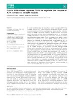
Fig. 2. Sequence analysis of the Rv1771
L-gulono-1,4-lactone dehydrogenase of
M. tuberculosis. The amino acids (16–168)
that form the FAD-binding domain (pfam
designation PF01565) are highlighted in
black. The
D-arabinono-1,4-lactone oxidase
domain (pfam designation PF04030) (amino
acids 172–427) is highlighted in gray. The
position of a potential transmembrane helice
(amino acids 205–227) is indicated by bold
italics. Tryptic peptides identified by MALDI
Q-TOF MS are underlined.
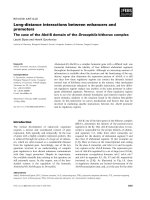
Fig. 1. Heterologous expression and purification of the recombinant
L-gulono-1,4-lactone dehydrogenase (Rv1771) of M. tuberculosis.
SDS ⁄ PAGE of the affinity-purified GST-tagged dehydrogenase (con-
taining an engineered enterokinase cleavage site) obtained from
the E. coli host after long (3 h) (lanes 2 and 3) and short (1 h) (lanes
4 and 5) periods of induction with IPTG. Fractions obtained with a
long period of induction before (lane 2) and after (lane 3) enterokin-
ase treatment were concentrated on Strataclean beads, as des-
cribed in Experimental procedures. Proteins were visualized by
Coomassie blue staining (lanes 1–4) and by western analysis (lane
5) using anti-GST IgG. Protein bands (lane 4) were identified by
MALDI-TOF MS of tryptic in-gel digests. Lane 1, molecular mass
standards.
B. A. Wolucka and D. Communi M. tuberculosis
L-gulono-1,4-lactone dehydrogenase
FEBS Journal 273 (2006) 4435–4445 ª 2006 The Authors Journal compilation ª 2006 FEBS 4437
activity (results not shown). These results suggest
that the mycobacterial dehydrogenase, like the cauli-
flower l-galactono-1,4-lactone dehydrogenase [12], is
not a flavoenzyme. The C-terminus of the Rv1771
protein contains a d-arabinono-1,4-lactone domain
(Pfam04030) (Fig. 2) that is present in all known ald-
onolactone oxidoreductases.
In order to determine the enzymatic activity of the
GST-tagged Rv1771 protein of M. tuberculosis, the
oxidase activity was tested in the presence of the l-gul-
ono-1,4-lactone and l-galactono-1,4-lactone substrates,
as described [24], but no activity could be detected.
However, the enzyme could oxidize l-gulono-1,4-lac-
tone aerobically by using exogenous cytochrome c from
horse heart as an electron acceptor (Table 1). l-Galac-
tono-1,4-lactone and other sugar derivatives did not
serve as substrates in the dehydrogenase reaction
(Table 1). Interestingly, phenazine methosulfate could
substitute for cytochrome c and was about three times
more efficient as a direct electron acceptor than the
latter (Table 1). 2,6-dichloroindophenol alone could
not serve as electron acceptor in the dehydrogenase
reaction (not shown). Thus, like the plant l-gulono-1,4-
lactone and l-galactono-1,4-lactone dehydrogenases
[9,12], the mycobacterial enzyme acts exclusively as a
dehydrogenase and does not use molecular oxygen as
an electron acceptor. In contrast to the animal and
plant l-gulono-1,4-lactone oxidoreductases, the myco-
bacterial enzyme is specific for l-gulono-1,4-lactone
and has no activity towards l-galactono-1,4-lactone
(Table 1). The steady-state parameters of the recombin-
ant l-gulono-1,4-lactone dehydrogenase of M. tuber-
culosis were determined. The dehydrogenase obeys
Michaelis–Menten kinetics with l-gulono-1,4-lactone
and cytochrome c as substrates (Fig. 3A,B). The appar-
ent K
m
values for l-gulono-1,4-lactone and cytochrome c
were determined to be 5.5 mm (Fig. 3A) and 4.7 lm
(Fig. 3B), respectively. The V
max
value was determined
to be 2.44 lmolÆh
)1
Æmg protein
)1
(Fig. 3A). The kinetic
parameters of the recombinant GST-tagged mycobacte-
rial l-gulono-1,4-lactone dehydrogenase are therefore
similar to those reported for the plant l-galactonolac-
tone dehydrogenase (K
m
values equal 3.3 mm and
3.6 lm for l-galactono-1,4-lactone and cytochrome c,
respectively) [12,25]. These results suggest that the
mycobacterial enzyme could operate efficiently in vivo.
Optimal conditions for the mycobacterial dehydroge-
nase activity were determined. The optimal pH for the
dehydrogenase reaction is between 7.5 and 8 (Fig. 4A).
At higher pH values, enzyme activity rapidly
decreased, probably because of hydrolysis of the lac-
tone substrate. As for the mammalian l-gulono-1,4-
lactone oxidases [26], the temperature optimum for the
dehydrogenase reaction was relatively high (39 °C)
(Fig. 4B). Preincubation at 60 °C for 5 min resulted in
only partial inactivation of the enzyme (53% of con-
trol), thus indicating that the dehydrogenase is relat-
ively heat-stable.
The enzyme was completely inhibited by 1 mm
N-ethylmaleimide, Cu
2+
and Zn
2+
(results not shown).
These effects suggest the involvement of sulfhydryl
group(s) in the catalytic activity of the mycobacterial
enzyme, as observed for the plant l-galactono-1,4-lac-
tone dehydrogenase [12,25]. No dehydrogenase activity
could be measured in the presence of 1 mm potassium
cyanide. Mg
2+
and Ca
2+
had no effect on the dehy-
drogenase activity, and 1 mm Mn
2+
slightly inhibited
the enzyme (21% inhibition). However, the mycobacte-
rial dehydrogenase requires for its activity trace
amounts of a divalent metal ion, because the enzyme
was inactive in the presence of 1 mm EDTA.
Presence of
L-gulono-1,4-lactone dehydrogenase
activity in M. bovis BCG strain Copenhagen
Crude extracts of exponentially growing M. bovis BCG
were prepared as described in Experimental proce-
dures. The dehydrogenase activity could be measured
in the soluble extracts [0.17 mUÆmg protein
)1
], but not
in the insoluble fraction, because of interfering con-
taminants. The determined activity of the mycobacteri-
al enzyme was comparable with that reported for
crude preparations of plant l-galactono-1,4-lactone
dehydrogenase [12,27], Thus, in agreement with previ-
ous results [28,29], the Rv1771 protein is expressed in
the M. tuberculosis complex; it is probably loosely
associated with the cell membrane [29], and is enzy-
matically active. In spite of the presence of dehydroge-
nase activity, ascorbic acid could not be detected in
Table 1. Substrate specificity of the recombinant GST-tagged L-gul-
ono-1,4-lactone dehydrogenase of M. tuberculosis. ND, not deter-
mined. All measurements were made in triplicate. The limit of
detection was 0.3 mUÆmg protein
)1
. Mean values ± SD are given.
Substrate
(50 m
M)
Enzyme specific activity with different
electron acceptors (mUÆmg protein
)1
)
Cytochrome c
(121 l
M)
Phenazine methosulfate
(2.5 mM)
L-Gulono-1,4-lactone 66.7 ± 4.0 249 ± 17.4
L-Galactono-1,4-lactone 0
a
ND
D-Glucurono-3,6-lactone 0 ND
D-Glucuronic acid 0 ND
D-Arabinose 0 ND
D-Xylose 0 ND
a
Measured values were equal to or below the detection limit.
M. tuberculosis
L-gulono-1,4-lactone dehydrogenase B. A. Wolucka and D. Communi
4438 FEBS Journal 273 (2006) 4435–4445 ª 2006 The Authors Journal compilation ª 2006 FEBS
the acid extracts obtained from M. bovis BCG Copen-
hagen or M. tuberculosis H37Rv cells (results not
shown). Possible explanations are that the levels of
extracted ascorbic acid were below the detection limit
of the HPLC method, or that M. tuberculosis cells did
not synthesize vitamin C in the in vitro culture condi-
tions used in this study.
Discussion
In the present work, we identified a novel l-gulono-
1,4-lactone dehydrogenase (Rv1771) of M. tuberculosis
that catalyzes the reaction depicted in Fig. 5. The
Rv1771 gene was difficult to express in E. coli, and
only small quantities of the corresponding GST-tagged
protein could be obtained (Fig. 1). The enzyme has an
absolute specificity for the l-gulono-1,4-lactone sub-
strate (K
m
5.5 mm) (Fig. 3A) and shows no activity
with l-galactono-1,4-lactone (Table 1). Thus, the
mycobacterial enzyme differs from the known l-gul-
ono-1,4-lactone oxidases (EC 1.1.3.8), which oxidize
both l-gulono-lactone and l-galactono-1,4-lactone
[13,17], and also from plant [12], yeast [15] and trypan-
osomal [19] l-galactono-1,4-lactone oxidoreductases,
which are inactive towards l-gulono-1,4-lactone.
Because l-galactono-1,4-lactone is not a substrate for
the mycobacterial dehydrogenase, we presume that
d-arabinono-1,4-lactone, a five-carbon homolog of
l-galactono-1,4-lactone, is not a substrate either. Thus,
the mycobacterial dehydrogenase is unusual in its
selectivity for l-gulono-1,4-lactone. Our preparations
of the recombinant dehydrogenase of M. tuberculosis
y = 0,1371x + 28,762
0
5
10
15
20
25
30
35
40
4
5
-300 -250 -200 -150 -100 -50 0 50 100 150
1/[cyt c] (µM)
-1
B
y = 135,11x + 24,829
0
10
20
30
40
50
60
-0,3 -0,2 -0,1 0 0,1 0,2 0,3
1/[L-gulono-1,4-lactone] (mM
-1
)
1/V
0
1/V
0
A
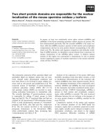
Fig. 3. Characterization of the recombinant GST-tagged L-gulono-1,4-lactone dehydrogenase (Rv1771) of M. tuberculosis. Steady-state param-
eters of the mycobacterial dehydrogenase determined for: (A) the
L-gulono-1,4-lactone substrate in the presence of 121 lM cytochrome c;
and (B) the cytochrome c substrate in the presence of 50 m
ML-gulono-1,4-lactone. Double-reciprocal Lineweaver–Burke plots are shown. V
0
is lmol of L-gulono-1,4-lactone oxidized per min and per mg of the recombinant dehydrogenase (UÆmg protein
)1
). L-Gulono-1,4-lactone con-
centrations ranged from 5 to 25 m
M, whereas cytochrome c concentrations ranged from 24 to 145 lM (B). All measurements were made in
duplicate in three independent experiments; the values obtained in a representative experiment are shown.
0
0.02
0.04
0.06
0.08
0.1
5.5 6 6.5 7 7.5 8 8.5 9
pH
Enzyme activityEnzyme activity
(% of control)
A
B
0
100
200
300
400
500
20 30 40 50
Temperature (°C)
Fig. 4. Effects of pH (A) and temperature (B) on the activity of the
recombinant GST-tagged
L-gulono-1,4-lactone dehydrogenase of
M. tuberculosis. Measurements were made in duplicate; mean
values ± SD are shown. In (A), the dehydrogenase activity is
expressed as DA
550
per min.
O
CH
2
OH
O
H
H O H
O H O H
L-gulono-1,4-lactone
O
CH
2
OH
O
H
H O H
O H O H
L-ascorbic acid
2 cyt c
ox
2 cyt c
red
Fig. 5. Reaction catalyzed by the L-gulono-1,4-lactone dehydroge-
nase of M. tuberculosis.
B. A. Wolucka and D. Communi M. tuberculosis
L-gulono-1,4-lactone dehydrogenase
FEBS Journal 273 (2006) 4435–4445 ª 2006 The Authors Journal compilation ª 2006 FEBS 4439
had low specific activity, ranging from 40 to
66 mUÆmg protein
)1
under the nonoptimal tempera-
ture conditions of the enzyme assay (24 °C). However,
taking into account that the enzyme activity is about
three-fold higher at 39 °C (Fig. 4B) and that the GST-
tagged Rv1771 protein represented only a portion of
the GST affinity-purified fraction (Fig. 1, lane 4), the
specific activity of the recombinant dehydrogenase
could be at least 10-fold higher [400–660 mUÆmg pro-
tein
)1
]. The relatively low activity of the recombinant
M. tuberculosis enzyme could be due to impaired pro-
tein folding, proteolytic degradation and ⁄ or the lack of
a mycobacterial cofactor in the E. coli expression sys-
tem. Another possibility is that the mycobacterial
dehydrogenase might require a specific post-transla-
tional modification that occurs inefficiently in the
E. coli host. As far as we know, the specific activities
of related recombinant enzymes of plant origin have
not been reported. Moreover, huge differences in the
specific activities of purified native aldonolactone
oxidoreductases, ranging from 760 mUÆmg protein
)1
[17] up to 51 000 UÆmg protein
)1
[12], have been
observed. In particular, specific activity values deter-
mined for the native l-galactono-1,4-lactone dehydro-
genase of sweet potato [30] were 1000-fold higher than
those reported for the same enzyme by others [27], but
no explanation for this discrepancy was provided.
l-Gulonolactone dehydrogenase activity could be
measured in the soluble fraction of the M. bovis BCG
Copenhagen strain, and its specific activity was compar-
able to that reported for crude preparations of the rela-
ted l-galactono-1,4-lactone dehydrogenase of plant
origin [9,12,27]. Altogether, our results suggest that the
mycobacterial enzyme could operate efficiently in vivo .
Other proteomic studies have demonstrated that the
Rv1771 protein is relatively abundant in the M. bovis
BCG strain [28] and also in M. tuberculosis H37Rv,
especially in the cell envelope fraction [29]. Indeed,
the Rv1771 protein contains a potential transmem-
brane helix (amino acids 205–227), as predicted by
the tmpred program ( />software/TMPRED_form.html) (Fig. 2). Thus, like all
the known aldonolactone oxidoreductases, the Rv1771
protein may be membrane-associated, in agreement
with the enzyme behavior in the presence of a detergent.
In contrast to the related aldonolactone oxidases
[13,16,19], but similar to the plant l-galactono-1,4-lac-
tone dehydrogenases (EC 1.3.2.3) [12], the mycobacteri-
al enzyme does not use molecular oxygen as an electron
acceptor, and has dehydrogenase activity (Table 1;
Fig. 3B). Interestingly, a dehydrogenase activity of the
rat l-gulono-1,4-lactone oxidase was reported in the
early literature [26,31], but the activity was not studied
further. Nowadays, the l-gulono-1,4-lactone oxidase
enzymes are considered exclusively as oxidases, the
reaction products of which are, paradoxically, L-AA
and hydrogen peroxide [8]. Dehydrogenase-to-oxidase
conversion is well known for another antioxidant (uric
acid)-producing enzyme, xanthine oxidoreductase [32].
Perhaps a similar molecular mechanism might be
responsible for the dehydrogenase-to-oxidase switch of
mammalian l-gulono-1,4-lactone oxidase proteins and
play a role in the metabolism of L-AA.
We showed that in vitro both cytochrome c
(K
m
4.7 lm) (Fig. 3B) and phenazine methosulfate
(Table 1) can serve as electron acceptors for the
l-gulono-1,4-lactone dehydrogenase of M. tuberculosis.
Remarkably, the phenazine methosulfate acceptor was
even more efficient than cytochrome c at saturation
(Table 1). Phenazines, ‘secondary metabolites’ of cer-
tain soil and pathogenic bacteria, are redox-active,
flavin-like low-molecular-weight compounds that can
produce ROS and play a role in quorum sensing and
biofilm formation in Pseudomonas aeruginosa lung
infection [33]. It is possible, therefore, that an unknown
phenazine-like, low-molecular-weight compound might
serve as an endogenous electron acceptor for the
Rv1771 dehydrogenase of M. tuberculosis.
Despite the presence of l-gulono-1,4-lactone dehy-
drogenase activity in the M. bovis BCG Copenhagen
strain, ascorbic acid could not be detected in the M. bo-
vis BCG and M. tuberculosis cells grown in vitro, either
because its concentrations were below the detection
limit or because of the lack of the l-gulono-1,4-lactone
substrate in these cells. l-Gulono-1,4-lactone can be
formed by the C1 reduction of d-glucuronic acid or
d-glucurono-3,6-lactone [8]. An NADPH-dependent
d-glucurono-3,6-lactone reductase activity is present in
cellular extracts of M. bovis BCG (B. A. Wolucka,
unpublished results) and could supply l-gulono-1,4-lac-
tone for the dehydrogenase reaction. In the animal
pathway for vitamin C, free d-glucuronic acid is
derived from UDP-d-glucuronate either directly by the
recently proposed abortive reaction catalyzed by a
UDP-glucuronosyl transferase [34] or via some poorly
characterized hydrolytic steps [8]. UDP-Glucuronate,
in turn, is synthesized from UDP-glucose by a UDP-
glucose dehydrogenase. Mycobacterium tuberculosis
possesses a gene encoding a putative UDP-glucose
dehydrogenase (Rv0322) that is necessary for UDP-
glucuronic acid formation. Moreover, the pathogen
synthesizes some unknown d-glucuronate-containing
glycoconjugates, as demonstrated by immunochemical
methods [35], and therefore must express UDP-glucu-
ronosyltransferase(s). Accordingly, a complete pathway
for vitamin C synthesis, similar to the animal route
M. tuberculosis L-gulono-1,4-lactone dehydrogenase B. A. Wolucka and D. Communi
4440 FEBS Journal 273 (2006) 4435–4445 ª 2006 The Authors Journal compilation ª 2006 FEBS
[8,34], may exist in M. tuberculosis but operate only in
specific conditions. Examples of differential regulation
of gene expression and metabolic reprogramming in
M. tuberculosis are known [36,37]. Some earlier steps in
a pathway for ascorbic acid might be inducible, e.g.
during the intracellular growth of the pathogen in its
host. This could explain the absence of the l-gulono-
1,4-lactone dehydrogenase reaction product in the
M. tuberculosis cells grown in vitro.
The Rv1771 l-gulono-1,4-lactone dehydrogenase of
M. tuberculosis is a specific enzyme for the biosynthesis
of L-AA. As far as we know, this is the first report of a
specific biosynthetic enzyme for vitamin C in bacteria.
To detect related aldonolactone dehydrogenases ⁄
oxidases in other bacterial genomes, we used the rat
l-gulono-1,4-lactone oxidase as a query sequence in
blast searches of the protein database. These searches
retrieved, for a limited number of bacterial species,
additional putative aldonolactone oxidase orthologs
that display significant sequence identity (about 30%)
with the rat l-gulono-1,4-lactone oxidase protein, and
are closely related to the known and predicted
l-gulono-1,4-lactone oxidase-like proteins of animals,
plants and fungi (Fig. 6). Surprisingly, an important
number of bacterial species that contain a vitamin C
biosynthetic gene belong to the Actinomycetales
[M. tuberculosis, M. bovis, Thermobifida fusca, Streptomy-
ces coelicolor and Streptomyces avermitilis (NP_ 823585)].
It is worth noting that members of the Actinomycetales
(Streptomyces verticillus and Saccharothrix mutabilis)
possess orthologs of another vitamin C-related enzyme,
namely the plant GDP-mannose-3¢,5¢-epimerase [38].
Ancestral soil-based organisms might therefore play a
role in horizontal transfer of vitamin C-related genes.
In the process of microbial adaptation, horizontal gene
transfer is essential for the dissemination and assembly
of detoxification pathways that can form part of
genomic islands and have both pathogenicity and
degradation functions [39]. Remarkably, several l-gul-
ono-1,4-lactone oxidase-positive bacteria are known
pathogens [M. tuberculosis, M. bovis, Burkholderia
cepacia, Bacillus anthracis (NP_654628) and Pseudo-
monas aeruginosa (NP_254014)] that infect human
lungs. On the other hand, photosynthetic cynobacteria
that were largely believed to be able to synthesize vita-
min C do not, apparently, contain l-gulono-1,4-lactone
oxidase homologs, with the exception of Nostoc puncti-
forme. These surprising findings raise important ques-
tions about the role of aldonolactone oxidoreductases
in prokaryotic organisms and the evolution of vitamin
C biosynthetic pathways in general.
What could be the physiologic role of the Rv1771
protein in M. tuberculosis? Mycobacterium tuberculosis
has coevolved with its human host, and may persist
for years in a strange symbiosis known as latent
infection. According to World Health Organisation
estimates ( />fs104/en/), about one-third of the world’s population
is infected with M. tuberculosis but only 5–10% of
the infected persons will develop active disease. The
Rv1771 gene is apparently not essential, because
transposon mutants of the gene could be obtained
[40]. The gene is well conserved within the M. tuber-
culosis complex, except for the M. bovis BCG Pasteur
(1173P2) strain, which lost the gene due to the dele-
tion of chromosomal region RD14 [41]. Ironically,
whereas the pathogen’s ortholog is well conserved,
the l-gulono-1,4-lactone oxidase gene of tuberculosis-
prone species, such as humans and guinea pigs, is
nonfunctional because of mutations accumulated dur-
ing evolution [8]. These facts strongly suggest that
the l-gulonolactone dehydrogenase of M. tuberculosis
could play a role in virulence, pathogenesis and ⁄ or
survival of the parasite within its host. In agreement
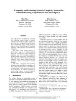
Fig. 6. Sequence relationship between L-gulono-1,4-lactone dehy-
drogenase of M. tuberculosis and previously identified or predicted
L-gulono-1,4-lactone oxidase-like proteins. The unrooted neighbor-
joining (N-J) tree was generated () on the
basis of the amino acid sequences of proteins that show at least
30% identity with the rat
L-gulono-1,4-lactone oxidase. The acces-
sion numbers of the sequences used were: M. tuberculosis
L-gulono-1,4-lactone dehydrogenase, NP_216287; Streptomyces
coelicolor, NP_629980; Thermobifida fusca, ZP_00059445; Oceano-
bacillus iheyensis, NP_692632; Bacillus cereus, NP_830486;
Burkholderia cepacia, ZP_00218082; Saccharomyces cerevisiae
D-arabinono-1,4-lactone oxidase (ALO), P54783; Candida albicans
D-arabinono-1,4-lactone oxidase (ALO), O93852; Neurospora crassa,
Q7SGY1; Gibberella zeae, XP_388870; Arabidopsis thaliana
L-galac-
tono-1,4-lactone dehydrogenase (GLDH), At3g47930; Arabidopsis
thaliana putative
L-gulono-1,4-lactone dehydrogenase, At2g46740;
Sus scrofa
L-gulono-1,4-lactone oxidase (GLO), Q8HXWO; Rattus
norvegicus
L-gulono-1,4-lactone oxidase (GLO), P10867.
B. A. Wolucka and D. Communi M. tuberculosis
L-gulono-1,4-lactone dehydrogenase
FEBS Journal 273 (2006) 4435–4445 ª 2006 The Authors Journal compilation ª 2006 FEBS 4441
with this, a deficiency in d-arabinono-1,4-lactone
oxidase (ALO1), which catalyzes the last step in the
biosynthesis of erythroascorbic acid in yeasts, resulted
in attenuated virulence of the Candida albicans
mutant [42]. If synthesized by the Rv1771 l-gulono-
1,4-lactone dehydrogenase, L-AA may represent a
novel weapon in the antioxidative arsenal of the
pathogen at least in some, although still unknown,
stages of M. tuberculosis infection. Interestingly, the
promoter of the cell wall catalase-peroxidase ⁄ NADH
oxidase [43] gene katG of M. tuberculosis, which is
important for virulence and for the activation of the
antimycobacterial prodrug isoniazid, is induced by
ascorbic acid [44]. Moreover, M. tuberculosis possesses
an ascorbic acid-dependent isomerase that converts
a-acetohydroxyacids to the corresponding a-ketoacids
in the pathway for branched-chain amino acids [45].
These observations suggest that L-AA could act in
M. tuberculosis as a modulator of gene expression
and an enzyme cofactor, in addition to its possible
antioxidant function.
In summary, the l-gulono-1,4-lactone dehydroge-
nase of M. tuberculosis is a new and distinct member
of the family of l-gulono-1,4-lactone dehydrogenase ⁄
oxidases that have been characterized up to now, and
the first example of a specific, vitamin C biosynthetic
enzyme of bacterial origin. Further studies will be
necessary to elucidate the role of the Rv1771
l-gulono-1,4-lactone dehydrogenase in M. tuberculosis
infection.
Experimental procedures
Chemicals
GST affinity and StrataClean resins were obtained from
Stratagene (Madison, WI). l-Gulono-1,4-lactone, cyto-
chrome c from horse heart (oxidized form), phenazine
methosulfate and 2,6-dichloroindophenol were purchased
from Sigma-Aldrich (St Louis, MO). All reagents were of
analytical grade.
Plasmid construction
The ORF corresponding to the mycobacterial Rv1771
l-gulonolactone dehydrogenase (1287 bp) was PCR ampli-
fied from the genomic DNA of M. tuberculosis H37Rv.
Oligonucleotide primers were designed with attB1 or attB2
sites for insertion into the Gateway donor vector
pDONR201 (Invitrogen, Gaithersburg, MD) by homolog-
ous recombination. A sequence (GATGACGACGACAAG)
corresponding to the enterokinase cleavage site (DDDDK)
was included within the forward primer immediately
upstream of the start codon (ATG). Primers with the fol-
lowing sequences were synthesized by Proligo (Paris,
France): 5forGulox (forward), 5¢-GGGGACAAGTTT
GTACAAAAAAGCAGGCTTCGATGACGACGACAAG
ATGAGCCCGATATGGAGTAATTGGCCT-3¢; and 3rev-
Gulox (reverse), 5¢-GGGGACCACTTTGTACAAGAAA
GCTGGGTCTCAGGGACCGAGAACGCGCCGGGTGT
A-3¢. The PCR product was cloned into the pDONR201
vector, and the resulting plasmid, pENTR_Rv1771, was
used to transfer the gene sequence into pDEST15 (Invitro-
gen) (GST tag N-terminal fusion) by means of homologous
recombination. The pDEST15_Rv1771 (GST fusion) plas-
mid obtained was used for expression of the protein in
E. coli BL21(DE3) cells.
Heterologous expression of the recombinant
L-gulono-1,4-lactone dehydrogenase
Escherichia coli cells carrying the pDEST15_Rv1771 plas-
mid were grown at 37 °C to an optical density of 0.8 at
600 nm in 200 mL of culture volume, and then IPTG was
added to a final concentration of 0.1 mm for induction, and
the fusion protein was produced for 1 h or 3 h, as indica-
ted. The cells were washed, resuspended in three volumes of
100 mm phosphate buffer (pH 7.3) containing 0.1% Triton
X-100, 1 mm dithiothreitol, 1 mm phenylmethanesulfonyl
fluoride and 20% glycerol, and sonicated. After centrifuga-
tion at 13 000 g for 15 min at 4 °C on an Eppendorf 5402
centrifuge (Eppendorf, Hamburg, Germany), the superna-
tant was loaded onto a GST affinity column.
GST affinity chromatography of the recombinant
L-gulono-1,4-lactone dehydrogenase of
M. tuberculosis
A crude extract containing the GST-tagged dehydrogenase
was applied to a 1 mL GST affinity column equilibrated
with 50 mm phosphate buffer (pH 7.3) containing 0.1%
Triton X-100, 1 m m phenylmethanesulfonyl fluoride and
20% glycerol (buffer A). The column was washed with 15
volumes of buffer A, and the recombinant dehydrogenase
was eluted with three volumes of 10 mm glutathione
(reduced form) in buffer A. Fractions containing the recom-
binant dehydrogenase were pooled. For measurements of
the l-gulono-1,4-lactone dehydrogenase activity, glutathi-
one and dithiothreitol were removed by gel filtration of
the pooled GST affinity fractions on a prepacked NAP-25
column (Amersham Pharmacia Biotech, Uppsala, Sweden).
Enterokinase cleavage of the GST-tagged
L-gulono-1,4-lactone dehydrogenase
In order to remove the GST tag, an aliquot of the affinity-
purified recombinant dehydrogenase was incubated for 24 h
M. tuberculosis L-gulono-1,4-lactone dehydrogenase B. A. Wolucka and D. Communi
4442 FEBS Journal 273 (2006) 4435–4445 ª 2006 The Authors Journal compilation ª 2006 FEBS
at 22 °C with 2 units of enterokinase (Stratagene) in
500 lL (final volume) of 20 mm Tris ⁄ HCl buffer (pH 8.0)
containing 50 mm NaCl and 2 mm CaCl
2
. One unit of
enterokinase is the amount of enzyme required to cleave
100 lg of the CBP-EK-JNK fusion protein (Stratagene) to
90% completion at 21 °C in 16 hours.
Concentration of protein fractions on
Strataclean beads
To pooled GST affinity fractions, 10 lL of Strataclean
resin suspension was added. After overnight incubation at
4 °C, samples were centrifuged at 13 000 g for 5 min at
room temperature on a Microfuge 18 centrifuge (Beckman-
Coulter, Fullerton, CA), and the supernatants discarded.
The adsorbed proteins were recovered by heating the beads
in three volumes of the five-times concentrated sample buf-
fer for 5 min at 100 °C. After centrifugation at 13 000 g for
5 min at room temperature on a Microfuge 18 centrifuge
(Beckman-Coulter), the concentrated proteins were ana-
lyzed by SDS ⁄ PAGE.
Preparation of crude enzyme extracts from
M. bovis BCG
The M. bovis BCG strain Copenhagen cultures were
grown to mid-exponential phase in Middlebrook 7H9
medium containing ADC enrichment (Becton Dickinson,
San Jose, CA) at 37 °C, without agitation. Cells were collec-
ted by centrifugation at 1500 g for 15 min at 4 °C, on a
Sorvall RC5B plus centrifuge (Sorvall, Ashville, NC), resus-
pended in 100 mm phosphate buffer (pH 7.3) containing
1mm phenylmethanesulfonyl fluoride, and disrupted by so-
nication. Unbroken cells were removed by centrifugation at
500 g for 15 min at 4 °C, and the supernatant was further
centrifuged at 25 000 g as described above. The obtained
soluble extract and the insoluble cell envelope fraction, pre-
viously resuspended in the extraction buffer, were used
for measurements of l-gulono-1,4-lactone dehydrogenase
activity.
Assay of L-gulono-1,4-lactone dehydrogenase
The reaction mixture (1 mL) contained 25 mml-gulono-
1,4-lactone, 0.121 mm cytochrome c and an aliquot of the
affinity-purified dehydrogenase in 50 mm phosphate buffer
(pH 7.3). Because dithiothreitol and glutathione interfered
with the enzyme assay, they were removed by gel filtration
prior to measurements. l-Gulono-1,4-lactone dehydro-
genase activity was measured spectrophotometrically at
550 nm by following the l-gulono-1,4-lactone-dependent
reduction of cytochrome c [46]. When indicated, 2.5 mm
phenazine methosulfate was used as a direct electron accep-
tor in the presence of 100 lm 2,6-dichloroindophenol, and
the decrease in absorbance at 610 nm due to the reduction
of 2,6-dichloroindophenol was measured, as described [47].
PAGE
Proteins were separated by SDS ⁄ PAGE, using 10% mini-
gels and the buffer system described by Laemmli [48]. Gels
were stained with Coomassie Brilliant Blue R-250.
Immunoblotting
Samples fractionated by SDS ⁄ PAGE were transferred to a
nitrocellulose membrane by electroblotting (Bio-Rad, Her-
cules, CA) according to the manufacturer’s protocol. Mem-
branes were incubated with goat anti-GST IgG (1 : 1000
dilution; Amersham Pharmacia Biotech), and antibody
binding was detected using anti-goat IgG conjugated to
alkaline phosphatase (1 : 5000 dilution; Sigma-Aldrich) and
the 5-bromo-4-chloro-3-indolyl-phosphate ⁄ nitroblue tetra-
zolium reagent (Promega, Madison, WI).
MS
MALDI quadrupole TOF (MALDI Q-TOF) MS analysis
of in-gel-digested protein bands was performed on a
Q-TOF Ultima Global mass spectrometer equipped with a
MALDI source (Micromass, Waters Corporation, Milford,
MA), as described [49].
Protein determination
Protein concentration was determined by the method of
Bradford [50], using BSA as standard.
Ascorbic acid determination
Mycobacterial cells were extracted with 5% m-phosphoric
acid [51] or 5% perchloric acid [52], as described. Ascorbic
acid was measured by the HPLC method [51] by using an
Alliance separation module equipped with an M2996 pho-
todiode array detector and the empower chromatography
software (Waters Corporation).
Acknowledgements
We wish to thank Dr P. Jungblut (Max Planck Institute
for Infection Biology, Berlin) for a gift of the M. bovis
BCG strain Copenhagen. We thank Virginie Imbault
for technical assistance in MS. This work was partially
supported by the Fonds de la Recherche Scientifique
Me
´
dicale, Belgium (convention no. 3.4.626.05.F to
BAW). DC is Research Associate at the Fonds
National de la Recherche Scientifique (FNRS).
B. A. Wolucka and D. Communi M. tuberculosis L-gulono-1,4-lactone dehydrogenase
FEBS Journal 273 (2006) 4435–4445 ª 2006 The Authors Journal compilation ª 2006 FEBS 4443
References
1 Schwager J & Schulze J (1998) Modulation of interleu-
kin production by ascorbic acid. Vet Immunol Immuno-
pathol 64, 45–57.
2 Margittai E, Banhegyi G, Kiss A, Nagy G, Mandl J,
Schaff Z & Csala M (2005) Scurvy leads to endoplasmic
reticulum stress and apoptosis in the liver of guinea
pigs. J Nutr 135, 2530–2534.
3 Dhar-Mascareno M, Carcamo JM & Golde DW
(2005) Hypoxia–reoxygenation-induced mitochondrial
damage and apoptosis in human endothelial cells are
inhibited by vitamin C. Free Radic Biol Med 38, 1311–
1322.
4 Fischer H, Schwarzer C & Illek B (2004) Vitamin C
controls the cystic fibrosis transmembrane conductance
regulator chloride channel. Proc Natl Acad Sci USA
101, 3691–3696.
5 Brown LAS & Jones DP (1997) Antioxidant action of
vitamin C in the lung. In Vitamin C in Health and Dis-
ease (Packer L & Fuchs J, eds), pp. 265–278. Marcel
Dekker, New York.
6 May JM, Huang J & Qu ZC (2005) Macrophage uptake
and recycling of ascorbic acid: response to activation by
lipopolysaccharide. Free Radic Biol Med 39, 1449–1459.
7 Valpuesta V & Botella MA (2004) Biosynthesis of
l-ascorbic acid in plants: new pathways for an old
antioxidant. Trends Plant Sci 9 , 573–577.
8 Nishikimi M & Yagi K (1996) Biochemistry and mole-
cular biology of ascorbic acid biosynthesis. Subcell
Biochem 25, 17–39.
9 Wolucka BA & Van Montagu M (2003) GDP-mannose
3¢,5¢-epimerase forms GDP-l-gulose, a putative interme-
diate for the de novo biosynthesis of vitamin C in plants.
J Biol Chem 278, 47483–47490.
10 Shigeoka S, Nakano Y & Kitaoka S (1979) Some prop-
erties and subcellular localization of l-gulono-c-lactone
dehydrogenase in Euglena gracilis z. Agric Biol Chem
43, 2187–2188.
11 Mapson LW & Breslow E (1958) Biological synthesis of
ascorbic acid: l-galactono-c-lactone dehydrogenase.
Biochem J 68, 395–406.
12 Ostergaard J, Persiau G, Davey MW, Bauw G & Van
Montagu M (1997) Isolation of a cDNA coding for
l-galactono-c-lactone dehydrogenase, an enzyme
involved in the biosynthesis of ascorbic acid in plants.
Purification, characterization, cDNA cloning, and
expression in yeast. J Biol Chem 272, 30009–30016.
13 Kiuchi K, Nishikimi M & Yagi K (1982) Purification
and characterization of l-gulonolactone oxidase from
chicken kidney microsomes. Biochemistry 21, 5076–
5082.
14 Loewus FA (1999) Biosynthesis and metabolism of
ascorbic acid in plants and of analogs of ascorbic acid
in fungi. Phytochemistry 52, 193–210.
15 Bleeg HS & Christensen F (1982) Biosynthesis of ascor-
bate in yeast. Purification of l-galactono-1,4-lactone
oxidase with properties different from mammalian
l-gulonolactone oxidase. Eur J Biochem 127, 391–
396.
16 Huh WK, Kim ST, Yang KS, Seok YJ, Hah YC &
Kang SO (1994) Characterisation of d-arabinono-1,4-
lactone oxidase from Candida albicans ATCC 10231.
Eur J Biochem 225, 1073–1079.
17 Nishikimi M, Noguchi E & Yagi K (1978) Occurrence
in yeast of l-galactonolactone oxidase which is similar
to a key enzyme for ascorbic acid biosynthesis in
animals, l-gulonolactone oxidase. Arch Biochem Biophys
191, 479–486.
18 Huh WK, Lee BH, Kim ST, Kim YR, Rhie GE, Baek
YW, Hwang CS, Lee JS & Kang SO (1998) D-Ery-
throascorbic acid is an important antioxidant molecule
in Saccharomyces cerevisiae. Mol Microbiol 30, 895–
903.
19 Wilkinson SR, Prathalingam SR, Taylor MC, Horn D
& Kelly JM (2005) Vitamin C biosynthesis in trypano-
somes: a role for the glycosome. Proc Natl Acad Sci
USA 102, 11645–11650.
20 Koshizaka T, Nishikimi M, Ozawa T & Yagi K (1988)
Isolation and sequence analysis of a complementary
DNA encoding rat liver l-gulono-c-lactone oxidase, a
key enzyme for l-ascorbic acid biosynthesis. J Biol
Chem 263, 1619–1621.
21 Sugisawa T, Ojima S, Matzinger PK & Hoshino T
(1995) Microbial production of l-ascorbic acid from
d-sorbitol, l-sorbose, l-gulose, and l-sorbosone by
Ketogulonicigenium vulgare DSM 4025. Biosci Biotech-
nol Biochem 59, 190–196.
22 Nakagawa H, Asano A & Sato R (1975) Ascorbate-
synthesizing system in rat liver microsomes. II. A peptide-
bound flavin as the prosthetic group of l-gulono-c-lac-
tone oxidase. J Biochem (Tokyo) 77, 221–232.
23 Nishikimi M, Kiuchi K & Yagi K (1977) Detection of
l-gulono-c-lactone oxidase on SDS-polyacrylamide gels
by the fluorescence of its covalently bound flavin. FEBS
Lett 81, 323–325.
24 Kito M, Ohishi N & Yagi K (1991) Micro-determina-
tion of l-gulono-
c-lactone oxidase activity. Biochem Int
24, 131–135.
25 Yabuta Y, Yoshimura K, Takeda T & Shigeoka S
(2000) Molecular characterization of tobacco mitochon-
drial l-galactono-c-lactone dehydrogenase and its
expression in Escherichia coli. Plant Cell Physiol 41,
666–675.
26 Eliceiri GL, Lai EK & McCay PB (1969) Gulonolactone
oxidase. Solubilization, properties, and partial purifica-
tion. J Biol Chem 244, 2641–2645.
27 Imai T, Karita S, Shiratori G, Hattori M, Nunome T,
Oba K & Hirai M (1998) l-Galactono-c-lactone
dehydrogenase from sweet potato: purification and
M. tuberculosis L-gulono-1,4-lactone dehydrogenase B. A. Wolucka and D. Communi
4444 FEBS Journal 273 (2006) 4435–4445 ª 2006 The Authors Journal compilation ª 2006 FEBS
cDNA sequence analysis. Plant Cell Physiol 39, 1350–
1358.
28 Schmidt F, Donahoe S, Hagens K, Mattow J, Schaible
UE, Kaufmann SH, Aebersold R & Jungblut PR (2004)
Complementary analysis of the Mycobacterium tubercu-
losis proteome by two-dimensional electrophoresis and
isotope-coded affinity tag technology. Mol Cell Proteo-
mics 3, 24–42.
29 Mawuenyega KG, Forst CV, Dobos KM, Belisle JT,
Chen J, Bradbury EM, Bradbury AR & Chen X (2005)
Mycobacterium tuberculosis functional network analysis
by global subcellular protein profiling. Mol Biol Cell 16,
396–404.
30 Oba K, Ishikawa S, Nishikawa M, Mizuno H &
Yamamoto T (1995) Purification and properties of
l-galactono-c-lactone dehydrogenase, a key enzyme for
ascorbic acid biosynthesis, from sweet potato roots.
J Biochem (Tokyo) 117, 120–124.
31 Kar NC, Ghosh NC & Guha BC (1963) Microsomal
l-gulonolactone dehydrogenase. Nature 197, 494–495.
32 Kuwabara Y, Nishino T, Okamoto K, Matsumura T,
Eger BT & Pai EF (2003) Unique amino acids cluster
for switching from the dehydrogenase to oxidase form
of xanthine oxidoreductase. Proc Natl Acad Sci USA
100, 8170–8175.
33 Price-Whelan A, Dietrich LE & Newman DK (2006)
Rethinking ‘secondary’ metabolism: physiological roles
for phenazine antibiotics. Nat Chem Biol 2, 71–78.
34 Linster CL & Van Schaftingen E (2006) Glucuronate,
the precursor of vitamin C, is directly formed from
UDP-glucuronate in liver. FEBS J 273, 1516–1527.
35 Wolucka BA, McNeil MR, Kalbe L, Cocito C & Bren-
nan PJ (1993) Isolation and characterization of a novel
glucuronosyl diacylglycerol from Mycobacterium smeg-
matis. Biochim Biophys Acta 1170, 131–136.
36 Fenhalls G, Stevens L, Moses L, Bezuidenhout J, Betts
JC, Helden PP, Lukey PT & Duncan K (2002) In situ
detection of Mycobacterium tuberculosis transcripts in
human lung granulomas reveals differential gene expres-
sion in necrotic lesions. Infect Immun 70, 6330–6338.
37 McKinney JD, Honer zu Bentrup K, Munoz-Elias EJ,
Miczak A, Chen B, Chan WT, Swenson D, Sacchettini
JC, Jacobs WR Jr & Russell DG (2000) Persistence of
Mycobacterium tuberculosis in macrophages and mice
requires the glyoxylate shunt enzyme isocitrate lyase.
Nature 406, 735–738.
38 Wolucka BA, Persiau G, Van Doorsselaere J, Davey
MW, Demol H, Vandekerckhove J, Van Montagu M,
Zabeau M & Boerjan W (2001) Partial purification and
identification of GDP-mannose 3¢,5¢-epimerase of Ara-
bidopsis thaliana, a key enzyme of the plant vitamin C
pathway. Proc Natl Acad Sci USA 98, 14843–14848.
39 Van der Meer JR & Sentchilo V (2003) Genomic islands
and the evolution of catabolic pathways in bacteria.
Curr Opin Biotechnol 14, 248–254.
40 Lamichhane G, Zignol M, Blades NJ, Geiman DE,
Dougherty A, Grosset J, Broman KW & Bishai WR
(2003) A postgenomic method for predicting essential
genes at subsaturation levels of mutagenesis: application
to Mycobacterium tuberculosis. Proc Natl Acad Sci
USA 100, 7213–7218.
41 Behr MA, Wilson MA, Gill WP, Salamon H, Schoolnik
GK, Rane S & Small PM (1999) Comparative genomics
of BCG vaccines by whole-genome DNA microarray.
Science 284, 1520–1523.
42 Huh WK, Kim ST, Kim H, Jeong G & Kang SO (2001)
Deficiency of D -erythroascorbic acid attenuates hyphal
growth and virulence of Candida albicans. Infect Immun
69, 3939–3946.
43 Singh R, Wiseman B, Deemagarn T, Donald LJ,
Duckworth HW, Carpena X, Fita I & Loewen PC
(2004) Catalase-peroxidases (KatG) exhibit NADH
oxidase activity.
J Biol Chem 279, 43098–43106.
44 Mulder MA, Zappe H & Steyn LM (1999) The Myco-
bacterium tuberculosis katG promoter region contains a
novel upstream activator. Microbiology 145, 2507–2518.
45 Allaudeen HS & Ramakrishnan T (1970) Biosynthesis
of isoleucine and valine in Mycobacterium tuberculosis
H37Rv. II. Purification and properties of acetohydroxy
acid isomerase. Arch Biochem Biophys 140, 245–256.
46 O
ˆ
ba K, Fukui M, Imai Y, Iriyama S & Nogami K
(1994) l-Galactono-c-lactone dehydrogenase: partial
characterization, induction of activity and role in the
synthesis of ascorbic acid in wounded potato tuber tis-
sue. Plant Cell Physiol 35, 473–478.
47 Nakagawa H & Asano A (1970) Ascorbate synthesizing
system in rat liver microsomes. I. Gulonolactone-reduci-
ble pigment as a prosthetic group of gulonolactone oxi-
dase. J Biochem (Tokyo) 68 , 737–746.
48 Laemmli UK (1970) Cleavage of structural proteins
during the assembly of the head of bacteriophage T4.
Nature 227, 680–685.
49 Wittamer V, Franssen JD, Vulcano M, Mirjolet JF,
Le Poul E, Migeotte I, Brezillon S, Tyldesley R,
Blanpain C, Detheux M et al. (2003) Specific recruit-
ment of antigen-presenting cells by chemerin, a novel
processed ligand from human inflammatory fluids.
J Exp Med 198, 977–985.
50 Bradford MM (1976) A rapid and sensitive method for
the quantitation of microgram quantities of protein util-
izing the principle of protein-dye binding. Anal Biochem
72, 248–254.
51 Wolucka BA, Davey MW & Boerjan W (2001) A high-
performance liquid chromatography radio method for
determination of l-ascorbic acid and guanosine
5¢-diphosphate-l-galactose, key metabolites of the plant
vitamin C pathway. Anal Biochem 294, 161–168.
52 Conklin PL, Pallanca JE, Last RL & Smirnoff N (1997)
L-ascorbic acid metabolism in the ascorbate-deficient
arabidopsis mutant vtc1. Plant Physiol 115, 1277–1285.
B. A. Wolucka and D. Communi M. tuberculosis L-gulono-1,4-lactone dehydrogenase
FEBS Journal 273 (2006) 4435–4445 ª 2006 The Authors Journal compilation ª 2006 FEBS 4445