Báo cáo khoa học: Stage specific expression of poly(malic acid)-affiliated genes in the life cycle of Physarum polycephalum Spherulin 3b and polymalatase potx
Bạn đang xem bản rút gọn của tài liệu. Xem và tải ngay bản đầy đủ của tài liệu tại đây (1.19 MB, 10 trang )
Stage specific expression of poly(malic acid)-affiliated
genes in the life cycle of Physarum polycephalum
Spherulin 3b and polymalatase
Nadthanan Pinchai, Bong-Seop Lee and Eggehard Holler
Institut fu
¨
r Biophysik und Physikalische Biochemie der Universita
¨
t Regensburg, Germany
Physarum polycephalum is a versatile organism, dis-
playing several alternative cell types and developmental
transitions [1]. Uninucleate amoebae and multinucleate
plasmodia constitute the two vegetative growth phases
in the life cycle. These two cell types differ in cellular
organization, behaviour and gene expression. In
adverse conditions, amoebae reversibly transform into
cysts. Usually, when the conditions are favourable,
amoebae mate and develop into plasmodia. Plasmodia
survive adverse conditions by transforming into
another kind of cysts, spherules. When starved in the
light, sporangia are formed. In favourable conditions,
spores hatch to release amoebae, thus completing the
cycle.
Of the various cell types in the life cycle of
P. polycephalum, only the plasmodium contains the
water soluble polymer, b-poly(l-malate) (PMLA)
[2–4]. The polymer is concentrated in the nuclei in
an amount comparable with that of DNA and hi-
stones [5]. Due to its structural similarity to the
backbone of nucleic acids, PMLA has been proposed
to bind nuclear proteins and function in a molecular
transporter system ([6] and references therein). Injec-
tion of PMLA into plasmodia increased the growth
rate and shortened cell cycle duration, indicating that
it could also be involved in the molecular events
responsible for the synchronization of events in the
plasmodium [7,8]. PMLA is synthesized from
l-malate derived from d-glucose through the glyco-
lytic pathway and the tricarboxylic acid cycle [9].
The polymer is released from the nuclei into cyto-
plasm and finally to the culture medium, where it is
Keywords
Physarum polycephalum; plasmodium;
polymalatase, polymalic acid; spherulin 3b
Correspondence
E. Holler, Institut fu
¨
r Biophysik und
Physikalische Biochemie der Universita
¨
t
Regensburg, D-93040 Regensburg,
Germany
Fax: +49 941 943 2813
Tel: +49 941 943 3030
E-mail: Eggehard.Holler@biologie.
uni-regensburg.de
(Received 4 November 2005, revised 4
January 2006, accepted 9 January 2006)
doi:10.1111/j.1742-4658.2006.05131.x
Polymalic acid is receiving interest as a unique biopolymer of the plasmodia
of mycetozoa and recently as a biogenic matrix for the synthesis of devices
for drug delivery. The acellular slime mold Physarum polycephalum is charac-
terized by two distinctive growth phases: uninucleated amoebae and multi-
nucleated plasmodia. In adverse conditions, plasmodia reversibly transform
into spherules. Only plasmodia synthesize poly(malic acid) (PMLA) and
PMLA-hydrolase (polymalatase). We have performed suppression subtrac-
tive hybridization (SSH) of cDNA from amoebae and plasmodia to identify
plasmodium-specific genes involved in PMLA metabolism. We found cDNA
encoding a plasmodium-specific, spherulin 3a-like polypeptide, NKA48
(spherulin 3b), but no evidence for a PMLA-synthetase encoding transcript.
Inhibitory RNA (RNAi)-induced knockdown of NKA48-cDNA generated a
severe reduction in the level of PMLA suggesting that spherulin 3b func-
tioned in regulating the level of PMLA. Unexpectedly, cDNA of poly-
malatase was not SSH-selected, suggesting its presence also in amoebae.
Quantitative PCR then revealed low levels of mRNA in amoebae, high levels
in plasmodia, and also low levels in spherules, in agreement with the expres-
sion under transcriptional regulation in these cells.
Abbreviations
DSDM, diluted semidefined medium; PMLA, b-poly(
L-malate); RNAi, inhibitory RNA; SDM, semidefined medium; Sph, spherulin; SSH,
suppression subtractive hybridisation.
1046 FEBS Journal 273 (2006) 1046–1055 ª 2006 The Authors Journal compilation ª 2006 FEBS
degraded to l-malate by a plasmodium-specific hy-
drolase (polymalatase) [4,5,10,11].
PMLA is a highly interesting polymer: applications
in pharmacy and medicine are proposed ([6] and refer-
ences therein); nanoconjugates of PMLA can be used
as drug delivery vehicles [12], the crystal structure is
being investigated [13]. However, little is known about
the regulation of the polymer at the genetic level of its
synthesis and degradation. The gene for polymalatase
has been sequenced (accession number AJ543320 [14]),
and distinct features of its sequenced promotor remain
to be assigned to transcription control. The mechanism
of PMLA synthesis has been studied in vivo, but
attempts to identify the PMLA synthesizing enzyme
system have been unsuccessful because of loss of syn-
thetic activity during rupture of plasmodia in the pre-
paration of extracts [15].
The PMLA synthetic capacity of plasmodia of the
yellow strains such as P. polycephalum MC
3
VII is
approximately 1 mgÆh
)1
Æg plasmodia
)1
[8,9] ([6] and
references therein), suggesting the presence of detect-
able amounts of PMLA synthetase-specific mRNA.
The absence of PMLA and polymalatase in other cell
types could be the result of cell specific gene expression
for synthesis and degradation. To gain deeper insight,
this investigation was aimed at identifying plasmo-
dium-specific genes, which are involved in the synthesis
of PMLA and ⁄ or its regulation, and to clarify whether
the stage-specific expression of polymalatase [4,10] is
regulated at the transcriptional or the translational
level. We report on the identification of plasmodium-
specific mRNAs on the basis of suppressive substrac-
tive hybridization (SSH) using cDNAs of plasmodial
extracts as tester and of amoebal extracts as driver. A
large number of transcripts were found, most of them
false-positive and only three true-positive. One had a
high degree of identity with spherulin 3a and appeared
to be involved in regulation of PMLA levels in vivo.
None of the SSH-generated DNAs showed similarity
with a sequence listed in the databases that would be
indicative of a PMLA synthetase. Quantitative PCR
revealed that polymalatase mRNA was expressed at
considerably lower levels in amoebae and spherules
than in plasmodia. This paralleled contents of poly-
malatase protein [4,10] suggesting regulated expression
at the transcriptional level.
Results
Isolation of differentially expressed cDNAs
After SSH, differentially expressed cDNAs were ana-
lysed after two rounds of PCR. The amplified products
from the secondary nested PCR were ligated with
pGEM
Ò
T-vector and were transformed into DH10B
competent cells. About 70 white colonies were
obtained in total, 52 of which were selected. Plasmid
DNAs were isolated and analysed after restriction
enzyme digestion. Each DNA sequence occurred only
once in agreement with the fact that 5¢-ends of
mRNAs had been isolated with the Capfinder oligo-
nucleotides. Restriction to 5¢-ends was thought to
reduce the complexity of bands after SSH and enhance
isolation of products. Nineteen of the plasmid prepara-
tions contained inserts of 150 bp and were sequenced.
PCR analysis indicated three true-positive subtracted
transcripts and all others to be false positives. The
high ratio of false- to true-transcripts was attributed to
the use of the different strains LU352 for amoebae
and M
3
CVII for plasmodia. Isolation of 5¢-ends of
mRNA by SSH using Capfinder oligonucleotides
responded in particular to variability in this region.
The three transcripts NKA8 (accession number
DQ017262), NKA49 (accession number DQ017263),
and NKA48 (accession number DQ017261) were plas-
modium-specific, as they were not detected in amoe-
bae. Fragment NKA8 contained an ORF encoding
257 amino acids and showed a putative conserved
domain in the NCBI data base termed DUF343 (or
gnI|CDD|26165 in the conserved domain data base),
found in various cellular organisms. Fragment NKA49
encoded 37 amino acids, and no alignments were
found. These two fragments were not considered fur-
ther. Although the high PMLA producing activity of
plasmodia had suggested the finding of an abundant
cDNA for PMLA-synthetase, no such cDNA could be
identified to date.
Transcript of NKA48 showed the highest abundance
and was further analysed. Nucleotide and deduced
amino acid sequences of NKA48 were compared with
the GenBank database. The results indicated a high
degree of identity on the levels of nucleotides (84%)
and amino acids (86%) with spherulin 3a (Figs 1 and
2), the most abundant encystment-specific protein [16],
and identities with sequences of bc-crystallins (Fig. 2).
The total number of amino acids is 103, correlating
with a calculated molecular mass of 11271.5 and a the-
oretical isoelectric point of 4.88. Because of the high
similarity, the polypeptide encoded by NKA48 was
named spherulin 3b.
Knockdown of mRNA to NKA48 (spherulin 3b)
Macroplasmodia were injected with dsRNA to
NKA48 (spherulin 3b) and harvested after 24 h. Two
negative controls were performed: macroplasmodia
N. Pinchai et al. Cell type expression of spherulin 3b and polymalatase
FEBS Journal 273 (2006) 1046–1055 ª 2006 The Authors Journal compilation ª 2006 FEBS 1047
without microinjection and macroplasmodia injected
with unspecific dsRNA (generated using part of the
pGEM
Ò
-5zf(+) vector as template [14]). The degree of
mRNA knockdown was analysed by real-time PCR
with actin mRNA as reference. Figure 3 shows that
the ratio of mRNA to NKA48 over that of actin was
significantly reduced to 1% (P<0.001). Control
microinjection with unspecific dsRNA showed no
effect on mRNA levels (P > 0.5), indicating that the
knockdown was specific. The fact that this low residual
level was obtained after 24 h suggested that the half-
life of spherulin 3b mRNA was in the range of one to
a few hours and much less than the half-life of 24–
36 h for spherulin 3a mRNA [16]. Inhibitory RNA
(RNAi) experiments were also carried out with dsRNA
to NKA8 but a reduction of only 25% of the mRNA
level was observed and this was considered insignifi-
cant (P > 0.1). For the relatively short NKA49, RNAi
inhibition was not attempted; this decision was based
on previous experience with short dsRNA.
Decreased levels of PMLA after microinjection
of dsRNA
PMLA was measured in the extracts of the above
NKA48-dsRNA injected and control macroplasmodia
harvested 24 h after microinjection and referenced to
the amount of protein in the same cells. Knockdown
of mRNA in Fig. 3A was found to be paralleled by a
severe reduction to 3.5 ± 0.5 lg PMLAÆmg
)1
protein
(12% with reference to uninjected macroplasmodia;
P < 0.002) (Fig. 3B). The control that had received
unspecific dsRNA amounted to 20 ± 4 lg PMLAÆmg
)1
protein (P > 0.05), not significantly lower than the
uninjected control, indicating that the reduction in
PMLA content was specifically referred to knockdown
of NKA48 mRNA. As suggested by the low level of
suppression of specific mRNA, no effects were notified
in experiments with dsRNA to NKA8.
Macroplasmodia were observed for several days
after microinjection, however, significant morphologi-
cal changes related to the depression of PMLA were
not observed.
Level of polymalatase mRNA at different stages
in the life cycle
The amount of polymalatase transcript at different sta-
ges in the life cycle was monitored by real-time PCR
using specific primers. Since the mRNA level of house-
keeping genes, such as of actin, varies from one cell
type to the other, a cloned fragment of the polymala-
tase gene was used as an external standard and subjec-
ted to the same treatment as the samples. In Fig. 3C,
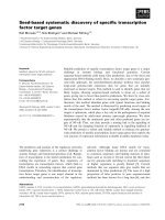
Fig. 1. Nucleotide sequence alignment of spherulin 3b (1) with spherulin 3a (2). Identical residues are highlighted in grey. Start and stop
codons are given in bold. Forward and reverse primer for RNAi experiments are underlined. Ac, Accession Number. The alignment was car-
ried out using
CLUSTALW from and BLAST from NCBI.
Cell type expression of spherulin 3b and polymalatase N. Pinchai et al.
1048 FEBS Journal 273 (2006) 1046–1055 ª 2006 The Authors Journal compilation ª 2006 FEBS
the level of cDNA of polymalatase (corresponding to
the level of mRNA) was very low for amoebae and
spherules in comparison with plasmodia (P<0.001).
The expression of the gene in amoebae and plasmodia
explained, why cDNA was absent after SSH screening
(see above). The presence of cDNA in the stages of the
life cycle indicated that the protein could have some
general function. High levels specifically in plasmodia
are consistent with a functional affiliation to PMLA
and with a regulation of gene expression at the tran-
scription level.
Discussion
Physarum polycephalum belongs to the mycetozoa, the
multicellular eukaryotes more closely related to ani-
mal–fungi than to green plants [17,18]. Mycetozoa dis-
play a life cycle including the microscopic amoebae
and the gigantic multinucleate plasmodium [1]. Of the
various cell types only the plasmodium contains the
water soluble polymer, PMLA [10]. The polymer is
concentrated in the nuclei, the level being under homeo-
static control, and the excess released continuously
into the culture medium [5]. Its presumed function is
to coordinate transport, delivery, and activity of cer-
tain proteins (DNA polymerases, histones, etc.) to nuc-
lei [3,7,10,19,20], and it has been suggested that it
participates in the maintenance of the observed high
degree of synchrony typical for plasmodia [8]. Strain
M
3
CVII is one of the high PMLA producers [8]. Sev-
eral other strains contain less PMLA, but no strain
has been found that was devoid of the polymer. In
contrast, PMLA contents of nuclei were similar in all
strains. Thus, although the treatment with RNAi to
spherulin 3b reported here suppressed the overall level
of PMLA, the remaining low level was probably suffi-
cient to support normal cell morphology.
Under adverse conditions, such as starvation and
desiccation in the dark, the plasmodium undergoes
reversible differentiation into smaller dehydrated sphe-
rules [21]. Each of the spherules contains several nuclei
that overexpress particular stress proteins. Four major
spherulation-specific mRNAs have been identified that
emerged 24 h after beginning of starvation-induced
spherulation of plasmodia and that then comprise
10% of the total mRNA [16]. Among them, spheru-
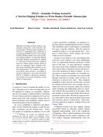
Fig. 2. Structural alignment of amino acid sequence by motifs, of spherulin 3b with spherulin 3a and other members of the bc-crystallin fam-
ily: (1) spherulin 3b from P. polycephalum; (2) spherulin 3a from P. polycephalum; (3) hypothetical protein YPTB2846 from Yersinia pseudo-
tuberculosis; (4) hypothetical protein YmolA_01000341 from Y. mollaretii; (5) hypothetical protein Y1348 from Y. pestis; (6) c-crystallin from
Danio rerio; and (7) development-specific protein S homologue from Myxococcus xanthis. The residues highlighted in black indicate glycines,
serines, and aromatics that are conserved in a bc-crystallin fold. The residues shown in grey indicate the side chains and backbone sites that
are involved in calcium binding [24]. The residues in the conserved tyrosine corners are in bold [24]. ‘Greek key’ motifs are highlighted with
underlines: single line, first motif; broken line, second motif; double line, third motif. Motif searches were performed using
PROSITE from
. Similarity search and multiple alignment were carried out using
CLUSTALW from and BLAST
from NCBI. Ac, Accession Number.
N. Pinchai et al. Cell type expression of spherulin 3b and polymalatase
FEBS Journal 273 (2006) 1046–1055 ª 2006 The Authors Journal compilation ª 2006 FEBS 1049
lin 3a is the most abundant mRNA. During differenti-
ation, synthesis of PMLA discontinues, and the
remaining polymer is exported into the extracellular
fluid and degraded. It is assumed that the PMLA syn-
thesizing enzyme is downregulated at the onset of
spherulation.
Despite considerable effort, knowledge of PMLA
synthetase activity and its regulation is still fragment-
ary [15]. To discover stage-specifc genes affiliated with
PMLA metabolism and ultimately to identify the syn-
thetase gene, SSH was used with mRNA of the plas-
modium as the tester and mRNA of amoebae as the
driver. The amoebae strain chosen was LU352, which
was not identical with plasmodia of strain M
3
CVII. It
was chosen because it allowed preparations of contam-
ination-free RNA that, for unknown reasons, had not
been possible for M
3
CVII amoebae. The choice of the
different strain had the principle disadvantage of gen-
erating a large portion of false-positive transcripts.
Assuming that mRNA would be abundant in the
plasmodium it was hoped that PMLA synthetase
cDNA could be identified by SSH using amoebae as
driver, which do not produce PMLA. While this
cDNA could not be identified, an abundant species
was revealed that encoded a 11.3-kDa polypeptide,
NKA48 (named spherulin 3b), which is structurally
highly related to spherulin 3a (85% identical amino
acids). While NKA48 occurs in plasmodia, spherulin
3a is only found in spherules [16]. Both proteins con-
tain the ‘Greek key’ typical of the bc-crystallin family
of proteins. While spheruline 3a like another two-
domain protein, protein S [22], responds in terms of
stress proteins [23] to extreme environmental condi-
tions, NKA48 has no such function.
bc-Crystallins are two-domain proteins found in
vertebrate eye lenses and have distant relatives in
microorganisms (e.g. the proteins in Fig. 2). The
bc-crystallin domain of spherulin 3a from P. poly-
cephalum, considered by some as a primitive organ-
ism, has been compared by X-ray crystallography
with the modern lens crystalline domain fold in order
to address the evolutionary origin of the vertebrate
bc-crystallins [24]. Typically, two successive Greek
key motives (underlined in Fig. 2, each approximately
40 amino acid residues) pair to form a domain. The
domain fold contains a pair of calcium binding sites.
While the bc-crystallins of lens (not shown) and lower
organisms in Fig. 2 contain two domain folds, spheru-
lin 3a and NKA48 contain only a single domain fold.
The stability of these two proteins is highly dependent
on calcium binding [25]. The typical domain motives
contain a ‘tyrosine corner’ in the domain centre as
seen in proteins 3–6 of Fig. 2 or slightly displaced as
A
B
C
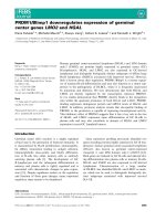
Fig. 3. Knockdown experiments and stage specific expression of
polymalatase mRNA. (A) Knockdown of NKA48 mRNA by specific
dsRNA. Levels of mRNA relative to that of actin are shown 24 h
after microinjection with dsRNA to NKA48 and with unspecific con-
trol dsRNA to pGEM-5zf(+) vector. Standard deviations refer to
experiments in triplicates. (B) PMLA content of plasmodia injected
with dsRNA to NKA48 in the RNAi experiment. The data are refer-
enced to protein contents. Standard deviations are shown for
measurements in triplicates. (C) mRNA levels of polymalatase in
different cell types during the life cycle. Levels were measured
in terms of cDNA by PCR referenced to a standard as described in
Experimental procedures. One-hundred per cent mRNA (plasmodia)
refers to 8.91 pgÆlL
)1
standard cDNA. Standard deviations are
shown for measurements in triplicates.
Cell type expression of spherulin 3b and polymalatase N. Pinchai et al.
1050 FEBS Journal 273 (2006) 1046–1055 ª 2006 The Authors Journal compilation ª 2006 FEBS
in Protein S (7) or spherulin 3a (2). NKA48 (1) dif-
fers from all of these proteins by not containing a
tyrosine in a corresponding position. In contrast to
NKA48, spherulin 3a is stabilized by forming dimers
through disulfide bonds. Dimerization is not possible
for NKA48, because it does not contain such cyste-
ines. It is concluded that NKA48 is more distant
from two-domain bc-crystallins as is spherulin 3a,
and has evolved from this gene by gene duplication,
as indicated by the high degree of sequence similarity
(Fig. 1). This resulted in a structure devoid of the
tyrosine corner and dimerization by disulfide forma-
tion. It is also different in structure from spherulin 3a
by 14 amino acid substitutions, eight of them located
in the first two b-strands of the N-terminal half of
NKA48, upstream of the homodimer interface and
accessible for interactions with other macromolecules.
It is to be shown how these mutations serve the par-
ticular function of NKA48 in the regulation of
PMLA synthesis.
Knockdown analysis of plasmodia with dsRNA to
NKA48 revealed a dramatic decrease in NKA48
mRNA to a residual 1% and a decrease in PMLA to
a residual 12% compared to the contents in reference
plasmodia. Because of the high sequence identity of
mRNA for spherulin 3a and spherulin 3b, knockdown
of spherulin 3a mRNA might have also occurred by
this dsRNA treatment. However, because spherulin 3a
is not transcribed in the plasmodium [16], this possibil-
ity could not have effected the suppression of PMLA
synthesis.
Among other possibilities, this effect on PMLA
synthesis could be the result of loss of induction at
the transcriptional level, of loss of activation of the
synthetase protein itself, or of derepression of
enzyme(s) catalysing PMLA degradation. An interest-
ing interplay of NKA48 with spherulin 3a could be
imagined if both proteins bound competitively at the
same loci but only NKA48 was an inducer and ⁄ or
activator. In a physiologically meaningful mechanism,
spherulin 3a would displace NKA48 during the onset
of spherulation and suppress PMLA synthesizing
activity.
Degradation of PMLA during the onset of spherula-
tion is catalysed by enzymatically active forms of
polymalatase in the extraplasmodial fluid [5,10,11].
During plasmodia growth, only catalytic amounts of
polymalatase are contained in the culture medium,
while large amounts of zymogen reside within the plas-
modium. Correspondingly, zymogen and polymalatase
with different functions have been proposed, namely a
PMLA hydrolysing variant in the exterior and a chap-
eroning adapter variant in the interior of plasmodia
[7,10,11]. Polymalatase activity depends on zymogen
activation [10] at the outer surface of plasmodia
(unpublished results). The enzymology has been inves-
tigated in detail [5,10,11,26,27].
The hydrolytically inactive form or zymogen of
polymalatase binds PMLA, chaperons it through the
intracellular fluid, thus functioning as an adapter by
connecting it with other proteins [7,10], and eventually
manages its export into the extracellular fluid (unpub-
lished data).
In agreement with these activities, the role of
polymalatase and its zymogen is correlated with the
synthesis of PMLA by the plasmodium. Our results of
real-time PCR measurements indicated high levels of
mRNA in plasmodia and low levels in both amoebae
and spherules. The differences parallel the occurrence
of high amounts of polymalatase protein in plasmodia,
very low levels in spherules, and the absence of
polymalatase protein in amoebae [10]. The correlation
suggested regulation of synthesis at the transcriptional
level.
Experimental procedures
Culture conditions for the growth of plasmodia
Microplasmodia of P. polycephalum strain M
3
CVII ATCC
204388 (American type Culture Collection, LGC Promo-
chem, Wesel, Germany) were grown axenically in semi-
defined medium (SDM) as described [28]. Cells were
harvested for SSH after 2 days. Macroplasmodia were
obtained by fusion of 400 lL of packed 2-day-old micro-
plasmodia on agar in 13.5-cm Petri dishes according to a
previously described method [29] and grown for 24 h in the
dark prior to microinjections. After injection, they were
grown for further 24 h and then harvested for the analyses
of mRNA and PMLA content.
Culture conditions for spherule preparation
Spherules were induced by the transfer of 2-day-old micro-
plasmodia to a non-nutrient salt medium and were shaken
in the dark for 2 days at 24 °C as described [30]. After
replacement with fresh salt medium, spherules were incuba-
ted at 24 °C for 1 day and were harvested for real-time
PCR.
Culture conditions for the growth of amoebae
DSPB plates (diluted SDM with phosphate buffer [28])
were inoculated with 3 · 10
5
amoebal cysts of the apogamic
strain LU352 [31], 100 lL formalin-killed bacteria, and
100 lL Millipore water. The plates were incubated at 24 °C
N. Pinchai et al. Cell type expression of spherulin 3b and polymalatase
FEBS Journal 273 (2006) 1046–1055 ª 2006 The Authors Journal compilation ª 2006 FEBS 1051
for 48 h to allow excystment and were then transferred to
30 °C. After 4 days at 30 °C the plates became confluent
and were harvested for SSH and real-time PCR.
RNA isolation
To isolate total RNA, amoebae and macroplasmodia were
harvested from the agar plates and immediately frozen in
liquid nitrogen. RNA isolation was carried out by using the
QIAGEN RNeasy
Ò
Mini Kit (Qiagen, Hilden, Germany)
and a maximum of 100 mg frozen cells.
PolyA
+
RNA was isolated using 85 lL Dynabeads
Ò
(Invitrogen, Karlsruhe, Germany) oligo(dT)
25
and 25 lg
total RNA and was eluted in 15 lL Tris ⁄ HCl (10 mm,
pH 7.5). The eluted mRNA was immediately used for the
first-strand cDNA synthesis.
cDNA synthesis
First-strand synthesis reactions were set up with each con-
taining 0.5 lg mRNA, 10 lm oligo(dT) primer and 10 lm
CapFinder oligonucleotide according to the protocol of
Clontech Laboratories 1996 (Mountain View, CA). Reverse
transcription was performed with Rnase H Minus
M-MuLV Reverse Transcriptase (MBI Fermentas, St.
Leon-Rot, Germany). After 1 h at 42 °C, 0.4 lL 100 mm
MnCl
2
was added, and the sample was incubated for a fur-
ther 15 min. The reaction was terminated by heating at
70 °C for 10 min, the first-strand product was purified
using QIAquick PCR purification Kit (QIAGEN).
Second-strand synthesis reactions were carried out using
Advantage
TM
2 PCR Kit (BD Biosciences Clontech, Moun-
tain View, CA) and long-distance PCR (BD SMART
TM
PCR cDNA Synthesis Kit User Manual).
Suppression subtractive hybridization
Differentially expressed cDNAs in plasmodia and amoebae
were identified following the SSH technique described by
Diatchenko et al. [32]. Plasmodial extract mRNA was used
as tester and amoebal extract mRNA as driver. Only
poly(A)
+
RNA was used for first-strand cDNA synthesis.
PCR reactions were optimized and performed in such a
way that syntheses remained in the exponential phase. Care
was taken that at least 25% of total cDNA was ligated
with adaptors on both ends. The success of SSH was tested
for an abundantly expressed housekeeping gene (actin
Ppa35 [33], accession number M21500), for a less abun-
dantly expressed gene lig1 [34], and for the known stage-
specific genes actin-fragmin kinase [35], fragmin A [36],
fragmin P [36], and polymalatase (accession no. AJ543320)
using primers to the published cDNA sequences. Also, the
efficiency of SSH was checked by comparing the number of
PCR cycles necessary to produce equal amounts of actin
cDNA in probes containing equal amounts of either sub-
stracted or unsubstracted DNA.
Subtracted PCR products were then ligated with pGEM
Ò
T-vectors (Promega, Mannheim, Germany) and were trans-
formed into DH10B competent cells. The plasmids were
isolated using Nucleospin
Ò
Plasmid Kit (Machery-Nagel,
Du
¨
ren, Germany) and were sent for sequencing (MWG
Biotech, Ebersberg, Germany). The blast program was
used for Databases analysis.
The stage specificity of the subtracted cDNA sequences
was verified by conventional PCR including 20 ng of the
above cloned cDNA from SSH, 1 · PCR buffer, 25 mm
MgCl
2
, 10 mN dNTP mix, Taq polymerase (2.5 U, MBI Fer-
mentas) and 10 lm of each of the following primers. For
NKA8: forward, 5¢-GTCTCCAGACGTCTCGAAC-3¢;
reverse, 5¢-CATCCAAGTCTTGGGAGCTC-3¢. For
NKA48: forward, 5¢-GATGCTAACTTCAGCGGAAAC
TC-3¢; reverse, 5¢-CACGATGATGGATGAAATGGCG
TC-3¢. For NKA49: forward, 5¢-CTTCCACGACGGAAAC
GATGAC-3¢; reverse, 5¢-CTCTCCAACACATGCTGACG
TAG-3¢. Cycling conditions were 94 °C for 2 min, followed
by 35 cycles of 94 °C for 30 s, 54 °C for 30 s, 72 °C for
2 min, and 72 °C for 10 min. The samples were then separ-
ated by electrophoresis through 2% agarose gel. Sequences
and primers of the other SSH products can be obtained on
request from the corresponding author.
RNA interference
RNAi experiments were carried out with dsRNA to
NKA48 by the method essentially as described by Haindl
and Holler [14]. Specific DNA template to NKA48 for
dsRNA synthesis was generated from first-strand cDNA
and the following primers (NKA48, accession number
DQ017261): 5¢-GATGCATAATACGACTCACTATAGG
GAAATGTCCGTCCAACAAGGAG-3¢ (forward) and 5¢-
GCCTTCTAATACGACTCACTATAGGGACCACGATG
ATGGATGAAATG-3¢ (reverse). Both primers contained
T7-polymerase promoter at the 5¢ terminus and were cus-
tom-synthesized by MWG-Biotech. The resulting 294-bp
DNA spanned the nucleotides 51–310 of the gene including
the origin of transcription, and was used as template for
in vitro dsRNA synthesis as described by Donze and Picard
[37]. In the case of NKA8, the forward primer was 5¢-GAT
GCATAATACGACTCACTATAGGGAGTGCCTTGCAA
GGAGTATTG-3¢ and the reverse primer was 5¢-GCCTTC
TAATACGACTCACTATAGGGAGCTCGTAATAGCTT
TTGGAC-3¢, the resulting DNA spanning nucleotides
21–536 of the gene (accession number DQ017262). For con-
trol injections, nonspecific dsRNA was generated by the
same method using a PCR-derived fragment with 592 bp,
nucleotides 142–734 from the vector pGEM(R)-5zf(+)
(Technical Servics, Promega Corporation, Madison, WI,
USA). Each knockdown experiment was carried out with
Cell type expression of spherulin 3b and polymalatase N. Pinchai et al.
1052 FEBS Journal 273 (2006) 1046–1055 ª 2006 The Authors Journal compilation ª 2006 FEBS
10 lg dsRNA, microinjected into the veins of macro-
plasmodia. After 24 h, the plasmodia were analysed by
real-time PCR.
Real-time PCR
The amount of NKA48-specific mRNA in the RNA inter-
ference experiment was measured with reference to mRNA
expressed for actin Ppa35 gene using the Roche-LightCycler
(Roche, Mannheim, Germany). cDNA was synthesized
from 2 lg total RNA of each sample and reference, and
2 lL of the purified product was subjected to real-time
PCR, each containing 10 lL2· SYBR Green Master Mix,
10 lm each primer and 6 lL RNase free water using the
following conditions: 15 min 95 °C activation of HotS-
tarTaq DNA Polymerase and 35 cycles (15 s 94 °C, 20 s
58 °C and 20 s 72 °C). For actin, primer pairs were
5¢-CATGTGCAAGGCTGGATTTGCTG-3¢ (forward) and
5¢-ACCGACGTATGAGTCCTTTTG-3¢ (reverse) and for
NKA48 5¢-GATGCTACTTCAGCGGAAACTC-3¢ (for-
ward), 5¢-CACTTGAGTGTTCTGCTCCAG-3¢ (reverse).
To compare mRNA expression levels of PMLA hydro-
lase in the amoebae, plasmodia and spherules, a PCR frag-
ment derived from the target sequence (polymalatase,
accession number AJ543320) was generated as a standard
for absolute quantification. To create the standard, 40 ng
of plasmodial cDNA was used in a conventional PCR
including 1 · PCR buffer, 25 mm MgCl
2
,10mm dNTP
mix, Taq polymerase (MBI Fermentas) and 10 lm each
primer 5¢-CAAAGGGATTATGAGACAGCAG-3¢ (for-
ward) and 5¢-ACTGTGCCATCCGCCTTC-3¢ (reverse).
Cycling conditions were 94 °C for 2 min, followed by 35
cycles of 94 °C for 30 s, 55 °C for 30 s and 72 °C for
2 min. The amplified product was purified by electrophor-
esis on 2% agarose gel and using QIAquick
Ò
Gel Extrac-
tion Kit (QIAGEN). Fifty nanograms pGEM
Ò
T-vector
was ligated with 16 ng purified PCR product and was
transformed into DH10B competent cells (Bethesda
Research Laboratories, Frederick, MD). Plasmid isolation
was carried out using the Nucleospin
Ò
Plasmid Kit
(Machery-Nagel). For insert isolation, 12 lg plasmid DNA
was digested with NcoI and SpeIat37°C for 1.5 h and
was analysed on a 2% agarose gel. The purified DNA
fragment was used as standard.
Total RNA was isolated from cells of the different stages
in the life cycle, and first-strand cDNA synthesis was per-
formed in triplicate as above for mRNAs, but using 2 lg
total RNA. Real-time PCR was carried out with 40 ng
cDNA of each sample in parallel with five different
amounts of the standard DNA, using the same primer pair
for polymalatase as above and cycling conditions 95 °C for
15 min, followed by 35 cycles of 95 °C for 15 s, 58 °C for
20 s, and 72 °C for 20 s. Default settings of the Lightcycler
Software Version 3.5.3 and conditions in the linear range of
the PCR-reaction were ensured.
Quantitative analysis of PMLA
Macroplasmodia were harvested, weighed and transferred
into a glass homogenizer. Two vols lysis buffer (50 mm
Tris ⁄ HCl pH 7.5, 5 mm NaS
2
O
5
,50mm EGTA, 10 mm
MgCl
2
, 300 mm NaCl, 0.5% Triton X-100), 1 ⁄ 25 volume
of protease inhibitor (calculated from the volume of the
lysis buffer) and 1 ⁄ 1000 volume of mercaptoethanol were
added. The homogenate was transferred to a clean tube
and centrifuged at 20 000 g for 30 min. One hundred
microlitres of the clarified lysate was removed and ali-
quoted equally into two microcentrifuge tubes. One of
the two aliquots was hydrolysed with 50 lL2m sulfuric
acid and incubated at 95 °C for 1.5 h. Then the acid was
neutralized with 50 lLof4m NaOH. The other tube
was kept on ice and was used to measure the amount of
endogenous malate. Polymalic acid was assessed on basis
of the malate dehydrogenase reaction as described [8].
Protein was assayed according to the method of Bradford
[38].
Acknowledgements
The technical assistance of Hermine Reisner and Sonja
Fuchs is greately acknowledged.
References
1 Burland TG, Solnica KL, Bailey J, Cunningham DB &
Dove WF (1993) Patterns of inheritance, development
and the mitotic cycle in the protist Physarum
polycephalum. Adv Microb Physiol 35, 1–69.
2 Fischer H, Erdmann ES & Holler E (1989) An unusual
polyanion from Physarum polycephalum that inhibits
homologous DNA polymerase-alpha in vitro. Biochem-
istry 28, 5219–5226.
3 Doerhoefer S, Windisch C, Angerer B, Lavrik OI, Lee
B-S & Holler E (2002) The DNA-polymerase inhibiting
activity of poly (b-L-malic acid) in nuclear extract dur-
ing the cell cycle of Physarum polycephalum. Eur J
Biochem 269, 1253–1258.
4 Windisch C, Miller S, Reisner H, Angerer B, Achham-
mer G & Holler E (1992) Production and degradation of
b-poly-L-malate in cultures of Physarum polycephalum.
Cell Biol Internat Reports 16, 1211–1213.
5 Schmidt A, Windisch C & Holler E (1996) Nuclear
accumulation and homeostasis of the unusual polymer
b-poly (L-malate) in plasmodia of Physarum
polycephalum. Eur J Cell Bio 70, 373–380.
6 Lee B-S, Vert M & Holler E (2002) Water-soluble ali-
phatic polyesters: Poly(malic acid)s. In Biopolymers
Volume 3a Polyesters (Doi, Y & Steinbu
¨
chel, A, eds),
pp. 76–103. Wiley-VCH, Weinheim, Germany.
7 Karl M, Gasselmaier B, Krieg RC & Holler E (2003)
Localization of fluorescence-labeled poly(malic acid) to
N. Pinchai et al. Cell type expression of spherulin 3b and polymalatase
FEBS Journal 273 (2006) 1046–1055 ª 2006 The Authors Journal compilation ª 2006 FEBS 1053
the nuclei of the plasmodium of Physarum polycepha-
lum. Eur J Biochem 270, 1536–1542.
8 Karl M, Anderson R & Holler E (2004) Injection of
poly(b-L-malate) into the plasmodium of Physarum
polycephalum shortens the cell cycle and increases
growth rate. Eur J Biochem 271, 3805–3811.
9 Lee B-S & Holler E (1999) Effects of culture conditions
on beta-poly(L-malate) production by Physarum polyce-
phalum. Appl Microbiol Biotechnol 51, 647–652.
10 Karl M & Holler E (1998) Multiple polypeptides
immunologically related to b-poly(L-malate) hydrolase
(polymalatase) in the plasmodium of the slime
mold Physarum polycephalum. Eur J Biochem 251, 405–
412.
11 Korherr C, Roth M & Holler E (1995) Poly(beta-L-
malate) hydrolase from plasmodia of Physarum poly-
cephalum. Can J Microbiol 41, 192–199.
12 Lee B-S, Fujita M, Khazenzon NM, Wawrowsky KA,
Wachsman-Hogiu S, Farkas DL, Black KL, Ljubimova
JY & Holler E (2006) Polycefin, a new prototype of a
multifunctional nanoconjugate based on poly( b -L-
malic acid) for drug delivery. Bioconjugate Chem,
doi:10.1021/bc0502457.
13 Fernandez CE, Mancera M, Holler E, Bou JJ, Galbis
JA & Munoz-Guerra S (2005) Low-molecular-weight
poly(a-methyl b,L-malate) of microbial origin: synthesis
and crytallization. Macromol Biosci 5, 172–176.
14 Haindl M & Holler E (2005) The giant multinucleate
plasmodium of Physarum polycephalum to study RNA
interference in the myxomycete. Anal Biochem 342, 194–
199.
15 Willibald B, Bildl W, Lee B-S & Holler E (1999) Is
b-poly(L-malate) synthesis catalysed by a combination
of b-L-malyl-AMP-ligase and b-poly(L-malate) poly-
merase. Eur J Biochem 269, 1085–1090.
16 Bernier F, Lemieux G & Pallotta D (1987) Gene
families encode the major encystment-specific proteins
of Physarum polycephalum plasmodia. Gene 59, 265–
277.
17 Baldauf SL, Roger AJ, Wenk-Siefert I & Doolittle WF
(2004) A kingdom-level phylogeny of eukaryotes based
on combined protein data. Science 290, 972–977.
18 Baldauf SL & Doolittle WF (1997) Origin and evolution
of the slime molds (Mycetozoa). Proc Natl Acad Sci
USA 94, 12007–12012.
19 Angerer B & Holler E (2005) Large complexes of
b-poly(L-malate) with DNA polymerase a, histones,
and other proteins in nuclei of growing plasmodia
of Physarum polycephalum. Biochemistry 34, 14741–
14751.
20 Doerhoefer S, Khodyreva S, Safranov IV, Wlassoff
WA, Anarbaev R, Lavrik OI & Holler E (1998) Mole-
cular constituents of the replication apparatus in the
plasmodium of Physarum polycephalum: identification
by photoaffinity labelling. Microbiology 144, 3181–3193.
21 Chet I & Rush HP (1969) Induction of spherule forma-
tion in Physarum polycephalum by polyols. J Bacteriol
100, 674–678.
22 Wistow G, Summers L & Blundell T (2005) Myxococcus
xanthus spore coat protein S may have a similar struc-
ture to vertebrate lens beta-gamma-crystallins. Nature
315, 771–773.
23 de Jong WW, Caspers GJ & Leunissen JAM (1998)
Genealogy of the a crystallin-small heat shock protein
superfamily. Int J Biol Macromol 22, 151–162.
24 Clout NJ, Kretschmar M, Jaenicker R & Slingsby C
(2001) Crystal structure of the calcium-loaded spherulin
3a dimer sheds light on the evolution of the eye lens
bc-crystallin domain fold. Structure 9, 115–124.
25 Rosinke B, Renner C, Mayr E, Jaenicke R & Holak T
(1997) The solution structure of calcium loaded spheru-
lin 3a. J Mol Biol 272, 1–11.
26 Gasslmaier B, Krell CM, Seebach D & Holler E (2000)
Synthetic substrates and inhibitors of b-poly(L-malate)
-hydrolase (polymalatase). Eur J Biochem 267, 5101–
5105.
27 Gasslmaier B & Holler E (1997) Specificity and direc-
tion of depolymerization of b-poly(L-malate) catalysed
by polymalatase from Physarum polycephalum. Fluores-
cence labeling at the carboxy-terminus of b-poly
(L-malate). Eur J Biochem 250, 308–314.
28 Dee J, Foxon JL, Roberts EM & Walker MH (1997)
Contact with a solid substratum induces cysts in axenic
cultures of Physarum polycephalum amoebae: mannitol
induced detergent-resistent cells are not true cysts.
Microbiology 143, 1059–1069.
29 Nygaard OP, Guttes S & Rush HP (1960) Nucleic acid
metabolism in a slime mold with synchronous mitosis.
Biochim Biophys Acta 38, 298–306.
30 Daniel FW & Baldwin HH (1964) Methods of culture
for plasmodial myxomycetes. Meth Cell Physiol 1,
9–14.
31 Anderson R & Dee J (1990) Culture, development and
genetics of physarum polycephalum – a practical manual.
9th European Physarum Conference. Leicester, UK.
April 1990.
32 Diatchenko L, Lau VF, Campbell AP, Chenchik A,
Moqadam F, Huang B, Lukyanov S, Lukyanov K,
Gurskaya N, Sverdlov ED et al. (1996) Suppression
subtractive hybridization: a method for generating
differentially regulated or tissue-specific cDNA probes
and libraries. Proc Natl Acad Sci USA 93, 6025–
6030.
33 Hamelin M, Adam L, Lemieux G & Pallotta D (1988)
Expression of the three unlinked isocoding actin genes
of Physarum polycephalum. DNA 7, 317–328.
34 Kroneder R, Cashmore AR & Marwan W (1999)
Phytochrome-induced expression of lig1, a homologue
of the fission yeast cell-cycle checkpoint gene hus1,
is associated with the developemental switch in
Cell type expression of spherulin 3b and polymalatase N. Pinchai et al.
1054 FEBS Journal 273 (2006) 1046–1055 ª 2006 The Authors Journal compilation ª 2006 FEBS
Physarum polycephalum plasmodium. Curr Genet 36,
86–93.
35 Gettemans J, de Ville Y, Vandekerckhove J & Waelkens
E (1993) Purification and partial amino acid sequence
of actin-fragmin kinase from Physarum polycephalum.
Eur J Biochem 214, 111–119.
36 T’Jampens D, Bailey J, Cook L, Constantin B, Van-
deckerckhove J & Gettemans J (1999) Physarum
amoebae express a distinct fragmin-like actin-binding
protein that controls in vivo phosphorylation of actin by
the actin-fragmin kinase. Eur J Biochem 265, 240–250.
37 Donze O & Picard D (2002) RNA interference in mam-
malian cells using siRNAs synthesized with T7 RNA
polymerase. Nucleic Acids Res 46, 1–4.
38 Bradford M (1976) A rapid and sensitive method for
the quantitation of microgram quantities of protein util-
izing the principle of protein-dye binding. Anal Biochem
72, 248–254.
N. Pinchai et al. Cell type expression of spherulin 3b and polymalatase
FEBS Journal 273 (2006) 1046–1055 ª 2006 The Authors Journal compilation ª 2006 FEBS 1055