Báo cáo khoa học: Modulation of glucocorticoid receptor-interacting protein 1 (GRIP1) transactivation and co-activation activities through its C-terminal repression and self-association domains pptx
Bạn đang xem bản rút gọn của tài liệu. Xem và tải ngay bản đầy đủ của tài liệu tại đây (375.57 KB, 12 trang )
Modulation of glucocorticoid receptor-interacting
protein 1 (GRIP1) transactivation and co-activation
activities through its C-terminal repression and
self-association domains
Pei-Yao Liu
1
, Tsai-Yuan Hsieh
2
, Wei-Yuan Chou
1
and Shih-Ming Huang
1
1 Department of Biochemistry, National Defense Medical Center, Taipei, Taiwan
2 Department of Medicine, Division of Gastroenterology, Tri-Service General Hospital, National Defense Medical Center, Taipei, Taiwan
Members of the nuclear receptor (NR) superfamily are
ligand-inducible transcription factors. This family
includes the receptors for steroids, thyroid hormone
and vitamin D, as well as orphan receptors for which
no ligands have yet been identified [1,2]. Each receptor
has two activation functions (AFs), namely hormone
independent (AF-1) and hormone dependent (AF-2).
The relative importance of AF-1 and AF-2 varies
between different NRs and is influenced by ligand, cell
type and the target gene promoter [3,4]. The mechanism
by which DNA-bound NRs regulate transcription
appears to involve the recruitment of co-regulatory
proteins, including co-activators and co-repressors
[5–8]. Co-activators are not usually DNA-binding pro-
teins, but are recruited to the promoter through
protein–protein contact with transcriptional activators.
Transcriptional co-repression can involve competition
for limiting factors, displacement of positive factors, or
histone deacetylation to generate a chromatin structure
that limits promoter accessibility [7,8]. Therefore, the
latest working model regarding transcriptional regula-
tion by NRs is an initial association with transcriptional
Keywords
co-activation; GRIP1; HDAC1; nuclear
receptor; transactivation
Correspondence
S M. Huang, National Defense Medical
Center, Department of Biochemistry, 161,
Section 6, MinChuan East Road, Taipei,
Taiwan 114
Fax: +886 287924057
Tel: +886 227937318
E-mail:
(Received 11 December 2005, revised
13 March 2006, accepted 16 March 2006)
doi:10.1111/j.1742-4658.2006.05231.x
Glucocorticoid receptor-interacting protein 1 (GRIP1), a p160 family nuc-
lear receptor co-activator, possesses at least two autonomous activation
domains (AD1 and AD2) in the C-terminal region. AD1 activity appears
to be mediated by CBP ⁄ p300, whereas AD2 activity is apparently mediated
through co-activator-associated arginine methyltransferase 1 (CARM1).
The mechanisms responsible for regulating the activities of AD1 and AD2
are not well understood. We provide evidence that the GRIP1 C-terminal
region may be involved in regulating its own transactivation and nuclear
receptor co-activation activities through primary self-association and a
repression domain. We also compared the effects of the GRIP1 C terminus
with those of other factors that functionally interact with the GRIP1 C ter-
minus, such as CARM1. Based on our results, we propose a regulatory
mechanism involving conformational changes to GRIP1 mediated through
its intramolecular and intermolecular interactions, and through modulation
of the effects of co-repressors on its repression domains. These are the first
results to indicate that the structural components of GRIP1, especially
those of the C terminus, might functionally modulate its putative transacti-
vation activities and nuclear receptor co-activator functions.
Abbreviations
ACTR, activator for thyroid hormone and retinoid receptors; AD, activation domain; AF, activation function; AR, androgen receptor; CARM1,
co-activator-associated arginine methyltransferase 1; CoCoA, coiled-coil co-activator; ER, estrogen receptor; GAC63, GRIP1-associated
co-activator 63; GAL4DBD, Gal4 DNA-binding domain; GRIP1, glucocorticoid receptor-interacting protein 1; GST, glutathione S-transferase;
HA, hemagglutinin; HAT, histone acetyltransferase activity; HDAC1, histone deacetylase 1; HMT, histone methyltransferase; NR, nuclear
receptor; RLU, relative light unit; SRC-1, steroid receptor co-activator 1; TR, thyroid receptor; TSA, trichostatin A.
2172 FEBS Journal 273 (2006) 2172–2183 ª 2006 The Authors Journal compilation ª 2006 FEBS
co-repressors, followed by recruitment of co-activators
in response to ligands and other signals [9].
There are at least three families of NR co-activators:
CBP ⁄ p300; the p160 family; and p ⁄ CAF [7,10,11]. The
best characterized of these is a family of three structur-
ally related, but genetically distinct, 160 kDa proteins
called the NR co-activators or p160 co-activators
[12–18]. These three proteins are steroid receptor
co-activator 1 (SRC-1), glucocorticoid receptor-inter-
acting protein 1 (GRIP1, also called TIF2), and activ-
ator for thyroid hormone and retinoid receptors
(ACTR) (also called RAC3, pCIP, AIB1 and
TRAM1). These co-activators bind directly to the
DNA-bound NRs and apparently function by recruit-
ing secondary co-activators, such as CBP ⁄ p300,
co-activator-associated arginine methyltransferase 1
(CARM1), or related proteins, and possibly by acety-
lating or methylating histones or other proteins
involved in the transcription machinery [5,10,19–21].
Two separate domains of p160 co-activators can bind
to AF-1 and AF-2 of NRs. The p160 co-activators
contain at least three NR-interacting boxes or LXXLL
motifs (where L stands for leucine and X can be any
amino acid) in their central regions, which interact
directly with the highly conserved AF-2 domain of
NRs [22]. The C-terminal region of the p160 co-activa-
tor can interact with the AF-1 domain of some NRs
and enhance their AF-1 activities in the absence of lig-
ands [23–25].
Recent studies have identified three activation
domains (which transduce the activation signal) in the
p160 co-activator [23,26,27]. The enhancement of NR
activity by the p160 co-activator depends on the
CBP ⁄ p300 family, which is necessary for the function
of activation domain 1 (AD1) (amino acids 1075–1083
in GRIP1) [26]. AD1 receives an activating signal from
DNA-bound NRs and recruits CBP ⁄ p300 [23,27].
CBP ⁄ p300 may activate the transcription machinery
through its histone acetyltransferase (HAT) activity,
which acetylates histones and other proteins involved
in transcription [28]. The second activation domain of
the p160 co-activator, AD2, is located in its far C-ter-
minal region (amino acids 1305–1462 in GRIP1)
[23,26]. The mechanism of signalling by AD 2 may
involve the weak HAT activity found in two p160 fam-
ily members (SRC-1 and ACTR), but not in GRIP1
[14,20]. The importance of HAT activity for p160
co-activator function has not been established, and no
efficiently acetylated substrates have yet been reported.
CARM1 is a protein with histone methyltransferase
(HMT) activity. It mainly binds to the C-terminal
region of GRIP1 to stimulate its AD2 transactivation
function [19]. Furthermore, CBP and CARM1 also
support synergistic cross-talk through their HAT and
HMT specificities for histones and other transcrip-
tional factors [19,21]. A third activation domain,
AD3, was recently identified in the highly conserved
N-terminal bHLH-PAS domain of p160 co-activators
by recruitment of secondary co-activators, including
coiled-coil co-activator (CoCoA) and GRIP1-assoc-
iated co-activator 63 (GAC63) [29,30]. As CoCoA
and GAC63 have no obvious sequence homology, the
nature of their downstream targets and the specific
components of the transcriptional machinery remain
unknown.
The mechanisms by which the p160 co-activators
function in NR transcriptional activation, and how
they are regulated, are not fully understood, and their
components have not been identified in detail. It
remains to be established whether the functions of the
p160 co-activator are modulated by post-translational
events, such as self-association, protein modification,
or subcellular localization. In this article, we present
several lines of evidence that demonstrate the func-
tional roles of the GRIP1 C terminus in the regulation
of its own transactivation and of NR co-activator
activities, which are mediated through its repression
and self-association properties. Hence, our results pro-
vide insights into the regulatory mechanisms control-
ling the functional activities of GRIP1. They extend
our understanding of the importance of the structural
status of GRIP1 in modulating NR functions.
Results
Autoregulation of GRIP1 AD activities by its
C-terminal region
Previous studies have demonstrated that deletion of
the AD1orAD2 domain of GRIP1 results in selective
loss of its co-activator functions in the NR system,
affecting specific primary or secondary co-activator
functions [23,26]. We were interested in establishing
whether this involved the structural components of
GRIP1. Therefore, we created various GRIP1 frag-
ments fused with the yeast Gal4 DNA-binding domain
(Gal4DBD) and monitored Gal4-responsive reporter
(GK1 reporter) luciferase activity in HeLa cells to
assess the transactivation activity of the fragments
(Fig. 1A,B). In general, the reporter activity of full-
length GRIP1 (amino acids 5–1462) was negatively
regulated by its structural component (Fig. 1, histo-
gram 2, compare A and B). We performed western
blotting analysis to examine the expression levels of
Gal4 fusions and GRIP1 fragments and found poor
expression of full-length GRIP1 (Fig. 1C, lane 2),
P Y. Liu et al. Autoregulation of GRIP1 functions via C-terminal region
FEBS Journal 273 (2006) 2172–2183 ª 2006 The Authors Journal compilation ª 2006 FEBS 2173
which is also evident from Figs 2B and 5C. Although
the expression of GRIP1 fragments varied, their trans-
activation activities were primarily determined by
structural components. For example, a C-terminal
truncated GRIP1 (amino acids 5–1121) showed greater
reporter activity than one N-terminal truncated GRIP1
(GRIP1-563–1462) (Fig. 1A, compare histogram 3 with
histogram 4), suggesting a repression region in its
C terminus. Subsequently, the region encompassing
amino acids 1122–1304 was identified as the major
repression region in the GRIP1 C terminus (Fig. 1A,
compare histogram 5 with histogram 6). Furthermore,
GRIP1-1013–1121 induced maximal AD1 activity
(Fig. 1B, histogram 9, compare A and B), which sug-
gests that amino acids 563–1012 also constitute a
repression region for AD1 activity (compare histogram
5 with histogram 9 of Fig. 1B). Similar patterns of
transactivation activity were exhibited by these GRIP1
fragments in human embryonic kidney 293 cells, and
the identities of AD1, AD2, and at least two repres-
sion regions in amino acids 563–1012 and 1122–1304,
were consistent with our findings derived from HeLa
cells (data not shown).
HDAC1 is involved in the GRIP1 repression
complex
Having established the existence of a repression prop-
erty of GRIP1, we investigated whether deacetylase
activity mediated through the histone deacetylase
(HDAC) family was involved in the repression effect.
First, we treated HeLa cells with 100 ngÆmL
)1
trichost-
atin A (TSA), an inhibitor of HDAC activity [31],
and monitored the changes in reporter activity of
Gal4DBD fused with various GRIP1 fragments after
16 h of TSA treatment (Fig. 2A). TSA enhanced the
reporter activity of the GRIP1 C-terminal fragment
(amino acids 1122–1462) (4.5-fold) and suppressed that
of full-length GRIP1 (Fig. 2A). We then used glutathi-
one S-transferase (GST) pull-down analysis to examine
which of the co-repressor proteins, HDAC1, HDAC4,
mSin3a or SMRT-a, were involved in the repression
complex. We found that HDAC1, mSin3a and SMRT-
a interacted physically with two C-terminal fragments
(amino acids 1122–1462 or 1305–1462) (data not
shown). In addition, we examined the GRIP1–HDAC1
complex using a co-immunoprecipitation assay in
COS7 cells. We detected the GRIP1–HDAC1 complex
by immunoprecipitating GRIP1 using a hemagglutinin
(HA) antibody or by immunoprecipitating HDAC1
using a myc antibody (Fig. 2B). HA antibody immuno-
precipitation identified three HDAC1-interacting
regions, in GRIP1 residues 563–1121, 5–765, and
AD2
AD2
AD2
AD2
1122
1122
1305
1305
1462
1462
1462
1462
AD1
AD1
AD2
AD2
5
1462
1462
AD1
AD1
5
1121
1121
AD1
AD1
AD2
AD2
1462
1462
563
563
AD1
AD1
1121
1121
563
563
AD1
AD1
1304
1304
563
563
[Gal4DBD; pM vector]
[Gal4DBD; pM vector]
1
2
3
4
5
6
7
8
Luciferase Activity
5
(RLU 10 )
Luciferase Activity
(RLU 10 )
5
0
1 2
3 120
120
160
160
1x
1x
858x
858x
29000x
29000x
290x
290x
1
5
9
10
10
AD1
AD1
1013
1013
1121
1121
AD1
AD1
AD2
AD2
1462
1462
1013
1013
AD1
AD1
1121
1121
563
563
[Gal4DBD; pM vector]
[Gal4DBD; pM vector]
Luciferase Activity
4
(RLU 10 )
Luciferase Activity
(RLU 10 )
4
0 8
4
35
35
40
40
1x
1x
13x
13x
277x
277x
15x
15x
10x
10x
858x
858x
16x
16x
2x
2x
7
8
9
10
10
3
4
5 6
2
WB anti-Gal4DBD
WB anti-Gal4DBD
WB anti-HuR
WB anti-HuR
M
r
M
r
M
r
M
r
170
170
130
130
100
100
72
72
55
55
72
72
55
55
40
40
33
33
24
24
7
8
9
10
10
3
4
5 6
2
A
B
C
Fig. 1. Modulation of glucocorticoid receptor-interacting protein 1
(GRIP1) transactivation activity. (A, B) Expression vectors (0.5 lg)
for the indicated fragments of GRIP1 fused to the Gal4 DNA-bind-
ing domain (Gal4DBD) were transiently transfected into HeLa cells
together with the GK1 reporter gene (0.5 lg), which encodes lucif-
erase and is controlled by the Gal4 response element. The lucif-
erase activity of transfected cell extracts was determined.
Numbers beside the bars indicate fold activation compared with
that of the Gal4DBD alone. RLU, relative light units. These data are
the average of three experiments (mean ± SD; n ¼ 3). (C) COS-1
cells were co-transfected with various Gal4DBD.GRIP1 fragments
(2 lg) in a six-well plate. Cell lysates were subjected to western
blotting analysis and then immunoblotted with anti-Gal4DBD (upper
panel) to determine the GRIP1 expression level and anti-HuR
(bottom panel) to determine the loading control. Results shown are
representative of three independent experiments.
Autoregulation of GRIP1 functions via C-terminal region P Y. Liu et al.
2174 FEBS Journal 273 (2006) 2172–2183 ª 2006 The Authors Journal compilation ª 2006 FEBS
1122–1462 (Fig. 2B, compare lanes 1, 4, 6, and 7). The
myc immunoprecipitation also contained these GRIP1
fragments (data not shown). Our results with TSA
(Fig. 2A) suggested that the HDAC family might be
involved in repression through a deacetylase-independ-
ent pathway. Hence, we used a mutant HDAC1 protein
that lacks deacetylase activity and found that the parti-
ally repressive effect on the Gal4 reporter activity was
the same as with wild-type HDAC1 for both full-length
GRIP1 (amino acids 5–1462) and C-terminal GRIP1
(amino acids 1122–1462) in HeLa cells (Fig. 2C,
compare the histograms with open and grey columns).
Homo-oligomerization of GRIP1
We examined whether the GRIP1 C terminus can inter-
act inter- or intramolecularly with full-length GRIP1 to
modulate its transactivation response to other GRIP
C-terminal interacting proteins, such as CARM1,
Zac1 and ACTN2 [19,32,33]. A co-immunoprecipitation
assay in COS-7 cells showed that Gal4DBD fused to
the full-length GRIP1 (amino acids 5–1462) complexed
strongly with HA.GRIP1-563–1462 and weakly with
HA.GRIP1-5–765 or HA.GRIP1-563–1121 (Fig. 3A,
lanes 7, 5 and 8, respectively). Our co-immunoprecipi-
tation analysis suggested that the primary region of
GRIP1 self-association is located at its C terminus,
within amino acids 1122–1462 (Fig. 3A, compare lanes
5–8). In a parallel experiment, we were unable to detect
any HA-tag signal by immunoprecipitation using a
mouse anti-IgG antibody (Fig. 3A, lanes 9–12). Based
on the results in Fig. 3A, we used GST pull-down
assays to confirm this potential self-association motif
with different C-terminal regions of GRIP1 (amino
acids 1122–1462, 1305–1462, 1122–1304, 1305–1398,
1305–1462 and 1399–1462). These regions were fused to
GST and the fusion proteins were immobilized on
agarose beads. Their ability to bind to a synthesized
AD2
AD2
1122
1122
1462
1462
AD1
AD1
AD2
AD2
5
1462
1462
[Gal4DBD; pM vector]
[Gal4DBD; pM vector]
1
2
3
0
2 4
6
1
2
3
2x
2x
0.3x
0.3x
4.5x
HDAC1.myc
HDAC1.myc
WB by α-myc
WB by
α
-myc
WB by α-myc
WB by α-myc
IP by α-HA
IP by α-HA
Input (5%)
Input (5%)
HA
HA.GRIP1
5-1462
GRIP1
5-1121
HA.
HA.GRIP1
563-1121
HA.GRIP1
563-1462
HA.GRIP1
1122-1462
HA.GRIP1
5-765
HDAC1.myc
HA
HA.GRIP1
GRIP1
5-1462
5-1121
HA.
HA.GRIP1
HA.GRIP1
HA.GRIP1
HA.GRIP1
HDAC1.myc
563-1121
563-1462
1122-1462
5-765
++ + + + + +
+
+
+
+
+
+
+
WB by α-HA
WB by α-HA
66
66
46
46
30
30
97.6
97.6
220
220
kDa
kDa
1 2
3
4
5
6
7
HDAC1.myc
HDAC1.myc
Luciferase Activity
3
(RLU 10 )
Luciferase Activity
(RLU 10 )
3
0
1
2
3
Gal4DBD
Gal4DBD
Gal4DBD.
GRIP1
5-1462
Gal4DBD.
GRIP1
5-1462
Gal4DBD.
GRIP1
1122-1462
Gal4DBD.
GRIP1
1122-1462
4
none
none
HDAC1 wt
HDAC1 wt
HDAC1 mt
HDAC1 mt
A
B
C
Fig. 2. GRIP1 physically and functionally interacts with histone
deacetylase 1 (HDAC1). (A) Expression vectors (0.5 lg) for the indi-
cated fragments of GRIP1 fused to the Gal4 DNA-binding domain
(Gal4DBD) (pM vector) were transiently transfected into HeLa cells
along with the GK1 reporter gene (0.4 lg) in the absence or pres-
ence of 100 ngÆmL
)1
trichostatin A (TSA) for 16 h. Numbers above
the bars indicate fold activation compared with that of no TSA
treatment. (B) COS-7 cells were co-transfected with various
Gal4DBD.GRIP1 fragments (5 lg) and with HDAC1.myc (5 lg) in a
100 mm Petri dish. Cell lysates were subjected to immunoprecipi-
tation with anti-myc (upper panel) immunoglobulin and then immu-
noblotted with anti-hemagglutinin (middle panel) and anti-myc
(bottom panel) immunoglobulin for the loading control for GRIP1
and HDAC1 proteins. Results shown are representative of three
independent experiments. (C) Expression vectors (0.4 lg) for the
indicated fragments of GRIP1 fused to the Gal4DBD were transi-
ently co-transfected into HeLa cells, together with the GK1 reporter
gene (0.2 lg) with 0.2 lg of wild-type pcDNA3.HDAC1.flag (open
column) or the enzyme-dead HDAC1 mutant (grey column). The
luciferase activity of the transfected cell extracts was determined.
These data (A,C) are the average of three experiments (mean ±
SD; n ¼ 3).
P Y. Liu et al. Autoregulation of GRIP1 functions via C-terminal region
FEBS Journal 273 (2006) 2172–2183 ª 2006 The Authors Journal compilation ª 2006 FEBS 2175
GRIP1 C-terminal fragment (amino acids 1122–1462)
was measured in vitro (Fig. 3B). The results indicated
that amino acids 1305–1398 constitute the primary self-
association region in the GRIP1 C terminus (Fig. 3B,
compare lanes 6–10). The amount of protein pulled
down by GST–GRIP1-1305–1462 was greater than
that pulled down by GST–GRIP1-1122–1462 (Fig. 3B,
compare lane 3 with 4). GST–GRIP1-1305–1398 was
subsequently used to identify whether other GRIP1
regions interact with this C-terminal region in vitro.
GST–GRIP1-1305–1398 pulled down full-length
GRIP1 (amino acids 5–1462) and C-terminal GRIP1
fragments (amino acids 1122–1462) but not N-terminal
(amino acids 5–765) or central (amino acids 563–1121)
GRIP1 fragments (Fig. 3C). Thus, our in vivo and
in vitro results suggest that GRIP1 might form at least
a homodimer through its C-terminal region.
Enhancement of GRIP1 AD1 and AD2 activities by
an exogenously overexpressed GRIP1 C terminus
The recent identification of CARM1, Zac1 and
ACTN2 using GRIP1 amino acids 1122–1462 as bait
suggests that the GRIP1-dependent co-activation func-
tion of these factors might be mediated through a pro-
tein–protein interaction with the GRIP1 C terminus
[19,32,33]. Hence, we examined the effect of exogen-
ously overexpressed full-length GRIP1 (GRIP1-5–
1462), a C-truncated fragment (GRIP1-5–1121) and a
C-terminal fragment (GRIP1-1122–1462), on GRIP1
transactivation activity. We measured GRIP1 transac-
tivation using the Gal4 reporter activities of full-length
GRIP1 and a C-terminal GRIP1 fragment (amino
acids 1122–1462) fused with the Gal4DBD vector
(Fig. 4). The full-length and C-terminal GRIP1 frag-
ments expressed various levels of enhanced reporter
activities in the presence of all Gal4DBD-fused GRIP1
fragments (Fig. 4A,B). The C-terminal fragment,
GRIP1-1122–1462, expressed greater enhancement
than full-length GRIP1 only on the Gal4 reporter
activity fused with full-length GRIP1, not C-terminal
GRIP1 (Fig. 4, compare histogram 2 with histogram
4). This suggests that GRIP1-1122–1462 might mediate
its enhancement effect on full-length GRIP1 both
through its C terminus and through other regions. A
C-truncated GRIP1 had no or a little enhancement
effect on the Gal4 reporter activities (Fig. 4, compare
histogram 1 and histogram 3).
We then used a series of C-terminal truncations to
explore the importance of the GRIP1 C-terminal region
in the regulation of GRIP1 transactivation activity
(Fig. 5). The results suggested that residues 1161–1280
constitute the primary repression region for AD1 trans-
activation activity (Fig. 5A, compare histograms 6–9).
We also found that GRIP1-truncated fragments associ-
ated with full-length GRIP1 in a sequence-dependent
1122
1122
1462
1462
5
1462
1462
1121
1121
563
563
5
765
In
p
u
t
10
%
I
np
u
t
1
0
%
G
ST
GST
G
ST-G
R
IP1
1305-1398
GST-GRIP1
1305-1398
GRIP1
GRIP1
1 2
3
4
5
9
6
10
10
11
11
7
12
12
8
HA.GRIP1
563-1462
HA.GRIP1
563-1462
HA.GRIP1
5-765
HA.GRIP1
5-765
HA.GRIP1
5-1121
HA.GRIP1
5-1121
HA.GRIP1
563-1 121
HA.GRIP1
563-1 121
Gal4DBD.GRIP1
5-1462
Gal4DBD.GRIP1
5-1462
+
+
+
+
+ + + +
+
+
+
+
+ + + +
+
+
+
+
+ + + +
Input 5%
Input 5%
IP by
α-Gal4DBD
IP by
-Gal4DBDα
IP by
α-IgG
IP by
-IgGα
WB by α-HA
WB by α-HA
NS
NS
97.6
97.6
66
66
46
46
Input 10%
Input 10%
GST
I
nput 10%
I
n
p
u
t 10%
G
ST
G
ST
1122-1304
13
05-
1
398
1
305-
1
398
1305-1462
1305-1462
1399-1462
1399-1462
GST-GRIP1
GST-GRIP1
GRIP1
1122-1462
GRIP1
1122-1462
1122-1462
1122-1462
1305-1462
130
5-14
6
2
GST-GRIP1
GST-GRIP1
1 2
3
4
5
6
7
8
9
10
10
A
B
C
Fig. 3. GRIP1 forms a homodimer under in vitro and in vivo
conditions. (A) COS-7 cells were transfected with the Gal4 DNA-
binding domain (Gal4DBD). GRIP1
5)1462
(5 lg) in the pre-
sence of HA.GRIP1
5)765
, HA.GRIP1
5)1121
, HA.GRIP1
563)1462
,or
HA.GRIP1
563)1121
(5 lg, in a 100 mm Petri dish). Cell lysates were
subjected to immunoprecipitation with anti-Gal4DBD (lanes 5–8) or
control (normal mouse IgG) (lanes 9–12) immunoglobulin and then
immunoblotted with anti-HA immunoglobulin. (B) The protein for the
GRIP1 C-terminal region (amino acids 1122–1462) was translated
in vitro and incubated with bead-bound glutathione S-transferase
(GST)–GRIP1 (amino acids 1122–1462, 1305–1462, 1122–1304,
1305–1398, 1305–1462, and 1399–1462) fusion proteins or with GST
alone; bound proteins were eluted, separated by SDS ⁄ PAGE, and
visualized by autoradiography. (C) The proteins for the GRIP1 frag-
ments were translated in vitro and incubated with bead-bound GST–
GRIP1
1305)1398
fusion protein or GST alone; bound proteins were
eluted, separated by SDS ⁄ PAGE, and visualized by autoradiography.
Results shown are representative of three independent experiments.
Autoregulation of GRIP1 functions via C-terminal region P Y. Liu et al.
2176 FEBS Journal 273 (2006) 2172–2183 ª 2006 The Authors Journal compilation ª 2006 FEBS
manner in the mammalian two-hybrid analysis
(Fig. 5B), and that amino acids 1350–1400 constituted
the primary association site of GRIP1 (Fig. 5B, com-
pare histogram 4 with histogram 5). Hence, the
enhancement effect on transaction activities of these
C-terminal truncations by exogenous full-length or
1
2
3
4
0
1 2
3
4
5
1
2
3
4
0
2 4
6 8 10
10
[ pSG5.HA vector]
[ pSG5.HA vector]
5
1462
1462
5
1121
1121
1122
1122
1462
1462
AD1
AD1
AD2
AD2
AD2
AD2
AD1
AD1
Gal4DBD
5 1462
1462
AD1
AD1
AD2
AD2
Gal4DBD
1122
1122
1462
1462
AD2
AD2
1x
1x
69x
69x
2.8x
2.8x
111x
111x
1x
1x
39x
39x
1.3x
1.3x
13x
13x
1
2
3
4
Luciferase Activity
4
(RLU 10 )
Luciferase Activity
(RLU 10 )
4
Luciferase Activity
3
(RLU 10 )
Luciferase Activity
(RLU 10 )
3
AB
Fig. 4. The C-terminal region of GRIP1 is the primary regulatory region for GRIP1 transactivation activities. Expression vectors (0.4 lg) for
the indicated fragments of GRIP1 (A, amino acids 5–1462; and B, amino acids 1122–1462) fused to the Gal4 DNA-binding domain (Gal4DBD)
were transiently transfected into HeLa cells together with the GK1 reporter gene (0.2 lg) in the presence of 0.2 lg of pSG5.HA vector and
the indicated fragments of GRIP1 in the pSG5.HA vector. The actual luciferase activities measured for each histogram were as follows: for
Gal4DBD.GRIP1
5)1462
, 3.3 · 10
3
± 5 relative light units (RLU) and for Gal4DBD.GRIP1
1122)1462
, 1.7 · 10
2
± 18 RLU. Numbers above the
bars indicate fold activation compared with that of the ratio related pM.GRIP1 to pM vector. These data are the average of three experi-
ments (mean ± SD; n ¼ 3).
Luciferase Activity
3
(RLU 10 )
Luciferase Activity
(RLU 10 )
3
0 3 6
9
12
15
1
2
3
4
5
6
7
8
9
10
1x
2.7x
2.7x
3.3x
3.3x
2.1x
2.1x
2.6x
2.6x
4x
17x
17x
28x
28x
46x
46x
39x
39x
1
2
3
4
5
6
7
8
9
10
Gal4DBD
Gal4DBD
5
1462
1462
5
1430
1430
5
1400
1400
5
1350
1350
5
1280
1280
5
1240
1240
5
1200
1200
5
1160
1160
5
1121
1121
1
2
3
4
5
6
7
8
9
10
0 5 10 15 20 25
pVP16.GRIP1/pVP16
pVP16.GRIP1/pVP16
10
2
3
4
5 6
7
8
9
Gal4DBD.GRIP1 fragment
Gal4DBD.GRIP1 fragment
WB anti-Gal4DBD
WB anti-Gal4DBD
WB anti-HuR
WB anti-HuR
M
r
M
r
170
170
130
130
AB
C
Fig. 5. Residues 1161–1280 are the primary
repression region in the GRIP1 C terminus.
Expression vectors (0.4 lg) for the trun-
cated fragments of GRIP1 fused to the Gal4
DNA-binding domain (Gal4DBD) were transi-
ently transfected into HeLa cells together
with the GK1 reporter gene (0.2 lg) (A) in
the presence of 0.2 lg of pVP16 vector or
pVP16.GRIP1 (B). Luciferase activity of the
transfected cell extracts was determined.
Numbers beside the bars indicate fold acti-
vation compared with that of the Gal4DBD
vector. These data are the average of three
experiments (mean ± SD; n ¼ 3). (C) COS-1
cells were co-transfected with various
Gal4DBD.GRIP1 fragments (2 lg) in a six-
well plate. Cell lysates were subjected to
western blotting analysis and then immuno-
blotted with anti-Gal4DBD (upper panel) for
GRIP1 expression and anti-HuR (bottom
panel) immunolglobulin for the loading con-
trol. Results shown are representative of
three independent experiments.
P Y. Liu et al. Autoregulation of GRIP1 functions via C-terminal region
FEBS Journal 273 (2006) 2172–2183 ª 2006 The Authors Journal compilation ª 2006 FEBS 2177
C-terminal GRIP1 also depended on the sequence con-
stitution in the C-terminal region (data not shown).
Furthermore, the low expression levels of GRIP1 frag-
ments, such as amino acids 5–1462, 5–1430 and 5–1400
(Fig. 5C), suggest that the expression level was not the
primary factor because the GRIP1-5–1200 induced
higher transactivation activity than GRIP1-5–1350
(Fig. 5A,C, compare histograms and lanes 5 with 8).
GRIP1 C terminus functions as a
GRIP1-dependent NR co-activator in HeLa cells
Because the C-terminal region of GRIP1 is involved in
the repression of transactivation activity and self-
association of GRIP1 (Figs 1–5), we examined the
relationship between transactivation and co-activation
of GRIP1, using a series of C-truncations to monitor
its co-activator functions in the androgen receptor
(AR), estrogen receptor (ER) and thyroid receptor
(TR) systems (Fig. 6). Our previous study suggests that
GRIP1 AD2 activity is necessary for its co-activation
in the AR system, AD1 activity is necessary for its co-
activation in the TR system, and cross-talk between
AD1and AD2 activities is necessary for maximal co-
activation in the ER system [26]. We next examined
whether the GRIP1 C terminus itself functions as a
secondary (or GRIP1-dependent) co-activator, in a
manner similar to that of CARM1, in NR transcrip-
tional activation. The exogenously co-transfected
GRIP1 C terminus, or CARM1 with GRIP1, further
enhanced the co-activator function of GRIP1 on var-
ious NR transcriptional activations, including AR, ER
and TR (Fig. 6). In the AR system, the GRIP1 C ter-
minus had a stronger enhancement effect than
pSG5.HA
pSG5.HA
5
1462
1462
5
1400
1400
5
1304
1304
5
1280
1280
5
1160
1160
5
1121
1121
5
765
765
1
2
3
4
5
6
7
8
1
2
3
4
5
6
7
8
1
2
3
4
5
6
7
8
1
2
3
4
5
6
7
8
0 10 20 30 40 0 30 60
90
0 40 80 120
120
160
160
AR ER TR
Fold
Fold
Fold
Fold
Fold
Fold
none
none
GRIP1
1122-1462
GRIP1
1122-1462
CARM1
CARM1
4
5 6
7
8
2
3
WB anti-HA
WB anti-HA
WB anti-HuR
WB anti-HuR
170
170
130
130
100
100
72
72
ABC
D
M
r
M
r
Fig. 6. The GRIP1 C terminus serves as the GRIP-dependent nuclear receptor (NR) co-activator. HeLa cells were transfected with the repor-
ter plasmid [0.25 lg of MMTV-LUC vector for androgen receptor (AR) (A), EREII-LUC vector for estrogen receptor (ER) (B), and MMTV[TRE]-
LUC vector for thyroid receptor (TR) (C)] and the NR expression vector [0.15 lg of AR (A), 0.04 lg of ER vector (B) and 0.04 lg of TR vector
(C)]. Transfected cells were grown with 100 n
M dihydrotestosterone (A), 100 nM estradiol (B) or 100 nM 3,5,5¢-triido-L-thryonine (C). Expres-
sion vectors (0.35 lg) for the indicated fragments of GRIP1 fused to the pSG5.HA were transiently transfected into HeLa cells together with
GRIP1
1122)1462
(open column) or CARM1 (grey column). The luciferase activity of transfected cell extracts was determined. Numbers beside
the bars indicate fold activation compared with that of the pSG5.HA vector alone without co-activator co-transfection. These data are the
average of three experiments (mean ± SD; n ¼ 3). (D) COS-1 cells were co-transfected with various HA.GRIP1 fragments (2 lg) in a six-well
plate. Cell lysates were subjected to western blotting analysis and then immunoblotted with anti-HA (upper panel) for GRIP1 expression and
anti-HuR (bottom panel) immunoglobulin for the loading control. The results shown are representative of three independent experiments.
Autoregulation of GRIP1 functions via C-terminal region P Y. Liu et al.
2178 FEBS Journal 273 (2006) 2172–2183 ª 2006 The Authors Journal compilation ª 2006 FEBS
CARM1 (Fig. 6A, compare open with grey columns),
whereas CARM1 had a stronger effect on TR tran-
scriptional transactivation than the GRIP1 C terminus
(Fig. 6C, compare open with grey columns). No
GRIP1-dependent TR co-activator effect by the
GRIP1 C terminus was observed in GRIP1 fragments
containing amino acids 1161–1462 (Fig. 6C, compare
histograms 2–6, open columns). In the ER transcrip-
tional system, the particular sequence that was trun-
cated determined the effectiveness of the GRIP1
C terminus or CARM1 on GRIP1 co-activator func-
tion (Fig. 6B, compare open and grey columns). The
expression levels of various HA-tag fused GRIP1 frag-
ments were similar to those of the respective
Gal4DBD-tag fused GRIP1 fragments, including poor
full-length GRIP1 expression (Fig. 6D, lane 2). The
protein level of GRIP1 fragments was not the primary
factor for NR co-activator function, because amino
acids 5–765 could not serve as a NR co-activator, even
when present at a higher level (Fig. 6, compare histo-
grams, and lane 2 with lane 8).
Discussion
Autoregulation of GRIP1 transactivation activity
To date, some of the functions of the N- and C-ter-
mini of p160 co-activators were unclear. Recently,
Stallcup’s laboratory identified two new GRIP1 N-ter-
minal interacting proteins, CoCoA and GAC63
[29,30]. In this study, we investigated the regulation of
GRIP1 transactivation and co-activation activities by
its own C terminus through the repression and self-
association motifs. Our work showed that the major
masking effect of the GRIP1 C terminus on GRIP1
transactivation functions could be overcome by exo-
genous co-expression of the GRIP1 C terminus, but
not by the GRIP1 N-terminal fragment (Fig. 4). The
enhancement of GRIP1 transactivation activities of
AD1 and AD2 might be mediated either through trun-
cation or overexpression of its C-terminal region
(Figs 1, 4 and 5). These effects differed from those
induced by other general GRIP1-dependent co-activa-
tors, such as CBP and CARM1. Generally, CBP and
CARM1 regulate the co-activator functions of the
p160 co-activator in NR systems both through pro-
tein–protein interaction and through their catalytic
effects (acetylation and methylation, respectively) on
histones or other transcriptional factors [19,34–36].
There are no reports showing that the C-terminal
region of GRIP1 has specific enzymatic activity in
modulating basal transcriptional machinery. In addi-
tion, the effects of CBP or CARM1 on GRIP1 AD1
or AD2 activity differed from those of the GRIP1
C terminus (data not shown).
The repression region of the GRIP1 C terminus
might recruit the co-repressor family (Fig. 2B and data
not shown). The deacetylase inhibitor, TSA, only func-
tioned with the GRIP1 C-terminal fragment (amino
acids 1122–1462), and not with full-length GRIP1, sug-
gesting the existence of a mechanism that is different
from the deacetylase activity of HDAC1 (Fig. 2A).
The similarity between the repression effect on GRIP1
transactivation function by wild-type HDAC1 and its
enzyme-dead mutant suggested that a protein–protein
interaction was involved, not deacetylase activity
(Fig. 2B,C). GRIP1 associated with its C-terminal
region in the co-immunoprecipitation analysis and
GST pull-down, but it complexed with the N-terminal
and central regions only in the co-immunoprecipitation
analysis, not in the GST pull-down analysis (Fig. 3).
These findings supported the idea that the conforma-
tional change of GRIP1 might have resulted from
inter- and intramolecular interactions within its C-ter-
minal and other regions. Hence, the modulation of
GRIP1 transactivation and co-activation activities
through its C terminus or other exogenous factors
(HDAC1 or CARM1) might be mediated through pro-
tein–protein interaction, which change the local con-
formation of GRIP1 or have downstream effects on
basal transcriptional machinery for expressing full
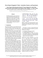
GRIP1 co-activator function. Figure 7 shows a work-
ing model based on our findings.
The functional roles of the GRIP1 C-terminal
region
In Figs 1–6, we present several lines of evidence to sup-
port the concept that the GRIP1 C-terminal region is
involved in the modulation of self-transactivation (AD1
and AD2) and co-activator (AR, ER and TR) functions
in HeLa cells. The outcome of the relationship between
GRIP1 transactivation and co-activator functions var-
ies according to the system under investigation (Figs 5
and 6). In the AR transcriptional system, the GRIP1
co-activator function was destroyed when GRIP1-trun-
cated fragments expressed higher transactivation activ-
ity because of the loss of intact AD2-dependent
function. In contrast, GRIP1 transactivation and co-
activation activities were correlated in the TR transcrip-
tional system and the relationship was independent in
the ER transcriptional system. We also found that the
GRIP1 co-activator function depends not only on the
existence of a repression domain or a protein–protein
interaction with identified and unidentified factors,
but also on the GRIP1 conformation under specific
P Y. Liu et al. Autoregulation of GRIP1 functions via C-terminal region
FEBS Journal 273 (2006) 2172–2183 ª 2006 The Authors Journal compilation ª 2006 FEBS 2179
conditions (Figs 5 and 6). Hence, the linking of repres-
sion and self-association motifs to the GRIP1 confor-
mation demonstrated in this study might be explained
by the effect of the co-expressing GRIP1 C terminus on
GRIP1 transactivation and co-activation activities.
Our western blotting analysis showed that the
amount of protein expressed by the exogenous GRIP1
fragment was also tightly regulated by its structural
component. These findings are consistent with a recent
study conducted by the Hager laboratory, which dem-
onstrated that the C terminus of GRIP1 is essential for
the formation of discrete nuclear foci and 26S protea-
some degradation in gene regulation [37]. Similarly to
the regulatory mechanism reported in p53 studies
[38,39], GRIP1 might form a more active conforma-
tion, determined by its relative concentration in cells.
The relative concentration of GRIP1 might depend on
its homo-oligomerization status, which is mainly deter-
mined by the involvement of its C-terminal region in
protein–protein interactions, including self-association,
repression by HDAC1 and other proteins, 26S protea-
some degradation, or translocalization. Taken together,
the effect of GRIP1 C-terminal interacting proteins as
a GRIP1-dependent secondary co-activator might, in
part, be mediated through conformational change of
the GRIP1 C terminus and subsequent exposure of a
working surface, with extra downstream signalling for
its transactivation and NR co-activator functions.
Experimental procedures
Plasmids
The pSG5.HA vectors coding for full-length GRIP1
(codons 5–1462), other GRIP1 fragments (codons 5–1121
and 1122–1462), and HA.CARM1 have been described
1
1013
1013
1122
1122
1305
1305
1398
1398
1462
1013
1013
1122
1122
1305
1305
1398
1398
1462
1462
1
1305
1305
1398
1462
1462
1122
1013
1122
1122
1305
1305
1398
1398
1462
1462
1122
1122
1305
1398
41
6
2
41
6
2
1122
1122
1305
1305
1398
162
4
1
6
24
1013
1013
1122
1122
1305
1305
1398
1398
1462
1
1
0
1
3
1
0
1
3
1
3
9
8
1
3
9
8
1013
1122
1122
1305
1305
1398
1398
1462
1122
1122
1305
1305
1398
1398
1
2
64
1
2
64
1
1013
1013
1122
1122
1305
1305
1398
1398
1462
1462
AD1
AD1
Repression domain
Repression domain
Association domain
Association domain
AD2
AD2
AD3
AD3
1305
1305
1305
1305
1398
1462
1462
1122
1122
+
?
+
16
24
16
2
4
1
1
1
1
I
II
III
III
IV
V
1122
1122
1
1013
1122
1305
1398
1398
1462
1462
+
?
1
1013
1013
1122
1305
1305
1398
1398
1462
+
Fig. 7. Dynamic model of the potential GRIP1 conformational change mediated through its C terminus. We propose that either monomeric
(I) or dimeric (or higher oligomeric) (II) GRIP1 might form a distinct conformation in cells. One repression (grey circle) and association (dotted
circle) are defined in this study. AD1 (slant circle), 2 (closed circle), and 3 (open circle) have been previously reported [23,26,28]. The expo-
sure of any GRIP1 C-terminal interacting protein, including the GRIP1 C terminus in this model, might alter GRIP1 conformation I through
intramolecular interaction into conformation III or conformation II through intermolecular interaction into conformation IV (first effect). In this
study, the exogenous GRIP1 C terminus dramatically enhanced GRIP1 transactivation activity through the repression and association
domains (or the titration of co-repressors), resulting in conformational changes from conformation I (or II) into III or IV. In contrast, the extra
downstream signal (second effect) of other GRIP1-dependent co-activators might be required for some full GRIP1 NR co-activator functions,
for example, the methyltransferase activity of co-activator-associated arginine methyltransferase 1 (CARM1) in this study. The question mark
indicates that further analyses are necessary to identify the involvement of the GRIP1 N terminus or the status of oligomerization in cells.
Autoregulation of GRIP1 functions via C-terminal region P Y. Liu et al.
2180 FEBS Journal 273 (2006) 2172–2183 ª 2006 The Authors Journal compilation ª 2006 FEBS
previously [19]; GRIP1-563–1121 was constructed by inserting
an EcoRI–SalI fragment of the appropriate PCR-amplified
GRIP1 cDNA into the EcoRI and XhoI sites of the
pSG5.HA vector. GRIP1-5–765 and GRIP1-1305–1462
were constructed by inserting EcoRI–XhoI fragments enco-
ding GRIP1
5)765
and GRIP1
1305)1462
into the pSG5.HA
vector; GRIP1-563–1462 was constructed by inserting an
XhoI–EcoRI (GRIP1
766)1462
) fragment from GRIP1
5)1462
into the pSG5.HA.GRIP1
563)1121
treated by XhoI diges-
tion. Vectors encoding Gal4DBD fused to various GRIP1
fragments were constructed by inserting EcoRI–SalI frag-
ments of the appropriate PCR-amplified GRIP1 cDNA or
EcoRI–XhoI GRIP1 fragments cut from respective
pSG5.HA.GRIP1s into the EcoRI and SalI sites of the pM
vector (Clontech, Mountain View, CA, USA), a vector for
expression of Gal4DBD fusion proteins from a constitu-
tive SV40 early promoter. C-terminal truncations of
pM.GRIP1
5)1462
were constructed by inserting XhoI–XbaI
fragments of the appropriate truncated PCR-amplified
GRIP1 (amino acids from 750 to indicated numbers) into
the XhoI and XbaI sites of the pM.GRIP1
5)1121
vector.
C-terminal truncations of pSG5.HA.GRIP1
5)1462
were con-
structed by inserting EcoRI–SalI fragments of the indicated
pM.GRIP1 truncations into the EcoRI and XhoI sites
of the pSG5.HA vector. Plasmid DNAs encoding
pCDNA3.1.HDAC1.myc [40] were gifts from M.A. Lazar
(University of Pennsylvania, Philadelphia, PA, USA), and
pCDNA3.HDAC1.flag wild type and H141A mutant were
gifts from T.P. Yao (Duke University, Durham, NC,
USA) [41]. Reporter genes MMTV-LUC, EREII-LUC
[GL45], MMTV[TRE]-LUC, and GK1, were as described
previously [42,43]. The expression of NRs in mammalian
cells and ⁄ or in vitro, vectors pSVAR
0
for human AR [44],
pHE0 for human ERa [43] and pCMX.hTR b 1 [9] for
human TRb1, were as described previously.
Bacterial expression vectors for GST fused to various
GRIP1 fragments (codons 1122–1462, 1305–1462, 1122–
1304, 1305–1398, 1305–1462 and 1399–1462) were con-
structed by inserting the appropriate PCR fragment into
pGEX-4T1 expression vector (GE HealthCare, Chicago,
IL, USA) via EcoRI–XhoI sites.
Cell culture and transient transfection assays
HeLa, COS-7 and COS-1 cells were grown in Dulbecco’s
modified Eagle’s medium (DMEM) supplemented with
10% charcoal ⁄ dextran-treated fetal bovine serum. The cells
in each well (a six- or a 24-well plate) were transfected with
SuperFect Transfection Reagent (Qiagen, Hilden, Ger-
many) or jetPEI (PolyPlus-transfection, Illkirch, France),
according to the manufacturer’s protocol; total DNA was
adjusted to 2.0 lg (six well) or 1.0 lg (24-well) by addition
of the empty vector pSG5.HA. Luciferase assays were per-
formed with the Promega Luciferase Assay kit (Madison,
WI, USA), and the measurement is expressed numerically
as relative light units (RLU). Luciferase activities are shown
as the mean and SD from two transfected sets. The results
shown are representative of at least three independent
experiments. Because some co-activators, including GRIP1
and CARM1, enhance the activities of so-called constitutive
promoters two- to ninefold, internal controls by co-trans-
fection of constitutive b-galactosidase expression vectors
were not used to normalize luciferase data. However, inter-
nal controls were used strategically to show that variation
in transfection efficiency was not a factor in the key results
(data not shown).
Immunoprecipitation and immunoblots
For analysis of the homo-oligomerization of GRIP1 and
the physical interaction between GRIP1 and HDAC1, these
expression vectors were transfected into COS-7 cells. After
transfection, cells were lysed in RIPA buffer (100 m m
Tris ⁄ HCl pH 8.0, 150 mm NaCl, 0.1% SDS, and 1% Tri-
ton 100) at 4 °C. Lysates were subjected to immunoprecipi-
tation with antibodies against Gal4 DBD or HA for 3 h,
followed by adsorption to Sepharose-coupled protein A ⁄ G
(Santa Cruz Biotechnology, Santa Cruz, CA, USA) for 3 h.
Immunoprecipitates were separated by SDS ⁄ PAGE and
analysed with immunoblots. For determination of total
protein levels of Gal4DBD- or HA-GRIP1 fragments,
aliquots of cell lysates were subjected to direct immuno-
blots. Immunoblots were performed as previously described
[23] using 10% of the extract from lysates for immunopre-
cipitation and monoclonal antibodies 3F10 against the HA
epitope (Roche, Mannheim, Germany), RK5C1 against
Gal4DBD, 3A2 against HuR, and normal mouse IgG
(Santa Cruz Biotechnology).
Protein–protein interaction assays
For GST pull-down assays,
35
S-labelled proteins were pro-
duced using the TNT T7-coupled reticulocyte lysate system
(Promega, Madison, WI, USA). GST fusion proteins were
produced in Escherichia coli BL21, eluted, and analysed by
gel electrophoresis, as previously described [23].
Acknowledgements
We thank Dr W. Feng (University of California, USA)
for expression vectors and reporter genes for TR;
P. Webb and P. J. Kushner (University of California,
USA) fro expression vectors and reporter genes for
ER; A. O. Brinkmann (Erasmus University, Rotter-
dam, the Netherlands) for AR expression vector;
M. A. Lazar (University of Pennsylvania, USA) for
pCDNA3.1.HDAC1.myc; and T. P. Yao (Duke
University, USA) for pCDNA3.HDAC1.flag (wild-type
and H141A mutant) expression vectors. This work
P Y. Liu et al. Autoregulation of GRIP1 functions via C-terminal region
FEBS Journal 273 (2006) 2172–2183 ª 2006 The Authors Journal compilation ª 2006 FEBS 2181
was supported by grants from the National Health
Research Institute and National Science Council,
Taiwan, Republic of China (NHRI-EX94-9224NC and
NSC 94-2320-B-016–044 to S. M. Huang).
References
1 Enmark E & Gustafsson JA (1996) Orphan nuclear
receptors – the first eight years. Mol Endocrinol 10,
1293–1307.
2 Tsai MJ & O’Malley BW (1994) Molecular mechanisms
of action of steroid ⁄ thyroid receptor superfamily mem-
bers. Annu Rev Biochem 63, 451–486.
3 Folkers GE, Van der Burg B & Van der Saag PT (1996)
A role for cofactors in synergistic and cell-specific acti-
vation by retinoic acid receptors and retinoid X recep-
tor. J Steroid Biochem Mol Biol 56, 119–129.
4 Metzger D, Ali S, Bornert JM & Chambon P (1995)
Characterization of the amino-terminal transcriptional
activation function of the human estrogen receptor
in animal and yeast cells. J Biol Chem 270, 9535–9542.
5 McKenna NJ, Xu J, Nawaz Z, Tsai SY, Tsai MJ &
O’Malley BW (1999) Nuclear receptor coactivators:
multiple enzymes, multiple complexes, multiple func-
tions. J Steroid Biochem Mol Biol 69, 3–12.
6 Korzus E, Torchia J, Rose DW, Xu L, Kurokawa R,
McInerney EM, Mullen TM, Glass CK & Rosenfeld
MG (1998) Transcription factor-specific requirements
for coactivators and their acetyltransferase functions.
Science 279, 703–707.
7 Glass CK, Rose DW & Rosenfeld MG (1997)
Nuclear receptor coactivators. Curr Opin Cell Biol 9,
222–232.
8 Xu L, Glass CK & Rosenfeld MG (1999) Coactivator
and corepressor complexes in nuclear receptor function.
Curr Opin Genet Dev 9, 140–147.
9 Feng W, Ribeiro RC, Wagner RL, Nguyen H, Apriletti
JW, Fletterick RJ, Baxter JD, Kushner PJ & West BL
(1998) Hormone-dependent coactivator binding to a
hydrophobic cleft on nuclear receptors. Science 280,
1747–1749.
10 Blanco JC, Minucci S, Lu J, Yang XJ, Walker KK,
Chen H, Evans RM, Nakatani Y & Ozato K (1998)
The histone acetylase PCAF is a nuclear receptor
coactivator. Genes Dev 12, 1638–1651.
11 Chakravarti D, LaMorte VJ, Nelson MC, Nakajima T,
Schulman IG, Juguilon H, Montminy M & Evans RM
(1996) Role of CBP ⁄ P300 in nuclear receptor signalling.
Nature 383, 99–103.
12 Takeshita A, Cardona GR, Koibuchi N, Suen CS &
Chin WW (1997) TRAM-1, a novel 160-kDa thyroid
hormone receptor activator molecule, exhibits distinct
properties from steroid receptor coactivator-1. J Biol
Chem 272, 27629–27634.
13 Li H, Gomes PJ & Chen JD (1997) RAC3, a
steroid ⁄ nuclear receptor-associated coactivator that is
related to SRC-1 and TIF2. Proc Natl Acad Sci USA
94, 8479–8484.
14 Chen H, Lin RJ, Schiltz RL, Chakravarti D, Nash A,
Nagy L, Privalsky ML, Nakatani Y & Evans RM (1997)
Nuclear receptor coactivator ACTR is a novel histone
acetyltransferase and forms a multimeric activation
complex with P ⁄ CAF and CBP ⁄ p300. Cell 90, 569–580.
15 Anzick SL, Kononen J, Walker RL, Azorsa DO,
Tanner MM, Guan XY, Sauter G, Kallioniemi OP,
Trent JM & Meltzer PS (1997) AIB1, a steroid receptor
coactivator amplified in breast and ovarian cancer.
Science 277, 965–968.
16 Voegel JJ, Heine MJ, Zechel C, Chambon P & Grone-
meyer H (1996) TIF2, a 160 kDa transcriptional media-
tor for the ligand-dependent activation function AF-2
of nuclear receptors. EMBO J 15, 3667–3675.
17 Hong H, Kohli K, Trivedi A, Johnson DL & Stallcup
MR (1996) GRIP1, a novel mouse protein that serves
as a transcriptional coactivator in yeast for the hormone
binding domains of steroid receptors. Proc Natl Acad
Sci USA 93, 4948–4952.
18 Onate SA, Tsai SY, Tsai MJ & O’Malley BW (1995)
Sequence and characterization of a coactivator for the
steroid hormone receptor superfamily. Science 270,
1354–1357.
19 Chen D, Ma H, Hong H, Koh SS, Huang SM, Schurter
BT, Aswad DW & Stallcup MR (1999) Regulation of
transcription by a protein methyltransferase. Science
284, 2174–2177.
20 Spencer TE, Jenster G, Burcin MM, Allis CD, Zhou J,
Mizzen CA, McKenna NJ, Onate SA, Tsai SY, Tsai
MJ et al. (1997) Steroid receptor coactivator-1 is a his-
tone acetyltransferase. Nature 389, 194–198.
21 Bannister AJ & Kouzarides T (1996) The CBP co-activa-
tor is a histone acetyltransferase. Nature 384, 641–643.
22 Heery DM, Kalkhoven E, Hoare S & Parker MG (1997)
A signature motif in transcriptional co-activators med-
iates binding to nuclear receptors. Nature 387, 733–736.
23 Ma H, Hong H, Huang SM, Irvine RA, Webb P, Kush-
ner PJ, Coetzee GA & Stallcup MR (1999) Multiple sig-
nal input and output domains of the 160-kilodalton
nuclear receptor coactivator proteins. Mol Cell Biol 19,
6164–6173.
24 Bevan CL, Hoare S, Claessens F, Heery DM & Parker
MG (1999) The AF1 and AF2 domains of the androgen
receptor interact with distinct regions of SRC1. Mol
Cell Biol 19, 8383–8392.
25 Webb P, Nguyen P, Shinsako J, Anderson C, Feng W,
Nguyen MP, Chen D, Huang SM, Subramanian S,
McKinerney E et al. (1998) Estrogen receptor activation
function 1 works by binding p160 coactivator proteins.
Mol Endocrinol 12, 1605–1618.
Autoregulation of GRIP1 functions via C-terminal region P Y. Liu et al.
2182 FEBS Journal 273 (2006) 2172–2183 ª 2006 The Authors Journal compilation ª 2006 FEBS
26 Huang SM & Cheng YS (2004) Analysis of two CBP
(cAMP-response-element-binding protein-binding
protein) interacting sites in GRIP1 (glucocorticoid
receptor-interacting protein), and their importance for
the function of GRIP1. Biochem J 382, 111–119.
27 Voegel JJ, Heine MJ, Tini M, Vivat V, Chambon P &
Gronemeyer H (1998) The coactivator TIF2 contains
three nuclear receptor-binding motifs and mediates
transactivation through CBP binding-dependent and
-independent pathways. EMBO J 17, 507–519.
28 Li J, O’Malley BW & Wong J (2000) p300 requires its
histone acetyltransferase activity and SRC-1 interaction
domain to facilitate thyroid hormone receptor activation
in chromatin. Mol Cell Biol 20, 2031–2042.
29 Chen YH, Kim JH & Stallcup MR (2005) GAC63, a
GRIP1-dependent nuclear receptor coactivator. Mol
Cell Biol 25, 5965–5972.
30 Kim JH, Li H & Stallcup MR (2003) CoCoA, a nuclear
receptor coactivator which acts through an N-terminal
activation domain of p160 coactivators. Mol Cell 12,
1537–1549.
31 Yoshida M, Kijima M, Akita M & Beppu T (1990)
Potent and specific inhibition of mammalian histone
deacetylase both in vivo and in vitro by trichostatin A.
J Biol Chem 265, 17174–17179.
32 Huang SM & Stallcup MR (2000) Mouse Zac1, a tran-
scriptional coactivator and repressor for nuclear recep-
tors. Mol Cell Biol 20, 1855–1867.
33 Huang SM, Huang CJ, Wang WM, Kang JC & Hsu
WC (2004) The enhancement of nuclear receptor tran-
scriptional activation by a mouse actin-binding protein,
alpha actinin 2. J Mol Endocrinol 32, 481–496.
34 Demarest SJ, Martinez-Yamout M, Chung J, Chen H,
Xu W, Dyson HJ, Evans RM & Wright PE (2002) Mutual
synergistic folding in recruitment of CBP ⁄ p300 by p160
nuclear receptor coactivators. Nature 415, 549–553.
35 Chevillard-Briet M, Trouche D & Vandel L (2002)
Control of CBP co-activating activity by arginine
methylation. EMBO J 21, 5457–5466.
36 Chen D, Huang SM & Stallcup MR (2000) Synergistic,
p160 coactivator-dependent enhancement of estrogen
receptor function by CARM1 and p300. J Biol Chem
275, 40810–40816.
37 Baumann CT, Ma H, Wolford R, Reyes JC,
Maruvada P, Lim C, Yen PM, Stallcup MR & Hager
GL (2001) The glucocorticoid receptor interacting
protein 1 (GRIP1) localizes in discrete nuclear foci
that associate with ND10 bodies and are enriched in
components of the 26S proteasome. Mol Endocrinol
15, 485–500.
38 Waterman JL, Shenk JL & Halazonetis TD (1995) The
dihedral symmetry of the p53 tetramerization domain
mandates a conformational switch upon DNA binding.
EMBO J 14, 512–519.
39 Marston NJ, Jenkins JR & Vousden KH (1995)
Oligomerisation of full length p53 contributes to the
interaction with mdm2 but not HPV E6. Oncogene 10,
1709–1715.
40 Miska EA, Karlsson C, Langley E, Nielsen SJ, Pines J
& Kouzarides T (1999) HDAC4 deacetylase associates
with and represses the MEF2 transcription factor.
EMBO J 18, 5099–5107.
41 Ito A, Kawaguchi Y, Lai CH, Kovacs JJ, Higashimoto
Y, Appella E & Yao TP (2002) MDM2-HDAC1-
mediated deacetylation of p53 is required for its degra-
dation. EMBO J 21, 6236–6245.
42 Umesono K & Evans RM (1989) Determinants of target
gene specificity for steroid ⁄ thyroid hormone receptors.
Cell 57, 1139–1146.
43 Green S, Issemann I & Sheer E (1988) A versatile
in vivo and in vitro eukaryotic expression vector for
protein engineering. Nucleic Acids Res 16, 369.
44 Brinkmann AO, Faber PW, van Rooij HC, Kuiper GG,
Ris C, Klaassen P, van der Korput JA, Voorhorst MM,
van Laar JH, Mulder E et al. (1989) The human andro-
gen receptor: domain structure, genomic organization
and regulation of expression. J Steroid Biochem 34,
307–310.
P Y. Liu et al. Autoregulation of GRIP1 functions via C-terminal region
FEBS Journal 273 (2006) 2172–2183 ª 2006 The Authors Journal compilation ª 2006 FEBS 2183