Báo cáo khoa học: Crosstalk between Src and major vault protein in epidermal growth factor-dependent cell signalling docx
Bạn đang xem bản rút gọn của tài liệu. Xem và tải ngay bản đầy đủ của tài liệu tại đây (414.81 KB, 12 trang )
Crosstalk between Src and major vault protein in
epidermal growth factor-dependent cell signalling
Euikyung Kim
1
*, Seunghwan Lee
1
, Md Firoz Mian
2
, Sang Uk Yun
2
, Minseok Song
2
, Kye-Sook Yi
2
,
Sung Ho Ryu
2
and Pann-Ghill Suh
2
*
1 Institue of Animal Medicine, College of Veterinary Medicine, Gyeongsang National University, Jinju, Korea
2 Department of Life Science, Pohang University of Science and Technology, Pohang, Korea
The major vault protein (MVP) is the predominant
component of a large cytoplasmic ribonucleoprotein
particle, the vault complex [1,2]. The vault particle was
originally identified as a barrel shaped body in prepa-
rations of clathrin-coated vesicles and named after its
morphology reminiscent of the vaulted ceilings of
cathedrals [3]. Vaults exist in thousands of copies per
cell and are widely expressed in all eukaryotic organ-
isms [4–8]. In both structure and composition vaults
are highly conserved throughout evolution in diverse
phylogenetic lineages including mammals, avians,
amphibians and slime moulds [9]. They represent
multimeric protein complexes with one predominant
member, the MVP which constitutes more than 70%
of the total complex. The remaining mass comprises
vault RNA and two high molecular weight proteins,
vault poly(ADP-ribose) polymerase (VPARP) and
telomerase-associated protein 1 (TEP1) [10,11]. The
Keywords
ERK signaling pathway; MVP; Src; Src
activity; tyrosine phophorylation
Correspondence
E. Kim, Institue of Animal Medicine, College
of Veterinary Medicine, Gyeongsang
National University, Jinju, 660-701, Korea
Fax: +82 55 751 5803
Tel: +82 55 751 5812
E-mail:
P G. Suh, Department of Life Science,
Pohang University of Science and
Technology, 790-784, Korea
Fax: +82 54 283 4613
Tel: +82 54 279 2293
E-mail:
*Note
E. Kim and P G. Suh contributed equally to
this work.
(Received 14 November 2005, revised
13 December 2005, accepted 19 December
2005)
doi:10.1111/j.1742-4658.2006.05112.x
Vaults are highly conserved, ubiquitous ribonucleoprotein (RNP) particles
with an unidentified function. For the three protein species (TEP1,
VPARP, and MVP) and a small RNA that comprises vault, expression of
the unique 100-kDa major vault protein (MVP) is sufficient to form the
basic vault structure. To identify and characterize proteins that interact
with the Src homology 2 (SH2) domain of Src and potentially regulate Src
activity, we used a pull-down assay using GST–Src–SH2 fusion proteins.
We found MVP as a Src–SH2 binding protein in human stomach tissue.
Interaction of Src and MVP was also observed in 253J stomach cancer
cells. A subcellular localization study using immunofluorescence micros-
copy shows that epidermal growth factor (EGF) stimulation triggers MVP
translocation from the nucleus to the cytosol and perinuclear region where
it colocalizes with Src. We found that the interaction between Src and
MVP is critically dependent on Src activity and protein (MVP) tyrosyl
phosphorylation, which are induced by EGF stimulation. Our results also
indicate MVP to be a novel substrate of Src and phosphorylated in an
EGF-dependent manner. Interestingly, purified MVP inhibited the in vitro
tyrosine kinase activity of Src in a concentration-dependent manner. MVP
overexpression downregulates EGF-dependent ERK activation in Src over-
expressing cells. To our knowledge, this is the first report of MVP interact-
ing with a protein tyrosine kinase involved in a distinct cell signalling
pathway. It appears that MVP is a novel regulator of Src-mediated signal-
ling cascades.
Abbreviations
EGF, epidermal growth factor; GST, glutathione S-transferase; MVP, major vault protein; PAP, potato acid phosphatase; PTEN, phosphatase
and tensin homologue deleted on chromosome 10; SH2, Src homology 2; TCL, total cell lysate; TEP1, telomerase-associated protein 1;
VPARP, vault poly(ADP-ribose) polymerase.
FEBS Journal 273 (2006) 793–804 ª 2006 The Authors Journal compilation ª 2006 FEBS 793
expression of the unique 100 kDa MVP is sufficient
to form the basic vault structure. Although many
molecular features of vault particles have been charac-
terized, the function of this large ribonucleoprotein
particle remains enigmatic. The identification of lung
resistance-related protein (LRP) as the human MVP
shed new light on putative cellular functions of vaults
[7]. Numerous multidrug resistance cancer cells fre-
quently overexpress MVP and increased MVP mRNA
expression was found to correlate strongly with a pre-
dictive value of a multidrug resistance phenotype
[12,13]. An early postulate of vault function was nucle-
ocytoplasmic transport [1,14]. A recent study using
MVP knockout mice has shown that MVP ⁄ vaults are
not directly involved in the resistance to cytostatic
agents [15]. Vaults have been proposed to constitute
the transporter or central plug of the nuclear pore
complex, controlling bi-directional exchange between
nucleus and cytoplasm [16]. Major vault protein has
been coimmunoprecipitated with human oestrogen
receptor in oestradiol dependent interaction and might
be involved in nucleocytoplasmic shuttle for modula-
tion of signal transduction of steroid hormone [17].
Another recent study showed that MVP physically
interacts with phosphatase and tensin homologue
deleted on chromosome 10 (PTEN) and the interaction
is Ca
2+
dependent [18]. However the physiological role
of MVP hitherto remains elusive.
The Src tyrosine kinase participates in multiple sig-
nalling pathways that regulate diverse cellular func-
tions, including proliferation, differentiation, motility,
adhesion and architecture [19,20]. The subcellular
localization of Src in part determines its substrate
specificity and function. One example of a Src sub-
strate, Sam68, an RNA binding protein [21], whose
phosphorylation by Src appears to determine specific
functions of Src. Src phosphorylates Sam68 during
mitosis, presumably after breakdown of the nuclear
envelope. Src appears to be important for cell cycle
progression via Sam68, particularly during the late
mitosis and possibly during G
1
⁄ S transition. Identifi-
cation of Src binding proteins has led to a better
understanding of Src regulation and has provided
clues about the function of Src in normal and trans-
formed cells [22]. Compelling evidence indicates that
Src-binding proteins can regulate Src activity [23].
While a number of interacting proteins that upregu-
late Src activity have been identified; however, only
a few that downregulate Src activity have been
known. It is important to elucidate the molecular
mechanisms that inactivate c-Src. Recently Caveolin,
a 22 kDa integral membrane protein [24–26] and a
receptor for activated C kinase (RACK1) [27] were
shown to bind Src and suppress its tyrosine kinase
activity. Domains within Src kinases target the
enzyme to specific subcellular locations where they
bind to regulatory and ⁄ or substrate proteins and are
integrated into cell signalling pathways and cell cycle
events [23]. The UD, Src homology 3 and Src
homology 2 (SH2) domains in Src are key binding
sites for proteins that regulate Src activity and integ-
rate Src into important signalling pathways and cell
cycle events. The aim of the present study was to
identify and characterize Src interacting proteins that
potentially regulate Src activity. We focused on pro-
tein interactions that involve the SH2 domain of Src
using a glutathione S-transferase (GST)–SH2 fusion
pull-down assay and identified MVP as a Src–SH2
binding protein. We observed that MVP interacts
with Src in mammalian cells and inhibits the activity
of Src tyrosine kinase.
Results
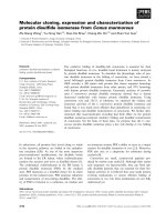
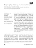
Isolation of MVP as Src–SH2 interacting protein
by GS–SH2 fusion pull-down assay
To isolate proteins that regulate cancer-specific cell sig-
nalling, we incubated GST fusion–SH2 domains of
various Src SH2 domain-containing proteins with cell
lysates from human stomach cancer tissues or normal
stomach tissues. The protein complexes were collected
on glutathione-agarose beads and resolved in
SDS ⁄ PAGE followed by silver staining (Fig. 1A). The
targeted protein bands were then analysed by
MALDI-TOF MS. A 100 kDa protein that bound
strongly with the SH2 domain was identified as MVP
(Fig. 1B, Table 1). It was also verified by immunoblot-
ting with polyclonal anti-MVP IgG (Fig. 1C). Both the
MS analysis and immunoblotting with polyclonal anti-
MVP IgG showed that MVP strongly bound to the
SH2 domain of Src, but not to the SH2 domains of
other proteins tested (Fig. 1C). Thus MVP interacted
specifically with the SH2 domain of Src, but not with
the SH2 domains of PLCc1, Grb2, STAT3 or Crk.
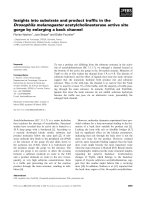
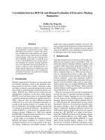
MVP associates with Src endogenously in
253J cells and exogenously in cotransfected
293T cells
MVP constitutes about 70% of the total molecular
mass of vault particles and is capable of assembling
into the characteristic vault structure in the absence of
other vault components (TEP1, VPARP or vRNA).
To examine whether the MVP can interact with full-
length Src in vivo, we prepared MVP containing lysates
MVP interacts with Src tyrosine kinase E. Kim et al.
794 FEBS Journal 273 (2006) 793–804 ª 2006 The Authors Journal compilation ª 2006 FEBS
from 253J cells and immunoprecipitated c-Src using
a polyclonal antibody. Western blot analysis of the
Src-immunoprecipitates with MVP antibody showed
that MVP ⁄ vault interacted with Src in vivo in 253J
cells (Fig. 2A). If MVP interacts with Src in other
established mammalian cell lines was examined by
cotransfecting flag-tagged MVP and c-Src into 293T
cells. Coimmunoprecipitation and western blot analyses
of the immunoprecipitates were performed using anti-
FLAG IgG or Src antibody. Figure 2B shows that
Flag–MVP immune complex contains c-Src (lane 2).
The reciprocal experiment confirmed the interaction as
shown in Fig. 2B, lane-3 that MVP was coimmunopre-
cipitated with Src.
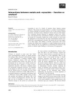
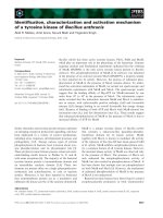
EGF enhances the MVP–Src interaction, which
can be blocked by src kinase inhibitor, PP2
To determine whether epidermal growth factor (EGF)
can activate Src and influences the association between
MVP and Src, we treated serum starved fibroblasts
T
SG
ylno
-TSG
-
C
L
P
γ1
2
H
Sn
-TSG
2b
r
G
2
H
S
-T
S
G
-CLP γ1
2HSc
-T
S
G
-
CL
P γ
1
22H
S
C
N
-CN-
C
N
-C
N
-
CN-
-
TSG
3
ta
t
S
2H
S
-T
S
G
kr
C
2H
S
-
T
SG
crS
2
HS
:eu
s
siT
C
N
-
C
N
-
CN
-
noisuF
nie
t
orP
:
79
86
0
0
2
34
gniniatSrevl
i
S
TSG
enola
0
0
1p
-
T
SG
2
HS-crS
Mass (m/z)
0
05
0
0
52
%Intensity
0
5
A
B
C
0
000
20051
000
1
1
P
2
P
3
P
4P
5P
6P
7P
8P
9
P
01P
11P
2
1
P
31P
4
1P
p100
IB: αMVP
97
68
(kDa)
Input
GST only
Stat3 SH2
Crk SH2
Grb2 SH2
Src SH2
PLC
γ
1 nSH2
PLC
γ
1 cSH2
PLC
γ
1 SH22
GST-fusioned
Fig. 1. Major vault protein interacts with c-Src through the Src SH2
domain. (A) Stomach cancer tissue (C) and normal stomach tissue
(N) were obtained from cancer patients in a local hospital (Dongguk
University Pohang Hospital) and stored at )70 °C until use. The tis-
sue lysates were prepared and incubated with GST fusion proteins
of various SH2 domains. Formed protein complexes were isolated
by glutathione beads and washed three times with fresh TBS, and
analysed by SDS ⁄ PAGE and subsequent silver staining as des-
cribed in Experimental procedures. (B) p100 isolated from proteins
that markedly coprecipitated with the GST–Src-SH2 fusion protein
was in-gel digested with trypsin, and the resulting peptide mixture
was analysed by MALDI-TOF MS. The arrows indicate matched
peaks among the measured tryptic peaks of p100 with calculated
molecular masses of MVP within 50 p.p.m. The detailed descrip-
tions of each peptide analysed and used for protein identification
are shown in Table 1. (C) Binding proteins in stomach cancer tissue
to the GST–SH2 of various signaling proteins (Src, PLCc1, STAT3,
Grb2, Crk) which were tested were immunoblotted with polyclonal
anti-MVP IgG (from Dr Rome, UCLA, CA), confirming that MVP
specifically interacts with the SH2 domain of Src, and not with SH2
domains of other proteins. The input shows approximately 10% of
the tissue lysate that was applied for GST-fusion pulldown.
Table 1. Peptide sequences and masses from p100 by MALDI-TOF
MS.
M+H
+
(Da)
Observed Calculated
P1 VLFAPMR (43–49) 848.426 848.457
P2 SLQPLAPR (445–452) 880.513 880.513
P3 ELELVYAR (767–774) 991.518 991.533
P4 VSHQAGDHWLIR (349–360) 1417.666 1417.721
P5 VPHNAAVQVYDYR (462–474) 1530.741 1530.757
P6 AQALAIETEAELQR (748–761) 1541.774 1541.804
P7 EVEVVEIIQATIIR (156–169) 1610.853 1610.923
P8 DAQGLVLFDVTGQVR (68–82) 1616.809 1616.851
P9 KEVEVVEIIQATIIR (155–169) 1738.998 1739.018
P10 AQDPFPLYPGEVLEK (92–107) 1814.926 1814.944
P11 VAGDEWLFEGPGTYIPR (137–154) 2004.986 2004.994
P12 QLQLAYNWHFEVNDR (537–552) 2044.936 2045.011
P13 VIGSTYMLTQDEVLWEK (400–417) 2081.970 2082.033
P14 PPYHYIHVLDQNSNVSR (10–27) 2151.017 2151.085
E. Kim et al. MVP interacts with Src tyrosine kinase
FEBS Journal 273 (2006) 793–804 ª 2006 The Authors Journal compilation ª 2006 FEBS 795
that overexpress FLAG–MVP and Src with EGF
(100 ngÆmL
)1
) for various time periods. Then the cell
lysates were immunoprecipitated with anti-FLAG IgG
and the immune complexes were resolved by
SDS ⁄ PAGE followed by immunoblotting with anti-Src
IgG (Fig. 3A). We observed that EGF enhanced the
interaction between MVP and Src in time-dependent
manner with a peak after 3 min followed by gradual
decline and return to the basal level after 15 min
(Fig. 3A). The effect of EGF on MVP–Src interaction
was concentration dependent, with a maximal effect
achieved at 100 ngÆmL
)1
(data not shown). From the
current results, however, it is not clear whether only
the SH2 domain of Src is important for the Src–MVP
association in vivo. This could be addressed by examin-
ing whether an SH2 domain deletion mutant of Src
can still associate with MVP in vivo from a further
study. We also examined the effect of Src specific tyro-
sine kinase inhibitor (PP2) on EGF dependent Src–
MVP interaction (Fig. 3B). We treated serum starved
253J cells that express high levels of Src and MVP pro-
teins endogenously, with EGF for various time periods
and one group was pretreated with PP2 for 45 min
before EGF stimulation. We could observe that endo-
genous interaction between Src and MVP after EGF
stimulation was almost completely inhibited by PP2.
These results clearly show that EGF potentiates the
interactions between Src and MVP in a time-dependent
manner, which is abrogated by specific Src kinase
inhibitor.
Epidermal growth factor-dependent coimmunopre-
cipitation of Src and MVP prompted us to test if they
colocalize in any subcellular compartment on EGF sti-
mulation. As we expected, immunofluorescence micros-
copy showed EGF-dependent transient colocalization
of MVP and Src in the cytoplasmic region of 253J cells
that express high levels of Src and MVP proteins
endogenously (Fig. 3C). Interestingly, MVP predomin-
antly localized in the nucleus of quiescent cell seems to
translocate onto perinuclear and cytoskeletal compart-
ment where it overlaps with Src upon EGF treatment.
The kinetics of Src–MVP colocalization correlated well
with the biochemical data of protein complex forma-
tion as shown earlier. This result suggests that mole-
cular interaction between Src and MVP may play an
important role in EGF-dependent colocalization of the
two proteins. However, the detailed mechanism of
MVP translocation from nucleus to cytoplasm should
be further elucidated.
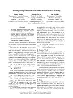
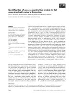
Tyrosine phosphorylation of MVP is important
for the binding of MVP with Src
MVP is known as a phosphoprotein as it is tyrosine
phosphorylated in vivo and phosphorylated by protein
kinase C (PKC) and casein kinase II (CKII) in vitro
[28,29]. To investigate the significance of MVP tyrosine
phosphorylation for the MVP–Src interaction, 293T
cells transfected with Src and FLAG-MVP were serum
starved, then treated with EGF for the indicated time
periods (Fig. 4A). Cell lysates were then immunopre-
cipitated with anti-FLAG IgG followed by immuno-
blotting with antiphosphotyrosine IgG (PY 20). We
observed that MVP phosphorylation reached the peak
level upon EGF stimulation for 3 min (Fig. 4A) that
was comparable to the time kinetics of Src–MVP inter-
action upon EGF stimulation as shown in Fig. 3. This
α PVMf
α crS-c
PI
PVMf
crS-c
BI
α PVMf α crS
+++
-
++
LCT
+++
-
++
:xfT
B
:PI
α crS-c
α PVM
BI
A
α crS-c
-noN
enummi
mures
tu
pni
Fig. 2. MVP interacts with Src in vivo in 253J cells and in cotrans-
fected 293T cells. (A) To confirm whether MVP interacts endogen-
ously with full-length Src, we prepared 253J (stomach cancer cell
line) cell lysates and immunoprecipitated with c-Src mAb or non-
immune serum, followed by immunoblotting with anti-MVP IgG or
anti-c-Src IgG. Thw upper panel indicates MVP that had been asso-
ciated with Src and the lower panel indicates immunoprecipitated
c-Src protein. The input shows approximately 10% of the tissue
lysate that was applied for immunoprecipitation. (B) Flag-tagged
MVP was prepared by generating the rat MVP cDNA construct
encoding Flag sequence at the N terminus. The flag-tagged MVP
cDNA and c-Src cDNA were cotransfected into 293T cells as indica-
ted (Tfx) in the result. The total cell lysates (TCL) were prepared
and immunoprecipitated with anti-Flag IgG or anti-Src IgG. The
immunoprecipitated complex and total cell lysates were run on
SDS ⁄ PAGE and transferred to nitrocellulose membrane, and west-
ern blotting was performed using anti-Flag IgG or anti-Src IgG. The
TCL show the overexpression of Flag-MVP and Src in transfected
cells, respectively.
MVP interacts with Src tyrosine kinase E. Kim et al.
796 FEBS Journal 273 (2006) 793–804 ª 2006 The Authors Journal compilation ª 2006 FEBS
finding suggests that MVP tyrosine phosphorylation
might be required for MVP–Src interaction. Interest-
ingly, in almost all cases, MVP seems to have some
basal level of tyrosine phosphorylation in our system
and it should be clarified in a further study. To further
address this result, we examined whether MVP phos-
:P
I
α
α
PVMgalF
α c
r
S-c
α c
rS-c
α PVMgalF
α PVMgalF
α HDPAG
PVMf
-
+
++
++
+crS-c
-
++++
’
51’
5’3
:)n
im
(
FGE
xf
A
T
et
a
syLlleClatoT
BI
BI
Merged
MVPSrc
C
Starved
EGF 3 min
EGF 20 min
B
PVM
c
r
S
P
V
M
c
r
S
PV
M
c
r
S
0:
)
niM
(
FGE
-
’1
-
’
3
-
’6
-
’01
-
’02
-
’54
-
’6
+
5(2PP
µ :
)
M
crS
:
PI
etas
y
Ll
l
e
C
la
t
oT
BI
BI
Fig. 3. EGF substantially enhances Src–MVP
interaction that was blocked by Src tyrosine
kinase inhibitor (PP2). (A) To determine
whether Src–MVP interaction is EGF signal-
dependent, we starved 293T cells which
were transfected with c-Src and ⁄ or Flag-
tagged MVP as indicated. After 24 h, the
293T cells were treated with EGF
(100 ngÆmL
)1
) for the indicated times, and
the prepared cell lysates were then immu-
noprecipitated with anti-Flag mAb. The sam-
ples were immunoblotted with anti-Flag IgG
or anti-c-Src IgG, showing that the inter-
action is EGF-signal dependently increased
then rapidly declined. (B) To see the effect
of EGF on endogenous MVP–Src inter-
action, 253J cells were serum starved and
stimulated with EGF for the indicated time
periods. One group after serum starvation
was pretreated with PP2 for 45 min fol-
lowed by EGF stimulation for 6 min. The
results showed that in vivo Src–MVP inter-
action was also EGF signal dependent and
Src tyrosine kinase inhibitor (PP2) blocked
the EGF induced interaction. (C) 253J cells
seeded onto coverslips in DMEM with 10%
heat-inactivated fetal bovine serum were
serum starved for 24 h in serum-free
DMEM media. After serum starvation, the
cells were treated with EGF (100 ngÆmL
)1
final concentration) at 37 °C for the indica-
ted times, then fixed and permeabilized as
described in Experimental procedures. Non-
specific bindings were blocked by incubating
the coverslips with 4% BSA in NaCl ⁄ P
i
,
then the coverslips were incubated with
mouse monoclonal anti-Src IgG and rabbit
polyclonal anti-MVP IgG. After washing
three times with NaCl ⁄ P
i
, the coverslips
were incubated with fluorescent probe-con-
jugated secondary antibodies (fluoresceine
isothiocyanate-conjugated goat anti-rabbit
IgG and rhodamine-conjugated goat anti-
mouse IgG) for another 1 h. After washing
with NaCl ⁄ P
i
, the coverslips were mounted
face down onto slides and examined under
confocal fluorescence microscopy.
E. Kim et al. MVP interacts with Src tyrosine kinase
FEBS Journal 273 (2006) 793–804 ª 2006 The Authors Journal compilation ª 2006 FEBS 797
phorylation is a prerequisite for MVP–Src association.
We overexpressed FLAG–MVP in 293T cells and the
cell lysates were incubated with potato acid phospha-
tase (PAP), a phosphotyrosyl-protein phosphatase, for
the indicated time periods. Then lysates were immuno-
precipitated with anti-Src IgG and immunoblotted
with anti-MVP IgG. Potato acid phosphatase treat-
ment resulted in a marked decrease in MVP–Src
complex formation on 45-min pretreated lysates. This
result provides proof that the Src–MVP interaction is
dependent on MVP tyrosine phosphorylation.
MVP inhibits Src kinase activity
To assess the effect of MVP on Src protein kinase
activity, we performed an in vitro Src kinase activity
assay. We overexpressed FLAG-tagged MVP in 293T
cells, immunoprecipitated cell lysates with anti-FLAG
monoclonal IgG (mAb) and MVP was eluted from
FLAG-immunoprecipitates by the addition of excess
FLAG peptides and used as purified MVP. We incuba-
ted rabbit muscle enolase, an exogenous Src substrate
and purified Src kinase (Santa Cruz Biotechnologies
Inc., Santa Cruz, CA) with [
32
P]ATP and MnCl
2
in
the presence or absence of purified MVP and measured
phosphorylation from in vitro Src kinase assay
(Fig. 5A). We observed autophosphorylation of Src in
the absence of MVP (Fig. 5A, lane 2). Interestingly,
we found that Src autophosphorylation was dramatic-
ally reduced by MVP in a dose-dependent manner
(Fig. 5A, lane 3 and 4). Enolase phosphorylation fol-
lowed the same trend as Src and acted as an excellent
control substrate for Src. The addition of 0.5-lg MVP
inhibited Src activity by 60% (measured from the
autoradiogram), whereas the addition of 1.0 lgof
MVP inhibited Src activity almost completely. These
results suggest that MVP has an intrinsic activity sup-
pressing Src kinase enzymatic activity. Next, we inves-
tigated whether MVP can be a substrate of and
phosphorylated by Src. We incubated purified MVP
and commercially obtained purified Src with [
32
P]ATP
in a kinase reaction mixture without enolase addition,
then examined the phosphorylation status of MVP.
Autoradiogram results showed that MVP was highly
phosphorylated by Src in vitro (Fig. 5B, lane 2). The
slight phosphorylation modification of MVP in the
absence of exogenous Src kinase (Fig. 5B, lane 3)
seems to be by endogenous Src, which is basally inter-
acting with and copurified with MVP in the immuno-
precipitation procedure.
MVP–Src interaction down regulates Src
mediated ERK/MAPK pathway
Src mediates diverse signals to a number of down-
stream effector molecules. To explore the physiological
significance of our finding that MVP inhibits Src
kinase activity, we examined the EGF-dependent Src
downstream signalling molecules. 293T cells were tran-
siently transfected with Src cDNA with or without
Flag-tagged MVP and treated with EGF for the
A
PVMp
PVMf
crS-c
++++-
+
-
+++
’51’3’1
:)nim(FGE
xfT
:
P
I
α
P
V
M
g
a
l
F
BI
α ryTp
α PVMgalF
:)
niM
(P
A
P
α
PVMgalF
α
crS
-c
:PI
:PI α -c
crS
crS
’5
4
’510
BI
B
α ryTp
PVMp
Fig. 4. MVP–Src interaction is dependent on the tyrosine phos-
phorylation of MVP. (A) 293T cells were transiently transfected with
c-Src cDNA, FLAG-MVP cDNA or c-Src and Flag-MVP cDNAs. The
cells were then serum-starved and EGF stimulated as indicated.
Cell lysates were then immunoprecipitated with anti-FLAG IgG and
immunoblotted with an anti-phosphotyrosine IgG (PY20). The
results showed an EGF dependent MVP tyrosyl phosphorylation,
which consistently correlated with the EGF dependent interaction
between Src and MVP. (B) We examined if the MVP–Src interac-
tion requires MVP tyrosine phosphorylation. For this, we performed
in vitro phosphatase treatment followed by coimmunoprecipitation
of the complex. Briefly, 293T cells were transiently cotransfected
with Flag-tagged MVP cDNA and c-Src cDNA, then the cellular
phosphotyrosyl-proteins were dephosphorylated for the indicated
times by incubating with PAP, a phosphotyrosyl-protein phospha-
tase. The dephosphorylated cell lysates were immunoprecipitated
with anti-Src monoclonal IgG. The coimmunoprecipitated com-
plexes were run on SDS ⁄ PAGE, transferred to nitrocellulose, then
immunoblotted with anti-Flag mAb and anti-Src mAb. The PAP
treatment markedly reduced the interaction between MVP and Src,
suggesting that the interaction is dependent on protein tyrosine
phosphorylation.
MVP interacts with Src tyrosine kinase E. Kim et al.
798 FEBS Journal 273 (2006) 793–804 ª 2006 The Authors Journal compilation ª 2006 FEBS
indicated time periods, cell lysates were immunoblotted
with phospho-ERK and phospho-Akt (S473) antibod-
ies. Immunoblotting of the cell lysates with antic-Src
and ERK antibodies indicated loading control for
equal amounts of proteins in gels. The results revealed
that MVP attenuated the EGF stimulated ERK activa-
tion which is probably mediated through inhibiting the
Src sinase activity (Fig. 6). However the Src–MVP
complex apparently had no effect on Akt (Fig. 6,
lower panel). Further studies will be required for the
detailed mechanism of MVP-mediated regulation of
ERK signalling pathway in the near future.
Discussion
The present study shows that SH2 domain of Src but
not the SH2 domains of STAT3, Grb2, Crk or PLCc1
interacts with MVP in tissue lysates from human stom-
ach cancer and normal stomach (Fig. 1). The Src–
MVP interaction, which is mediated, at least in part,
by the SH2 domain of Src, is enhanced by EGF stimu-
lation. As shown in Figs 3 and 4, there is a correlation
between tyrosine phosphorylation of MVP and its inter-
action with Src: (a) MVP is tyrosine phosphorylated
by Src in an EGF-dependent manner; (b) the Src
inhibitor, PP2 blocked the interaction between Src and
MVP; (c) dephosphorylation of MVP reduced its affin-
ity for Src. These results prompted us to speculate that
a signal (like epidermal growth factor receptor activa-
tion), which brings Src and MVP in close proximity to
each other, results in phosphorylation of MVP by Src
and, in turn, enhances binding of MVP to the SH2 Src
domain. We believe that tyrosine phosphorylation of
MVP may be an important ‘switch’ that links this
:crS
-++
++-
5.0( PVM µ
µ
:)g
PVM
PVM
ma
r
goidarotu
A
S
u
a
e
c
no
P
niat
s
B
margoidarotuA
SuaecnoP
niats
esalonE
crS
:esalonE
:crS
( PVM µ :)g
++++
+++-
0.15.0
esalonE
crS
A
:BI α crS-c
Fig. 5. MVP inhibits Src kinase activity in a concentration-depend-
ent manner. (A) The effect of MVP–Src interaction on Src kinase
activity was assessed by in vitro kinase assay. Briefly, 293T cells
were transiently transfected with Flag-tagged MVP, and the cell
lysate was immunoprecipitated using anti-Flag mAb. Then Flag-
tagged MVP proteins were eluted from FLAG-immunoprecipitates
by the addition of excess amounts of free Flag peptide to the
immunoprecipitation beads. The eluted MVP was concentrated
using Centricon
TM
(cutoff molecular weight > 50 kDa). The src tyro-
sine kinase assay was performed by incubating enolase (substrate)
with [
3
H]ATP and purified Src proteins (Upstate Biotechnology Inc.)
in the presence or absence of MVP as indicated. Src tyrosine kin-
ase activity was determined from the autoradiogram of the kinase
assay samples. The result shows that MVP potently suppresses
the Src kinase activity in vitro. (B) We examined if MVP is a sub-
strate of Src tyrosine kinase. The experiment was performed using
the same Src kinase assay as in (A), without enolase. The result
indicates that MVP is a substrate of Src tyrosine kinase in vitro as
well.
α 2/1KREp
α 2/1KRE
α
c
rS
-
c
++++++crS-c
’02:)nim(FGE’01’3’01
PVMf
++++
xfT
BI
α tkAp
α PVM
e
tasyLlleClatoT
Fig. 6. MVP attenuates Src-mediated ERK signalling pathway. To
assess the functional significance of the MVP–Src interaction, we
examined EGF-dependent Src downstream signalling pathways.
Briefly, 293T cells were transiently transfected by Src cDNA with
or without MVP cDNA. Those cells were serum-starved for the
next 24 h, then treated with EGF as indicated in the result. The cell
lysates were immunoblotted with anti-phospho ERK IgG, anti-ERK
IgG, anti-pAkt or Src IgG as indicated. The result suggests that
MVP may downregulate EGF-dependent ERK activation by inhibit-
ing Src activity via the EGF-dependent MVP–Src interaction.
E. Kim et al. MVP interacts with Src tyrosine kinase
FEBS Journal 273 (2006) 793–804 ª 2006 The Authors Journal compilation ª 2006 FEBS 799
molecule to other signalling molecules and relays sig-
nals across multiple pathways. This is particularly
interesting in that both Src and MVP has been inde-
pendently reported to be overexpressed in various
kinds of cancer cells. However, it is too premature to
speculate what is the clinical significance of the interac-
tion between Src and MVP in those cancer cells, which
are overexpressing these proteins. Furthermore, Src
may not be the only tyrosine kinase that could poten-
tially phosphorylate MVP in cells. There can be also
other factors, in addition to tyrosine phosphorylation
of MVP, which may regulate the interaction of Src
and MVP. With all the uncertainty and lack of infor-
mation, we strongly believe that elucidation of the
interplays between Src and MVP can be very import-
ant for deciphering their pathophysiological roles in
normal cells as well as in anticancer drug resistance
and oncogenesis. Two previous reports have indicated
that MVP is tyrosine phosphorylated in vivo in CHO
and PC12 cells and phosphorylated by PKC and casein
kinase II in vitro using specific kinase agonist and
inhibitors [31,32]. Although MVP has been recently
reported to interact with oestrogen receptor [17] and
PTEN [18] and SHP-2 [33] and the La-autoantigen [34],
this is so far the first report of MVP interacting with a
tyrosine kinase (Src) and signal-dependently modula-
ting the function of its downstream effector molecule.
In a variety of tumour types including those derived
from colon and breast, the Src nonreceptor tyrosine
kinase is either overexpressed or constitutively active
in a large percentage of the tumours. The activity of
Src is strongly associated with malignant phenotype
changes [35–38], and increased expression or activity
of Src correlates with the stage and metastatic poten-
tial of some neoplasia [39]. Although a number of
interacting proteins that upregulate Src activity have
been identified, only a few that downregulate Src activ-
ity have been known. Here we report the identification
of a protein, MVP, which appears to be an inhibitor
of Src activity. The likely explanation is that MVP
binds to the SH2 domain of Src and inhibits the
autophosphorylation at Tyr416 on Src, thereby block-
ing the enzymatic activity of Src. How does MVP inhi-
bit Src activity? In the inactive state, Src folds up with
phosphorylated Tyr527 in the C-terminal tail binding
to the SH2 domain. The ligand binding surfaces of the
SH2 and SH3 domains are tucked inside, thus present-
ing an inert surface to the outside environment [40–
42]. Thus it is possible that MVP inhibits Src activity
by clamping down on Src and holding it in the closed,
inactive, conformational state. Once MVP is tyrosine
phosphorylated, it binds to the SH2 domain of Src
and in turn, regulates its activity. Once the precise
binding sites on Src and MVP have been identified, we
may better understand the mechanism by which MVP
inhibits Src activity.
The Src tyrosine kinase is necessary for activation of
extracellular signal-regulated kinases (ERKs) for cell
growth or proliferation. To examine the downstream
consequences of Src-dependent signalling in 293T cells,
we measured ERK activation using pERK antibody.
The finding that MVP inhibits Src kinase (Fig. 5) and
downregulates the EGF stimulated ERK pathway
(Fig. 6) suggests a role for MVP in Src-mediated mito-
genic signalling. A clear correlation exists between the
suppression of Src activities by MVP and suppression
of Src-mediated ERK activation upon EGF stimula-
tion. Thus it is tempting to suggest that the two are
linked, and that it is in part through the repression of
Src kinases that MVP inhibits Erk phosphorylation. It
is also likely to suggest that MVP exerts its influence
on Src activity at the G
1
⁄ S boundary, where the acti-
vation of Src is required for EGF-induced G
1
⁄ S trans-
ition and DNA synthesis [43,44]. One recent study has
shown that PTEN, a tumour suppressor gene, associ-
ates with MVP [18]. But the physiological function
of the association between PTEN and vault is not
explored. PTEN has been implicated in regulating
many cellular events including growth, adhesion,
migration, invasion and apoptosis [45]. Therefore, elu-
cidation of the physiological function of the PTEN–
MVP interaction and effects on PTEN activity may
shed new light on the role of MVP in cells. In a more
recent study, SH2 domain-containing tyrosine phos-
phatase, SHP-2, was shown to be associated via its
SH2 domains with tyrosyl phosphorylated MVP [34].
They showed that MVP can be a substrate of SHP-2
in vitro and form enzyme–substrate complex in vivo.
The study suggested the function of MVP as a scaffold
protein for both SHP-2 and Erk for the cell survival
signalling. This previous report and our current finding
strongly suggest that MVP may have important roles
in ERK-related signalling pathways.
Accumulated evidence showed that MVP and vault
particles are frequently upregulated in multidrug resist-
ant cancer cells [46]. Several other studies have impli-
cated that the vaults are involved in nucleocytoplasmic
transport [47]. However, a recent study using MVP
knockout mice have clearly shown that MVP ⁄ vaults
are not directly involved in the resistance of cytostatic
agents, and the activities of the ABC transporters
P-glycoprotein, multidrug resistance-associated protein
and breast cancer resistance protein were unaltered on
MVP deletion in these cells [15]. Our present study
reveals that MVP downregulates Src-mediated ERK
signalling pathways, indicate the role of MVP ⁄ vaults
MVP interacts with Src tyrosine kinase E. Kim et al.
800 FEBS Journal 273 (2006) 793–804 ª 2006 The Authors Journal compilation ª 2006 FEBS
not as multidrug-resistant inducers rather implicating
the importance of MVP in cell growth regulation. We
also examined the expression of MVP in various
cancer cells and drug-resistant cancer cells (cisplatin,
vincristin and adriamycin resistant leukaemia lympho-
blast cells) by immunoblotting with MVP antibody.
However we could not observe MVP overexpression in
any of the drug-resistant cancer cell lines (data not
shown). Therefore, our data consistently correlates the
findings of Mossink et al. [15] that MVP is not directly
related to drug resistance in cancer cells.
In summary, we have shown that MVP interacts
with the SH2 domain of Src, as well as with full-length
Src kinase in mammalian cells. The binding of MVP
to Src is enhanced by EGF stimulation and tyrosine
phosphorylation of MVP. We believe that tyrosine
phosphorylated MVP plays an important role in pro-
tein–protein interactions and signal transduction path-
ways. Moreover, MVP inhibits the activities of Src
tyrosine kinases and attenuates the Src-mediated activ-
ity of ERK pathways. Thus MVP is involved in the
regulation of Src function and cell growth.
Experimental procedures
Cell culture
253J cells were cultured in Dulbecco’s modified Eagle’s
medium (DMEM) (Biowhittaker, Baltimore, MD) supple-
mented with 10% heat-inactivated fetal bovine serum in a
humidified 5% CO
2
atmosphere at 37 °C. 293T cells were
maintained in DMEM containing 10% fetal bovine serum
under the same atmosphere as 253J cells.
Antibodies and materials
Affinity-purified polyclonal antibody against rat MVP and
rat MVP cDNA clone were the generous gifts from Dr
L. H. Rome (UCLA, CA). Flag-tagged MVP was prepared
by generating the rat MVP cDNA construct encoding Flag
sequence at the N terminus. Monoclonal antibody against
MVP (LRP56) was the generous gift of Dr G. L. Scheffer
(Free University Medical Center, Amsterdam, the Nether-
lands). Chicken c-Src cDNA was the generous gift of Dr
G. S. Martin (UC Berkeley, CA). Anti-Src mAb used for
immunoprecipitation was from Oncogene Research Prod-
ucts Inc. and c-Src polyclonal antibody used for immuno-
blotting was from Santa Cruz Biotechnology Inc. (Santa
Cruz, CA). Rabbit muscle enolase for in vitro Src kinase
assay, anti-FLAG monoclonal antibody and anti-FLAG
M2 agarose were from Sigma (St. Louis, MO). Anti-phos-
photyrosine mAb (clone 4G10) and purified Src enzyme for
in vitro Src kinase assay were from Upstate Biotechnology
(Lake Placid, NY). Anti-phospho ERK polyclonal antibody
was from Santa Cruz Biotechnology Inc.
GST-fusion pull down assay
Cultures of Escherichia coli DH5a containing pGEX-Src–
SH2, Grb2-SH2, PLCc1-nSH2 and cSH2, STAT3-SH2,
and Crk-SH2 plasmids were induced with 0.1 mm isopro-
pyl-b-d-thiogalactopyranoside (United States Biochemical,
Cleveland, OH) for 3 h at 30 °C. Bacteria were harvested,
resuspended in Tris-buffered saline (TBS) containing 1%
Triton X-100, 100 mm EDTA, sonicated then lysed by soni-
cation and clarified by centrifugation at 15 000 g for
20 min. The GST fusion proteins were purified by incuba-
ting the bacterial supernatants with glutathione–agarose
beads (Pharmacia Biotech Inc., Piscataway, NJ) for 3 h at
4 °C, and the beads were washed three times with fresh
TBS. Stomach cancer tissue and normal stomach tissue
were obtained from cancer patients in a local hospital
(Dongguk University Pohang Hospital) and frozen stored
at )70 °C until use. The frozen tissues were thawed and
chopped, then homogenized using Polytron (3 · 30 s with
1-min interval) in 10 · (v ⁄ w) ice cold Triton-X lysis buffer
[1% Triton X-100, 150 mm NaCl, 20 mm Tris ⁄ HCl pH 7.4,
20 mm NaF, 200 m sodium orthovanadate, 1 mm phenyl-
methylsulphonyl fluoride (PMSF), 3 lgÆ mL
)1
protease
inhibitor cocktail (Sigma)]. The tissue homogenates were
centrifuged (100 000 g, 1 h), and the supernatants were
incubated with purified GST fusion proteins (1–5 lg)
immobilized on glutathione–agarose beads in a final volume
of 1 mL lysis buffer for 3 h at 4 °C. The GST fusion pro-
tein ⁄ bead ⁄ cell lysate complexes were washed three times
with fresh TBS prior to adding SDS ⁄ PAGE sample buffer.
Associated protein complexes were dissociated by heating
in SDS sample buffer and resolved by SDS ⁄ PAGE. The
proteins were visualized by silver staining, and the protein
bands were analysed by MALDI-TOF MS.
Protein identification by peptide mass
fingerprinting analysis
Silver stained candidate bands were excised from the gel
and digested with trypsin as described [28]. A 1-lL aliquot
of the total digest (total volume, 30 lL) was used for pep-
tide mass fingerprinting [29,30]. The masses of the tryptic
peptides were measured with a Bruker REFLEX III
time-of-flight mass spectrometer (Bruker Daltonics Inc.,
Billerica, MA). Matrix-assisted laser desorption ⁄ ionization
was performed with -cyano-4-hydroxycinnamic acid as the
matrix. Trypsin autolysis products were used for internal
calibration. Delayed ion extraction resulted in peptide mas-
ses with better than 50 p.p.m. mass accuracy on average.
Comparison of the mass values against the NCBInr data-
base was performed using peptide search.
E. Kim et al. MVP interacts with Src tyrosine kinase
FEBS Journal 273 (2006) 793–804 ª 2006 The Authors Journal compilation ª 2006 FEBS 801
Protein extractions, transfection and immuno-
precipitations
cDNA encoding full length rat MVP with a FLAG epitope
at the N terminus (FLAG-tagged-MVP) was cloned into
the pFLAG CMV
TM
-2 (Sigma) mammalian expression vec-
tor. The plasmid DNA was transiently transfected into
293T cells by the use of Lipofectamine (Gibco-BRL,
Gaithersburg, MD) according to the manufacturer’s proto-
col. Briefly, 2 · 10
5
cells were cultured in 60-mm dishes
16–20 h before transfection to obtain 40–50% confluency at
the time of transfection. Transfections were performed with
serum-free DMEM containing 1.0 lg FLAG-MVP and ⁄ or
1.0 lg Src and 12 lL lipofectamine. After 36 h, the medium
was replaced with fresh DMEM containing 10% fetal
bovine serum. For EGF treatment, cells were serum starved
for 24 h and then treated with EGF. Then cells were
washed twice with NaCl ⁄ P
i
and lysed in ice cold Triton-X
lysis Buffer (1% Triton X-100, 150 mm NaCl, 20 mm
Tris ⁄ HCl pH 7.4, 20 mm NaF, 200 m sodium orthovana-
date, 1 mm PMSF, 3 lgÆmL
)1
protease inhibitor cocktail).
The samples were vigorously vortexed for 15 s, kept on ice
20 min and centrifuged at 20 000 g for 20 min at 4 °C. The
resulting supernatants were harvested, the protein concen-
tration assayed by the Bradford method and subjected to
immunoprecipitation. 253J cells were washed once with ice-
cold NaCl ⁄ P
i
and lysed with Buffer B [20 mm Hepes
pH 7.9, 100 mm KCl, 2 mm MgCl
2,
1mm dithiothreitol,
15% glycerol, 10% sucrose, 1% Nonidet P-40 and EDTA
free protease inhibitor (mix)] and centrifuged at 20 000 g
for 15 min at 4 °C. The lysates were incubated for 2–3 h
with 20 lL anti-FLAG M2 agarose (Sigma) or with 2 lg
Src mAb coupled to 20 lL protein A–agarose beads (Phar-
macia). The protein complexes were then washed four times
with lysis buffer, eluted with SDS sample buffer and
resolved by SDS ⁄ PAGE on 7% polyacrylamide gels to
achieve maximum separation of the 60 kDa Src and
55 kDa IgG heavy chain.
Immunoblot analysis
FLAG or Src immunoprecipitates were resolved by
SDS ⁄ PAGE on 8% polyacrylamide gels (acrylamide–bisa-
crylamide ratio, 20 : 1). Proteins were transferred to polyvi-
nylidene fluoride membranes (Millipore, Billerica, MA) in
transfer buffer (25 mm Tris ⁄ HCl pH 7.4, 192 mm glycine,
15% methanol) with a transblot apparatus (Bio-Rad, Her-
cules, CA) for 1.5 h at 60 V. The membrane was blocked
for 2 h or overnight in blocking buffer (5% skimmed milk
in TBS containing 0.05% Tween-20). Membranes were
incubated with anti-FLAG mAb (0.2 lgÆmL
)1
), antic-Src
polyclonal Ab, antiphosphotyrosine monoclonal antibody
(PY20), Anti-ERK or antiphospho-ERK IgG for 2–3 h,
washed in Tween 20 containing Tris-buffered saline (TTBS,
50 mm Tris-HCl, pH 7.4, 0.05% Tween 20, 150 mm NaCl),
with changes every 10 min for 45 min, and incubated with
horseradish peroxidase-conjugated goat antimouse IgG
(Bio-Rad) or goat antirabbit IgG. Proteins were detected
by enhanced chemiluminescence (Amersham Pharmacia
Biotech, Piscataway, NJ) according to the manufacturer’s
protocol.
Immunocytochemistry
253J cells were seeded on glass coverslips and cultured
overnight then serum starved for the next 24 h in serum-
free DMEM. The cells were treated with EGF
(100 ngÆmL
)1
final concentration) at 37 °C for the indicated
times. From this, the cells were washed three times on ice
with ice-cold NaCl ⁄ P
i
between each step. After EGF treat-
ment, they were fixed with 3% paraformaldehyde in
NaCl ⁄ P
i
for 20 min, and then permeabilized in 0.1% Triton
X-100 in NaCl ⁄ P
i
for 20 min. The cells were then prepared
at room temperature. After blocking nonspecific binding with
4% BSA in NaCl ⁄ P
i
for 1 h, the cells were incubated
with primary antibodies for 1 h, then washed three times
with NaCl ⁄ P
i
and incubated with fluorescent probe-conju-
gated secondary antibodies for another 1 h. After washing
with NaCl ⁄ P
i
, the coverslips were mounted face down onto
slides and examined by fluorescence microscopy.
In vitro Src kinase activity assay
Flag-CMV plasmid containing MVP overexpressed 293T
cell lysates were immunoprecipitated with anti-FLAG IgG.
The immunoprecipitates were washed three times with cell
lysis buffer and once with kinase buffer (20 mm Pipes
pH 7.0, 10 mm MnCl
2
,20lgÆmL
)1
aprotinin, 100 lm
ATP). The MVP was eluted from the immunoprecipitates
by adding excess FLAG peptide, and then concentrated
using Centricon
TM
(Millipore, MA). This MVP was used as
purified MVP. Rabbit muscle enolase (Sigma), used as an
exogenous substrate of Src, was denatured with 50 mm
acetic acid for 10 min at 30 °C and buffered with Pipes
pH 7.0. The kinase reaction mixture containing kinase buf-
fer (20 mm Pipes pH 7.0, 10 mm MnCl
2,
20 lgÆmL
)1
aproti-
nin, 100 lm ATP, 1 l Ci [c-
32
P]ATP, 2.0 lg acid denatured
enolase as a substrate, 5 U purified recombinant human
c-Src (Upstate Biotechnology) and purified MVP (0.5 lgor
1.0 lg) were incubated at 30 °C for 20 min. The reaction
was stopped by the addition of electrophoresis sample buf-
fer. Samples were then boiled, resolved by SDS ⁄ PAGE,
and visualized by autoradiography.
Acknowledgements
We are grateful for the generous gifts of polyclonal anti-
MVP IgG and rat MVP cDNA from Dr Rome
(UCLA, CA). We also appreciate the generous gifts of
MVP interacts with Src tyrosine kinase E. Kim et al.
802 FEBS Journal 273 (2006) 793–804 ª 2006 The Authors Journal compilation ª 2006 FEBS
monoclonal anti-MVP IgG (LRP56) from Dr Scheffer
(Free University Medical Center, Amsterdam, the Neth-
erlands) and chicken c-Src cDNA from Dr G.S. Martin
(UC Berkeley, CA). We are also greatly indebted to Dr
Wiemer (Erasmus Medical Center, the Netherlands) for
his valuable comments and advice for this manuscript.
References
1 Rome LH, Kedersh NL & Chugani D (1991) Unlocking
vaults: organelles in search of a function. Trends Cell
Biol 1, 47–50.
2 Kickhoefer VA, Vasu SK & Rome LR (1996) Vaults
are the answer, what is the question? Trends Cell Biol 6,
174–178.
3 Kedersha NL & Rome LH (1986) Isolation and charac-
terization of a novel ribonucleoproetin particle: large
structures contain a single species of small RNA. J Cell
Biol 103, 699–709.
4 Kedersha NL, Miquel MC, Bittner D & Rome LH
(1990) Vaults. II. Ribonucleoprotein structures are
highly conserved among higher and lower eukaryotes.
J Cell Biol 110, 895–901.
5 Vasu SK, Kedersha NL & Rome LH (1993) cDNA
cloning and disruption of the major vault protein alpha
gene (mvpA) in Dictyostelium discoideum. J Biol Chem
268, 15356–15360.
6 Kickhoefer VA & Rome LH (1994) The sequence of
cDNA encoding the major vault protein from Rattus
novegicus. Gene 151, 257–260.
7 Scheffer GL, Wijngaard PLJ, Flens MJ, Izquierdo MA,
Slovak ML, Pinedo HM, Meijer CJLM, Clevers HC &
Scheper RJ (1995) The drug resistance-related protein
LRP is the human major vault protein. Nature Med 1,
578–582.
8 Hermann C, Golkaramnay E, Inman E, Rome L &
Volkandt W (1999) Recombinant major vault protein is
targeted to neuritic tips of PC12 cells. J Cell Biol 144,
1163–1172.
9 Hermann C, Zimmermann H & Volkandt W (1997) Ana-
lysis of cDNA encoding the major vault protein from the
electric ray Discopyge ommata. Gene 188, 85–90.
10 Kickhoefer VA, Stephen AG, Harrington L, Robinson
MO & Rome LR (1999) Vaults and telomerase share a
common subunit TEP1. J Cell Biol 274, 32712–32717.
11 Kickhoefer VA, Poderycki MJ, Chan EKI & Rome LH
(2002) The La RNA binding protein interacts with the
vault RNA and is a vault associated protein. J Biol
Chem 277, 41252–41268.
12 Hu Y, Stephen AG, Cao J, Tanzer LR, Slapak CA,
Harrison SD, Devanarayan V, Dantizig AH, Starling
JJ, Rome LR & Moore RE (2002) A very early induc-
tion of major vault protein accompanied by increased
drug resistance in U-937 cells. Int J Cancer 97, 149–156.
13 Kickhoefer VA, Rajavel KS, Scheffer GL, Dalton WS,
Scheper RJ & Rome LH (1998) Vaults are up-regulated
in multidrug-resistant cancer cell lines. J Biol Chem 273,
8971–8974.
14 Chugani DC, Rome LH & Kedersh NL (1993) Locali-
zation of the vault particles to the nuclear pore com-
plex. J Cell Sci 106, 23–29.
15 Mossink MH, van Zon A, Franzel-Luiten E, Schoester
M, Kickhoefer VA, Scheffer GL, Scheper RJ, Sonneveld
P & Wiemer EA (2002) Disruption of major vault pro-
tein (MVP ⁄ LRP) gene does not induce hypersensitivity
to cytostatics. Cancer Res 62, 7298–7304.
16 Van Zon A, Mossink MH, Schoester M, Scheffer GL,
Scheper RJ, Sonneveld P & Wiemer EA (2002) Struc-
tural domains of Vault proteins: a role for the coiled-
coil domain in vault assembly. Biochem Biophys Res
Comm 291, 535–541.
17 Abbondanza C, Rossi V, Roscigno A, Gallo L, Belsito
A, Piluso G, Medici N, Nigor V, Molinari AM, Mon-
charmont B & Puca GA (1998) Interaction of Vault
particles with estrogen receptor in the MCF-7 breast
cancer cell. J Cell Biol 141, 1301–1310.
18 YuZ, Fatouhi-Ardakani N, Wu L, Maoui M, Wang S,
Banville D & Shen SH (2002) PTEN associates with the
vault particles in HeLa cells. J Biol Chem 277, 40247–
40252.
19 Thomas JW, Ellis B, Renee JB, Knight WB, Gilbert
CW & Schaller MD (1998) SH2 and SH3 mediated
interactions between focal adhesion kinase and Src.
J Biol Chem 273, 577–583.
20 Schwartzberg PL (1998) The many faces of Src: multiple
functions of a prototypical tyrosine kinase. Oncogene
17, 1463–1468.
21 Thomas SM & Brugge JS (1997) Cellular functions
regulated by Src family kinases. Annu Rev Cell Dev Biol
273, 513–609.
22 Chang BY, Chiang M & Cartwright CA (2001) The
interaction of Src and RACK1 is enhanced by activa-
tion of Protein kinase C and tyrosine phosphorylation
of RACK1. J Biol Chem 276, 20346–20356.
23 Brown MT & Cooper JA (1996) Regulation, substrate
and function of Src. Biochem Biophys Acta 128 , 121–
149.
24 Li S, Couet J & Lisanti MP (1996) Phosphorylation of
Caveolin by Src tyrosine kinases. J Biol Chem 271,
3863–3868.
25 Li S, Okamotot T, Chun M, Sargiacomo M, Casanova
JE, Hansen SH, Nishimoto I & Lisanti MP (1996) Evi-
dence of a regulated interaction between heterotrimeric
G proteins and caveolin. J Biol Chem 270, 15693–15701.
26 Li S, Couet J & Lisanti MP (1996) Src tyrosine kinases,
G-alpha subunits and H-ras share a common mem-
brane-anchored scaffolding protein, caveolin. J Biol
Chem 271, 29182–29190.
E. Kim et al. MVP interacts with Src tyrosine kinase
FEBS Journal 273 (2006) 793–804 ª 2006 The Authors Journal compilation ª 2006 FEBS 803
27 Chang BY, Conroy KB, Machleder EM & Cartwright
CA (1998) RACK1, a receptor for activated C kinase
and a homolog of the beta subunit of G proteins, inhi-
bits activity of Src tyrosine kinases and growth in NIH
3T3 cells. Mol Cell Biol 18, 3245–3256.
28 Ehrnsperger C & Volkandt W (2001) Major vault pro-
tein is a substrate of endogenous protein kinases in
CHO and PC12 cells. Biol Chem 382, 1463–1471.
29 Hermann C, Kellner R & Volkandt W (1998) Major
vault protein of electric ray is a phosphoprotein. Neuro-
chem Res 23, 39–46.
30 Jensen ON, Vorm O & Mann M (1996) Sequence pat-
terns produced by incomplete enzymatic digestion or
one-step Edman degradation of peptide mixtures as
probes for protein database searches. Electrophoresis 17,
938–944.
31 Mortz E, Vorm O, Mann M & Roepstorff P (1994)
Identification of proteins in polyacrylamide gels by mass
spectrometric peptide mapping combined with database
search. Biol Mass Spectrom 23, 249–261.
32 Shevchenko A, Wilm M, Vorm O & Mann M (1996)
Mass spectrometric sequencing of proteins silver-stained
polyacrylamide gels. Anal Chem 68, 850–858.
33 Kolli S, Zito CI, Mossink MH, Wiemer EA & Bennett
AM (2004) The major vault protein is a novel substrate
for the tyrosine phosphatase SHP-2 and scaffold protein
in epidermal growth factor signaling. J Biol Chem 279,
29374–29385.
34 Kickhoefer VA, Poderycki MJ, Chan EK & Rome LH
(2002) The La RNA-binding protein interacts with the
vault RNA and is a vault-associated protein. J Biol
Chem 277, 41282–41286.
35 Cartwright CA, Kamps MP, Meisler AI & Ekhart W
(1987) Cell transformation by pp60
c–src
mutated in the
carboxyl terminal regulatory domain. Cell 49, 83–91.
36 Kmiecik TE & Shalloway D (1987) Activation and sup-
pression of pp60
c–src
transforming ability by mutation
of its primary sites of tyrosine phosphorylation. Cell 49,
65–73.
37 Piwnica-Worms H, Saunders KB, Roberts TM, Smith
AE & Cheng SH (1987) Tyrosine phosphorylation
regulates the biochemical and biological properties of
pp60
c-Src
. Cell 49, 75–82.
38 Reynolds AB, Vila J, Lansing TJ, Potts WM, Weber
MJ & Parsons JT (1987) Activation of the oncogenic
potential of the avian cellular Src protein by specific
structural alteration of the carboxy terminus. EMBO J
6, 2359–2364.
39 Russello SV & Shore SK (2003) Src in human carcino-
genesis. Front Biosci 8, s1068–s1073.
40 Mayer BJ (1997) Signal transduction: clamping down
on Src activity. Curr Biol 7, R295–R298.
41 Sicheri F, Moarefi I & Kuriyan J (1997) Crystal struc-
ture of the Src family tyrosine kinase Hck. Nature 385,
602–609.
42 Xu W, Harrisson SC & Eck MJ (1997) Three-dimen-
sional structure of the tyrosine kinase c-Src. Nature 385,
595–602.
43 Roche S, Koegl M, Barone MV, Roussel MF & Court-
neidge SA (1995) DNA synthesis induced by some but
not all growth factors requires Src family tyrosine
kinases. Mol Cell Biol 15, 1102–1109.
44 Twamley-Stein GM, Pepperkok R, Ansorge W &
Courtneidge SA (1993) The Src family tyrosine kinases
are required for PDGF mediated signal transduction in
NIH3T3 cells. Proc Natl Acad Sci USA 90, 7696–7700.
45 Stambolic V, Suzuki A, de la Pompa JL, Brothers GM,
Mirtsos C, Sasaki T, Ruland J, Penninger JM, Siderovski
DP & Mak TW (1998) Negative regulation of PKB ⁄
Akt-dependent cell survival by the tumor suppressor
PTEN. Cell 95, 29–39.
46 Mossink MH, van Zon A, Scheper RJ, Sonneveld P &
Wiemer EA (2003) Vaults: a ribonucleoprotein particle
involved in drug resistance? Oncogene 22, 7458–7467.
47 Hamill DR & Supernant KA (1997) Characterization of
the sea urchin major vault protein: a possible role of
vault ribonucleoprotein particles in nucleocytoplasmic
transport. Dev Biol 190, 117–128.
MVP interacts with Src tyrosine kinase E. Kim et al.
804 FEBS Journal 273 (2006) 793–804 ª 2006 The Authors Journal compilation ª 2006 FEBS