Báo cáo khóa học: SUT2 is a novel multicopy suppressor of low activity of the cAMP/protein kinase A pathway in yeast docx
Bạn đang xem bản rút gọn của tài liệu. Xem và tải ngay bản đầy đủ của tài liệu tại đây (241.45 KB, 8 trang )
SUT2
is a novel multicopy suppressor of low activity of the
cAMP/protein kinase A pathway in yeast
Michael Ru¨ tzler*, Andre
´
Reissaus, Magdalena Budzowska and Wolfhard Bandlow
Ludwig-Maximilians-Universita
¨
tMu
¨
nchen, Department Biologie I, Bereich Genetik, Munich, Germany
SUT2 was found in a screen for multicopy suppressors of the
synthetic slow growth phenotype of a Dras2 Dgpa2 double
deletion mutant. It failed, however, to cure the lethal
phenotype of a Dras1 Dras2 mutant suggesting that it acts
upstream of Ras or in a parallel pathway. By testing cAMP-
dependent reactions including the accumulation of storage
carbohydrates, pseudohyphal differentiation, entry of mei-
osis as well as the measurement of FLO11 reporter activity
we show that Sut2p modulates the activity of protein kinase
A (PKA). Additionally, we demonstrate that cellular levels
of Ras2p are affected by Sut2p and that Sut2-GFPp accu-
mulates significantly in the nucleus. Based on the observed
influence of high SUT2 gene dosage on PKA activity as well
as Sut2p’s homology to the presumptive transcription factor
Sut1p, we suggest that Sut2p contributes to regulation of
PKA activity at the level of transcription.
Keywords: Saccharomyces cerevisiae; cAMP/PKA pathway;
suppressors; genetic screen.
In the yeast Saccharomyces cerevisiae two distinct GTP-
binding (G) protein systems have been found to activate
adenylate cyclase: (a) The Ras proteins Ras1p and Ras2p
[1], which are members of the highly conserved family of
small GTP-binding proteins and (b) the Gpr1p/Gpa2p
carbohydrate receptor system consisting of the G protein
coupled receptor (GPCR), Gpr1p, and its coupled hetero-
trimeric G protein composed of Gpa2 (Ga) [2] and the
atypical Gb and Gc subunits, Gpb1p, Gpb2p and Gpg1p,
respectively [3,4]. ras1 ras2 Double mutants are not viable,
indicating a specific role of the Ras proteins that cannot be
complemented by Gpa2p, whereas Dgpa2 Dras2 mutants
display a very slow growth phenotype [5]. cAMP activates
the three protein kinase A (PKA) catalytic subunits, Tpk1p,
Tpk2p and Tpk3p [6], via binding to the PKA regulatory
subunit, Bcy1p [7] and controls several nutrient-related
processes such as glycogen and trehalose homeostasis, entry
of meiosis and progression through the G1 phase of the cell
cycle [8]. In addition, Ras2p has been found to control a
mitogen-activated protein kinase (MAPK) cascade, thereby
regulating filamentous growth [9]. Both the cAMP/PKA
pathway and the MAPK cascade activity converge at the
promoter of FLO11 [10], a key element in establishing
filamentation, suggesting that Ras is a major switch in this
process.
The GPCR system has been shown to control the level of
intracellular cAMP in response to glucose or sucrose [2].
The exact downstream signaling events controlled by the
carbohydrate receptor system are, however, as yet not well
understood. It has been proposed that Gpa2 acts in parallel
to the Ras proteins to activate adenylate cyclase to
synthesize cAMP. However, no physical interaction of
Gpa2p or its recently identified b/c-like interaction partners
[3,4] has been described as yet.
To gain further insight into the signaling pathway
downstream of Gpr1p/Gpa2p and Ras we made use of
the synthetic slow growth phenotype displayed by Dgpa2
Dras2 double deletion mutants [5]: we constructed a Dgpa2
Dras2 strain in the CEN.PK2 genetic background and
transformed it with a high copy yeast genomic library in
order to identify high gene dosage suppressors. In addition
to several known suppressors of low Ras/cAMP pathway
activity, we have identified SUT2, a homologue of a
presumptive transcription activator of sterol biosynthetic
genes [11] and describe a possible linkage between cAMP/
PKA activity and SUT2.
Experimental procedures
Strains and plasmids
The S. cerevisiae strains and plasmids used in this study are
listed in Table 1.
To construct the Dras2 strains MB1 (CEN.PK2) and
MR211 (S1278b) the kanMX cassette from plasmid
pUG6 [12] was amplified by polymerase chain reaction
(PCR) using the primers disRAS2fwd 5¢-TAACCGT
TTTCGAATTGAAAGGAGATATACAGAAAAAA
AACAGCTGAAGCTTCGTACGC-3¢ and disRAS2rev
Correspondence to M. Ru
¨
tzler, Department of Biological Sciences,
6270 Medical Research Building III, Vanderbilt University,
Station B 3582, Nashville, TN 37235–3582 USA.
Fax: + 1 6159360129, Tel.: +1 6153433718,
E-mail:
Abbreviations:DAPI,4¢,6-diamidino-2-phenylindole; ECL, enhanced
chemiluminescence; FOA, 5-fluoroorotic acid; GFP, green fluorescent
protein; GPCR, G protein coupled receptor; MAPK, mitogen-
activated protein kinase; PKA, protein kinase A; PVDF,
poly(vinylidene difluoride).
*Present address: Department of Biological Sciences, 6270 Medical
Research Building III, Vanderbilt University, Station B 3582,
Nashville, TN 37235–3582 USA.
(Received 8 December 2003, revised 2 February 2004,
accepted 9 February 2004)
Eur. J. Biochem. 271, 1284–1291 (2004) Ó FEBS 2004 doi:10.1111/j.1432-1033.2004.04034.x
5¢-AGAGTTCTTTTCGTCTTAGCGTTTCTACAACT
ATTTCCTTTTTATTAGCATAGGCCACTAGTGGAT
CTG-3¢ and both wild-type strains were transformed with
the DNA fragment. For disruption of SUT2 we utilized the
loxP-S. pombe his5
+
-loxP cassette [13] (the Schizosaccaro-
myces pombe his5 gene can complement a S. cerevisiae his3
mutation, but due to sequence divergence integration is
preferred at the intended disruption locus). The cassette was
amplified from the plasmid pUG27 [13] using the primers
disSUT2fwd 5¢-TGACGCTCACCAAGCTATTGGTTT
GTTTGGATCAATCGTCAGATATGAAGGCATAG
GCCACTAGTGGATCTG-3¢ and disSUT2rev 5¢-TAT
TAATATTCCTATATTTTACATAGGAGGAAATTA
CATGCATGAAACCTACAGCTGAAGCTTCGTAC
GC-3¢, respectively. The plasmid pFL38-RAS2 was con-
structed by ligating the 3 kb HindIII/EcoRI-RAS2 frag-
ment from plasmid YCplac22-RAS2 [14] to the respective
sites of pFL38. Plasmid p426MET25-RAS2 was a gift from
B. Klebl (Functional Genomics Center Martinsried, Aventis
Pharma Deutschland GmbH, Martinsried, Germany). The
plasmid was used to transform MB1 Dras2 ura3 prior to
disruption of GPA2 in order to reduce the appearance
of spontaneous second site suppressors after the disruption
of GPA2. GPA2 was disrupted with a TRP1-containing
construct allowing deletion of basepairs 237–870 of the
GPA2 open reading frame to yield TG1. For construction
of MR349 (CEN.PK2 Dras1 Dras2), MB1 [p426MET25-
RAS2] was deleted for RAS1 utilizing the S.pombe
his5
+
construct amplified from plasmid pUG27 as
described above using the primers disRAS1fwd
5¢-TTCACGATTGAACAGGTAAACAAAATTTTCC
CTTTTTAGAACGACATGCAGCTGAAGCTTCGTA
CGC-3¢ and disRAS1rev CAAAACCATGTCATAT
CAAGAGAGCAGGATCATTTTCAACAAATTATGC
ATAGGCCACTAGGGATCTG-3¢. YEp351-SUT2 was
constructed to contain SUT2 as the only open reading
frame present in the plasmid in order to confirm the role of
SUT2 as a high copy suppressor of the synthetic phenotype
Table 1. S. cerevi siae strains and plasmids used in this study.
Characteristics Source
Yeast strains
MB1 CEN.PK2; MATa ras2::kanMX ura3–52 leu2–3, 112 his3-D1 trp1–289 MAL2–8
c
SUC2 This study
TG1 CEN.PK2; MATa gpa2::TRP1 ras2::kanMX ura3–52 leu2–3, 112 his3-D1 trp1–289 MAL2–8
c
SUC2 [p426MET25-RAS2]
This study
MR349 CEN.PK2; MATa ras1::S.pombe his5
+
ras2::kanMX ura3–52 leu2–3, 112 his3-D1 trp1–289
MAL2–8
c
SUC2 [pFL38-RAS2]
This study
AR1 S1278b; MATa ura3–52 his3:hisG leu2::hisG sut2:: S.pombe his5
+
This study
YHUM214 S1278b; MATa ura3–52 his3:hisG trp1::hisG H.U. Moesch
YHUM216 S1278b; MATa ura3–52 his3:hisG leu2::hisG H.U. Moesch
MR161 S1278b; MATa/a ura3–52/ura3–52 his3:hisG/his3::hisG trp1::hisG/trp1::hisG This study
MR211 S1278b; MATa ura3–52 his3:hisG leu2::hisG ras2::kanMX
MR298 S1278b; MATa/a ras2::kanMX/ras2::kanMX ura3–52/ura3–52 his3:hisG/his3:hisG
leu2::hisG leu)2::hisG
This study
AR2 S1278b; MATa/a sut2::S.pombe his5
+
/sut2::S.pombe
his5
+
ura3–52/ura3–52 his3:hisG/his3:hisG leu2::hisG/LEU2 TRP1/trp1::hisG
This study
MR287 S1278b; MATa ura3–52 his3:hisG leu2::hisG FLO11::lacZ This study
AR3 S1278b; MATa ura3–52 his3:hisG leu2::hisG ras2::kanMX FLO11::lacZ This study
AR4 S1278b; MATa ura3–52 his3:hisG leu2::hisG sut2::S.pombe his5
+
FLO11::lacZ This study
Plasmids
pFL38 Low copy number, URA3 marker [30]
YEp351 High copy number, LEU2 marker [31]
p426MET25-RAS2 High copy number, URA3 marker, RAS2 – ORF in p426MET25[32] B. Klebl
YEp351-GPA2 High copy number, LEU2 marker, 1.5 kb genomic Sau3A fragment containing
full length GPA2
This study
YEp351-RAS1 High copy number, LEU2 marker, 4 kb genomic Sau3A fragment containing full length RAS1 This study
YEp351-RAS2 High copy number, LEU2 marker, 1.5 kb genomic Sau3A fragment containing
full length RAS2
This study
pFL38-RAS2 Low copy number, URA3 marker, RAS2 – ORF plus endogenous regulatory regions This study
YEp 351-SUT2 High copy number, LEU2 marker, 1.9 kb genomic ScaI–PstI SUT2 fragment,
SmaI–PstI in YEp351
This study
YEp 351-SUT2-GFP High copy number, LEU2 marker, SUT2 with in-frame C-terminal yEGFP fusion This study
YEp 351-TPK2 High copy number, LEU2 marker, 5 kb genomic Sau3A fragment
containing full length TPK2
This study
pYLZ-6int-Flo11 integration plasmid, URA3 marker, contains 950 bp of FLO11 upstream region
in pYLZ-6[15]8]
This study
GPA2DTRP plasmid, containing a SmaI-SmaI fragment for deletion of bp 237–870
of the GPA2 open reading; sequence see supplementary material
This study
YEp351-library BamHI-Sau3A yeast genomic library, LEU2 marker, insert size range 0.5–5 kb in YEp351 E. Bogengruber
Ó FEBS 2004 SUT2, a multicopy suppressor of low PKA activity (Eur. J. Biochem. 271) 1285
of Dgpa2 Dras2 strains (Results). Hence, a 1.9-kb PstI-ScaI
genomic fragment containing SUT2 was ligated into
YEp351 (PstI-SmaI). To construct an SUT2-GFP fusion,
yEGFP was amplified from pUG35 (U. Gu
¨
ldener & J. H.
Hegemann, Institut fu
¨
r Mikrobiologie, Heinrich Heine
Univ., Du
¨
sseldorf, Germany; unpublished results; plasmid
information available online at />yeast/info/tools/hegemann/gfp.html) using the primers
SUT-GFPfwd 5¢-GACTGTCGATGATTATGGTTGCC
CGCTGGCTTCCAAACCCTTATCGATACCGTCGA
CCC-3¢ and SUT-GFPrev 5¢-AACAATTTCACACACA
GGAAACAGCTATGACCATGATTACGCTATAGG
GCGAATTGGGTA-3¢, respectively. YEp351-SUT2
was linearized with SphI and co-transformed with the
yEGFP PCR fragment into YHUM216. Both SUT-GFP
primers provide fragments overlapping with SUT2 and
YEp351, respectively, thereby allowing recombination
resulting in restoration of a replicating plasmid. Positive
recombination was identified by selection for LEU2 and
GFP fluorescence. In frame recombination of GFP
C-terminal to SUT2 was verified by DNA nucleotide
sequence analysis of isolated plasmids. To generate
Flo11-b galactosidase reporter strains, a 950 bp fragment
upstream of the FLO11 open reading frame was amplified
using the primers Flo11_lacZ_fwd 5¢-GTTTAGAA
TTCGATTGTAGGCAGAA-3¢ and Flo11_lacZ_rev
5¢-AGGATCCAAATAAGCGAGTAGAAAT-3¢,respec-
tively. Plasmid pYLZ-6 was converted to an integration
plasmid, as suggested [15], and the amplified FLO11-
fragment was ligated to the resulting plasmid pYLZ-6int
via EcoRI/BamHI-sites, subsequently. The resulting plasmid
pYLZ-6int-Flo11 was linearized with XbaI and subse-
quently used to transform YHUM216, creating MR287.
Two individual transformants were then used to obtain the
corresponding Dras2 (AR3) and Dsut2 (AR4) reporter
strains by standard genetic methods.
High copy suppressor screen
The Yep351-based yeast genomic library used for the
suppressor screen was a gift from E. Bogengruber (Institute
for Genetics and General Biology, University of Salzburg,
Austria). The insert size ranges from 0.5 to 5 kb. All yeast
transformations were performed by a modified lithium
acetate method [12]. Transformation efficiency was opti-
mized to yield 500–1000 colonies per plate to facilitate
subsequent identification of suppressors. After 2–3 days of
growth on selective medium, colonies where replica-plated
onto 5-fluoroorotate (FOA) containing medium (0.075%,
BioVectra, Canada) to select against plasmid p426MET25-
RAS2 URA3. After an additional 2 days of growth, plasmids
where isolated from colonies that had formed. To distinguish
from spontaneous genomic suppressor mutants, plasmids
that accelerated growth of Dgpa2 Dras2 cells were identified
after re-transformation into TG1 and FOA-selection against
the RAS2 and URA3-harboring plasmid.
Yeast culture, sample preparation, biochemical analysis,
immunoblots and invasive growth assay
For determination of endogenous glycogen or trehalose
levels, yeast strains were cultivated overnight in SC
medium (0.17% yeast nitrogen base, ammonium free;
0.5% ammonium sulfate; 2% glucose) with required
amino acid supplements (0.002%) to a final D
600
of 6
(± 0.5). A fraction of these cultures (equaling approx.
50 mg of wet weight cells) was collected to determine the
level of storage carbohydrates after entry of stationary
phase. The remainder of the cultures was used to inoculate
fresh SC-medium to an D
600
of 0.5. Aliquots from these
cultures were collected at the time points indicated in
Fig. 3 and the storage carbohydrate levels were determined
as described by Lillie and Pringle [16]. Culture conditions
for immunoblots were as described for storage carbohy-
drate determination. Cells equal to 5 D
600
units (1 unit ¼
1 D
600
ÆmL
)1
) were harvested by centrifugation, washed
once in ice-cold water and resuspended in 1.5% SDS, 1
M
2-mercaptoethanol and disrupted with acid-washed glass
beads (0.45–0.55 mm) by vortexing for 3· 1 min between
1-min intervals of cooling at 0 °C. Samples were centri-
fuged for 1 min at 800 g, and subsequently supernatants
were assayed for protein content by determining A
280
.All
sampleswerethendilutedtoanA
280
of 20, 1/2 volume
sample buffer (15% glycerol, bromophenol blue, 66 m
M
Tris/HCl, pH 6.8) was added, and 30 lLofeachsample
was subjected to SDS/PAGE and transferred to a
poly(vinylidene difluoride) (PVDF)-membrane. Ras pro-
teins were detected using ECL after incubation of the
membranes with monoclonal anti-H-Ras antibody (259)
and peroxidase-labeled chicken anti-rat antibody (both
from Santa Cruz Biotechnology, Heidelberg, Germany).
Each blot was re-incubated with chicken anti-Aky2p Ig
[17] and peroxidase coupled anti-chicken secondary Ig
(Sigma-Aldrich, Taufkirchen, Germany) as a loading
control.
To determine sporulation efficiency, diploid yeast cells
were cultured in YPD medium overnight. Aliquots were
washed and transferred to sporulation medium (1%
potassium acetate, 0.1% yeast extract, 0.05% glucose).
Formation of asci was monitored after 3 days in a
Thoma cell counting chamber. Invasive growth was
assayed after 3 days of growth on YPD medium at
30 °C by washing nonadhering cells from the plates with
a squeeze bottle.
b-Galactosidase assays
Yeast cells were grown in YPD, harvested at an D
600
of
2.5–3, disrupted with glass beads (diameter: 0.5–0.75 mm,
Braun, Melsungen, Germany) and total protein concentra-
tion was determined as described by Bradford [18].
For b-galactosidase assays, an appropriate amount of pro-
tein was incubated in Z-buffer (5· Z-buffer ¼ 300 m
M
Na
2
HPO
4
,200m
M
NaH
2
PO
4
,50m
M
KCl, 5 m
M
MgSO
4
,
250 m
M
2-mercaptoethanol) with 0.7 mgÆmL
)1
o-nitro-
phenyl-b-
D
-galactopyranoside (ONPG) as substrate. After
30–40 min of incubation at 30 °C, the reaction was
terminated by adding 1
M
Na
2
CO
3
[19]. The amount of
hydrolyzed ONPG was determined (A
420
) and activity of
b-galactosidase (as U per mg protein) calculated as follows:
DA
420
· 1000/0.0045 · total protein (lg) · incubation time
(min). For statistical analysis of sporulation efficiency and
lacZ reporter expression, the
SPSS
11.0 software package
(SPSSInc.,Chicago,IL,USA)wasused.
1286 M. Ru
¨
tzler et al. (Eur. J. Biochem. 271) Ó FEBS 2004
Microscopy
For fluorescence microscopy, YHUM216 [YEp351-SUT2-
GFP] cells were grown in selective medium to an D
600
of 1.
Cell suspension (100 lL) were added to 1 mL of 70%
ethanol ()20 °C), mixed, spun down and re-suspended in
mounting solution (0.1
M
Pipes/KOH, pH 6.9, 5 m
M
EGTA, 5 m
M
MgCl
2
, 50% glycerol, 0.01 mg DAPI).
Images were taken with a Zeiss Axioscop equipped with a
Spot RT Monochrome CCD camera (Diagnostic Instru-
ments Inc., USA) and evaluated by using the
SPOT
3.02
software (Diagnostic Instruments Inc., USA).
Results
SUT2 is a high copy suppressor of synthetic slow growth
in D
gpa2
D
ras2
strains
To identify high copy suppressors of the synthetic slow
growth phenotype of Dgpa2 Dras2 we produced the
double deletion genotype in a CEN.PK2 background. To
avoid emergence of spontaneous genomic suppressors, the
parental strain TG1 contained plasmid p426MET25-
RAS2 URA3 carrying a RAS2 wild-type copy to allow
propagation after disruption of the genomic RAS2 copy
and a URA3 marker. After transformation with a yeast
genomic YEp351-based DNA library, p426MET25-RAS2
URA3 was removed by selection against the URA3
marker using FOA [20]. Using this experimental setup we
screened a total of 35 000 colonies and thereby identified
a set of plasmids that contained several suppressors
(RAS1, RAS2, GPA2, TPK2, SCH9) (Fig. 1A and data
not shown) whose relation to the cAMP/PKA pathway
has been described before [1,6,21–23]. In addition, we
identified a plasmid containing the SUT2 gene, which
after sequence analysis was putatively characterized as a
homologue of the sterol uptake, biosynthesis and traf-
ficking regulator SUT1 [11]. In the present work, we
investigate how SUT2 might be linked to the Ras/cAMP
pathway.
High SUT2 gene dosage does not suppress lethality
of a
Dras1 Dras2
strain
In order to investigate the interaction of Sut2p with the
Ras/cAMP pathway, we constructed a strain (denoted as
MR349) that was deleted for both RAS genes. This
genetic combination is lethal. To test whether SUT2 is
able to complement the lethal phenotype of Dras1 Dras2
the Dras2 strain was transformed with a low copy URA3-
selectable plasmid construct encoding RAS2,priorto
deletion of RAS1. In addition, this mutant strain carried
SUT2 on a LEU2 plasmid. Again, we used selection
against the RAS2 URA3 plasmidwithFOAtotestfor
complementation of lethality by SUT2 (see Fig. 1B). In
contrast to TPK2 or RAS2, high copy SUT2-containing
plasmids were incapable of rescuing the lethality caused
by the Dras1 Dras2 double mutant. Because SUT2 was
able to suppress the Dgpa2 Dras2, but not the Dras1
Dras2 phenotype, we concluded that SUT2 action
requires at least one of the Ras proteins to sustain its
effect on the cAMP pathway.
SUT2 modulates PKA-dependent processes
We then addressed the question as to whether high SUT2
gene dosage suppresses the slow growth phenotype of
Dgpa2 Dras2 cells by increasing PKA activity. Low
activity of the PKA pathway leads to accumulation of
glycogen and trehalose, arrest of the cell cycle in G0
phase and alleviates entry of meiosis in diploids. Upon
partial nutrient limitation, invasive growth may occur in
haploids [24] which in the wild type requires high PKA
activity. These consequences of PKA-pathway activity
were tested subsequently in order to examine the influence
of high copy SUT2 on PKA activity. We found that high
copy SUT2 significantly reduced glycogen and trehalose
levels in a Dras2 mutant background, whereas there was
apparently no influence on carbohydrate content in wild
type CEN.PK2 cells (Fig. 2, top). We did not investigate
the influence of high SUT2 gene dosage on PKA-
dependent processes in Dgpa2 Dras2 cells, as it was not
possible to propagate these cells without a suppressor-
plasmid, such as p426MET25-RAS2.WhenDgpa2 Dras2
cells were grown without a suppressor-plasmid, we
noticed frequent appearance of spontaneous mutations
that accelerated growth and consequently might have
affected PKA activity.
As CEN.PK2 has been previously reported to have a
mutation in the adenylate cyclase gene that renders it largely
insensitive to stimulation by Gpa2p and Ras2p [25], we
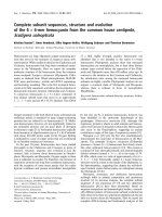
Fig. 1. Complementation of CEN.PK2 Dgpa2 Dras2 (A) or CEN.PK2
Dras1 Dras2 (B) by different high copy plasmids. Droplets containing
the indicated cell numbers (top) of high-copy transformants were
applied onto FOA medium. Plasmid inserts are indicated on the left
margin. e.p., empty plasmid (YEp351). Note that the parental strains
CEN.PK2 Dgpa2 Dras2 and CEN.PK2 Dras1 Dras2 contained plasmid
p426MET25-RAS2 (2l plasmid) and pFL38-RAS2 (centromeric
plasmid), respectively, prior to FOA selection.
Ó FEBS 2004 SUT2, a multicopy suppressor of low PKA activity (Eur. J. Biochem. 271) 1287
re-examined storage carbohydrate levels in the independent
genetic background of strain S1278b. In this case, high
SUT2 dosage conferred a reduction in PKA activity as
judged from storage carbohydrate levels in S1278b cells
during vegetative growth on glucose (Fig. 2, bottom)
indicating that the strong, nutrient responsive PKA activity
of S1278b [26] is modulated by Sut2p.
It is reasonable to suppose that the observed differences
between the two strains both containing high dosage of
SUT2 could be due to the reported difference in PKA
pathway activity. On this basis, we hypothesized that (a)
high SUT2 gene dosage might increase PKA activity only
when it is low, but that (b) it acts inhibitory when PKA
activity is high. To gain further insight into SUT2 function
we generated a sut2D strain for subsequent analyses. To
challenge the first part of the above hypothesis, we used
diploid S1278b cells and determined sporulation efficiency,
which is inversely correlated with PKA activity [27]. We
found that high SUT2 gene dosage drastically diminished
sporulation efficiency and, thus, mimics a phenotype of
increased PKA activity in S1278b wild type and slightly less
in Dras2 cells. While the deletion of SUT2 in wild-type cells
slightly decreased sporulation efficiency the difference was
not statistically significant (Fig. 3).
High SUT2 gene dosage reduces invasive growth
To address the assumption that a high level of SUT2
reduces PKA activity only when PKA activity is high we
investigated the influence of SUT2 on invasive growth,
which requires high PKA activity. We found that after three
days of growth on YPD medium, S1278b cells carrying a
high copy SUT2 plasmid did not at all adhere to the agar
medium in agreement with low cellular PKA activity. On
the other hand, S1278b wild-type control cells showed the
normal, strong invasive growth. In fact, the extent of the
reduction of invasive growth by high copy SUT2 was
similar to a Dras2 strain (Fig. 4A). To quantify this
observation, we generated a set of FLO11::lacZ reporter
strains, which allowed us to study expression of FLO11,the
major indicator of invasive growth. In agreement with the
observed effect during invasive growth, high SUT2 gene
dosage reduced b-galactosidase expression in wild type to a
level similar to Dras2 cells. Consistently, Dsut2 displayed an
increase in reporter gene expression, suggesting that Sut2p,
in fact, negatively regulates high PKA activity (Fig. 4B).
Based on the genetic evidence that high SUT2 gene
dosage is sufficient to sustain growth in Dgpa2 Dras2 but not
Dras1 Dras2 cells and considering that Sut2p has initially
been identified as a homologue of the putative transcription
factor Sut1p [11], we reasoned that Sut2p might act to
modify the transcription of one or both RAS genes.
Therefore, we determined the level of total Ras protein in
extracts of cells grown as described for the carbohydrate
determination experiments using Western blots with an
anti-H-RAS (259) antibody which detects both, yeast Ras1p
and Ras2p [14]. We found that high SUT2 gene dosage had
only a limited effect on Ras protein levels when cells were
harvested in stationary phase. However, in cells shifted to
fresh glucose medium and re-grown for an additional 4 h,
Ras protein levels were strongly reduced by high SUT2 gene
dosage (Fig. 4C), thus providing a possible explanation for
the observed phenotype of reduced invasive growth sug-
gestive of low PKA activity.
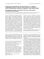
Sut2p-GFP localizes to the nucleus
In order to support the possibility that SUT2 encodes a
transcription factor, like its anaerobically expressed isozyme
Sut1p [11], we determined the subcellular localization of
GFP fused to the C-terminus of Sut2p. Expression was
controlled by the authentic SUT2 promoter on a high copy
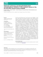
Fig. 2. Levels of glycogen and trehalose in CEN.PK2 Dras2 (top) and
R1278b Dras2 (bottom) transformed with different plasmids. Cultures
where grown to stationary phase in selective glucose medium over
night (D
600
5.5) and shifted to fresh glucose medium (SC 2% glucose)
at time point 0. (j) YEp351 + pFL38; (d) YEp351 + pFL38-
RAS2;(h) YEp351-SUT2 +pFL38;(s) YEp351-SUT2 + pFL38-
RAS2. Experiments were performed three times with similar results.
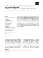
Fig. 3. Sporulation efficiency of R1278b. SUT2 carried on a high copy
plasmid reduces sporulation efficiency in wild-type (P<0.001) and
Dras2 (P<0.05) cells (Tukey HSD; n ¼ 4). Error bars show ± 1.0
SD; columns show mean values. WT, MR161; h.c. SUT2, YEp351-
SUT2; Dras2, MR298; Dsut2,AR2.
1288 M. Ru
¨
tzler et al. (Eur. J. Biochem. 271) Ó FEBS 2004
plasmid. We found that Sut2p-GFP localizes to the entire
cytoplasm of the cell with some accumulation in the nucleus
of most cells (Fig. 5). To verify that the observed fluorescent
signal was, indeed, mainly localized to the nucleus, we
carried out DAPI staining of ethanol fixed cells. Cells which
retain GFP fluorescence in this procedure show a clear
colocalization of DAPI and green fluorescence in agreement
with a possible involvement of Sut2p in transcription
regulation.
Discussion
In this report we describe the isolation of SUT2 in a screen
for high copy suppressors of the synthetic slow growth
phenotype of Dgpa2 Dras2. In addition to SUT2, a number
of other suppressors were identified that have been impli-
cated to function in the RAS/cAMP pathway. These include
the two disrupted genes, GPA2 and RAS2, the second RAS
gene RAS1, SCH9, a protein kinase A homologue, which
previously has been described as a high copy suppressor of a
number of defects in the RAS/cAMP pathway [23] and
TPK2, one of the three catalytic subunits of PKA [6].
Interestingly, this screen did not yield any plasmids that
contained TPK1 or TPK3, the other two genes encoding
catalytic subunits of PKA. Plasmids that contained either
RAS1, RAS2 or SUT2 were isolated frequently in the screen
(50–100 times each), whereas GPA2, SCH9 and TPK2 were
isolated < 10 times. This may indicate that the yeast
genomic library utilized in this study did not contain a
perfectly random array of DNA fragments and hence the
screen was probably not comprehensive.
Sut2p has been described previously as a homologue
of the putative transcriptional activator Sut1p. When
expressed under the control of a strong heterologous
promoter both proteins enhance uptake of sterols and, at
least Sut1p, also increases the biosynthesis of sterol
precursors [11]. In contrast to the exclusively anaerobically
expressed SUT1, expression of SUT2 is apparently not
controlled by oxygen [28]. In order to establish an
epistatic relationship between SUT2 and elements of the
RAS/cAMP pathway we determined the effect of high
SUT2 gene dosage on the lethal double-deletion of both
RAS-genes. In contrast to concomitant deletion of GPA2
and RAS2,highSUT2 gene dosage did not rescue the
RAS1 RAS2-double deletion, suggesting that SUT2 either
acts upstream or, alternatively, in a parallel pathway to
RAS. To further investigate the relation between SUT2
and the RAS/cAMP pathway we studied the influence of
high SUT2 gene dosage on storage-carbohydrate homeo-
stasis, which is controlled by PKA. Consistent with
SUT2’s function as a high copy suppressor of the syn-
thetic Dgpa2 Dras2 phenotype, high SUT2 gene dosage
resulted in decreased storage-carbohydrate levels in a
CEN.PK2 background. However, this effect was only
observed in Dras2 mutants.
Surprisingly, re-evaluation of this result in an independent
genetic background (S1278b) yielded a different result: high
SUT2 gene dosage led to increased storage carbohydrate
levels, which suggests reduced PKA activity. PKA activity in
CEN.PK2 is reduced due to a mutation in adenylate cyclase
[25] whereas S1278b is known to contain a particularly
strong PKA pathway [26]. We therefore hypothesized that
Sut2p may represent a new element in PKA feedback-
regulation and, hence, affects these two strains differently:
increased SUT2 gene dosage stimulates low PKA activity but
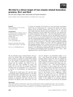
Fig. 5. Localization of the Sut2-GFPp chimeric protein. Cells were fixed
in 70% ethanol for DAPI staining and imaged as described in
Experimental procedures.
Fig. 4. Influence of Sut2p on invasive growth, FLO11 expression and
Ras protein level in R1278b. (A)CellsweregrownonYPDmediumfor
3 days at 30 °C (top, left) and then washed off the plates with a squeeze
bottle to determine invasive growth (top, right). Genotypes are as
indicated (center, left). (B) To quantify the influence of Sut2p on
invasive growth, Flo11-b galactosidase reporter assays were carried
out as detailed in Experimental procedures. YEp351-SUT2 reduces
lacZ reporter expression in WT (P<0.01). In contrast, deletion of
SUT2 increased reporter expression (P ¼ 0.001; Tukey HSD; n ¼ 9
and 3 for Dras2, respectively. Error bars show ± 1.0 SD. Columns
represent mean values. (C) Immunoblot with anti-H-Ras (259) or anti-
Aky2 Ig: Total protein was prepared from cells grown overnight in SC
medium (stat.) or after shift to fresh SC medium (growth) as indicated
in Experimental procedures. WT, YHUM216 and MR287 (lacZ);
Dras2, MR211 and AR3 (lacZ); Dsut2, AR1 and AR4 (lacZ); h.c.
SUT2, YEp351-SUT2.
Ó FEBS 2004 SUT2, a multicopy suppressor of low PKA activity (Eur. J. Biochem. 271) 1289
inhibits high PKA activity. Subsequent experiments on
sporulation efficiency and invasive growth of S1278b
supported this hypothesis. Sporulation in S. cerevisiae is
facilitated by starvation-conditions that result in low PKA
activity. High SUT2 gene dosage yielded diminished spor-
ulation efficiency, which indicates increased PKA activity
relative to wild type. In contrast, invasive growth in haploids
requires strong PKA activity and is assayed on rich medium
[24]. Under these growth conditions, high SUT2 gene dosage
resulted in a strong reduction of agar invasion in a RAS2-
wild-type strain, and this reduction was similar to the
phenotype of Dras2 mutants. We consistently found that
duringgrowthinrichmedium,highSUT2 gene dosage
resulted in diminished expression of FLO11, a surface
flocculin whose expression is stimulated by PKA and that
is essential for invasive growth [10]. As it has been proposed
that Sut1p and Sut2p act as transcription factors [11] we
investigated if SUT2 affects expression of elements of the
RAS/cAMP pathway. Indeed we found that high SUT2 gene
dosage reduced Ras2p expression in cells that had been
grown in rich medium. Importantly, no reduction in Ras2p
was observed in cultures approaching stationary phase,
which correlates with a reduction in PKA-activity.
Interestingly, a link between sterol-biosynthesis and RAS
has previously been established: if yeast cells were starved
for mevalonate, an early precursor of isoprenoids and
sterols, levels of both RAS-mRNAs were decreased [29].
One possible explanation for the effect of high SUT2 gene
dosage on the RAS/cAMP pathway is that Dgpa2 Dras2
and possibly additional mutants reducing PKA activity are
simultaneously down-regulated for isoprenoid/sterol bio-
synthesis, thereby reducing Ras1p abundance and, hence,
impairing the residual G-protein stimulating adenylate
cyclase. High copy expression of SUT2 then could, in
analogy to its homologue SUT1, relieve this sterol precur-
sor-starvation by increasing sterol-biosynthesis. Therefore it
will be interesting to determine if genes of the sterol
biosynthetic pathway are subject to regulation by Sut2p,
which may yield a better understanding of the proposed
connection between the nutrient-sensitive activity of PKA
and sterol biosynthesis.
Acknowledgements
This work was supported by a grant from the Deutsche Forschungs-
gemeinschaft to W. B. (Ba415/24–1). We thank H. U. Mo
¨
sch,
Go
¨
ttingen, for the gift of strains and plasmids. We are also indebted
to B. Klebl, Martinsried, for the gift of plasmid p426MET25-Ras2. M.
Angermayr and G. Strobel are acknowledged for their help with yeast
genetics and valuable discussions. We also acknowledge L. J. Zwiebel
for reading the manuscript. Finally, we thank T. Grimm for
construction of strain CEN.PK2 Dgpa2 Dras2.
References
1. Kataoka, T., Powers, S., McGill, C., Fasano, O., Strathern, J.,
Broach,J.&Wigler,M.(1984)GeneticanalysisofyeastRAS1
and RAS2 genes. Cell 37, 437–445.
2. Rolland, F., De Winde, J.H., Lemaire, K., Boles, E., Thevelein,
J.M. & Winderickx, J. (2000) Glucose-induced cAMP signalling in
yeast requires both a G-protein coupled receptor system for
extracellular glucose detection and a separable hexose kinase-
dependent sensing process. Mol. Microbiol. 38, 348–358.
3. Harashima, T. & Heitman, J. (2002) The Galpha protein Gpa2
controls yeast differentiation by interacting with kelch repeat
proteins that mimic Gbeta subunits. Mol. Cell 10, 163–173.
4. Batlle,M.,Lu,A.,Green,D.A.,Xue,Y.&Hirsch,J.P.(2003)
Krh1p and Krh2p act downstream of the Gpa2p G (alpha) sub-
unit to negatively regulate haploid invasive growth. J. Cell Sci.
116, 701–710.
5. Xue, Y., Batlle, M. & Hirsch, J.P. (1998) GPR1 encodes a putative
G protein-coupled receptor that associates with the Gpa2p Gal-
pha subunit and functions in a Ras-independent pathway. EMBO
J. 17, 1996–2007.
6. Toda, T., Cameron, S., Sass, P., Zoller, M. & Wigler, M. (1987)
Three different genes in S.cerevisiaeencode the catalytic subunits
of the cAMP-dependent protein kinase. Cell 50, 277–287.
7. Toda, T., Cameron, S., Sass, P., Zoller, M., Scott, J.D., McMul-
len, B., Hurwitz, M., Krebs, E.G. & Wigler, M. (1987) Cloning
and characterization of BCY1, a locus encoding a regulatory
subunit of the cyclic AMP-dependent protein kinase in
Saccharomyces cerevisiae. Mol. Cell. Biol. 7, 1371–1377.
8. Thevelein, J.M. & de Winde, J.H. (1999) Novel sensing mechan-
isms and targets for the cAMP-protein kinase A pathway in the
yeast Saccharomyces cerevisiae. Mol. Microbiol. 33, 904–918.
9. Mo
¨
sch, H.U., Roberts, R.L. & Fink, G.R. (1996) Ras2 signals via
the Cdc42/Ste20/mitogen-activated protein kinase module to
induce filamentous growth in Saccharomyces cerevisiae. Proc. Natl
Acad. Sci. USA 93, 5352–5356.
10. Rupp, S., Summers, E., Lo, H.J., Madhani, H. & Fink, G. (1999)
MAP kinase and cAMP filamentation signaling pathways con-
verge on the unusually large promoter of the yeast FLO11 gene.
EMBO J. 18, 1257–1269.
11. Ness, F., Bourot, S., Regnacq, M., Spagnoli, R., Berges, T. &
Karst, F. (2001) SUT1 is a putative Zn[II]2Cys6-transcription
factor whose upregulation enhances both sterol uptake and syn-
thesis in aerobically growing Saccharomyces cerevisiae cells. Eur. J.
Biochem. 268, 1585–1595.
12. Gu
¨
ldener, U., Heck, S., Fielder, T., Beinhauer, J. & Hegemann,
J.H. (1996) A new efficient gene disruption cassette for repeated
use in budding yeast. Nucleic Acids Res. 24, 2519–2524.
13. Gu
¨
ldener, U., Heinisch, J., Koehler, G.J., Voss, D. & Hegemann,
J.H. (2002) A second set of loxP marker cassettes for Cre-mediated
multiple gene knockouts in budding yeast. Nucleic Acids Res.
30, e23.
14. Mo
¨
sch, H.U., Ku
¨
bler, E., Krappmann, S., Fink, G.R. & Braus,
G.H. (1999) Crosstalk between the Ras2p-controlled mitogen-
activated protein kinase and cAMP pathways during invasive
growth of Saccharomyces cerevisiae. Mol. Biol. Cell 10, 1325–1335.
15. Hermann, H., Ha
¨
cker, U., Bandlow, W. & Magdolen, V. (1992)
pYLZ. vectors: Saccharomyces cerevisiae/Escherichia coli shuttle
plasmids to analyze yeast promoters. Gene 119, 137–141.
16. Lillie, S.H. & Pringle, J.R. (1980) Reserve carbohydrate metabo-
lism in Saccharomyces cerevisiae: responses to nutrient limitation.
J. Bacteriol. 143, 1384–1394.
17. Schricker, R., Angermayr, M., Strobel, G., Klinke, S., Korber, D.
& Bandlow, W. (2002) Redundant mitochondrial targeting signals
in yeast adenylate kinase. J. Biol. Chem. 277, 28757–28764.
18. Bradford, M.M. (1976) A rapid and sensitive method for the
quantitation of microgram quantities of protein utilizing the
principle of protein-dye binding. Anal. Biochem. 72, 248–254.
19. Guarente, L. (1983) Yeast promoters and lacZ fusions designed to
study expression of cloned genes in yeast. Methods Enzymol. 101,
181–191.
20. Boeke, J.D., Trueheart, J., Natsoulis, G. & Fink, G.R. (1987)
5-Fluoroorotic acid as a selective agent in yeast molecular genetics.
Methods Enzymol. 154, 164–175.
21. Colombo, S., Ma, P., Cauwenberg, L., Winderickx, J., Crauwels,
M.,Teunissen,A.,Nauwelaers,D.,deWinde,J.H.,Gorwa,M.F.,
1290 M. Ru
¨
tzler et al. (Eur. J. Biochem. 271) Ó FEBS 2004
Colavizza, D. & Thevelein, J.M. (1998) Involvement of distinct
G-proteins, Gpa2 and Ras, in glucose- and intracellular acid-
ification-induced cAMP signalling in the yeast Saccharomyces
cerevisiae. EMBO J. 17, 3326–3341.
22. Toda, T., Uno, I., Ishikawa, T., Powers, S., Kataoka, T., Broek,
D., Cameron, S., Broach, J., Matsumoto, K. & Wigler, M. (1985)
In yeast, RAS proteins are controlling elements of adenylate
cyclase. Cell 40, 27–36.
23. Toda, T., Cameron, S., Sass, P. & Wigler, M. (1988) SCH9, a gene
of Saccharomyces cerevisiae that encodes a protein distinct from,
but functionally and structurally related to, cAMP-dependent
protein kinase catalytic subunits. Genes Dev. 2, 517–527.
24. Gancedo, J.M. (2001) Control of pseudohyphae formation in
Saccharomyces cerevisiae. FEMS Microbiol. Rev. 25, 107–123.
25. Vanhalewyn, M., Dumortier, F., Debast, G., Colombo, S., Ma,
P., Winderickx, J., Van Dijck, P. & Thevelein, J.M. (1999)
AmutationinSaccharomyces cerevisiae adenylate cyclase,
Cyr1K1876M, specifically affects glucose- and acidification-
induced cAMP signalling and not the basal cAMP level. Mol.
Microbiol. 33, 363–376.
26. Stanhill, A., Schick, N. & Engelberg, D. (1999) The yeast ras/cyclic
AMP pathway induces invasive growth by suppressing the cellular
stress response. Mol. Cell. Biol. 19, 7529–7538.
27. Matsumoto, K., Uno, I. & Ishikawa, T. (1983) Initiation of
meiosis in yeast mutants defective in adenylate cyclase and cyclic
AMP-dependent protein kinase. Cell 32, 417–423.
28. ter Linde, J.J., Liang, H., Davis, R.W., Steensma, H.Y., van
Dijken, J.P. & Pronk, J.T. (1999) Genome-wide transcriptional
analysis of aerobic and anaerobic chemostat cultures of Saccharo-
myces cerevisiae. J. Bacteriol. 181, 7409–7413.
29. Dimster-Denk,D.,Schafer,W.R.&Rine,J.(1995)Controlof
RAS mRNA level by the mevalonate pathway. Mol. Biol. Cell 6,
59–70.
30. Bonneaud, N., Ozier-Kalogeropoulos, O., Li, G.Y., Labouesse,
M., Minvielle-Sebastia, L. & Lacroute, F. (1991) A family of
low and high copy replicative, integrative and single-stranded
S. cerevisiae/E. coli shuttle vectors. Yeast 7, 609–615.
31. Hill, J.E., Myers, A.M., Koerner, T.J. & Tzagoloff, A. (1986)
Yeast/E. coli shuttle vectors with multiple unique restriction sites.
Yeast 2, 163–167.
32. Mumberg, D., Muller, R. & Funk, M. (1995) Yeast vectors for the
controlled expression of heterologous proteins in different genetic
backgrounds. Gene 156, 119–122.
Ó FEBS 2004 SUT2, a multicopy suppressor of low PKA activity (Eur. J. Biochem. 271) 1291