Tài liệu Báo cáo khoa học: Complete subunit sequences, structure and evolution of the 6 · 6-mer hemocyanin from the common house centipede, Scutigera coleoptrata pptx
Bạn đang xem bản rút gọn của tài liệu. Xem và tải ngay bản đầy đủ của tài liệu tại đây (407.73 KB, 9 trang )
Complete subunit sequences, structure and evolution
of the 6 · 6-mer hemocyanin from the common house centipede,
Scutigera coleoptrata
Kristina Kusche*, Anne Hembach, Silke Hagner-Holler, Wolfgang Gebauer and Thorsten Burmester
Institute of Zoology, Molecular Animal Physiology, University of Mainz, Germany
Hemocyanins are large oligomeric copper-containing pro-
teins that serve for the transport of oxygen in many arth-
ropod species. While studied in detail in the Chelicerata and
Crustacea, hemocyanins had long been considered unnec-
essary in the Myriapoda. Here we report the complete
molecular structure of the hemocyanin from the common
house centipede Scutigera coleoptrata (Myriapoda: Chilo-
poda), as deduced from 2D-gel electrophoresis, MALDI-
TOF mass spectrometry, protein and cDNA sequencing,
and homology modeling. This is the first myriapod hemo-
cyanin to be fully sequenced, and allows the investigation of
hemocyanin structure–function relationship and evolution.
S. coleoptrata hemocyanin is a 6 · 6-mer composed of
four distinct subunit types that occur in an approximate
2 : 2 : 1 : 1 ratio and are 49.5–55.5% identical. The cDNA
of a fifth, highly diverged, putative hemocyanin was
identified that is not included in the native 6 · 6-mer
hemocyanin. Phylogenetic analyses show that myriapod
hemocyanins are monophyletic, but at least three distinct
subunit types evolved before the separation of the Chilo-
poda and Diplopoda more than 420 million years ago. In
contrast to the situation in the Crustacea and Chelicerata,
the substitution rates among the myriapod hemocyanin
subunits are highly variable. Phylogenetic analyses do not
support a common clade of Myriapoda and Hexapoda,
whereas there is evidence in favor of monophyletic
Mandibulata.
Keywords: hemocyanin; subunit diversity; structure; Arthro-
poda; evolution.
Oxygen transport in the body fluid of many arthropod and
molluscan species is mediated by large copper-containing
proteins that are referred to as hemocyanins [1–3]. Mollu-
scan hemocyanins, however, are not significantly related to
the arthropod proteins and are most likely of divergent
evolutionary origin [3–5]. Arthropod hemocyanins typically
form hexamers or oligo-hexamers (up to 8 · 6) of subunits
with about 620–660 amino acids [1,2,6]. Oxygen binding
is mediated by a pair of copper atoms that are coordinated
by six histidine residues per monomer.
In the past 30 years, hemocyanins have been studied
thoroughly in the Chelicerata and Crustacea in terms of
function, structure, sequence and evolution [1,2,6]. By
contrast, little is known about hemocyanins in Onycho-
phora, Myriapoda and Hexapoda. Oxygen supply in these
taxa is mediated via trachea [7]. Therefore, the presence of
respiratory proteins had long been considered unnecessary,
with the exception of some insect species that live under
hypoxic conditions and possess extracellular hemoglobins
[8, but also see 9]. A putative hemocyanin, however, has
been identified in the embryonic hemolymph of the
grasshopper Schistocerca americana [10], although this
respiratory protein is absent in adult animals and hemocy-
anins were lost later in hexapod evolution [6]. Hemocyanins
also occur in the Onychophora, suggesting an ancient
evolutionary origin of this type of respiratory protein [11].
Mangum et al. [12] were the first who unambiguously
demonstrated the occurrence of hemocyanins in the Myr-
iapoda. The centipede Scutigera coleoptrata (Chilopoda)
possesses a 36-mer (6 · 6) hemocyanin that closely resem-
bles other arthropod hemocyanins [12,13]. This protein is
composed of four electrophoretically and immunologically
distinct subunits (termed a : b : c : d) in the range of 74–
80 kDa, which occur in a ratio of approximately a/b/c/d ¼
2 : 2 : 1 : 1 [14]. Structurally similar 6 · 6-mer hemocya-
nins also occur in at least in one family of the Diplopoda, i.e.
the Spirostreptidae, suggesting that despite the well-devel-
oped tracheal system hemocyanins are widespread among
the Myriapoda [15,16].
Myriapod, crustacean, and chelicerate hemocyanins
strikingly differ in subunit composition and quaternary
structure [1]. In previous analyses, the complete subunit
sequences of one crustacean [17–19] and two chelicerate
hemocyanins [20,21] have been determined. Based on
sequence comparison and immunological studies, a remark-
ably different pattern of hemocyanin subunit evolution has
been observed in these two arthropod subphyla [4,6,22], but
little was known about the Myriapoda. Here we report the
complete cDNA-cloning and biochemical analyses of the
Correspondence to T. Burmester, Institute of Zoology,
University of Mainz, D-55099 Mainz, Germany.
Fax: + 49 6131 3924652, Tel.: + 49 6131 3924477,
E-mail:
Abbreviation: Hc, hemocyanin.
*Present address: Institute of Animal Physiology, University of
Mu
¨
nster, Germany.
(Received 21 March 2003, revised 7 May 2003,
accepted 12 May 2003)
Eur. J. Biochem. 270, 2860–2868 (2003) Ó FEBS 2003 doi:10.1046/j.1432-1033.2003.03664.x
hemocyanin subunits from S. coleoptrata, being the first
myriapod hemocyanin of which the complete molecular
structure is known. This allows us to compare structure, and
intra- and intermolecular evolution of hemocyanins from
Myriapoda, Crustacea and Chelicerata.
Materials and methods
Protein biochemistry
Scutigera coleoptrata (Myriapoda, Chilopoda, Scutigero-
morpha) were captured on Lesbos, Greece. The hemolymph
was withdrawn from the dorsal intersegmental regions
withasyringeandstoredfrozenat)20 °C until use. The
hemocyanin was purified by ultracentrifugation in a Beck-
man airfuge for 16 h at 120 000 g.SDS/PAGEand
Western blotting were carried out using standard methods
as previously described [14,15]. Two-dimensional gel elec-
trophoresis with pH 3.5–10 ampholines was performed
according to O’Farrell et al. [23,24]. For antibody prepar-
ation, the hemocyanin subunits were separated by SDS/
PAGE and stained by Coomassie brilliant blue. The two
hemocyanin bands were cut out and dispersed. About
100 lg of hemocyanin was used for the immunization of
rabbits. For determination of the N-terminal ends, samples
were separated by SDS/PAGE and transferred to a
poly(vinylidene difluoride) membrane by electroblotting.
The hemocyanin bands were excised and submitted to
Edman degradation. MALDI-TOF mass spectrometry
experiments were performed by Proteosys (Mainz,
Germany) on hemocyanin subunits that had been separated
by 2D PAGE, stained with Coomassie brilliant blue and
digested with trypsin. The MALDI-TOF data were evalu-
ated using the program
PEPTIDE
-
MASS
at the ExPASy web
server ().
Cloning of
S. coleoptrata
hemocyanin cDNA
The total RNA was extracted from a single specimen and
poly(A)
+
RNA was purified from total RNA by the aid of
the PolyATract kit (Promega). About 5 lg poly(A)
+
RNA
were used to construct a directionally cloned cDNA
expression library applying the Lambda ZAP-cDNA syn-
thesis kit (Stratagene). The library was amplified using the
material provided by Stratagene and screened with the anti-
(S. coleoptrata hemocyanin) Igs. Positive phage clones were
converted into pBK-CMV plasmid vectors and sequenced
by a commercial sequencing service (Genterprise, Mainz,
Germany). The missing 5¢ ends of two clones [hemocyanin
(Hc)B and HcD; see below] were obtained from the library
by a PCR approach using two nested clone-specific
oligonucleotides primers and the T3 vector primer. The
sequences were obtained after the cloning of the PCR
products into the pCR4-TOPO TA vector (Invitrogen).
Protein structure modeling
Homology models of the S. coleoptrata hemocyanin sub-
units were built applying the online facility
SWISSMODEL
(GlaxoWellcome) [25] at the following address: http://
www.expasy.ch/spdbv/using the known arthropod hemo-
cyanin crystal structures [26,27] as templates.
Sequence data analyses and phylogenetic studies
The tools provided with Genetics Computer Group (GCG)
Software Package 10 and by the ExPASy Molecular
Biology Server of the Swiss Institute of Bioinformatics
() were used for the analyses of DNA
and amino acid sequences. Signal peptides were predicted
using the online version of
SIGNALP
V1.1 [28]. The amino
acid sequences of the S. coleoptrata hemocyanins were
added to a previously published alignment of the arthropod
hemocyanin superfamily [4,11] by the aid of
GENEDOC
2.6
[29]. Only selected hemocyanin and phenoloxidase
sequences were used for phylogenetic inference. The signal
peptides were eliminated from the final data set. Distances
between pairs of sequences were calculated using the PAM
matrix [30] implemented in the
PHYLIP
3.6a2 package [31].
Tree constructions were performed by the neighbor-joining
method. The reliability of the trees was tested by the
bootstrap procedure with 100 replications [32]. Replacement
rates were estimated from the PAM distances assuming that
Chilopoda and Diplopoda diverged 450 million years ago
[33,34]. Alternative models of sequence evolution were
tested using T
REE
-P
UZZLE
[35], and the WAG model [36]
was chosen on the basis of the highest likelihood values.
Bayesian phylogenetic analyses were performed with
MRBAYES
3.0 [37]. The WAG model with gamma distribu-
tion of rates was applied. Metropolis-coupled Markov chain
Monte Carlo sampling was performed with four chains that
were run for 200 000 generations. Prior probabilities for
all trees were equal, starting trees were random, tree
sampling was performed every 10 generations. Posterior
probability densities were estimated on 5000 trees (burnin ¼
15 000).
Results
Purification and analyses of
S. coleoptrata
hemocyanin
The 6 · 6-mer hemocyanin of S. coleoptrata (55S) [12] was
purified from total hemolymph by ultracentrifugation. After
separation by SDS/PAGE, the hemocyanin fraction shows
two distinct polypeptide bands with apparent molecular
masses in the range of 75 kDa, but no other contaminating
protein detectable by Coomassie staining, suggesting that
the preparation results in >95% pure protein (Fig. 1).
Antibodies raised against the hemocyanin fraction show
staining of the two 75 kDa bands in Western blot, but do
not recognize any other proteins of the hemolymph.
Although we expected that each of these bands contains
two distinct polypeptides [14], we determined their
N-terminal sequences. In fact both are heterogeneous, but
in each case a predominant sequence was obtained: The first
18 amino acids of the major sequence in the upper band are
DQEPAVPADTKDKLEKIL, the lower band gave rise to
the sequence DKEPXATD(QKI)EAKQKXMLE.
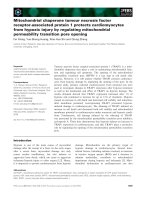
By 2D gel electrophoresis a total of 10 distinct protein
spots were identified in the hemocyanin sample. The appar-
ent molecular masses are about 75–80 kDa, the isoelectric
points (pI) range from 5 to 6.5 (Fig. 2). MALDI-TOF
analyses of tryptic peptides show that the spots represent
four distinct subunit types, as deduced from the pattern of
identical peptide masses. The subunits were named HcA to
Ó FEBS 2003 Centipede hemocyanin structure (Eur. J. Biochem. 270) 2861
HcD, in agreement with the previous data on Scutigera
hemocyanin subunit masses and charges [14]. HcA and
HcD were each found to form two spots that slightly differ
in their sizes, whereas each HcB and HcC form three spots
that probably represent charge variants.
cDNA-sequences of
S. coleoptrata
hemocyanin subunits
A cDNA expression library was constructed from about
5 lg poly(A)
+
RNA extracted from an adult S. coleoptrata
specimen. The library was screened with the antibodies
raised against the S. coleoptrata hemocyanin. In a total of
about 20 clones, five distinct hemocyanin subunit cDNAs
were identified and fully sequenced (Fig. 3). The cDNA
sequences were assigned to distinct subunits (HcA to HcD;
Fig. 2) by the aid of the MALDI-TOF data. About 30
peptides that cover a total of at least 40% of each subunit
could be unambiguously identified for each subunit. How-
ever, the fifth cDNA sequence could not be allocated to any
of the spots found in the native 6 · 6-mer hemocyanin, and
thus has been termed HcX.
As deduced from the N-terminal sequences obtained by
conventional protein sequencing and from comparison with
other arthropod hemocyanins, the cDNAs cover the
complete coding regions for the four hemocyanin subunits
and HcX, plus 6–45 bp of the respective 5¢ untranslated
regions and the entire 3¢ untranslated regions. The standard
polyadenylation signals (AATAAA) and the poly(A)-tails
of different lengths are present in each clone. The open
reading frames of the five sequences translate into distinct
polypeptides of 656–685 amino acids (Table 1; Fig. 3).
Signal peptides required for the transmembrane excretion
into the hemolymph were found in all sequences and cover
18–20 amino acids, as predicted by the
SIGNALP
computer
program [28]. The estimated molecular masses of the native
subunits (without signal peptides; 74.4–77.7 kDa) and the
theoretical isoelectric points (pI 5.44–6.18) agree well with
those observed in SDS/PAGE and in 2D PAGE (Figs 1 and
2; Table 1).
The N-terminal amino acid sequences (see above) allow
the assignment of the HcA clone to the major sequence of
the upper hemocyanin band in the SDS/PAGE gel (Fig. 1).
Two nonmatching amino acids at positions 3 (Glu instead
of Cys) and 5 (Ala instead of Pro) may be explained by the
presence of an additional subunit in the sample that most
likely corresponds to HcC, as also inferred from the minor
sequence in the sample (not shown). The lower protein band
was assigned to HcB, whereas minor amino acid peaks most
likely derive from HcD. Again, no indication for the
presence of a HcX-like subunit was found.
Sequence comparison
The four S. coleoptrata hemocyanin subunits (HcA-D)
share 49.5–55.5% of the amino acids (Table 2) with 240
amino acids ( 35%) being strictly conserved (Figs 3 and
4). The HcX sequence is highly diverged, as deduced from
the lower identity scores (42.1–48.1% identity with HcA to
HcD) and several short sequence insertions (Table 2;
Fig. 3). It is not included in the calculations below.
Arthropod hemocyanins are divided into three structural
domains [26,27]. While 119 strictly conserved residues
( 52%) have been found in domain 2 of subunits HcA
to D, there is less sequence conservation in domains 1 (51
conserved amino acids; 30%) and 3 (70 conserved
residues; 27%). As expected, the Scutigera hemocyanin
subunits (HcA-D) show the highest degree of sequence
similarity to the previously determined hemocyanin from
the diplopod Spirostreptus (44.9–51.0% identity). Lower
scores were observed with the chelicerate hemocyanins
(38–46%), the onychophoran hemocyanin ( 34–37%), the
phenoloxidases of Crustacea and insects ( 31–38%), the
crustacean hemocyanins ( 27–35%) and the insect hemo-
cyanin ( 34–38%).
Secondary and tertiary structure of
Scutigera
hemocyanin subunits
The S. coleoptrata hemocyanin subunits contain the six
copper-coordinating histidines necessary for copper binding
(Fig. 3), which are strictly conserved in all arthropod
hemocyanins [4,6,38]. No long insertions or deletions were
observed upon comparison with the other hemocyanins.
Fig. 2. Identification of S. coleoptrata hemocyanin subunits. About
20 lg of purified hemocyanin was separated by two-dimensional
PAGE. The anode (+) and cathode (–) are indicated, the molecular
mass marker is on the right side. The spots were submitted to MALDI-
TOF analysis and assigned to distinct subunits.
Fig. 1. SDS/PAGE and immunoblotting of S. coleoptrata hemocyanin.
About 10 lg of total hemolymph protein (HL) and 3 lgofpurified
hemocyanin (Hc) were separated on SDS/PAGE and stained with
Coomassie brilliant blue R-250. A Western blot analysis of total
hemolymph using anti-(S. coleoptrata hemocyanin) Ig is shown on the
rightside(Western).Themolecularmassmarkerproteinsareonthe
left side (kDa).
2862 K. Kusche et al. (Eur. J. Biochem. 270) Ó FEBS 2003
Fig. 3. Alignment of the myriapod hemocyanins. The conserved amino acids are shaded, the signal peptides are underlined and the potential
N-glycosylation sites are in italics/bold face. Above the alignment: Copper-binding histidines of the CuA and CuB sites (*); putative disulfide
bridges (c). Below the alignment: Secondary structure elements derived from a modeling approach of ScoHcA. The nomenclature follows the
standard convention for hemocyanin structure [26,27]; a, a helix; b, b sheet. Note that a helix 1.2 is missing. The abbreviations used are: ScoHcA to
D, S. coleoptrata hemocyanin subunits A to D; ScoHcX, S. coleoptrata hemocyanin X; SpiHc1, Spirostreptus spec. hemocyanin subunit 1.
Table 1. Properties of myriapod hemocyanin subunits. Accession numbers are the EMBL/GenBank DNA data accession number. CDNA lengths
refer to the sequence without the poly(A) tail; protein length is including the signal peptide but molecular mass and pI are calculated without the
signal peptide.
Subunit Accession number cDNA (bp)
Protein
(amino acids)
Molecular
mass (kDa) pI
Replacement rates
(per site per year)
ScoHcA AJ344359 2258 656 74.44 5.44 0.66 · 10
)9
ScoHcB AJ512793 2283 659 73.93 5.57 0.92 · 10
)9
ScoHcC AJ431379 2236 673 75.91 5.68 1.07 · 10
)9
ScoHcD AJ344360 2271 668 74.75 6.18 0.89 · 10
)9
ScoHcX AJ431378 2209 685 77.74 5.79 1.89 · 10
)9
SpiHc1 AJ297738 2082 653 73.74 6.16 1.13 · 10
)9
Ó FEBS 2003 Centipede hemocyanin structure (Eur. J. Biochem. 270) 2863
Similar to the Onychophora and Chelicerata, a-helix 1.2
is missing in the myriapod hemocyanins. Tentative 3D
structures of the subunits were constructed by a homology
modeling approach using the hemocyanins of Limulus
polyphemus and Panulirus interruptus as templates [26,27].
The modeling process was straightforward, with the excep-
tion of the first 20–30 amino acids of subunits HcC and
D, which could not be recovered. The positions of the
amino acids conserved among all subunits were super-
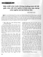
imposed on the model of subunit HcA (Fig. 4). Strong
conservation was found around the copper-binding sites,
most notably in the a-helices 2.1 and 2.2 (CuA site) and 2.5
and 2.6 (CuB site). The subunits all contain the cysteines
forming a disulfide bridge that stabilize domain 3 [27]
(Figs3and4).One(HcA,HcB,HcC)ortwo(HcDand
HcX) potential N-glycosylation sites (NXT/S) are present,
which are, however, not conserved in any other arthropod
hemocyanin subunit. Nevertheless, the glycosylation site at
a-sheet 2E (Fig. 3) is located at the surface of the putative
hemocyanin hexamer, as deduced from the comparison
with the L. polyphemus and P. interruptus hemocyanin
structures.
Hemocyanin molecular phylogeny
Phylogenetic trees were calculated using an amino acid
alignment of the six myriapod hemocyanins, 24 selected
hemocyanins from other arthropod species and eight
prophenoloxidase sequences [4]; the prophenoloxidases are
considered as an outgroup [4,39]. Various tree-building
methods were applied; the results of a neighbor-joining and
a Bayesian approach are presented in Fig. 5. The mono-
phyly of the myriapod hemocyanins was recovered by all
types of analyses with 100% support. There is solid
bootstrap support (82%) and Bayesian posterior probability
(0.87) for an association of the myriapod hemocyanins with
the crustacean and insect hemocyanins. All analyses support
a pancrustacean taxon of the insect and crustacean proteins
that excludes the myriapod hemocyanins. Within the clade
of myriapod hemocyanins, there is strong support that
Scutigera HcA is associated with the Spirostreptus hemocy-
anin 1. This branch joins a clade formed by HcB and HcX,
whereas the branch leading to HcC and HcD splits earlier.
Replacement rates of myriapod hemocyanins were diffi-
cult to calculate because the time of divergence of the
Chilopoda and Diplopoda is essentially unknown. Both
chilopod and diplopod fossils that already belong to distinct
extant orders have been identified in lower Devonian and
upper Silurian strata [33,34]. Thus these taxa must have
diverged more than 410–420 million years ago, although
this date may be underestimated [40]. For further calcula-
tions we assumed a time of divergence of 450 million years
ago. Then the estimated replacement rates of the different
myriapod hemocyanin subunits show a large variance,
Table 2. Comparison of myriapod hemocyanin subunits. Amino acid
identities (%) are given.
ScoHcB ScoHcC ScoHcD ScoHcX SpiHc1
ScoHcA 55.5 53.7 53.1 44.6 51.0
ScoHcB 53.0 49.5 48.1 46.0
ScoHcC 54.4 42.1 44.9
ScoHcD 43.3 45.3
ScoHcX 38.9
Fig. 4. Stereo view of a model of S. coleoptrata hemocyanin subunit A. The structure was deduced by comparative modeling. Positions strictly
conserved in all S. coleoptrata subunits are displayed in red, the copper atoms are blue, the coordinating histidines green and the disulfide bridge-
forming cysteines yellow.
Fig. 5. Phylogenetic analysis of the arthropod hemocyanins. A simpli-
fied phylogenetic tree was calculated by the neighbor-joining method
of the amino acids based on PAM distances [30]. The numbers at the
branches represent the confidence limits computed by the bootstrap
procedure (left number) [32] or the Bayesian posterior probabilities
(right number) [37] based on the WAG model [36].
2864 K. Kusche et al. (Eur. J. Biochem. 270) Ó FEBS 2003
ranging from 0.66 · 10
)9
(HcA) to 1.89 · 10
)9
(HcX)
substitutions per site per year (Table 1).
Discussion
In contrast to previous assumptions, hemocyanins are
present in the hemolymph of various myriapod species.
Hemocyanins have been identified in the Scutigeramorpha
(Chilopoda) Scutigera longicornis [41] and S. coleoptrata
[12], as well as in various Spirostreptidae (Diplopoda)
[15,16], suggesting a universal occurrence of these respirat-
ory proteins among the Myriapoda. While hemocyanins
have been studied in great detail in the Chelicerata and
Crustacea [1,2,6,22], our knowledge on function, structure
and evolution of myriapod hemocyanins is sparse.
S. coleoptrata
hemocyanin is a 36-mer with four
subunit types
The hemocyanin of S. coleoptrata has a unique structure
of 6 · 6 subunits that is unknown in other arthropod
subphyla [1,12,13]. Gebauer and Markl [14] identified four
distinct subunit types by means of immunological studies,
native and denaturating PAGE. Our MALDI-TOF experi-
ments and cDNA sequencing confirm their results. By
comparison of the patterns of the SDS, native and 2D
electrophoresis, we assign HcA and HcB to the a and b
polypeptides (that could not be separated in [14]), respect-
ively, HcC to the c subunit, and HcD to the d subunit. We
observed charge variants for HcA, HcB and HcC, as well
as slight variations in the masses of HcA and HcD (Fig. 2).
These differences are most likely to be explained by post-
translational modifications such as differential phosphory-
lation or glycosylation. Gebauer and Markl [14] calculated
that the four subunit types occur in a stoichiometric ratio of
2 : 2 : 1 : 1. This is also in basic agreement with the
intensities of the spots in the 2D gel, indicating that the
native 36-mer is composed of 12 HcA, 12 HcB, six HcC
and six HcD polypeptides. Thus the basic building block of
the hemocyanin, the hexamer, most likely contains two
copies of each HcA and HcB, and single copies of HcC and
HcD.
Three structural domains can be recognized in an
arthropod hemocyanin subunit [26,27,38]. As already
observed by the comparison of other hemocyanin sequences
[4], most conservation among the four myriapod subunits
occurs in the second domain (52% strictly conserved
residues), which includes the two copper-sites required for
oxygen-binding [6,20,21,38]. The lower degree of conserva-
tion in domains 1 and 3 (30% and 27% conserved amino
acids) may be explained by less selectional pressure imposed
on these sequences, but also by positive selection that have
formed distinct protein surfaces required for subunit
assembly and interaction [42].
An additional subunit not included in the native
hemocyanin
The presence of the putative hemocyanin subunit (HcX) in
the cDNA library that could not be discovered in the native
6 · 6-mer hemocyanin is surprising. This additional hemo-
cyanin is highly diverged, with a substitution rate about two
to three times higher than in the other S. coleoptrata
hemocyanin subunits (Table 1; Fig. 5). However, all key
determinants of arthropod hemocyanins, such as the six
copper-coordinating histidines, are strictly conserved. Thus
it is unlikely that this protein acts as hemocyanin derived,
copper-less storage protein, similar to the crustacean
pseudo-hemocyanins or cryptocyanins [43,44], or the insect
hexamerins [45]. Further studies are required to elucidate
the significance of this sequence.
Hemocyanin phylogeny and its implication
for arthropod evolution
Arthropod hemocyanins belong to a protein superfamily
that also includes phenoloxidases, crustacean pseudo-
hemocyanins, insect hexamerins and hexamerin receptors
[4,6,38,46]. Sequences from the arthropod hemocyanin
superfamily have been successfully used to infer both
protein and species evolution [4,11,16,47]. Phylogenetic
analyses show that the myriapod hemocyanin sequences
form a robust common clade. There is no evidence for a
paraphyly of the Myriapoda in respect of the insects, as
suggested by some morphological studies [48]. Moreover,
the myriapod and insect hemocyanins do not form a
common clade, as it may be assumed by the ÔTracheataÕ
hypothesis [7,48,49]. As proposed by the ÔPancrustaceaÕ
concept [50], there is a well-supported common branch of
the crustacean and insect sequences. Such topology is also
supported by a number of molecular studies using other
markers [40,51–54]. However, conflicting molecular evi-
dence on the exact position of the Myriapoda exists, either
supporting a common branch with the Pancrustacea [52,54]
or with the Chelicerata [40,53]. In contrast to previous
calculations using only the single Spirostreptus hemocyanin
[16], the inclusion of five additional myriapod sequences
resulted in a solid support of a Myriapoda + Pancrustacea
clade (Fig. 5). This renders the Mandibulata monophyletic,
with the chelicerates being an early offshoot of the
euarthropod clade. Thus our results are in agreement with
studies using nuclear markers [41,44], although the support
values in the present are significantly higher. This observa-
tion supports the notion of the usefulness of hemocyanin
sequences for the resolution of the arthropod phylogenetic
tree [4,6].
Myriapod hemocyanin subunit evolution and diversity
One of the most striking features of crustacean and
chelicerate hemocyanins is their enormous diversity of
subunit sequences [4] and subunit assembly [1,22]. Hemo-
cyanin subunit composition and evolution have been
studied in detail in the Chelicerata and decapod Crustacea
by immunological means and by sequence comparison
[6,22]. Most chelicerates have highly conserved 4 · 6-mer or
8 · 6-mer hemocyanins with seven distinct subunit types
that separated 550–420 million years ago. Crustacean
hemocyanins typically are 6-mers or 2 · 6-mer proteins,
whereas higher aggregation states have rarely been observed
[1,22]. Three distinct subunit types have been identified in
the decapod Crustacea that diverged only some 220 million
years ago [6,55], and assemble to quaternary structures that
may even differ within species [22].
Ó FEBS 2003 Centipede hemocyanin structure (Eur. J. Biochem. 270) 2865
Although the clades leading to the Diplopoda and
Chilopoda diverged at least 420 million years ago [28,29],
similar 6 · 6 hemocyanins are present in both the Scutiger-
amorpha (Chilopoda) and the Spirostreptidae (Diplopoda).
This observation suggests that such quaternary structure is
an ancient feature of the myriapod hemocyanins. This also
applies, at least in part, to myriapod hemocyanin subunit
sequence diversity. There is consistent support that the
hemocyanin subunit 1 of Spirostreptus and S. coleoptrata
HcA are orthologous and have a more recent common
ancestor than the other four S. coleoptrata hemocyanin
(Fig. 5). The topology demonstrates that the diversification
of the hemocyanin subunits commenced before the Chilo-
poda and Diplopoda split more than 420 million years ago
and supports the notion of a universal occurrence of
hemocyanins in the Myriapoda [15,16]. At least three
hemocyanin subunits were present at the time of divergence
of the Chilopoda and Diplopoda (HcA, HcB, and HcC/D).
According to the N-terminal sequences [16], the second
Spirostreptus hemocyanin subunit may be orthologous to
Scutigera HcB, while there is no indication for the presence
of HcC or HcD-like subunits in the Diplopoda [15,16].
Given the structural similarities of Scutigera and Spirostrep-
tus hemocyanins, it may be assumed that HcA and HcB
form the core-subunits that are necessary to build a 6 · 6-
mer hemocyanin structure.
The variability of estimated amino acid replacement rates
among the myriapod hemocyanin subunits is unusual
among the arthropod hemocyanins. Chelicerate hemo-
cyanins evolved with rather constant rates of about
0.5–0.6 · 10
)9
substitutions per site per year [4,6,20],
whereas crustacean rates are a little more variable and
range from 1.2 to 1.5 · 10
)9
[4,6,45,55]. By contrast, the
evolution rates among the myriapod hemocyanins differ
by a factor of more than two, ranging from about
0.7–1.9 · 10
)9
(Table 1). It must be considered, however,
that these values may be in fact overestimated, because we
assumed – based on fossil data [33,34] – that Diplopoda and
Chilopoda diverged 450 million years ago, whereas mole-
cular estimates hint to a more ancient date [40]. Moreover,
the extraordinarily high evolution rate of HcX (1.9 · 10
)9
substitutions per site per year) may be in fact explained by
its, as yet unknown, divergent function. Ignoring HcX, the
replacement rates of the other myriapod hemocyanins are in
the range of 0.66–1.13 · 10
)9
, with HcA being the most
conservative sequence. The reasons for this still unusual
large variability in amino acid replacement rates are
essentially unknown, but it may be speculated that different
structural or functional constraints have been imposed on
the subunits during evolution.
Distinct hemocyanin structure and function
in Chilopoda and Diplopoda
Both the chilopod and diplopod hemocyanins display
virtually identical quaternary structures, i.e. 6 · 6 subunits
in similar arrangements [13–15], however, the oxygen-
binding characteristics differ strikingly. S. coleoptrata hemo-
cyanin displays a low oxygen affinity of 55 Torr and
allosteric behavior with very high cooperativity (Hill
coefficient: h ¼ 8.9) [12]. Such features are typical for an
efficient oxygen carrier with large functional plasticity
[56,57]. By contrast, the Spirostreptus hemocyanin shows a
higher oxygen affinity (4.7 Torr) but low cooperativity
(h ¼ 1.3) [15], as typical for an efficient oxygen storage
protein.
It is known from various Crustacea that an increase in the
number of subunits decreases oxygen affinity but increase
cooperativity of hemocyanins [1]. Consequently the higher
functional plasticity of S. coleoptrata hemocyanin may be
related to the presence of two additional subunits (HcC and
HcD) in the Scutigera hemocyanin, which cannot be found
in Spirostreptus [15,16] and were lost during the evolution of
the Spirostreptidae (Fig. 5). The reason for different hemo-
cyanin function and thus structure of Scutigera and
Spirostreptus may be found in their distinct life styles. In
the Spirostreptidae, an oxygen storage protein that may be
used for continuous supply of oxygen can be easily related
to the moderate hypoxic environment of the subterrestrial
habitats in which the animals burrow during daytime. By
contrast, the Scutigeramorpha are very fast animals that
may require bursts of oxygen in active phases. Both high
cooperativity and low affinity guarantee that oxygen is
easily released upon demand.
Given their occurrence in the Scutigeramorpha and
Spirostreptida, it may be assumed that hemocyanins are
present in a wide range of myriapod taxa. Future studies
may shed light on the occurrence, functional differences,
subunit diversity and evolution of hemocyanins in the
Myriapoda, which may turn out to be as complex as in the
Chelicerata and Crustacea [1,4,6,22].
Acknowledgements
We wish thank J. Markl for his generous support, G. Pass for animals,
H. Heid for the determination of the N-terminal sequences, C.
Hunzinger for the MALDI-TOF analyses, C. Bache and J. Hermanns
for their help with the cloning experiments, and J. R. Harris for
correcting the language. The nucleotide sequences reported in this
paper have been deposited at the GenBank
TM
/EMBL databases with
the accession numbers AJ344359 (HcA), AJ512793 (HcB), AJ431379
(HcC), AJ344360 (HcD), and AJ431378 (HcX). This work is supported
by the Deutsche Forschungsgemeinschaft (Bu956/3 and Bu956/5) and
the Feldbauschstiftung Mainz.
References
1. Markl, J. & Decker, H. (1992) Molecular structure of the
arthropod hemocyanins. Adv. Comp. Environ. Physiol. 13, 325–376.
2. van Holde, K.E. & Miller, K.I. (1995) Hemocyanins. Adv. Protein
Chem. 47, 1–81.
3. van Holde, K.E., Miller, K.I. & Decker, H. (2001) Hemocyanins
and invertebrate evolution. J. Biol. Chem. 276, 15563–15566.
4. Burmester, T. (2001) Molecular evolution of the hemocyanin
superfamily. Mol. Biol. Evol. 18, 184–195.
5. Lieb, B., Altenhein, B., Markl, J., Vincent, A., van Olden, E.,
van Holde, K.E. & Miller, K.I. (2001) Structures of two molluscan
hemocyanin genes: significance for gene evolution. Proc. Natl
Acad.Sci.USA98, 4546–4551.
6. Burmester, T. (2002) Origin and evolution of arthropod hemo-
cyanins and related proteins. J. Comp. Physiol. B 172, 95–117.
7.Brusca,R.C.&Brusca,G.J.(1990)Invertebrates.Sinauer
Associates, Sunderland, MA, USA.
8. Weber, R.E. & Vinogradov, S.N. (2001) Nonvertebrate hemo-
globins: functions and molecular adaptations. Physiol. Rev. 81,
569–628.
2866 K. Kusche et al. (Eur. J. Biochem. 270) Ó FEBS 2003
9. Hankeln, T., Jaenicke, V., Kiger, L., Dewilde, S., Ungerechts, G.,
Schmidt, M., Urban, J., Marden, M., Moens, L. & Burmester, T.
(2002) Characterization of Drosophila hemoglobin: evidence for
hemoglobin-mediated respiration in insects. J. Biol. Chem. 277,
29012–29017.
10. Sa
´
nchez, D., Ganfornina, M.D., Gutie
´
rrez, G. & Bastiani, M.J.
(1998) Molecular characterization and phylogenetic relation-
ship of a protein with oxygen-binding capabilities in the grass-
hopper embryo: a hemocyanin in insects? Mol. Biol. Evol. 15,
415–426.
11. Kusche, K., Ruhberg, H. & Burmester, T. (2002) A hemocyanin
from the Onychophora and the emergence of respiratory proteins.
Proc.NatlAcad.Sci.USA99, 10545–10548.
12. Mangum, C.P., Scott, J.L., Black, R.E.L., Miller, K.I. & van
Holde, K.E. (1985) Centipedal hemocyanins: Its structure and
implication for arthropod phylogeny. Proc.NatlAcad.Sci.USA
82, 3721–3725.
13. Boisset, N., Taveau, J C. & Lamy, J N. (1990) An approach to
the architecture of Scutigera coleoptrata hemocyanin by electron
microscopy and image processing. Biol. Cell 86, 73–84.
14. Gebauer, W. & Markl, J. (1999) Identification of four distinct
subunit types in the unique 6x6 hemocyanin of the centipede
Scutigera coleoptrata. Naturwissenschaften 86, 445–447.
15. Jaenicke, E., Decker, H., Gebauer, W., Markl, J. & Burmester, T.
(1999) Identification, structure and properties of hemocyanins
from diplopod Myriapoda. J. Biol. Chem. 274, 29071–29074.
16. Kusche, K. & Burmester, T. (2001) Diplopod hemocyanin
sequence and the phylogenetic position of the Myriapoda. Mol.
Biol. Evol. 18, 1566–1573.
17. Bak, H.J. & Beintema, J.J. (1987) Panulirus interruptus hemo-
cyanin: the elucidation of the complete amino acid sequence of
subunit a. FEBS Lett. 204, 141–144.
18. Jekel, P.A., Bak, H.J., Soeter, N.M., Verejken, J.M. & Beintema,
J.J. (1988) Panulirus interruptus hemocyanin: the amino acid
sequence of subunit b and anomalous behaviour of subunits a and
b on polyacrylamide gel electrophoresis in the presence of SDS.
Eur. J. Biochem. 178, 403–412.
19. Neuteboom, B., Jekel, P.A. & Beintema, J.J. (1992) Primary
structure of hemocyanin subunit c from Panulirus interruptus.
Eur. J. Biochem. 206, 243–249.
20. Voit, R., Feldmaier-Fuchs, G., Schweikardt, T., Decker, H. &
Burmester, T. (2000) Complete sequence of the 24mer hemocyanin
of the tarantula Eurypelma californicum: structure and intra-
molecular evolution of the subunits. J. Biol. Chem. 275, 39339–
39344.
21. Ballweber, P., Markl, J. & Burmester, T. (2002) Complete
hemocyanin subunit sequences of the hunting spider Cupiennius
salei: recent hemocyanin-remodelling in entelegyne spiders. J. Biol.
Chem. 277, 14451–14457.
22. Markl, J. (1986) Evolution and function of structurally diverse
subunits in the respiratory protein hemocyanin from arthropods.
Biol. Bull. 171, 90–115.
23. O’Farrell, P.H. (1975) High resolution two-dimensional electro-
phoresis of proteins. J. Biol. Chem. 250, 4007–4021.
24. O’Farrell, P.Z., Goodman, H.M. & O’Farrell, P.H. (1977) High
resolution two-dimensional electrophoresis of basic as well as
acidic proteins. Cell 12, 1133–1141.
25. Guex, N. & Peitsch, M.C. (1997) SWISS-MODEL and the Swiss-
PdbViewer: an environment for comparative protein modeling.
Electrophoresis 18, 2714–2723.
26. Gaykema, W.P.J., Hol, W.G.J., Vereifken, J.M., Soeter, N.M.,
Bak, H.J. & Beintema, J.J. (1984) 3.2 A
˚
structure of the copper-
containing, oxygen-carrying protein Panulirus interruptus hemo-
cyanin. Nature 309, 23–29.
27. Hazes, B., Magnus, K.A., Bonaventura, C., Bonaventura, J.,
Dauter, Z., Kalk, K.H. & Hol, W.G.J. (1993) Crystal structure of
deoxygenated Limulus polyphemus subunit II hemocyanin at
2.18 A
˚
resolution: clues for a mechanism for allosteric regulation.
Protein Sci. 2, 597–619.
28.Nielsen,H.,Engelbrecht,J.,Brunak,S.&vonHeijne,G.
(1997) Identification of prokaryotic and eukaryotic signal
peptides and prediction of their cleavage sites. Protein Eng. 10,
1–6.
29. Nicholas, K.B. & Nicholas, H.B. Jr (1997) GeneDoc: analysis and
visualization of genetic variation. />genedoc/.
30. Dayhoff, M.O., Schwartz, R.M. & Orcutt, B.C. (1978) A model of
evolutionary change in proteins. In Atlas of Protein Sequence
Structur, 5 (Suppl. 3) (Dayhoff, M.O., ed.). National Biomedical
Research Foundation, Washington DC, USA.
31. Felsenstein, J. (2001) P
HYLIP
(Phylogeny Inference Package),
Version 3.6alpha2. Distributed by the author. Department of
Genetics, University of Washington, Seattle.
32. Felsenstein, J. (1985) Confidence limits on phylogenies: an
approach using the bootstrap. Evolution 39, 783–791.
33. Almond, J.E. (1985) The Silurian–Devonian fossil record of the
Myriapoda. Phil. Trans. Roy. Soc. Lond. B 309, 227–238.
34. Shear, W.A. (1997) The fossil record and evolution of the Myr-
iapoda. In Arthropod Relationships (Fortey, R.A. & Thomas,
R.H., eds), pp. 211–219. Systematic Association Special Volume
Series 55. Chapman & Hall, London, UK.
35. Strimmer, K. & von Haeseler, A. (1996) Quartet puzzling: a
quartet maximum likelihood method for reconstructing tree
topologies. Mol. Biol. Evol. 13, 964–969.
36. Whelan, S. & Goldman, N. (2001) A general empirical model of
protein evolution derived from multiple protein families using a
maximum-likelihood approach. Mol. Biol. Evol. 18, 691–699.
37. Huelsenbeck, J.P. & Ronquist, F. (2001) M
R
B
AYES
: Bayesian
inference of phylogenetic trees. Bioinformatics 17, 754–755.
38. Linzen, B., Soeter, N.M., Riggs, A.F., Schneider, H.J., Schartau,
W.,Moore,M.D.,Behrens,P.Q.,Nakashima,H.,Takagi,T.,
Nemoto, T., Vereijken, J.M., Bak, H.J., Beintema, J.J., Volbeda,
A., Gaykema, W.P.J. & Hol, W.G.J. (1985) The structure of
arthropod hemocyanins. Science 229, 519–524.
39. Burmester, T. & Scheller, K. (1996) Common origin of arthropod
tyrosinase, arthropod hemocyanin, insect hexamerin and dipteran
arylphorin receptor. J. Mol. Evol. 42, 713–728.
40. Friedrich, M. & Tautz, D. (1995) Ribosomal DNA phylogeny of
the major extant arthropod classes and the evolution of myr-
iapods. Nature 376, 165–167.
41. Sundara-Rajulu, G. (1969) Presence of hemocyanin in the blood
of a centiped Scutigera longicornis (Chilopoda: Myriapoda). Curr.
Sci. (Bangalore) 7, 168–169.
42.Kusche,K.,Hembach,A.,Milke,C.&Burmester,T.(2003)
Molecular characterisation and evolution of the hemocyanin from
the European spiny lobster, Palinurus elephas. J. Comp. Physiol. B.
in press.
43. Burmester, T. (1999) Identification, molecular cloning and phylo-
genetic analysis of a non-respiratory pseudo-hemocyanin of
Homarus americanus. J. Biol. Chem. 274, 13217–13222.
44. Terwilliger, N.B., Dangott, L.J. & Ryan, M.C. (1999) Crypto-
cyanin, a crustacean molting protein: evolutionary links to
arthropod hemocyanin and insect hexamerins. Proc. Natl Acad.
Sci. USA 96, 2013–2018.
45. Burmester, T. (1999) Evolution and function of the insect
hexamerins. Eur. J. Entomol. 96, 213–225.
46. Beintema, J.J., Stam, W.T., Hazes, B. & Smidt, M.P. (1994)
Evolution of arthropod hemocyanins and insect storage proteins
(hexamerins). Mol. Biol. Evol. 11, 493–503.
47. Burmester, T., Massey, H.C. Jr, Zakharkin, S.O. & Benes, H.
(1998) The evolution of hexamerins and the phylogeny of insects.
J. Mol. Evol. 47, 93–108.
Ó FEBS 2003 Centipede hemocyanin structure (Eur. J. Biochem. 270) 2867
48. Kraus, O. (1997) Phylogenetic relationships between higher taxa
of tracheate arthropods. In: Arthropod Relationships (Fortey, R.A.
& Thomas, R.H., eds), pp. 295–303. Systematic Association
Special Volume Series 55, Chapman & Hall, London.
49. Snodgrass, R.E. (1938) Evolution of annelida, onychophora and
arthropoda. Smithson. Miscellaneous Coll. 138, 1–77.
50. Zrzavy´ ,J.,Hyps
ˇ
a, V. & Vla
´
s
ˇ
kova
´
, M. (1997) Arthropod phylo-
geny: taxonomic congruence, total evidence and conditional
combination approaches to morphological and molecular data
sets. In Arthropod Relationships (Fortey, R.A. & Thomas, R.H.,
eds), pp. 97–107. Systematic Association Special Volume Series 55,
Chapman & Hall, London, UK.
51. Shultz, J.W. & Regier, J.C. (2000) Phylogenetic analysis of
arthropods using two nuclear protein-encoding genes supports a
crustacean + hexapod clade. Proc.R.Soc.Lond.BBiol.Sci.267,
1011–1019.
52. Giribet, G., Edgecombe, G.D. & Wheeler, W.C. (2001) Arthropod
phylogeny based on eight molecular loci and morphology. Nature
413, 157–161.
53. Hwang,U.W.,Friedrich,M.,Tautz,D.,Park,C.J.&Kim,W.
(2001) Mitochondrial protein phylogeny joins myriapods with
chelicerates. Nature 413, 154–157.
54. Regier, J.C. & Shultz, J.W. (2001) A phylogenetic analysis of
Myriapoda (Arthropoda) using two nuclear protein-encoding
genes. Zool. J. Linn. Soc. 132, 469–486.
55. Kusche, K. & Burmester, T. (2001) Sequence and evolution of
lobster hemocyanins. Biochem. Biophys. Biochem. Biophys. Res.
Commun. 282, 887–892.
56. Brower, M. (1992) Oxygen carriers as molecular models of allo-
steric behavior. Adv. Comp. Environ. Physiol. 13, 1–21.
57. Truchot, J.P. (1992) Respiratory function of arthropod hemo-
cyanins. Adv. Comp. Environ. Physiol. 13, 377–410.
2868 K. Kusche et al. (Eur. J. Biochem. 270) Ó FEBS 2003