Báo cáo khoa học: Programmed cell death Apoptosis and alternative deathstyles pot
Bạn đang xem bản rút gọn của tài liệu. Xem và tải ngay bản đầy đủ của tài liệu tại đây (258.88 KB, 13 trang )
REVIEW ARTICLE
Programmed cell death
Apoptosis and alternative deathstyles
Cinthya Assunc¸a
˜
o Guimara
˜
es* and Rafael Linden
Instituto de Biofı
´
sica da UFRJ, Rio de Janeiro, Brazil
Programmed cell death is a major component of both
normal development and disease. The roles of cell death
during either embryogenesis or pathogenesis, the signals
that modulate this event, and the mechanisms of cell
demise are the major subjects that drive research in this
field. Increasing evidence obtained both in vitro and
in vivo supports the hypothesis that a variety of cell death
programs may be triggered in distinct circumstances.
Contrary to the view that caspase-mediated apoptosis
represents the standard programmed cell death, recent
studies indicate that an apoptotic morphology can be
produced independent of caspases, that autophagic exe-
cution pathways of cell death may be engaged without
either the involvement of caspases or morphological signs
of apoptosis, and that even the necrotic morphology of
cell death may be consistently produced in some cases,
including certain plants. Alternative cell death programs
may imply novel therapeutic targets, with important
consequences for attempts to treat diseases associated with
disregulated programmed cell death.
Keywords: programmed cell death; apoptosis; autophagy;
necrosis; neurodegenerative diseases.
Introduction
Programmed cell death is a major component of both
normal development and disease [1–11]. The roles of cell
death during either embryogenesis or pathogenesis, the
signals that induce or regulate this event, and the mecha-
nisms of cell demise are common subjects that drive research
in this field [12–17]. The purpose of this article is to review
major morphological, biochemical and molecular hallmarks
of distinct forms of programmed cell death, and to examine
the limits of some prevailing views of cell death modes and
mechanisms.
The classical ultrastructural studies of Kerr and coworkers
[18] provided evidence that cells may undergo at least two
distinct types of cell death: The first type is known as necrosis,
a violent and quick form of degeneration affecting extensive
cell populations, characterized by cytoplasm swelling,
destruction of organelles and disruption of the plasma
membrane, leading to the release of intracellular contents
and inflammation. A remarkably distinct type of cell death
was called apoptosis, identified in single cells usually
surrounded by healthy-looking neighbors, and characterized
by cell shrinkage, blebbing of the plasma membrane,
maintenance of organelle integrity, and condensation and
fragmentation of DNA, followed by ordered removal
through phagocytosis [18,19]. During the last 30 years, cell
death has usually been classified within this dichotomy.
The work of Kerr and collaborators stirred interest in
programmed cell death, both because it provided a visible
object (the apoptotic profile) to be consistently approached
in experimental studies prior to the disappearance of the
dead cells, as well as due to the evidence provided for
controlled events that justify the operational definition of
ÔprogrammedÕ [20]. At a first approximation, necrosis was
attributed to accidental, uncontrolled degeneration, whereas
apoptosis presented the defining characteristics of a cell
death program. Indeed, many research groups began to
consider apoptosis and programmed cell death as a single
entity, despite the knowledgeable criticism of pioneers in
the field [21]. Nonetheless, this simplified and generalized
scheme neglects the exceptions; for example, morphologies
of cell death that do not fit in the original classification
(reviewed in [22]). On the other hand, evidence is now
available for multiple alternative cell death pathways, as
well as for cross-talk of intracellular mechanisms involved in
distinct aspects of cell degeneration. This review will focus
on the growing evidence that, besides apoptosis, autophagic
and necrotic forms of cell degeneration may be pro-
grammed, and underlie cell death either in isolation or
combined with mechanisms of apoptosis.
Correspondence to R. Linden, Instituto de Biofı
´
sica da UFRJ,
Centro de Cieˆ ncias da Sau´ de, bloco G, Cidade Universita
´
ria,
21949–900, Rio de Janeiro, Brazil. Tel.: + 55 21 25626553,
Fax: + 55 21 22808193, E-mail:
Abbreviations: AIF, apoptosis inducing factor; DAPk, death-associ-
ated protein kinase; DRP, DAPk-related protein kinase; FADD,
Fas-associated protein with death domain; IAP, inhibitor of
apoptosis; LEI, leucocyte elastase inhibitor; L-DNase II,
LEI-DNase II; MPT, mitochondrial permeability transition;
PCD, programmed cell death; PI3K, phosphatidylinositol-3-kinase;
TNFa, tumor necrosis factor-alpha; TUNEL, TdT-mediated
biotin-dUDP nick-end labeling.
Note: a website is available at />*Present address: The Hebrew University of Jerusalem, Department of
Biological Chemistry, Institute of Life Sciences, The Edmond J. Safra
Campus, Givat Ram, Jerusalem 91904 Israel.
(Received 12 January 2004, revised 17 February 2004,
accepted 10 March 2004)
Eur. J. Biochem. 271, 1638–1650 (2004) Ó FEBS 2004 doi:10.1111/j.1432-1033.2004.04084.x
Defining features of programmed cell death
Despite the tremendous impact of research in apoptosis
upon the understanding both of cellular and molecular
mechanisms of cell demise, as well as of mechanisms of
degenerative diseases, the confusion between apoptosis and
programmed cell death has somewhat obscured the field.
Regardless of whether this paradox is attributable to either
disconnection of modern science from its philosophical
foundations [23] or to a more trivial neglect of classical
papers (reviewed in [20]), it is likely that progress in the
identification and understanding of nonapoptotic forms
of programmed cell death may have been unnecessarily
delayed.
Indeed, well before the upsurge in the understanding of
mechanisms of apoptosis, a clear warning had been issued
to avoid confusion between the form of cell death called
apoptosis, and the concept of programmed cell death as a
sequence of events, but not necessarily those that led to the
morphology of apoptosis [21].
Although the original work that led to the concept of
programmed cell death was carried out in developing
organisms [24–29], there is nothing intrinsically develop-
mental in the concept. The apoptotic form has been long
identified in adult tissues [18], and there is no evidence that
any particular form of cell death, much less the operational
concept of programmed cell death, can be attributed
exclusively to either developing or mature cells. Conversely,
it has been argued that apoptotic forms of cell death induced
by cytotoxic drugs or physical stimuli could not be taken as
programmed cell death because the latter represents normal
degeneration that is part of the life of an organism [30].
However, those instances of induced degeneration reflected
no less an orderly sequence of cellular events than naturally
occurring cell death in the form of apoptosis found in
developing organisms.
Thus, a simple and noncommittal definition of pro-
grammed cell death as Ôa sequence of events based on
cellular metabolism that lead to cell destructionÕ is likely
both to preserve the concept as originally defined, as well as
to discard decorative qualifications based on particular
experimental findings. A disturbing example of the latter is
the requirement for protein synthesis [31,32], that had a
great impact in the acceptance of cell death as controlled by
gene expression (and thus Ôgenetically programmedÕ). Not
only do many cells die under inhibition of either transcrip-
tion or translation in a controlled way indistinguishable
from that underlying cell death dependent on protein
synthesis [33–35], but the rapid progress in the understand-
ing of post-translational mechanisms of cell death has
largely overshadowed transcriptional control and even the
classical requirement for protein synthesis [36,37]. The
caveat that the sequence of events in programmed cell death
must be based on cell metabolism allows for the irony that
even the time between hitting a cell with a hammer and the
death of the former is finite, notwithstanding that the
intervening events are not resolvable with current tech-
niques.
Acceptance of the minimalist concept should help
attribute appropriate weight to alternative forms of pro-
grammed cell death, despite the overwhelming dominance
of apoptosis in the literature.
Multiple mechanisms of apoptosis
Cell death with apoptotic morphology can be triggered
by several stimuli, including intracellular stress and
receptor-mediated signaling. These signals feed into an
evolutionarily conserved intracellular machinery of execu-
tion [36,38], the mechanisms of which have mainly been
traced to the activity of the caspase family of cysteine-
proteases [39–41].
Caspase-mediated apoptotic cell death has been
extensively reviewed, e.g. [16,36,38,42–44]. Briefly, the
caspases are synthesized as zymogens and upstream signals
convert these precursors into mature proteases. Initiator
caspases (caspase-1, -2, -4, -5, -8, -9, -10 and -14] are
activated via oligomerization-induced autoprocessing
[45–50], while effector caspases [caspase-3, -6 and -7] are
activated by other proteases, including initiator caspases
and granzyme B. Proteolytic cleavage of cellular substrates
by effector caspases largely determines the features of
apoptotic cell death ([51–53]; reviewed in [54–56]).
Three major pathways have been identified according to
their initiator caspase: the death receptor pathway involving
caspase-8 [57], the endoplasmic reticulum stress pathway
attributed to activation of caspase-12 [58], and the mito-
chondrial pathway, in which various signals can trigger
the release of harmful proteins by mitochondria into the
cytoplasm, leading to activation of caspase-9 and down-
stream cleavage of caspase-3, -7 or -6 [46,59–62].
Although caspase-3 is widely involved in the execution of
apoptosis [63], its effector functions may be dispensable for
apoptotic-like cell death [64,65]. The use of either pharma-
cological inhibitors or knockout animals further showed
that cells can trigger alternative mechanisms of cell demise.
For example, sympathetic and dorsal root ganglion neurons
deprived of nerve growth factor (NGF) die in a caspase-2-
dependent manner, but the same neurons derived from
caspase-2 knockout mice still die following nerve growth
factor deprivation, this time depending on activation of
caspase-9, which does not occur in wild-type mice [66].
Thus, rather than a single linear mechanism, alternative
caspase-mediated pathways may be activated for apoptotic
cell death, depending on whether a preferential caspase is
blocked. It is likely that the network of intrinsic regulatory
pathways that impinge upon the activity of caspases, such as
the inhibitors of apoptosis (IAPs) and IAP-binding proteins
[67], may regulate the choice between alternative pathways
in normal cells, depending on metabolic state, stage of
differentiation and other conditions.
In addition, caspase inhibition fails to block programmed
cell death with apoptotic morphology in several experimental
models [68–72]. For example, the ultrastructural features of
apoptosis inducing factor (AIF)-induced cell death represent
an example of a slight variation from the standard pattern of
apoptotic morphology, which appears to be independent of
caspase activation ([73]; see also [74]). Cell death pathways
independent of caspase activation have been described, for
example, even in some forms of cell death induced either
by the Bcl-family protein Bax [75], as well as in cell death
involving the activation of other proteases, such as calpain
[76], proteasome [77] and serine proteases.
The latter enzymes have an important role in early
chromatin cleavage [78], and are activated in the classical
Ó FEBS 2004 Alternative pathways of programmed cell death (Eur. J. Biochem. 271) 1639
model of apoptosis of thymocytes induced by glucocortic-
oids [79]. Serine proteases participate in a cell death pathway
that involves the activation of the endonuclease leucocyte
elastase inhibitor (LEI)-DNase II (L-DNase II), and is not
inhibited in HeLa cells by pancaspase inhibitors [80].
Activation of L-DNase II was first described in lens cell
differentiation, which is related to apoptosis [81]. The
activation of this enzyme also occurs under other physio-
logical conditions, such as the death of retinal cells during
development [82].
The key molecule of this pathway is LEI, which is a
member of the superfamily of protease inhibitors called
serpins (serine protease inhibitors). In its active form, LEI
inhibits elastase, cathepsin G and probably other proteases
[83]. LEI can undergo post-translational modifications
either under acidic pH or by the action of proteases,
including elastase. Once LEI is exposed to these conditions,
a decrease in the molecular mass is observed simultaneously
with the appearance of endonuclease activity [84]. The
DNase generated by the action of the serine protease
elastase was named L-DNase II, as it shows dependence on
the same ions and pH required by DNase II.
Recent reports shows that the serine protease Omi/HtrA2
is a mitochondrial direct X-chromosome-linked inhibitor of
apoptosis protein (XIAP)-binding protein, which is released
from mitochondria upon induction of apoptosis together
with cytochrome c and Smac/Diablo ([85,86]; reviewed in
[87]), and its release can be inhibited by Bcl-2 [88]. These
data suggest that in some cases there may be a cooperative
action between serine proteases and caspases in the execu-
tion of cell death.
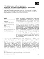
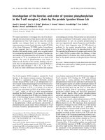
The data show that the classically defined apoptotic
morphology can be achieved either by activation of
caspases, or through the mediation of other families of
proteases [Fig. 1], although the exact cytological features
of cell demise may vary slightly among these various forms
of apoptosis.
Autophagy and autophagic cell death
As part of normal development, cells depend on a strictly
regulated balance of protein synthesis and degradation, as
well as organelle biogenesis and dismantlement. While
proteasome-mediated degradation is responsible for most of
the protein recycling, the turnover of organelles is mainly
attributed to autophagy [89].
Autophagy occurs in many eukaryotic cell types, where
organelles and other cell components are sequestered into
lysosomes and degraded. The lysosome is a cellular
compartment enriched in hydrolases able to cleave proteins,
lipids, nucleic acids and carbohydrates that may lead
to organelle degradation through macroautophagy [90].
Autophagy has been described both as a means to resist
starvation, and as part of cellular remodeling during
differentiation, metamorphosis, aging, cell transformation,
physiological whole-organ changes such as growth of the
uterus during pregnancy and its atrophy after childbirth, as
well as in the removal of anomalous cellular components
that accumulate following toxic insults or during cell death
[91]. In the nervous system, for example, morphological
signs of autophagy are observed in physiological processes,
such as the removal of outer segments of retinal photo-
receptors by the pigment epithelium [92], which is not
associated with cell death.
Notwithstanding, autophagic profiles identified by ultra-
structural features have been associated with cell death in
certain circumstances [22]. Cells in the early stages of
autophagy contain several autophagic vacuoli, and both the
nucleoplasm and the cytoplasm appear slightly darkened,
although nuclear structure still appears normal. Mitochon-
dria and the endoplasmic reticulum are sometimes dilated,
and the Golgi apparatus is often enlarged. The plasma
membrane loses specializations such as microvilli and
junctional complexes, and blebbing can occur. In several
cases, an intense endocytosis is observed, and this probably
Fig. 1. Multiple pathways to apoptosis, both dependent and independent of the activation of caspases. The diagram summarizes the major components
of the pathways reported to underlie cell death in various types of cells and tissues.
1640 C. Assunc¸ a
˜
o Guimara
˜
es and R. Linden (Eur. J. Biochem. 271) Ó FEBS 2004
leads to a reduction in the area of the plasma membrane.
During late stages, both the number and size of vacuoli
increase, and many of them contain myelin figures or are
filled with lipids, which appear as pale gray inclusions in the
cytoplasm [22].
The nucleus of a cell undergoing autophagic cell death
can become pyknotic and identifiable as such by light
microscopy, either in early or in late stages of the
degenerative process. Nevertheless, this nuclear condensa-
tion is neither as common nor as remarkable as that of
apoptosis. The late autophagic cell debris is frequently
removed by heterophagy, but this tends to occur in very late
stages, and seems to be less conspicuous than the clearance
of apoptotic bodies [22].
Autophagic cell death is not an exclusive feature of
multicellular organisms. In the protozoan pathogen Leish-
mania donovani, treatment with antimicrobial peptides
induced cytoplasmic vacuolization and dismantling of the
cellular organization without disruption of the plasma
membrane, with no nuclear fragmentation or DNA
laddering, and independent of caspase-like activity. Instead,
monodansylcadaverine, a biochemical marker of auto-
phagy, specifically labeled the vacuoles induced by anti-
microbial peptides [93].
Endostatin, an inhibitor of angiogenesis, was shown to
induce the formation of autophagic vacuoles in endothelial
cells. Cell death was not prevented by antioxidants or
caspase inhibitors, but was reduced by 3-methyladenine, a
specific inhibitor of autophagy, and serine and cysteine
lysosomal protease inhibitors [94]. Neuregulin (NRG; a
ligand of ErbB), also activates ErbB-2/ErbB-3 heterodimers
and induces cell death of prostate cancer LNCaP cells.
Neuregulin-induced cell death was not inhibited by broad-
spectrum caspases inhibitors, but was blocked by 3-methyl-
adenine [95].
Ionizing radiation induced a dose-dependent suppression
of cell proliferation and autophagic cell changes in several
glioblastoma multiform cell lines [96]. Arsenic trioxide, an
agent that causes remission in patients with acute promyelo-
cytic leukemia and multiple myeloma without severe side-
effects, was shown to inhibit proliferation of glioma cell
lines. The G2/M arrest was accompanied by ultrastructural
features of autophagy, and was inhibited by the autophagy
inhibitor bafilomycin A1, whereas general caspase inhibitors
did not block As
2
O
3
-induced cell death [97]. In neurobla-
stoma cells, dopamine leads to autophagic changes charac-
terized by the presence of numerous cytoplasmic vacuoles
with inclusions, and accompanied by mitochondrial aggre-
gation, activation of the stress-response kinases SAPK/JNK
and p38, and increased a-synuclein expression. Both cell
viability and the increase in a-synuclein expression were
prevented by antioxidants, by the specific inhibitors of
p38 and SAPK/JNK, and by 3-methyladenine [98]. Thus,
various agents can lead to autophagic cell death in tumor
cell lines.
Indeed, Bursch and collaborators [99] had long since
shown that the MCF-7 breast carcinoma cell line, which
does not express caspase-3 [100], undergoes autophagic cell
death upon treatment with tamoxifen. More recent work
showed that in apoptotic cell death induced by tyrphostin
A25 in the human colon cancer cell line HT29/HI1, early
stages of the death process are associated with depolymer-
ization of actin and degradation of intermediate filaments.
In contrast, during tamoxifen-induced autophagic cell death
of MCF-7 cells, intermediate and microfilaments are
redistributed, but largely preserved even beyond the stage
of nuclear collapse [101]. These data support the concept
that autophagic cell death is a separate form of programmed
cell death that is distinctly different from apoptosis.
In keeping with this interpretation, intensive irradiation
led to up to 30% cell death in MCF-7 cells without any signs
of apoptosis. In this case, cell death was accompanied by the
formation of acidic vesicular organelles and lamellar
structures, which was prevented by 3-methyladenine. How-
ever, following low-dose irradiation, the presence of acidic
vesicular organelles correlated with an increased chance of
survival, suggesting that moderate signs of autophagy may
be associated with a defensive reaction of nonlethally
damaged cells [102]. The data are consistent with the view
that nonlethal injury can trigger an autophagic defensive
reaction, whereas harsh treatment of certain cells can lead to
cell death largely dependent on autophagy itself.
The first step of autophagy is the formation of an
autophagosome, which occurs when a portion of the
cytoplasm is engulfed by a double membrane vacuole that
does not contain either acid phosphatases or aryl-sulphatase
activity. The double membrane is derived from ribosome-
free areas of rough endoplasmic reticulum [91]. After a
maturation period that includes the acidification of the
vacuole, hydrolases are inserted into the autophagosome by
fusion with pre-existing lysosomes or elements deriving
from the Golgi complex. This process appears to involve
mannose-6-phosphate receptors located in the autophago-
some membrane, resulting in the formation of a degradative
vacuole limited by a single membrane named an autolyso-
some [103]. Vacuole formation can be regulated by amino
acids and hormones and by stress [104,105].
Similarly to yeast [90], autophagy in mammalian cells is
highly dependent on phosphorylation events. In mammalian
hepatocytes, the phosphorylation of the ribosomal protein
S6 correlates strongly with inhibition of macroautophagy
[106]. The activity of the p70S6-kinase is regulated by the
mTor kinase, and inhibition of S6 phosphorylation caused
by inactivation of mTor with rapamycin induces autophagy
even under nutrient-rich conditions. In yeast, inhibition of
the Tor2 kinase results in activation of protein phosphatase
2A and induction of autophagy [107]. Also, in hepatocytes,
the effect of the phosphatase inhibitor okadaic acid upon
protein phosphatase 2A inhibits the autophagic process
[108]. Furthermore, various classes of phosphatidylinositol-
3-kinase (PI3K) control the autophagic pathway in distinct
ways: class IA PI3K inhibits cytoplasm sequestration and
degradation, while class III stimulates the sequestration of
cytoplasm, implicating the PI3K family as key regulators of
the autophagic pathway [109].
In a recent study, Inbal and coworkers [110] showed that
the expression of death-associated protein kinase (DAPk)
and DAPk-related protein kinase (DRP)-1, members of a
family of Ca
2+
/calmodulin-regulated Ser/Thr death kinases,
triggered two major caspase-independent cytoplasmic
events. These were membrane blebbing, a feature common
to various forms of cell death, and extensive autophagy,
which is typical of autophagic cell death. Furthermore,
either the expression of the dominant negative mutant of
Ó FEBS 2004 Alternative pathways of programmed cell death (Eur. J. Biochem. 271) 1641
DRP-1 or DAPk antisense mRNA reduced autophagy
induced by antiestrogens, amino acid starvation, or admin-
istration of interferon-c. The finding of DRP-1 inside the
autophagic vesicles suggests a direct involvement of this
kinase in the process of autophagy.
Liang and collaborators [111] showed that beclin-1, a
protein that interacts with bcl-2, promotes autophagy in
both an autophagy-defective yeast cell line and in the
MCF-7 cell line, which normally does not express detectable
levels of beclin-1. It was also shown that beclin-1 expression
is frequently low in epithelial breast carcinomas, but is
widely expressed in normal tissue. The authors suggested
that beclin-1 may be a mammalian gene for autophagy, and
that it inhibits tumorigenesis through activation of this cell
death pathway [111]. Kihara and coworkers [112] suggested
that beclin-1 is a component of the PI3K complex, which is
also required for autophagy, and that beclin-1 and PI3K
control autophagy as a complex at the Trans Golgi network
[112]. However, in contrast with the poor autophagic
response of MCF-7 cells to amino acid deprivation [111],
strong autophagic responses were elicited in this cell line
bothbytamoxifen[101]andbyirradiation[102].Thus,
either beclin-1 can be replaced by other autophagy-inducing
proteins, or alternative pathways of autophagy may operate
under various stimuli.
The death of cerebellar Purkinje cells in lurcher animals is
due to a mutation in GluRd2 that results in its constitutive
activation (GluRd2-Lc). Yue and collaborators [113]
showed that GluRd2, nPIST and beclin-1 interact, and that
autophagy can be induced by nPIST and beclin-1 synergy
and by the mutated GluRd2-Lc, but not by the wildtype
GluRd2. Dying lurcher Purkinje cells displayed morpholo-
gical features of autophagy in vivo, providing evidence both
for a direct link between GluRd2-Lc receptor and the
autophagic pathway in these cells [113], and that beclin-1
can exert its autophagic functions through interaction with
multiple proteins in the cells [111–113].
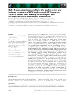
Current evidence therefore indicates that that in at least
some instances, autophagy may lead to a caspase-inde-
pendent program of cell demise that fits the concept of
programmed cell death, subject to complex, multivariate
control [Fig. 2]. The next section will examine its relation-
ship with apoptosis.
Apoptosis vs. autophagy
Notwithstanding the abundant evidence for a role for both
autophagy and apoptosis in various diseases, their interplay
in those pathologies is not yet fully understood. In
Parkinson’s disease, the ultrastructural study of neurons
of the substantia nigra of affected patients showed signs of
autophagy, as well as apoptosis [114]. However, it has been
shown that expression of a-synuclein mutants, a condition
frequently found among certain families with Parkinson’s
disease, induces autophagic cell death with no caspase
activation, due to alterations of the ubiquitin-dependent
protein degradation system [115]. This suggests that there
may be no obligatory interdependence between apoptosis
and autophagy in the pathology of this disease.
In Alzheimer’s disease, the endosomal–lysosomal system
was found to be enlarged and highly activated, showing that
endocytosis and/or autophagy are accelerated in the neu-
rons of affected brains, even at early stages. The early
activation of this system could be related to major
Fig. 2. A summary diagram of the major
components identified in pathways leading to
autophagy. These may lead to either cell death
or recovery from an insult such as aminoacid
deprivation.
1642 C. Assunc¸ a
˜
o Guimara
˜
es and R. Linden (Eur. J. Biochem. 271) Ó FEBS 2004
etiological factors of the disease, such as defective mem-
brane proteins, apolipoprotein E function, or altered
processing of the amyloid precursor protein [116]. In
Huntington’s disease, it was suggested that the accumula-
tion of a mutant isoform of the protein huntingtin in
lysosomal compartments can activate cell death by auto-
phagy [117,118]. Finally, in experimental prion disease,
bovine or mice brains inoculated with scrapie-infected brain
tissue showed signs of autophagy, suggesting that the
accumulation of the protease-resistant prion protein iso-
form can lead to sequestration and autophagy of portions of
the cytoplasm and eventually to neuron loss [119]. Further-
more, giant vacuoli were observed in experimental prion
disease induced in hamsters. The incidence of these vacuoli
correlates with the inoculation pathway, the intensity of the
process and the incubation period, and they are most
numerous following intracerebral inoculation. The emer-
gence of vacuoli was chronologically related to the appear-
ance of fibrils associated with prion disease, suggesting that
this process may be related to disturbed protein turnover
and processing of the prion protein [120]. Despite the
evidence for autophagy, there are several reports of
apoptotic cells detected by in situ nick-end DNA labeling
in human cases of all Alzheimer’s, Huntington’s and
Creutzfeldt–Jakob diseases [121–123].
The mode of cell death in neurodegenerative disorders
remains a matter of controversy [124], and it is possible that
both apoptotic and nonapoptotic cell death coexist in the
brains of affected patients.
This problem is further complicated by claims that the
TdT-mediated biotin-dUDP nick-end labeling (TUNEL)
technique may stain not only apoptotic cells [125,126]. For
example, cell death of keratinocytes induced by 5-fluoruracil
exhibits autophagic ultrastructural features, such as cyto-
plasm vacuolation and chromatin detachment from the
nuclear membrane, alongside apoptotic features such as
TUNEL staining and both a decrease in size and increase
in granularity observed by cytometric analysis [127]. The
authors suggested that in this case the TUNEL-positive cells
were dying by autophagy, and cast doubt on the identifi-
cation of apototic cell death solely using nick-end labeling
techniques, such as in some of the neuropathological
surveys cited above. The data show that the incidence of
mixed cell death features may be more common among
various cell types than predicted by previous studies.
The same stimulus can sometimes lead to the activation
of distinct and independent cell signaling pathways.
Tumor necrosis factor-alpha (TNFa)-induced cell death
in an acute T-lymphoblastic leukaemic cell line developed
with an apoptotic pattern that was preceded by auto-
phagy. The compound 3-methyladenine, which inhibits the
formation of autophagosomes, also inhibited the cytolysis
and DNA fragmentation induced by TNFa.However,
inhibition of the fusion of lysosomes with autophago-
somes by asparagine did not block TNFa-induced apop-
tosis, and amino acid and protein deprivation enhanced
TNFa-induced autophagy, but not apoptosis. These data
suggest that, at least in this case, early stages of autophagy
are required for, but do not necessarily result in, TNF-a
induced apoptosis [128], and also that a cell can switch
between apoptosis and autophagy as the dominant form of
cell death.
However, there are clear examples of interdependence
between apoptosis and autophagy. Following damage to
peripheral nerves in adult rats, oligodendrocytes undergo
ultrastructural features of apoptotic cell death, but mem-
brane-bound cytoplasmic organelles typical of autophagy
were also noticed in the cytoplasm of these cells [129]. In
sympathetic neurons from the superior cervical ganglia,
substantial autophagic activity was activated by pro-apop-
totic factors, and treatment of these cells with 3-methyl-
adenine decreased their rate of apoptosis [130]. This was
also observed in serum-deprived PC12 cells, and was
accompanied by changes in the activity of lysosomal
proteases, particularly cathepsins B and D [131].
Alternative pathways of execution of cell death induced
by blockade of protein synthesis were shown among a
relatively homogenous population of postmitotic undiffer-
entiated cells in the developing retinal tissue. Inhibition of
protein synthesis induced in the immature retina various
post-translational, mitochondria-dependent pathways of
cell death. In one pathway autophagy precedes the sequen-
tial activation of caspases-9 and -3, and DNA fragmenta-
tion, while, in parallel, caspase-6-dependent mechanisms led
to a TUNEL-negative form of cell death. Evidence was also
provided for additional cell death mechanisms dependent
on caspase-9 activity, which may be engaged upon selective
inhibition of execution caspases [132]. These results support
the hypothesis of interdependence of autophagy and
caspase-dependent cell death pathways.
Camougrand and coworkers [133] showed in yeast that
the expression of Bax induces cell death with characteristics
of both apoptosis and autophagy, providing a newly
identified function of Bax in autophagic cell death [133].
Developmental cell death of motoneurons in the hawkmoth
Manduca sexta depends on caspase activation and the loss
of mitochondrial function, showing an accumulation of
autophagic bodies and vacuoles. Motoneurons displayed a
normal nuclear ultrastructure, without chromatin conden-
sation, although they were found to be TUNEL-positive,
which is diagnostic of fragmented DNA. These results
indicate that the steroid-induced, caspase-dependent moto-
neuron cell death exhibits intermingled features of both
autophagy and apoptosis [134]. In the fruitfly Drosophila,
autophagic cell death depends on steroid-regulated genes
encoding transcription regulators, which appear to activate
other genes involved in cell degeneration. The latter include
genes that function in apoptosis, such as caspases, showing
that caspase function is required for autophagic cell death
during Drosophila development (for a review of apoptosis
and autophagy interplay during Drosophila development
see [135,136]).
The autophagy inhibitor 3-methyladenine also increased
the sensitivity of HT-29 colon cancer cells to apoptosis
induced by sulindac sulfide, an inhibitor of cyclo-oxygenase.
Mutants that have a low rate of autophagy were more
sensitive to sulindac sulfide-induced apoptosis than parental
HT-29 cells, and the rate of cytochrome c release was higher
in mutant cells than in HT-29 cells, suggesting that
autophagy could delay apoptosis by sequestering mito-
chondrial death-promoting factors such as cytochrome c
[137]. In this context, autophagy may represent an attempt
of the cell to recover from a noxious stimulus, rather than a
necessary stage of execution of cell death.
Ó FEBS 2004 Alternative pathways of programmed cell death (Eur. J. Biochem. 271) 1643
The available data thus indicate that cells may die by a
caspase-independent autophagic process either with or
without showing signs of apoptosis. In some cases, cells
may choose between the autophagic and the apoptotic
execution pathways. Nevertheless, several experiments show
that the limits between apoptosis and autophagy may be
tenuous, and suggest either considerable overlap or inter-
dependence of both programs of cell demise.
Mitochondria and cell deaths
Mitochondria have a central role both in cellular homeo-
stasis and pathological conditions, as not only do they
serve as the major energy factory of living cells, but they can
also either trigger or amplify the signals that lead to cell
death [46,59–62]. Permeabilization of the mitochondrial
membrane has been associated with cell death by apoptosis
[59,138], by mechanisms that are not yet completely
elucidated (reviewed in [139,140]). Induction of permeability
transition in the inner mitochondrial membrane may be
accompanied by release of cytochrome c, Smac/Diablo,
AIF and endonuclease G, all of which lead to mitochon-
dria-dependent apoptotic forms of cell death [141–146].
Notwithstanding, the collapse of the mitochondrial trans-
membrane potential as a consequence of permeability
transition is not a universal early feature of apoptosis
[147–149].
Although mitochondrial involvement in apoptosis is
broadly documented, permeabilization of mitochondria
seems to be an event also shared by autophagy and
necrosis. Mitochondria from hepatocytes spontaneously
depolarize after nutrient deprivation before their capture
by an acid lysosomal compartment, suggesting that a
permeability transition occurs before the normal autopha-
gic process [150]. The authors proposed that cells respond
to mitochondrial permeability transition (MPT) in a
graded manner. When MPT occurs only in a few
mitochondria, autophagy is activated, leading to lysosomal
degradation of the affected organelles and cessation of the
signals that stimulate autophagy. When a larger number of
mitochondria are permeabilized apoptosis occurs, probably
due to the higher concentration of molecules such as
cytochrome c and AIF in the cytoplasm. And finally, when
virtually all mitochondria in the cell are affected, MPT
promotes necrosis, attributed to the uncoupling of oxida-
tive phosphorylation and accelerated ATP hydrolysis by
mitochondrial ATPase [151]. This interpretation is consis-
tent with the hypothesis that autophagy, apoptosis and
necrosis are part of a continuum of degenerative events
[152,153].
Programmed necrosis
The death and elimination of cells by apoptosis remains
unnoticed by the body’s immune system, while the release of
the intracellular content of necrotic cells into the extracel-
lular space induces an inflammatory response, constituted
by the activation of resident phagocytes and attraction of
leukocytes into the necrotic area [18]. Until recently,
necrosis has often been viewed as an accidental and
uncontrolled cell death process. Nevertheless, there is
growing evidence that necrotic and apoptotic forms of cell
death may share more similarities than originally thought
(reviewed in [16,154–156]).
Necrosis has indeed been found to be a potential
substitute for apoptosis during development. Develop-
ment-related loss of spinal cord and brainstem neurons
was not impaired upon genetic deletion of both caspase-3
and caspase-9, although the morphology of the forebrain
suffered a marked perturbation [157]. Furthermore, the loss
of interdigital cells in the mouse embryo, a prototype of
programmed cell death, still occurs by necrosis either upon
caspase inhibition by drugs, or in mice bearing a mutation in
the apoptosis protease-activating factor 1 (APAF-1) gene.
Such necrotic cell death was also observed in normal
wild-type mice [158].
Several apoptosis-related molecules have been implicated
in necrosis-like forms of cell death. The antiapoptotic
proteins Bcl-2 and Bcl-xL as well as caspase inhibitors
delay necrotic cell death induced by cyanide, rotenone or
antimycin A [159]. Bcl-2 can also protect neural cells from
necrotic cell death induced by the depletion of glutathione
[160]. These results indicate that Bcl-2/Bcl-xL and caspases
modulate not only apoptotic but also some forms of
necrotic cell death.
In a Jurkat-derived cell line (JB-6) that is deficient in
caspase-8, the forced multimerization of Fas-associated
protein with death domain (FADD) induced caspase-
independent cell death. No DNA fragmentation was
observed and dying cells showed neither condensation nor
fragmentation of cells and nuclei, but the cells and nuclei
swelled in a manner similar to that seen in necrosis [161]. In
other studies, caspases-3 and -7 were activated in cell death
of murine L929 fibrosarcoma cells transfected with human
Fas receptor, but peptide inhibitors of caspase-1 and -3
failed to block cell degeneration, and microscopical analysis
showed features of necrosis after a delay of 3 h [162]. These
results suggest the existence of two distinct pathways of cell
death triggered by the engagement of Fas receptors, which
may lead either to classical caspase-dependent apoptosis or
to necrosis.
Chi and coworkers [163] showed that the expression of
oncogenically mutated ras gene in human glioma and
gastric cancer cell lines causes a necrotic-like cell death
characterized by cytoplasmic vacuoles derived from lyso-
somes. Dying cells had relatively well-preserved nuclei that
were negative for TUNEL staining. This oncogenic Ras-
induced cell death occurred in the absence of caspase
activation and was not inhibited by the overexpression of
Bcl-2 [163].
These morphological descriptions of cell death recall the
recently suggested concept of necrosis-like programmed cell
death (PCD), where this term was used to Ô… define PCD in
the absence of chromatin condensation, or at best with
chromatin clustering to specklesÕ [16]. It seems that the cell
death often classified as aborted apoptosis, where PCD
is initiated in the presence of caspase inhibitors, also meets
this requirement because cells end up dying by alternative
routes that are independent of caspase activation (reviewed
in [164]).
Biochemical mechanisms of the execution of necrosis are
beginning to be identified in plants. Recessive genetic
mutations in the Rn locus in soybean lead to progressive
browning of the root, accompanied by incidence of necrotic
1644 C. Assunc¸ a
˜
o Guimara
˜
es and R. Linden (Eur. J. Biochem. 271) Ó FEBS 2004
as well as apoptotic cell death within the same tissue, but in
distinct cells [165]. The appearance of necrotic cells in the
root preceded visible browning and lesion formation at the
macroscopic level, exhibiting a flocculent nucleoplasm,
increased vacuolation, condensed cytoplasm and presence
of swollen malformed organelles [165].
Rcr3 is a plant disease-resistance protein that recognizes
pathogens and activates plant defenses. This protein is a
secreted papain-like cysteine endopeptidase and is specific-
ally required for Cf-2 function, e.g. resistance to pathogens
in tomato, a hypersensitive response that results in cell
death. Genetic analysis showed that Rcr3 is allelic to the Ne
(Necrosis) gene, which suppresses the Cf-2-dependent auto-
necrosis induced by plant pathogens [166]. These data may
represent, at least in plants, the first examples of genes
related to the induction of necrotic-like PCD.
It is not known whether any of the necrosis-related genes
of plants have homologs in animal cells. However, compo-
nents of a necrotic program are beginning to emerge.
Integration of the BH3 protein BNIP3 to mitochondria
triggers a necrotic-like form of PCD [167]. The necrotic-like
cell death triggered by engagement of the Fas/FADD signal
transduction system in mammalian cells was reported to
depend on the kinase RIP as an effector molecule [168].
These data support the hypothesis that, besides uncontrol-
lable necrosis that may occur following either massive
mechanical aggression or harsh chemical treatment of
tissues, a program of cell demise with morphological
characteristics of necrosis may be found with components
largely distinct from either apoptosis or autophagy.
Conclusion and perspectives
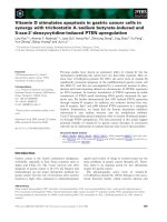
Increasing evidence obtained in many model systems both
in vitro and in vivo supports the hypothesis that a variety of
cell death programs may be triggered in distinct circum-
stances [Fig. 3]. Contrary to the widely held view that
caspase-mediated apoptosis represents the standard PCD,
recent studies indicate that an apoptotic morphology can be
produced without the involvement of caspases, that auto-
phagic execution pathways of cell death may be engaged
without either the involvement of caspases or morphological
signs of apoptosis, and that even the necrotic morphology of
cell death may be consistently produced in some cases,
including the natural history of at least some plants.
Particularly in the case of autophagic cell death, but to a
lesser extent also in the case of controlled necrosis,
components have been identified at the molecular level that
justify the assumption of an intracellular program mediating
either form of cell death under upstream command. Distinct
from the idea that a requirement for protein synthesis
defines genetically programmed cell death, the ÔprogramsÕ
appear in general to be ready for action. Together with
abundant evidence that the apoptosis execution pathways
are essentially independent of protein synthesis, both
autophagy and programmed necrosis have so far been
traced to post-translational signal transducers, such as
protein kinases and phosphatases. In all of these cases, it
would appear that only the simplest definition of PCD,
much like the original concept of Ôa sequence of events …
that lead to death of the cellÕ [20] would resist close scrutiny.
There is much to be said in favor of undecorated concepts,
which more often than not represent the essential features
upon which myriad variations can be identified, quite
typical of biological systems.
Despite the long-standing evidence for alternative cell
death strategies, it is still often assumed that caspase-
mediated apoptosis is either the major, or the most frequent,
form of PCD. Although statistics may eventually prove this
point, this is by no means warranted. Indeed, the dominance
of apoptosis among the published examples of PCD may be
due to the fact that apoptosis is the only form of cell death
that has long been categorized not only on the basis of its
defining ultrastructural changes, but also on the basis of
Fig. 3. A summary diagram of the various
forms of degeneration that may follow cell stress
or damage. Distinct forms of programmed cell
death are indicated in italics within the boldly
outlined boxes. Cell demise is the usual out-
come of either the somewhat variable forms of
apoptosis mediated by multiple, alternative
pathways, or of programmed necrosis.
Autophagy may lead to cell death either
directly or through apoptosis, as well as
mediate cellular recovery.
Ó FEBS 2004 Alternative pathways of programmed cell death (Eur. J. Biochem. 271) 1645
cytological features recognizable by either conventional
light microscopy or by relatively simple histochemical or
immunohistochemical techniques. Reviewers frequently
demand TUNEL or caspase assays as tests of the nature
of programmed cell death, and are usually satisfied with a
simple positive, unqualified response, even though this
positive identification is often achieved for only some of the
dying cells in either naturally occurring or experimentally
induced cell death.
More thorough examination of the forms of cell death in
each circumstance is needed to assess, for example, the view
that morphologically distinct pathways of cell death may be
part of a continuum of degenerative events. Only recently,
techniques such as the monodansylcadaverine assay have
become available to test for autophagic cell death in cell
populations [169] and no simple assay is as yet available for
the positive identification of necrosis. Thus, it remains to be
evaluated how frequently the alternative cell death forms
also occur where apoptosis has been positively identified,
whether it is caspase-mediated or not.
A further and pressing issue is how the autophagic and
apoptotic pathways interact. Evidence is available both for
independent as well as for tandem programs for each of
these forms of cell dismantlement. The issue is complicated
by the fact that the presence of autophagic vacuoles per se is
not enough evidence that the cell is committed to degener-
ation, because autophagy may be part of a cell defense
mechanism.
Admittedly, the rather unpopular view that apoptosis,
or at least caspase-mediated apoptosis, may well have
been grossly overrated has a significant chance to
underlie a minority of cases, after all. But it has already
been worthwhile in the form of increasing awareness
concerning alternative cell death styles. These must be
critically evaluated, in lieu of the prevailing assumption
that the overwhelming and excellent science behind
apoptosis may have definitively set the scene for all the
hot stories related to programmed cell death. Alternative
cell death programs most probably imply novel thera-
peutic targets, and have important consequences for
attempts to treat diseases associated with disregulated
programmed cell death.
Acknowledgements
Research in the authors’ lab was supported by CNPq, FAPERJ,
PRONEX-MCT, and a fellowship from the John Simon Guggenheim
Foundation to R.L. We are indebted to Richard Lockshin for critical
reading of this manuscript.
References
1. Bijl, M., Limburg, P.C. & Kallenberg, C.G. (2001) New insights
into the pathogenesis of systemic lupus erythematosus (SLE): the
role of apoptosis. Neth. J. Med. 59, 66–75.
2. Cecconi, F. & Gruss, P. (2001) Apaf1 in developmental apoptosis
and cancer: how many ways to die? Cell. Mol. Life Sci. 58,
1688–1697.
3. Clarke, G., Lumsden, C.J. & McInnes, R.R. (2001) Inherited
neurodegenerative diseases: the one-hit model of neurodegen-
eration. Hum. Mol. Genet. 10, 2269–2275.
4. Debatin, K.M. (2001) Cell death in T- and B-cell development.
Ann. Hematol. 80 (Suppl. 3), B29–B31.
5. Domen, J. (2000) The role of apoptosis in regulating
hematopoiesis and hematopoietic stem cells. Immunol. Res. 22,
83–94.
6. Dumont, R.J., Okonkwo, D.O., Verma, S., Hurlbert, R.J.,
Boulos, P.T., Ellegala, D.B. & Dumont, A.S. (2001) Acute spinal
cord injury, part I: pathophysiologic mechanisms. Clin. Neuro-
pharmacol. 24, 254–264.
7. Gerber, H.P. & Ferrara, N. (2000) Angiogenesis and bone
growth. Trends Cardiovasc. Med. 10, 223–228.
8. Joaquin, A.M. & Gollapudi, S. (2001) Functional decline in
aging and disease: a role for apoptosis. J.Am.Geriatr.Soc.49,
1234–1240.
9. Mayr, M. & Xu, Q. (2001) Smooth muscle cell apoptosis in
arteriosclerosis. Exp Gerontol. 36, 969–987.
10. Monk, C.S., Webb, S.J. & Nelson, C.A. (2001) Prenatal neuro-
biological development: molecular mechanisms and anatomical
change. Dev. Neuropsychol. 19, 211–236.
11. Pru, J.K. & Tilly, J.L. (2001) Programmed cell death in the ovary:
insights and future prospects using genetic technologies. Mol.
Endocrinol. 15, 845–853.
12. Bortner, C.D. & Cidlowski, J.A. (2002) Cellular mechanisms for
the repression of apoptosis. Annu. Rev. Pharmacol. Toxicol. 42,
259–281.
13. Buendia, B., Courvalin, J.C. & Collas, P. (2001) Dynamics of the
nuclear envelope at mitosis and during apoptosis. Cell. Mol. Life
Sci. 58, 1781–1789.
14. Bursch, W. (2001) The autophagosomal-lysosomal compartment
in programmed cell death. Cell Death Differ. 8, 569–581.
15. Henson, P.M., Bratton, D.L. & Fadok, V.A. (2001) Apoptotic
cell removal. Curr. Biol. 11, R795–R805.
16. Leist, M. & Jaattela, M. (2001) Four deaths and a funeral: from
caspases to alternative mechanisms. Nat.Rev.Mol.CellBiol.2,
589–598.
17. Zo
¨
rnig, M., Hueber, A O., Baum, W. & Evan, G. (2001)
Apoptosis regulators and their role in tumorigenesis. Biochim.
Biophys. Acta 1551, F1–F37.
18. Kerr, J.F.R., Wyllie, A.H. & Currie, A.R. (1972) Apoptosis: a
basic biological phenomenon with wide-ranging implications in
tissue kinetics. Br. J. Cancer 26, 239–257.
19. Wyllie, A.H., Kerr, J.F.R. & Currie, A.R. (1981) Cell death: the
significance of apoptosis. International, Version. Cytol. 68,
251–305.
20. Lockshin, R.A. & Zakeri, Z. (2001) Programmed cell death
and apoptosis: origins of the theory. Nat.Rev.Mol.CellBiol.2,
545–550.
21. Lockshin, R.A. & Zakeri, Z. (1991) Programmed cell death and
apoptosis. In: Tomei, L.D. & Cope, F.O., eds. Apoptosis: the
Molecular Basis of Cell Death. New York, Cold Springer.Harbor
Laboratory Press, pp. 47–60.
22. Clarke, P.G.H. (1990) Developmental cell death: morphological
diversity and multiple mechanisms. Anat. Embryol. 181, 195–213.
23. Sloviter, R.S. (2002) Apoptosis: a guide for the perplexed. Trends
Pharmacol. Sci. 23, 19–24.
24. Lockshin, R.A. & Williams, C.M. (1964) Programmed cell death.
II. Endocrine potentiation of the breakdown of intersegmental
muscles of silkmoths. J. Insect. Physiol. 10, 643–649.
25. Lockshin, R.A. & Williams, C.M. (1965a) Programmed cell
death. I. Cytology of degeneration in the intersegmental muscles
of the Pernyl silkmoth. J. Insect. Physiol. 11, 123–133.
26. Lockshin, R.A. & Williams, C.M. (1965b) Programmed cell
death. III. Neural control of the breakdown of intersegmental
muscles of silkmoths. J. Insect. Physiol. 11, 601–610.
27. Lockshin, R.A. & Williams, C.M. (1965c) Programmed cell
death. IV. The influence of drugs of the breakdown of
the intersegmental muscles of silkmoths. J. Insect. Physiol. 11,
803–809.
1646 C. Assunc¸ a
˜
o Guimara
˜
es and R. Linden (Eur. J. Biochem. 271) Ó FEBS 2004
28. Lockshin, R.A. & Williams, C.M. (1965d) Programmed cell
death. V. Cytolytic enzymes in relation to the breakdown of
the intersegmental muscles of silkmoths. J. Insect. Physiol. 11,
833–844.
29. Saunders, J.W. Jr (1996) Death in embryonic systems. Science
154, 604–612.
30. Martin, S.J., Green, D.R. & Cotter, T.G. (1994) Dicing with
death: dissecting the components of the apoptosis machinery.
Trends Biochem. Sci. 19, 26–30.
31. Tata, J.R. (1966) Requirement for RNA and protein synthesis
for induced regression of the tadpole tail in organ culture. Dev.
Biol. 13, 77–94.
32. Lockshin, R.A. (1969) Programmed cell death. Activation of lysis
by a mechanism involving the synthesis of protein. J. Insect.
Physiol. 15, 1505–1516.
33. Martin, S.J., Lennon, S.V., Bonham, A.M. & Cotter, T.G. (1990)
Induction of apoptosis (programmed cell death) in human leu-
kemic HL-60 cells by inhibition of RNA or protein synthesis.
J. Immunol. 145, 1859–1867.
34. Martin, S.J. (1993) Protein or RNA synthesis inhibition induces
apoptosis of mature human CD4+ T cell blasts. Immunol. Lett.
35, 125–134.
35. Rehen, S.K., Varella, M.H., Freitas, F.G., Moraes, M.O.
& Linden, R. (1996) Contrasting effects of protein synthesis
inhibition and of cyclic AMP on apoptosis in the developing
retina. Development 122, 1439–1448.
36. Hengartner, M.O. (2000) The biochemistry of apoptosis. Nature
407, 770–776.
37. Chang, S.H., Cvetanovic, M., Harvey, K.J., Komoriya, A.,
Packard, B.Z. & Ucker, D.S. (2002) The effector phase of phy-
siological cell death relies exclusively on the posttranslational
activation of resident components. Exp. Cell Res. 277, 15–30.
38. Green, D.R. (2000) Apoptotic pathways: paper wraps stone
blunts scissors. Cell 102, 1–4.
39. Cryns, V. & Yuan, J. (1998) Proteases to die for. Genes Dev. 12,
1551–1570.
40. Yuan, J., Shaham, S., Ledoux, S., Ellis, H.M. & Horvitz, H.R.
(1993) The C. elegans death gene ced-3 encodes a protein similar
to mammalian interleukin-1 beta-converting enzyme. Cell 75,
641–652.
41. Zhivotovsky, B., Burgress, D.H., Vanags, D.M. & Orrenius, S.
(1997) Involvement of cellular proteolytic machinery in apopto-
sis. Biochem. Biophys. Res. Commun. 230, 481–488.
42. Ravagnan, L., Roumier, T. & Kroemer, G. (2002) Mitochondria,
the killer organelles and their weapons. J. Cell Physiol. 192,
131–137.
43. Wajant, H. (2002) The Fas signaling pathway: more than a
paradigm. Science 296, 1635–1636.
44. Martin, S.J. (2002) Destabilizing influences in apoptosis: sowing
the seeds of IAP destruction.Cell 109, 793–796.
45. Butt, A.J., Harvey, N.L., Parasivam, G. & Kumar, S. (1998)
Dimerization and autoprocessing of the Nedd2 (caspase-2) pre-
cursor requires both the prodomain and the carboxyl-terminal
regions. J. Biol. Chem. 273, 6763–6768.
46. Li, P., Nijhawan, D., Budihardjo, I., Srinivasula, S.M., Ahmad,
M.,Alnemri,E.S.&Wang,X.(1997)CytochromecanddATP-
dependent formation of Apaf-1/caspase-9 complex initiates an
apoptotic protease cascade. Cell 91, 479–489.
47. Martin, D.A., Siegel, R.M., Zheng, L. & Lenardo, M.J.
(1998) Membrane oligomerization and cleavage activates the
caspase-8 (FLICE/MACH-1) death signal. J. Biol. Chem. 273,
4345–4349.
48. Muzio, M., Chinnaiyan, A.M., Kischkel, F.C., O’Rourke, K.,
Shevchenko, A., Ni, J., Scaffidi, C., Bretz, J.D., Zhang, M.,
Gentz, R., Mann, M., Krammer, P.H., Peter, M.E. & Dixit,
V.M. (1996) FLICE, a novel FADD-homologous ICE/CED-3-
like-protease, is recruited to teh CD95 (Fas/ APO-1) death-
inducing signaling complex. Cell 85, 817–827.
49. Srinivasula, S.M., Ahmad, M., Fernandes-Alnemri, T. &
Alnemri, E.S. (1998) Autoactivation of procaspase-9 by Apaf-1-
mediated oligomerization. Mol. Cell. 1, 949–957.
50. Yang, X., Chang, H.Y. & Baltimore, D. (1998) Autoproteolytic
activation of procaspases by oligomerization. Mol. Cell 1,
319–325.
51. Liu, X., Li, P., Widlak, P., Zou, H., Luo, X., Garrard, W.T. &
Wang, X. (1998) The 40-kDa subunit of DNA fragmentation
factor induces DNA fragmentation and chromatin condensation
during apoptosis. Proc. Natl. Acad. Sci. USA 95, 8461–8466.
52. Sakahira, H., Enari, M. & Nagata, S. (1998) Cleavage of CAD
inhibitor in CAD activation and DNA degradation during
apoptosis. Nature 391, 96–99.
53. Zhang,J.,Liu,X.,Scherer,D.C.,vanKaer,L.,Wang,X.&Xu,
M. (1998) Resistance to DNA fragmentation and chromatin
condensation in mice lacking the DNA fragmentation factor 45.
Proc. Natl. Acad. Sci. USA 95, 12480–12485.
54. Green, D.R. (1998) Apoptotic pathways: the roads to ruin. Cell
94, 695–698.
55. Stroh, C. & Schulze-Osthoff, K. (1998) Death by a thousand cuts:
an ever increasing list of caspase substrates. Cell Death Differ. 5,
997–1000.
56. Wolf, B.B. & Green, D.R. (1999) Suicidal tendencies: apoptotic
cell death by caspase family proteinases. J. Biol. Chem. 274,
20049–20052.
57. Medema, J.P., Scaffidi, C., Kischkel, F.C., Shevchenko, A.,
Mann, M., Krammer, P.H. & Peter, M.E. (1997) FLICE is
activated by association with the CD95 death-inducing signaling
complex (DISC). EMBO J. 16, 2794–2804.
58. Nakagawa, T., Zhu, H., Morishima, N., Li, E., Xu, J., Yankner,
B.A. & Yuan, J. (2000) Caspase-12 mediates endoplasmic-
reticulum-specific apoptosis and cytotoxicity by amyloid-beta.
Nature 403, 98–103.
59. Green, D.R. & Reed, J.C. (1998) Mitochondria and apoptosis.
Science 281, 1309–1312.
60. Grutter, M.G. (2000) Caspases: key players in programmed cell
death. Curr. Opinion Struct. Biol. 10, 649–655.
61. Li, H., Zhu, H., Xu, C.J. & Yuan, J. (1998) Cleavage of BID by
caspase 8 mediates the mitochondrial damage in the Fas pathway
of apoptosis. Cell 94, 491–501.
62. Luo, X., Budihardjo, I., Zou, H., Slaughter, C. & Wang, X.
(1998) Bid, a Bcl2 interacting protein, mediates cytochrome c
release from mitochondria in response to activation of cell sur-
face death receptors. Cell 94, 481–490.
63. Stennicke, H.R. & Salvesen, G.S. (2002) Caspases – controlling
intracellular signals by protease zymogen activation. Biochim.
Biophys. Acta. 1477, 299–306.
64. Kuida,K.,Zheng,T.S.,Na,S.,Kuan,C.,Yang,D.,Karasuy-
ama, H., Rakic, P. & Flavell, R.A. (1996) Decreased apoptosis
in the brain and premature lethality in CPP32-deficient mice.
Nature 384, 368–372.
65. Miyashita, T., Nagao, K., Krajewski, S., Salvesen, G.S., Reed,
J.C.,Inoue,T.&Yamada,M.(1998)Investigationofgluco-
corticoid-induced apoptotic pathway: processing of caspase-6 but
not caspase-3. Cell Death Differ. 5, 1034–1041.
66. Troy, C.M., Rabaccchi, A.S., Hohl, J.B., Angelastro, J.M.,
Greene, L.A. & Shelanski, M.L. (2001) Death in the balance:
alternative participation of the caspase-2 and caspase-9 pathways
in neuronal death induced by nerve growth factor deprivation.
J. Neurosci. 21, 5077–5016.
67. Salvesen, G.S. & Duckett, C.S. (2002) IAP proteins: blocking the
road to death’s door. Nat.Rev.Mol.CellBiol.3, 401–410.
68. Assefa, Z., Vantieghen, A., Garmyn, M., Declercq, W.,
Vandernabeele, P., Vandenheede, J.R., Bouillon, R., Merlevede,
Ó FEBS 2004 Alternative pathways of programmed cell death (Eur. J. Biochem. 271) 1647
W. & Agostinis, P. (2000) p38 mitogen-activated protein kinase
regulates a novel, caspase-independent pathway for the
mitochondrial cytochrome c release in ultraviolet b radiation-
induced apoptosis. J. Biol. Chem. 275, 21416–21421.
69. Carmody, R.J. & Cotter, T.G. (2000) Oxidative stress induces
caspase-independent retinal apoptosis in vitro. Cell Death Differ.
7, 282–291.
70. Lorenzo, H.K., Susin, S.A., Penninger, J. & Kroemer, G. (1999)
Apoptosis-inducing factor (AIF): a phylogenetically old, caspase-
independent effector of cell death. Cell Death Diff. 6, 516–524.
71. Mateo, V., Lagneaux, L., Bron, D., Biron, G., Armant, M.,
Delespesse, G. & Sarfati, M. (1999) CD47 ligation induces cas-
pase-independent cell death in chronic lymphocytic leukemia.
Nat. Med. 5, 1277–1284.
72. Mathiasen, I.S., Lademann, U. & Jaattela, M. (1999) Apoptosis
induced by vitamin D compounds in breast cancer cells is
inhibited by Bcl-2 but does not involve known caspases or p53.
Cancer Res. 59, 4848–4856.
73. Joza, N., Susin, S.A., Daugas, E., Stanford, W.L., Cho, S.K., Li,
C.Y.J., Sasaki, T., Elia, A.J., Cheng, H Y.M., Ravagnan, L.,
Ferri, K.F., Zamzami, N., Wakeham, A., Hakem, R., Yoshida,
H.,Kong,Y Y.,Mak,T.W.,Zu´ n
˜
iga-Pflu
¨
cker, J.C., Kroemer,
G. & Penninger, J.M. (2001) Essential role of the mitochondrial
apoptosis-inducing factor in programmed cell death. Nature 410,
549–554.
74. Arnoult, D., Gaume, B., Karbowski, M., Sharpe, J.C., Cecconi,
F. & Youle, R.J. (2003) Mitochondrial release of AIF and
EndoG requires caspase activation downstream of Bax/Bak-
mediated permeabilization. EMBO J. 22 (17), 4385–4399.
75. Jurgensmeier, J.M., Xie, Z., Deveraux, Q., Ellerby, L., Bredesen,
D. & Reed, J.C. (1998) Bax directly induces release of cyto-
chrome c from isolated mitochondria. Proc. Natl. Acad. Sci. 95,
4997–5002.
76. Squier, M.K., Miller, A.C., Malkinson, A.M. & Cohen, J.J.
(1994) Calpain activation in apoptosis. J. Cell Physiol. 159,
229–237.
77. Hirsch, T., Dallaporta, B., Zamzami, N., Susin, S.A., Ravagnan,
L., Marzo, I., Brenner, C. & Kroemer, G. (1998) Proteasome
activation occurs at an early, premitochondrial step of thymocyte
apoptosis. J. Immunol. 161, 35–40.
78. Hughes, F.M. Jr, Evans-Storms, R.B. & Cidlowski, J.A.
(1998) Evidence that non-caspase proteases are required for
chromatin degradation during apoptosis. Cell Death Differ. 5,
1017–1027.
79. Mann, C.L., Hughes, F.M. Jr & Cidlowski, J.A. (2000) Deli-
neation of the signaling pathways involved in glucocorticoid-
induced and spontaneous apoptosis of rat thymocytes.
Endocrinology 141, 528–538.
80. Torriglia, A., Negri, C., Chaudun, E., Prosperi, E., Courtois, Y.,
Counis, M.F. & Scovassi, A.I. (1999) Differential involvement of
Dnases in Hela cell apoptosis induced by etoposide and long term
culture. Cell Death Diff. 6, 234–244.
81. Counis, M.F., Chaudun, E., Arruti, C., Oliver, L., Sanwal, L.,
Courtois, Y. & Torriglia, A. (1998) Advances in nuclear
degradation during lens cell differentiation. Cell Death Diff. 5,
251–261.
82. Torriglia, A., Chaudun, E., Chany-Fournier, F., Jeanny, J.C.,
Courtois, Y. & Counis, M F. (2001) Involvement of Dnase II in
nuclear degeneration during chick retina development. Exp. Eye
Res. 72, 443–453.
83. Potempa, J., Dubin, A., Watorek, W. & Travis, J. (1988) An
elastase inhibitor from equine leucocyte cytosol belongs to the
serpin superfamily. Further characterization and aminoacid
sequence of the reactive center. J. Biol. Chem. 263, 7364–7369.
84. Torriglia,A.,Perani,P.,Brossas,J.Y.,Chaudun,E.,Tre
´
ton, J.,
Courtois, Y. & Counis, M.F. (1998) L-Dnase II, a molecule that
links proteases and endonucleases in apoptosis, derives from the
ubiquitous serpin Leukocyte Elastase Inhibitor. Mol. Cel. Biol.
18, 3612–3619.
85. Hegde, R., Srinivasula, S.M., Zhang, Z., Wassell, R., Mukattash,
R.,Cilenti,L.,DuBois,G.,Lazebnik,Y.,Zervos,A.S.,Fern-
andes-Alnemri, T. & Alnemri, E.S. (2002) Identification of
Omi/HtrA2 as a mitochondrial apoptotic serine protease that
disrupts inhibitor of apoptosis protein–caspase interaction.
J. Biol. Chem. 277, 432–438.
86. Martins, L.M., Iaccarino, I., Tenev, T., Gschmeissner, S., Totty,
N.F.,Lemoine,N.R.,Savopoulos,J.,Gray,C.W.,Creasy,C.L.,
Dingwall, C. & Downward, J. (2002) The serine protease Omi/
HtrA2 regulates apoptosis by binding XIAP through a reaper-
like motif. J. Biol. Chem. 277, 439–444.
87. Martins, L.M. (2002) The serine protease Omi/HtrA2: a second
mammalian protein with a Reaper-like function. Cell Death
Differ. 9, 699–701.
88. Van Loo, G., van Gurp, M., Depuydt, B., Srinivasula, S.M.,
Rodriguez, I., Alnemri, E.S., Gevaert, K., Vandekerckhove, J.,
Declercq, W. & Vandenabeele, P. (2002) The serine protease
Omi/HtrA2 is released from mitochondria during apoptosis. Omi
interacts with caspase-inhibitor XIAP and induces enhanced
caspase activity. Cell Death Differ. 9, 20–26.
89. Seglen, P.O. & Bohley, P. (1992) Autophagy and other vacuolar
protein degradation mechanisms. Experientia 48, 158–172.
90. Klionsky, D.J. & Emr, S.D. (2000) Autophagy as a regulated
pathway of cellular degradation. Science 290, 1717–1721.
91. Dunn, W.A. Jr (1990a) Studies on the mechanisms of autophagy:
formation of the autophagic vacuole. J. Cell Biol. 110, 1923–
1933.
92. Reme, C.E., Wolfrum, U., Imsand, C., Hafezi, F. & Williams,
T.P. (1999) Photoreceptor autophagy: effects of light history on
number and opsin content of degradative vacuoles. Invest. Oph-
thalmol. Vis. Sci. 40, 2398–2404.
93. Bera, A., Singh, S., Nagaraj, R. & Vaidya, T. (2003) Induction of
autophagic cell death in Leishmania donovani by antimicrobial
peptides. Mol. Biochem. Parasitol. 27, 23–35.
94. Chau, Y.P., Lin, S.Y., Chen, J.H. & Tai, M.H. (2003) Endostatin
induces autophagic cell death in EAhy926 human endothelial
cells. Histol. Histopathol. 18, 715–726.
95. Tal-Or, P., Di-Segni, A., Lupowitz, Z. & Pinkas-Kramarski, R.
(2003) Neuregulin promotes autophagic cell death of prostate
cancer cells. Prostate 55, 147–157.
96. Yao,K.C.,Komata,T.,Kondo,Y.,Kanzawa,T.,Kondo,S.
& Germano, I.M. (2003) Molecular response of human glio-
blastoma multiforme cells to ionizing radiation: cell cycle arrest,
modulation of the expression of cyclin-dependent kinase
inhibitors, and autophagy. J. Neurosurg. 98, 378–384.
97. Kanzawa, T., Kondo, Y., Ito, H., Kondo, S. & Germano, I.
(2003) Induction of autophagic cell death in malignant glioma
cells by arsenic trioxide. Cancer Res. 63, 2103–2108.
98. Gomez-Santos, C., Ferrer, I., Santidrian, A.F., Barrachina, M.,
Gil, J. & Ambrosio, S. (2003) Dopamine induces autophagic cell
death and alpha-synuclein increase in human neuroblastoma
SH-SY5Y cells. J. Neurosci. Res. 73, 341–350.
99. Bursch, W., Ellinger, A., Kienzl, H., Torok, L., Pandey, S.,
Sikorska, M., Walker, R. & Hermann, R.S. (1996) Active cell
death induced by the anti-estrogens tamoxifen and ICI 164 384 in
human mammary carcinoma cells (MCF-7) in culture: the role of
autophagy. Carcinogenesis 17, 1595–1607.
100.Janicke,R.U.,Sprengart,M.L.,Wati,M.R.&Porter,A.G.
(1998) Caspase-3 is required for DNA fragmentation and mor-
phological changes associated with apoptosis. J. Biol. Chem. 273,
9357–9360.
101. Bursch, W., Hochegger, K., Torok, L., Marian, B., Ellinger, A. &
Hermann, R.S. (2000) Autophagic and apoptotic types of
1648 C. Assunc¸ a
˜
o Guimara
˜
es and R. Linden (Eur. J. Biochem. 271) Ó FEBS 2004
programmed cell death exhibit different fates of cytoskeletal
filaments. J. Cell Sci. 113, 1189–1198.
102. Paglin, S., Hollister, T., Delohery, T., Hackett, N., McMahill,
M., Sphicas, E., Domingo, D. & Yahalom, J. (2001) A novel
response of cancer cells to radiation involves autophagy and
formation of acidic vesicles. Cancer Res. 61, 439–444.
103. Dunn, W.A. Jr (1990b) Studies on the mechanisms of autophagy:
maturation of the autophagic vacuole. J. Cell Biol. 110, 1935–
1945.
104. Lardeux, B.R. & Mortimore, G.E. (1987) Amino acid and hor-
monal control of macromolecular turnover in perfused rat liver.
Evidence for selective autophagy. J. Biol. Chem. 262, 14514–
14519.
105. Reunanen, H., Punnonen, E.L. & Hirsimaki, P. (1985) Studies in
vinblastine-induced autophagocytosis in mouse liver. His-
tochemistry 83, 513–517.
106. Blommaart, E.F., Luiken, J.J., Blommaart, P.J., van Woerkom,
G.M. & Meijer, A.J. (1995) Phosphorylation of ribosomal pro-
tein S6 is inhibitory for autophagy in isolated rat hepatocytes.
J. Biol. Chem. 270, 2320–2326.
107. Beck, T. & Hall, M.N. (1999) The TOR signalling pathway
controls nuclear localization of nutrient-regulated transcription
factors. Nature 402, 689–692.
108. Holen, I., Gordon, P.B. & Seglen, P.O. (1992) Protein kinase-
dependent effects of okadaic acid on hepatocytic autophagy and
cytoskeletal integrity. Biochem. J. 284, 633–636.
109. Petiot, A., Ogier-Denis, E., Blommaart, E.E.F.C., Meijer, A.J. &
Codogno, P. (2000) Distinct classes of phosphatidylinositol
3¢-kinase are involved in signaling pathways thet control
macroautophagy in HT-29 cells. J. Biol. Chem. 275, 992–998.
110. Inbal, B., Bialik, S., Sabanay, I., Shani, G. & Kimchi, A. (2002)
DAP kinase and DRP-1 mediate membrane blebbing and the
formation of autophagic vesicles during programmed cell death.
J. Cell. Biol. 157, 455–468.
111. Liang, X.H., Jackson, S., Seaman, M., Brown, K., Kempkes, B.,
Hibshoosh, H. & Levine, B. (1999) Induction of autophagy and
inhibition of tumorigenesis by beclin 1. Nature 402, 672–676.
112. Kihara, A., Kabeya, Y., Ohsumi, Y. & Yoshimori, T. (2001)
Beclin-phosphatidylinositol 3-kinase complex functions at the
trans-Golgi network. EMBO Report 2, 330–335.
113. Yue, Z., Horton, A., Bravin, M., DeJager, P.L., Selimi, F. &
Heintz, N. (2002) A novel protein complex linking the delta 2
glutamate receptor and autophagy: implications for neuro-
degeneration in lurcher mice. Neuron 35, 921–933.
114. Anglade, P., Vyas, S., Javoy-Agid, F., Herrero, M.T., Michel,
P.P., Marquez, J., Mouatt-Prigent, A., Ruberg, M., Hirsch,
E.C. & Agid, Y. (1997) Apoptosis and autophagy in nigral
neurons of patients with Parkinson’s disease. Histol. Histopathol.
12, 25–31.
115. Stefanis, L., Larsen, K.E., Rideout, H.J., Sulzer, D. & Greene,
L.A. (2001) Expression of A53T mutant but not wild-type alpha-
synuclein in PC12 cells induces alterations of the ubiquitin-
dependent degradation system, loss of dopamine release, and
autophagic cell death. J. Neurosci. 21, 9549–9560.
116. Cataldo,A.M.,Hamilton,D.J.,Barnett,J.L.,Paskevich,P.A.&
Nixon, R.A. (1996) Properties of the endosomal-lysosomal sys-
tem in the human central nervous system: disturbances mark
most neurons in populations at risk to degenerate in Alzheimer’s
disease. J. Neurosci. 16, 186–199.
117. Kegel, K.B., Kim, M., Sapp, E., McIntyre, C., Castan
˜
o, J.G.,
Aronin, N. & DiFiglia, M. (2000) Huntingtin expression stimu-
lates endosomal-lysosomal activity, endosome tubulation and
autophagy. J. Neurosci. 20, 7268–7278.
118. Petersen, A., Larsen, K.E., Behr, G.G., Romero, N., Przedbor-
ski, S., Brundin, P. & Sulzer, D. (2001) Expanded CAG repeats in
exon 1 of the Huntington’s disease gene stimulate dopamine-
mediated striatal neuron autophagy and degeneration. Hum.
Mol. Genet. 10, 1243–1254.
119. Jeffrey, M., Scott, J.R., Williams, A. & Fraser, H. (1992) Ultra-
structural features of spongiform encephalopathy transmitted to
mice from three species of bovidae. Acta Neuropathol. (Berl) 84,
559–569.
120. Boellaard, J.W., Kao, M., Schlote, W. & Diringer, H. (1991)
Neuronal autophagy in experimental scrapie. Acta Neuropathol.
(Berl) 82, 225–228.
121. Dragunow, M., Faull, R.L., Lawlor, P., Beilharz, E.J., Singleton,
K., Walker, E.B. & Mee, E. (1995) In situ evidence for DNA
fragmentation in Huntington’s disease striatum and Alzheimer’s
disease temporal lobes. Neuroreport 6, 1053–1057.
122. Thomas, L.B., Gates, D.J., Richfield, E.K., O’Brien, T.F.,
Schweitzer, J.B. & Steindler, D.A. (1995) DNA end labeling
(TUNEL) in Huntington’s disease and other neuropathological
conditions. Exp Neurol. 133, 265–272.
123. Gray, F., Chretien, F., Adle-Biassette, H., Dorandeu, A., Ereau,
T., Delisle, M.B., Kopp, N., Ironside, J.W. & Vital, C. (1999)
Neuronal apoptosis in Creutzfeldt-Jakob disease. J. Neuropathol.
Exp. Neurol. 58, 321–328.
124. Jellinger, K.A. (2001) Cell death mechanisms in neurodegenera-
tion. J. Cell. Mol. Med. 5, 1–17.
125. Charriaut-Marlangue, C. & Ben-Ari, Y. (1995) A cautionary
note on the use of the TUNEL stain to determine apoptosis.
Neuroreport 7, 61–64.
126. Grasl-Kraupp, B., Ruttkay-Nedecky, B., Koudelka, H., Buk-
owska, K., Bursch, W. & Schulte-Hermann, R. (1995) In situ
detection of fragmented DNA (TUNEL assay) fails to dis-
criminate among apoptosis, necrosis, and autolytic cell death: a
cautionary note. Hepatology 21, 1465–1468.
127. Bultzingslowen, I., Jontell, M., Hurst, P., Nannmark, U. &
Kardos, T. (2001) 5-Fluorouracil induces autophagic degenera-
tion in rat oral keratinocytes. Oral Oncol. 37, 537–544.
128. Jia,L.,Dourmashkin,R.R.,Allen,P.D.,Gray,A.B.,Newland,
A.C. & Kelsey, S.M. (1997) Inhibition of autophagy abrogates
tumour necrosis factor a-induced apoptosis in human T-lym-
phoblastic leukaemia cells. Br.J.Haematol.98, 673–685.
129. Delshad, A. & Al-Tiraihi, T. (2001) Ultrastructure of apoptotic
oligodendrocytes in the spinal cord of adult rat with long-
standing axotomised sciatic nerve. Folia Neuropathol. 39, 125–
128.
130. Xue, L., Fletcher, G.C. & Tolkovsky, A.M. (1999) Autophagy is
activated by apoptotic signaling in sympathetic neurons: na
alternative mechanism of death execution. Mol. Cell. Neurosci.
14, 180–198.
131. Uchiyama, Y. (2001) Autophagic cell death and its execution by
lysosomal cathepsins. Arch. Histol. Cytol. 64, 233–246.
132. Guimaraes, C.A., Benchimol, M., Amarante-Mendes, G.P. &
Linden, R. (2003) Alternative programs of cell death in devel-
oping retinal tissue. J. Biol. Chem. 278, 41938–41946.
133. Camougrand, N., Grelaud-Coq, A., Marza, E., Priault, M.,
Bessoule, J.J. & Manon, S. (2003) The product of the
UTH1 gene, required for Bax-induced cell death in yeast, is
involved in the response to rapamycin. Mol. Microbiol. 47 (2),
495–506.
134. Kinch, G., Hoffman, K.L., Rodrigues, E.M., Zee, M.C. &
Weeks, J.C. (2003) Steroid-triggered programmed cell death of a
motoneuron is autophagic and involves structural changes in
mitochondria. J. Comp. Neurol. 457, 384–403.
135. Baehrecke, E.H. (2002) How death shapes life during develop-
ment. Nat.Rev.Mol.CellBiol.3 (10), 779–787.
136. Baehrecke, E.H. (2003) Autophagic programmed cell death in
Drosophila. Cell Death Differ. 10 (9), 940–945.
137. Bauvy,C.,Gane,P.,Arico,S.,Codogno,P.&Ogier-Denis,E.
(2001) Autophagy delays sulindac sulfide-induced apoptosis in
Ó FEBS 2004 Alternative pathways of programmed cell death (Eur. J. Biochem. 271) 1649
the human intestinal colon cancer cell line HT-29. Exp Cell Res.
268, 139–149.
138. Kroemer, G. & Reed, J.C. (2000) Mitochondrial control of cell
death. Nature Med. 6, 513–519.
139. Tsujimoto, Y. & Shimizu, S. (2000) VDAC regulation by the Bcl-
2 family of proteins. Cell Death Diff. 7, 1174–1181.
140. Vieira, H.L.A., Haouzi, D., El Hamel, C., Jacotot, E., Belzacq,
A S., Brenner, C. & Kroemer, G. (2000) Permeabilization of
the mitochondrial inner membrane during apoptosis: impact of
the adenine nucleotide translocator. Cell Death Diff. 7, 1146–
1154.
141. Adrain, C. & Martin, S. (2001) The mitochondrial apoptosome: a
killer unleashed by the cytochrome seas. Trends Biochem. Sci. 26,
390–397.
142. Kluck, R.M., Bossy-Wetzelm, E., Green, D.R. & Newmeyer,
D.D. (1997) The release of cytochrome c from mitochondria: a
primary site for Bcl-2 regulation of apoptosis. Science 275, 1132–
1136.
143. Susin, S.A., Lorenzo, H.K., Zamzami, N., Marzo, I., Snow, B.E.,
Brothers, G.M., Mangion, J., Jacotot, E., Costantini, P., Loeffler,
M., Larochette, N., Goodlett, D.R., Aebersold, R., Siderovski,
D.P.,Penninger,J.M.&Kroemer,G.(1999)Molecularchar-
acterization of mitochondrial apoptosis-inducing factor. Nature
397, 441–446.
144. Li, L.Y., Luo, X. & Wang, X. (2001) Endonuclease G is an
apoptotic Dnase when released from mitochondria. Nature 412,
95–99.
145. Parrish, J., Li, L.Y., Klotz, K., Ledwich, D., Wang, X. & Xue, D.
(2001) Mitochondrial endonuclease G is important for apoptosis
in C. elegans. Nature 412, 90–94.
146. Van Loo, G., Schotte, P., van Gurp, M., Demol, H., Hoorelbeke,
B., Gevaert, K., Rodriguez, I., Ruiz-Carrillo, A., Vandekerck-
hove,J.,Declercq,W.,Beyaert,R.&Vandenabeele,P.(2001)
Endonuclease G: a mitochondrial protein released in apoptosis
and involved in caspase-independent DNA degradation. Cell
Death Differ. 8, 1136–1142.
147. Finucane, D.M., Waterhouse, N.J., Amarante-Mendes, G.P.,
Cotter, T.G. & Green, D.R. (1999) Collapse of the inner
mitochondrial transmembrane potential is not required for
apoptosis of HL60 cells. Exp. Cell Res. 251, 166–174.
148. Krohn, A.J., Wahlbrink, T. & Prehn, J.H. (1999) Mitochondrial
depolarization is not required for neuronal apoptosis. J. Neu-
rosci. 19, 7394–7404.
149. Shimizu, S. & Tsujimoto, Y. (2000) Proapoptotic BH3-only Bcl-2
family members induce cytochrome c release, but not
mitochondrial membrane potential loss, and do not directly
modulate voltage-dependent anion channel activity. Proc. Natl.
Acad. Sci. USA 97, 577–582.
150. Lemasters, J.J., Qian, T., Elmore, S.P., Trost, L.C., Nishimura,
Y.N.,Herman,B.,Bradham,C.A.,Brenner,D.A.&Nieminem,
A L. (1998b) Confocal microscopy of the mitochondrial per-
meability transition in necrotic cell killing, apoptosis and
autophagy. Biofactors 8, 283–285.
151. Lemasters, J.J., Nieminen, A.L., Qian, T., Trost, L.C., Elmore,
S.P., Nishimura, Y., Crowe, R.A., Cascio, W.E., Bradham, C.A.,
Brenner, D.A. & Herman, B. (1998a) The mitochondrial per-
meability transition in cell death: a common mechanism in
necrosis, apoptosis and autophagy. Biochim. Biophys. Acta 1366,
177–196.
152. Burlacu, A., Jinga, V., Gafencu, A.V. & Simionescu, M. (2001)
Severity of oxidative stress generates different mechanisms of
endothelial cell death. Cell Tissue Res. 306, 409–416.
153. Seoane, A., Dememes, D. & Llorens, J. (2001) Relationship
between insult intensity and mode of hair cell loss in the vestib-
ular system of rats exposed to 3,3¢-iminodipropionitrile. J. Comp.
Neurol. 439, 385–399.
154. Proskuryakov, S.Y., Gabai, V.L. & Konoplyannikov, A.G.
(2002) Necrosis is an active and controlled form of programmed
cell death. Biochemistry (Mosc) 67, 387–408.
155. Proskuryakov, S.Y., Konoplyannikov, A.G. & Gabai, V.L.
(2003) Necrosis: a specific form of programmed cell death? Exp.
Cell Res. 283, 1–16.
156. Syntichaki, P. & Tavernarakis, N. (2003) The biochemistry of
neuronal necrosis: rogue biology? Nat.Rev.Neurosci.4, 672–684.
157. Oppenheim, R.W., Flavell, R.A., Vinsant, S., Prevette, D., Kuan,
C.Y. & Rakic, P. (2001) Programmed cell death of developing
mammalian neurons after genetic deletion of caspases. J. Neu-
rosci. 21, 4752–4760.
158. Chautan, M., Chazal, G., Cecconi, F., Gruss, P. & Golstein, P.
(1999) Interdigital cell death can occur through a necrotic and
caspase-independent pathway. Curr. Biol. 9, 967–970.
159. Shimizu, S., Eguchi, Y., Kamiike, W., Waguri, S., Uchiyama, Y.,
Matsuda, H. & Tsujimoto, Y. (1996) Retardation of chemical
hypoxia-induced necrotic cell death by Bcl-2 and ICE inhibitors:
possible involvement of common mediators in apoptotic and
necrotic signal transductions. Oncogene 12, 2045–2050.
160. Kane, D.J., Ord, T., Anton, R. & Bredesen, D.E. (1995)
Expression of bcl-2 inhibits necrotic neural cell death. J. Neu-
rosci. Res. 40, 269–275.
161. Kawahara, A., Ohsawa, Y., Matsumura, H., Uchiyama, Y. &
Nagata, S. (1998) Caspase-independent cell killing by Fas-asso-
ciated protein with death domain. J. Cell Biol. 143, 1353–1360.
162. Vercammen, D., Brouckaert, G., Denecker, G., Van de Craen,
M., Declercq, W., Fiers, W. & Vandenabeele, P. (1998) Dual
signaling of the Fas receptor: initiation of both apoptotic and
necrotic cell death pathways. J. Exp. Med. 188, 919–930.
163. Chi, S., Kitanaka, C., Noguchi, K., Mochizuki, T., Nagashima,
Y., Shirouzu, M., Fujita, H., Yoshida, M., Chen, W., Asai, A.,
Himeno, M., Yokoyama, S. & Kuchino, Y. (1999) Oncogenic
Ras triggers cell suicide through the activation of a caspase-
independent cell death program in human cancer cells. Oncogene
18, 2281–2290.
164. Nicotera, P., Leist, M. & Manzo, L. (1999) Neuronal cell death: a
demise with different shapes. Trends Pharmacol. Sci. 20, 46–51.
165. Kosslak, R.M., Chamberlin, M.A., Palmer, R.G. & Bowen, B.A.
(1997) Programmed cell death in the root cortex of soybean root
necrosis mutants. Plant J. 11, 729–745.
166. Kruger, J., Thomas, C.M., Golstein, C., Dixon, M.S., Smoker,
M., Tang, S., Mulder, L. & Jones, J.D. (2002) A tomato cysteine
protease required for Cf-2-dependent disease resistance and
suppression of autonecrosis. Science 296, 744–747.
167. Vande Velde, C., Cizeau, J., Dubik, D., Alimonti, J., Brown, T.,
Israels, S., Hakem, R. & Greenberg, A.H. (2000) BNIP3 and
genetic control of necrosis-like cell death through the
mitochondrial permeability transition pore. Mol. Cell. Biol. 20,
5454–5468.
168.Holler,N.,Zaru,R.,Micheau,O.,Thome,M.,Attinger,A.,
Valitutti, S., Bodmer, J L., Schneider, P., Seed, B. & Tschopp, J.
(2000) Fas triggers an alternative, caspase-8-independent cell
death pathway using the kinase RIP as effector molecule. Nature
Immunol. 1, 489–495.
169. Munafo, D.B. & Colombo, M.I. (2001) A novel assay to study
autophagy: regulation of autophagosome vacuole size by amino
acid deprivation. J. Cell Sci. 114, 3619–3629.
1650 C. Assunc¸ a
˜
o Guimara
˜
es and R. Linden (Eur. J. Biochem. 271) Ó FEBS 2004