Báo cáo khoa học: Kinetics of the inhibition of neutrophil proteinases by recombinant elafin and pre-elafin (trappin-2) expressed in Pichia pastoris ppt
Bạn đang xem bản rút gọn của tài liệu. Xem và tải ngay bản đầy đủ của tài liệu tại đây (287.1 KB, 9 trang )
Kinetics of the inhibition of neutrophil proteinases by recombinant
elafin and pre-elafin (trappin-2) expressed in
Pichia pastoris
Marie-Louise Zani
1
, Shila M. Nobar
2
, Sandrine A. Lacour
1
, Soazig Lemoine
3
, Christian Boudier
2
,
Joseph G. Bieth
2
and Thierry Moreau
1
1
INSERM U618, University Franc¸ ois Rabelais, Tours, France;
2
Laboratory of Enzymology, INSERM U392, University Louis
Pasteur, Faculty of Pharmacy, Illkirch, France;
3
Laboratory of Marine Biology, Universite
´
Antilles-Guyane, Campus de Fouillole,
Pointe a
`
Pitre, Guadeloupe, France
Elafin and its precursor, trappin-2 or pre-elafin, are specific
endogenous inhibitors of human neutrophil elastase and
proteinase 3 but not of cathepsin G. Both inhibitors belong,
together with secretory leukocyte protease inhibitor, to the
chelonianin family of canonical protease inhibitors of serine
proteases. A cDNA coding either elafin or its precursor,
trappin-2, was fused in frame with yeast a-factor cDNA and
expressedinthePichia pastoris yeast expression system. Full-
length elafin or full-length trappin-2 were secreted into the
culture medium with high yield, indicating correct processing
of the fusion proteins by the yeast KEX2 signal peptidase.
Both recombinant inhibitors were purified to homogeneity
from concentrated culture medium by one-step cationic
exchange chromatography and characterized by N-terminal
amino acid sequencing, Western blot and kinetic studies.
Both recombinant elafin and trappin-2 were found to be fast-
acting inhibitors of pancreatic elastase, neutrophil elastase
and proteinase 3 with k
ass
values of 2–4 · 10
6
M
)1
Æs
)1
, while
dissociation rate constants k
diss
were found to be in the
10
)4
s
)1
range, indicating low reversibility of the complexes.
The equilibrium dissociation constant K
i
for the interaction
of both recombinant inhibitors with their target enzymes was
either directly measured for pancreatic elastase or calculated
from k
ass
and k
diss
values for neutrophil elastase and pro-
teinase 3. K
i
values were found to be in the 10
)10
molar range
and virtually identical for both inhibitors. Based on the
kinetic parameters determined here, it may be concluded
that both recombinant elafin and trappin-2 may act as
potent anti-inflammatory molecules and may be of thera-
peutic potential in the treatment of various inflammatory
lung diseases.
Keywords: elafin; enzyme kinetics; neutrophil proteinases;
Pichia pastoris; serine protease inhibitor.
Inflammatory lung diseases such as chronic obstructive
pulmonary disease, emphysema, acute respiratory distress
syndrome or cystic fibrosis have been known for a long time
to be the consequence of a protease-antiprotease imbalance.
The massive accumulation of stimulated polymorpho-
nuclear neutrophils (PMNs) at the site of inflammation is
accompanied by degranulation and/or lysis of these inflam-
matory cells resulting in the extracellular release of a variety
of hydrolases and oxidases, as well as reactive oxygen or
nitrogen species and antibacterial peptides. More specifi-
cally, three serine proteases including human leukocyte
elastase, cathepsin G and proteinase 3, are simultaneoulsy
released at high concentrations as active enzymes from
azurophilic granules of activated polymorphonuclear neu-
trophils where they are stored at concentrations reaching
millimolar range [1,2]. All three of these serine proteinases
participate in the destruction of lung tissues by degrading
numerous extracellular matrix proteins such as elastin,
type III, IV and VI collagens, fibronectin, laminin, etc.
[1,3]. In addition, these proteases stimulate mucous
secretion by submucosal gland serous cells and goblet cells
and also promote the synthesis of inflammatory cytokines,
and therefore have a major role in perpetuating the
inflammatory state. Though other degrading proteases
including metalloproteases may be released from neutrophils
[e.g. MMP-8 (neutrophil collagenase) and MMP-9 (92 kDa
gelatinase)], it is thought that serine proteases of neutrophil
origin have the greatest contribution to the protease-
antiprotease imbalance observed in lung inflammation [3].
In normal lung, the proteolytic activity of extracellular
neutrophil serine proteinases is efficiently regulated by at
least three natural protease inhibitors present in the lung
fluid, namely a
1
-proteinase inhibitor (a
1
-PI, also known as
Correspondence to T. Moreau, INSERM U618, University Franc¸ ois
Rabelais, 2bis Bd Tonnelle
´
, 37032 Tours Cedex, France.
Fax: + 33 247 366 046, Tel.: + 33 247 366 177,
E-mail:
Abbreviations: a
1
-PI, a
1
-proteinase inhibitor; SLPI, secretory leuko-
cyte proteinase inhibitor; HNE, human neutrophil elastase; PR3,
human neutrophil proteinase 3; PPE, porcine pancreatic elastase; Suc-
(Ala)
3
-p-NA, succinyl-Ala-Ala-Ala-p-nitroanilide; MeO-Suc-(Ala)
2
-
Pro-Val-p-NA, methoxysuccinyl-Ala-Ala-Pro-Val-p-nitroanilide;
MeO-Suc-Lys-(pico)-Ala-Pro-Val-TBE, methoxysuccinyl-Lys-
(2-picolinoyl)-Ala-Pro-Val-thiobenzyl ester; rec-elafin, recombinant
elafin; rec-trappin-2, recombinant trappin-2; E, enzyme;
I, inhibitor; S, substrate.
Enzymes: human neutrophil elastase (HNE; EC 3.4.21.37); human
neutrophil proteinase 3 (PR3; EC 3.4.21.76); porcine pancreatic
elastase (PPE; EC 3.4.21.36).
(Received 13 January 2004, revised 1 March 2004,
accepted 8 April 2004)
Eur. J. Biochem. 271, 2370–2378 (2004) Ó FEBS 2004 doi:10.1111/j.1432-1033.2004.04156.x
a
1
-antitrypsin), a member of the serpin superfamily and two
canonical, small inhibitors, secretory leukocyte proteinase
inhibitor (SLPI) and elafin. In acute or chronic inflamma-
tion, the imbalance is in favor of proteases which widely
overwhelm the inhibitory capacity of lung fluid. The
biological significance of this control mechanism has been
highlighted by the observation that the development of
emphysema in certain patients was related to an hereditary
deficiency of a
1
-PI, the major elastase inhibitor [4]. In cystic
fibrosis, another inflammatory lung disease, high levels of
active neutrophil elastase, cathepsin G and proteinase 3 are
usually found in pulmonary secretions and have been
correlated with the severity of the disease [5,6]. In addition
to participating to lung destruction, these proteases exhibit
various deleterious effects which contribute to maintaining
an inflammatory state and favor the persistence of microbial
infections. Taken together, these observations suggest that
increasing serine protease inhibitor levels in lungs, e.g. by
aerosol administration, would be beneficial to limit the
inflammation and therefore the progression of the disease.
Indeed, aerosol administration of recombinant SLPI to
patients with cystic fibrosis has been shown to markedly
decrease the level of active neutrophil elastase and the
number of neutrophil at the inflammatory sites due to the
reduction of elastase-induced secretion of IL-8 [7–9]. A
similar decrease in elastase levels was observed when a
1
-PI
was given in aerosol form to cystic fibrosis patients [10].
While development programs for recombinant SLPI have
been stalled, highly purified a
1
-PI produced in transgenic
animals has been obtained in huge quantities by pharma-
ceutical companies (PPL Therapeutics and Bayer), allowing
this molecule to enter clinical trials for its potential use as a
protein-based drug for cystic fibrosis. As an alternative to
a
1
-PI, other neutrophil elastase inhibitors are currently
under development [11–13] but, like a1-PI, they target only
elastase and not the similar neutrophil proteases, cathep-
sin G or proteinase 3. We hypothesized that elafin and/or
its precursor, trappin-2 or pre-elafin, might have interesting
therapeutic potential due to their capacity to inhibit elastase
and proteinase 3. Trappin-2 is a nonglycosylated 114 amino
acid protein comprising (a) an N-terminal domain (38
residues) containing several repeated motifs with the
consensus sequence Gly-Gln-Asp-Pro-Val-Lys or cemen-
toin domain [14] that can anchor the whole molecule by
transglutaminase-catalyzed cross-links and (b) a C-terminal
four-disulphide domain (56 residues) or whey acidic
protein corresponding to elafin, that is homologous to
SLPI. Elafin has been shown to be present in lung
secretions [15,16] or human epithelia [17] where it is
proteolytically released from its precursor trappin-2 by one
or several unknown protease(s). To further characterize the
maturation of elafin from trappin-2 and to compare the
antiproteolytic activity of both inhibitors, we have
expressed them in the Pichia pastoris expression system.
Using a genetic construct consisting of the yeast a-factor
signal sequence, stable transformants were obtained which
secrete full-length elafin or full-length trappin-2 in the
culture media. Production of elafin or trappin-2 using this
expression system allows the rapid purification of large
amounts of recombinant inhibitors which may be used for
further in vitro characterization and evaluation of their
therapeutic potential.
Experimental procedures
Materials
Human neutrophil elastase (HNE; EC 3.4.21.37) and
human neutrophil proteinase 3 (PR3; EC 3.4.21.76) were
obtained from Athens Research and Technology (Athens,
USA). Porcine pancreatic elastase (PPE; EC 3.4.21.36) was
purified as described previously [18]. The concentrations
of active enzymes were measured according to published
methods [19,20]. All the enzyme or inhibitor concentrations
mentioned in this article refer to active protein concen-
trations. Succinyl-Ala-Ala-Ala-p-nitroanilide [Suc-(Ala)
3
-
p-NA], methoxysuccinyl-Ala-Ala-Pro-Val-p-nitroanilide
[MeO-Suc-(Ala)
2
-Pro-Val-p-NA] and methoxysuccinyl-
Lys-(2-picolinoyl)-Ala-Pro-Val-thiobenzyl ester [MeO-Suc-
Lys-(pico)-Ala-Pro-Val-TBE] were from Bachem.
The cDNA coding full-length trappin-2 was a kind gift of
J. Schalkwijk (University of Nijmegen, the Netherlands).
The pPIC9 vector was from Invitrogen (Groningen, the
Netherlands) and restriction enzymes were from Life
Technologies.
Oligonucleotides
The following primers (Genset) were used for PCR
amplifications. Triplets correspond to amino acids;
restriction sites are underlined. Primer 1; 5¢-CGA
CTC
GAG AAA AGA GCT GTC ACG GGA GTT CCT-3¢,
restriction site XhoI. This primer fuses the trappin-2
mature protein immediately downstream of the a-peptide
sequence. Primer 2; 5¢-CGA
CTC GAG AAA AGA
GCG CAA GAG CCA GTC AA-3¢, restriction site
XhoI. This primer fuses the elafin mature protein
immediately downstream of the a-peptide sequence.
Primer 3; 5¢-CGA
GCGGCCGCCCCTC TCA CTG
GGG AAC-3¢, restriction site NotI. This primer corres-
ponds to the common C-terminal portion of elafin and
trappin-2.
Primers 1 and 2 fuse the trappin-2 and elafin mature
protein, respectively, immediately downstream of the
a-peptide sequence and downstream of the Lys-Arg dipep-
tide sequence which is removed by the yeast KEX2 protease
(Pichia pastoris Expression Kit manual, Invitrogen,
Groningen, the Netherlands).
Cloning of elafin and trappin-2 cDNA into pPIC9
Using the trappin-2 cDNA cloned into pGE-SKA-B/K
(20 ng) as a template, PCR amplification was run for 30
cycles of 10 s at 94 °C, 30 s at 55 °Cand45sat68°Cwith
primer combination 1 & 3 or 2 & 3. All the reactions were
performed using 1.5 pmol of each primer, 20 nmol of each
dNTP and 1 U Taq/Pwo polymerase (Expand High Fidelity
PCR system, Roche). Amplified fragments were digested
with XhoIandNotI, and cloned into the pPIC9 vector. The
constructs containing the yeast a-peptide cDNA sequence
fused to the mature elafin (pPIC9-elafin) or trappin-2
(pPIC9-trappin-2) cDNA sequence, were checked for the
absence of mutations in the coding sequence by sequencing
using an ABI PRISM A310 nucleotide sequencer (PE
Biosystems, Courtabeuf, France).
Ó FEBS 2004 Inhibitory activity of recombinant elafin and trappin-2 (Eur. J. Biochem. 271) 2371
Expression in
Pichia pastoris
About 10 lg of recombinant elafin or trappin-2 constructs,
previously linearized with SalI, were electroporated
(ECM399 electroporator, BTX Technologies, Hawthorne,
NY, USA) into P. pastoris strain GS115 (his4) competent
cells (Invitrogen). His
+
transformants were selected and
screened for elafin or trappin-2 production in small-scale
experiments. For the purification of large amounts of
recombinant elafin or trappin-2, positives clones were
grown in 2 L buffered minimal glycerol-complex medium
(BMGY) at 29 °C for two days, harvested and suspended in
300 mL buffered minimal methanol-complex medium
(BMMY) containing 1% (v/v) methanol to induce inhibitor
production. The supernatant (about 300 mL) was collected
after three (trappin-2) or seven (elafin) days of growth at
29 °C with constant methanol concentration (1%) and
concentrated 30-fold using a 3 kDa cutoff YM3 ultrafiltra-
tion membrane (Millipore, Paris, France).
Purification of secreted elafin and trappin-2
Concentrated supernatants containing secreted elafin or
trappin-2 were dialysed over a PD10 Pharmacia column
against 25 m
M
sodium phosphate, pH 6.0 (equilibration
buffer). Dialysed supernatant (200 lL) was then loaded
onto a mono SÒ column HR 5/5 (0.5 · 5 cm) equilibrated
with equilibration buffer using a Pharmacia FPLC chro-
matographic system. The column was washed with 6 mL of
equilibrium buffer to eliminate unbound proteins. Bound
elafin and bound trappin-2 were eluted at a flow rate of
1mLÆmin
)1
with a linear NaCl gradient of 0–0.2
M
in
equilibration buffer for 12 min and with a linear NaCl
gradient of 0–0.5
M
for 21 min, respectively. Absorbance
was monitored at 220 nm. The protein content of each peak
was analyzed using high resolution Tricine SDS/PAGE gels
according to Scha
¨
gger & von Jagow [21]. After several runs
performed using the conditions described above, fractions
containing elafin or trappin-2 were pooled, concentrated by
ultrafiltration with a YM3 membrane (Millipore) and
stored at )70 °C until further use. The N-terminal sequence
of the purified recombinant proteins was checked using an
automated amino acid sequencer (Applied Biosystems
477A) associated with an online model 120A analyzer for
the identification of phenylthiohydantoine derivatives.
Western blot analysis was performed using a goat anti-
elafin polyclonal antibody (Tebu-Bio SA, Le Perray en
Yvelines, France) according to the procedure described by
Zani et al. [22].
Kinetic measurements
Stock solutions of Suc-(Ala)
3
-p-NA were prepared in
N-methyl pyrrolidone. Other substrates and dithiodipyri-
dine (Sigma) were prepared in dimethylformamide.
Organic solvent final concentration was 1% (v/v). All
kinetic measurements were carried out at 25 °Cin0.05
M
Hepes 0.1
M
NaCl, a solution referred to as Ôthe bufferÕ.
Substrate breakdown was monitored by following the
changes of absorbance at 410 or 324 nm for para-
nitroanalide or thiobenzylester derivatives, respectively.
When the later substrate was used, 3 m
M
dithiodipyridine
was present in the reaction mixtures to assess the release
of benzylthiol.
Measurement of the active rec-elafin and rec-trappin-2
concentration. Recombinant elafin (rec-elafin) and recom-
binant trappin-2 (rec-trappin-2) preparations were active
site titrated using HNE. Reaction mixtures (990 lL)
containing constant amounts of enzyme (0.3 l
M
)and
increasing quantities of inhibitor were allowed to incubate
for 15 min in the thermostated cell holder of a computerized
Uvikon 943 spectrophotometer (Kontron Instruments,
Trappes, France) before measurement of the residual
enzymatic activity by addition of 10 lL of a 100 m
M
Suc-
(Ala)
3
-p-NA stock solution. Product release was continu-
ously recorded until a constant rate of paranitroaniline
production was reached (2–4 min), indicating that enzyme
(E), inhibitor (I), substrate (S), and their complexes are in
thermodynamic equilibrium. The active concentration of
both recombinant inhibitors was deduced from the volume of
inhibitor necessary to totally inhibit the enzyme assuming a
1 : 1 binding stoichiometry as suggested previously [23,24].
Thespecificactivityofrecombinantelafinandtrappin-2 (ratio
of active inhibitor vs. protein content) was found to be about
95% as inferred from active site titration experiments and
determination of the protein content by the Bradford method.
Determination of the equilibrium dissociation constant K
i
for the interaction between PPE and rec-elafin or rec-
trappin-2. Equilibrium dissociation constants governing
the interaction between PPE and rec-elafin or rec-trappin-
2 were determined using titration experiments. Increasing
concentrations (2–25 n
M
) of each inhibitor were reacted in
990 lL mixtures with 10 n
M
elastase for 20 min, a time
sufficient to ensure full enzyme–inhibitor association under
the present experimental conditions as checked by prelim-
inary experiments. The residual enzyme activity was
measured as mentioned above. To check the competitive
nature of the inhibition, 10 n
M
PPE was reacted with 10 n
M
rec-elafin or rec-trappin-2 in a total volume of 990 lL. After
20 min, 10 lLofeither20m
M
or 200 n
M
Suc-(Ala)
3
-p-NA
was added to measure the residual enzyme activity. Controls
without inhibitor were run in parallel.
Association kinetics. The reactions between rec-elafin or
rec-trappin-2 and PR3 or HNE were investigated using the
progress curve method [25]. At time zero, one volume of
inhibitor + substrate solution was rapidly mixed with one
volume of enzyme solution in the thermostated observation
cell of a stopped flow apparatus (SFM3, Bio-Logic, Claix,
France). Product formation was continuously recorded.
Data acquisition and analysis were performed with the
BIOKINE
software available from the manufacturer. All
experiments were done under pseudo-first order conditions,
that is, with [I]
0
¼ 10 · [E]
0
.
Kinetics of HNE and PR3 inhibition were studied in the
presence of 1.56 m
M
MeO-Suc-(Ala)
2
-Pro-Val-p-NA and
0.15 m
M
MeO-Suc-Lys-(pico)-Ala-Pro-Val-TBE, respect-
ively, using rec-elafin concentrations varying from 0.6 to
0.9 l
M
(HNE inhibition) and from 0.8 to 2.0 l
M
(PR3
inhibition) or rec-trappin-2 concentrations varying from
0.75 to 0.9 l
M
(HNE inhibition) and from 0.8 to 1.6 l
M
(PR3 inhibition).
2372 M L. Zani et al.(Eur. J. Biochem. 271) Ó FEBS 2004
Dissociation kinetics. Enzyme–inhibitor complexes were
obtained by reacting 1 l
M
rec-elafin or rec-trappin-2 with
the same concentration of HNE or PR3. At time 0, 10 lL
of enzyme–inhibitor complex solution was mixed in a
spectrophotometer cuvette with 990 lLof1.88m
M
MeO-
Suc-(Ala)
2
-Pro-Val-p-NA or 0.20 m
M
MeO-Suc-Lys-(pico)-
Ala-Pro-Val-TBE in the buffer. The substrate cleavage
was continuously monitored until a constant rate of
product formation was reached.
Results
Expression and purification of recombinant elafin
and trappin-2
The elafin cDNA and trappin-2 cDNA were cloned into the
yeast expression vector pPIC9, allowing the production of
both recombinant proteins in the P. pastoris expression
system. The cloning strategy was designed so that mature
proteins were secreted in the culture supernatant. Because
both proteins have a high proportion of basic residues with
predicted pI values of 8.51 for elafin and 9.15 for trappin-2
(
COMPUTE PI
/
MW
program at ), no tag
was introduced for further purification of each molecule by
cation-exchange chromatography. The engineered construct
contained the yeast a-peptide directly upstream the
N-terminus of either elafin or trappin-2 with a slight
modification of the linker region between the a-peptide and
the full-length protein. The EAEA sequence which corres-
ponds to the yeast STE13 protease cleavage site was
removed so that the KR dipeptide was now directly
upstream of the mature elafin or trappin-2 allowing their
release by the yeast KEX2 protease. Induction of protein
expression for seven days with methanol of positive yeast
clones expressing elafin resulted in a major form with a
molecular mass of 7 kDa consistent with mature elafin as
assessed by SDS/PAGE under reducing conditions and
Western blot analysis (not shown). Those conditions were
retained for large-scale production of recombinant elafin.
Culture of clones expressing trappin-2 in the same
conditions followed by SDS/PAGE analysis of secreted
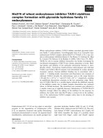
proteins in supernatants revealed the presence of three pro-
teins at 15, 13 and 11 kDa (Fig. 1), two of which (15 kDa
and 13 kDa) were immunoreactive with antibodies
directed against elafin (not shown). N-terminal sequence
analysis indicated that full-length trappin-2 correspon-
ded to the 15 kDa form whereas the 13 kDa protein
was a clipped form of trappin-2 (partial sequence:
GQDKVKAQE) resulting from a cleavage at the K32-
G33 sequence. The nonimmunoreactive 11 kDa protein
was believed to be a non related yeast protein and was
not further characterized. To limit the appearance of the
13 kDa clipped form of trappin-2 for the large-scale
production of trappin-2, the duration of fermentation was
reduced to three days. Under these conditions, no other
proteins except the 15 kDa form corresponding to mature
trappin-2 were detected in the supernatant by SDS/PAGE
analysis (Fig. 1).
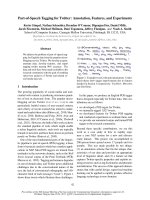
Recombinant elafin and trappin-2 were purified from
yeast culture supernatants by cation-exchange chromato-
graphy as described in Experimental procedures. For both
recombinant proteins, the elafin-immunoreactive material
was recovered in a single major peak (Fig. 2). An aliquot
from the main peak was analyzed by high-resolution Tricine
SDS/PAGE which revealed a single protein of about 7 kDa
and 12 kDa for elafin in reducing and nonreducing
conditions, respectively, and 12 kDa (reduced) and
15 kDa (nonreduced) for trappin-2, suggesting apparent
homogeneity of the purified proteins (Fig. 2). N-terminal
sequence analysis confirmed the identity of full-length
elafin (AQEPVKGPVS) and full-length trappin-2
(AVTGVPVKGQ).
Determination of the equilibrium dissociation constant
K
i
for the interaction between PPE and rec-trappin-2
or rec-elafin
The equilibrium dissociation constant K
i
for the interaction
of pancreatic elastase with recombinant elafin and trappin-2
was determined directly by adding substrate to an equilib-
rium mixture of protease and inhibitor, and measuring
spectrophotometrically the rate of release of the reaction
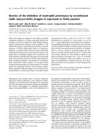
product. The concentration of both enzyme and inhibitor
was low enough to obtain a concave inhibition curve [25]
when incubating PPE with rec-elafin (Fig. 3). The best
estimates of K
i(app),
the substrate-dependent K
i
was obtained
by non linear regression analysis of the data based on the
following equation [25]:
a ¼
1 Àð½E
0
þ½I
0
þ K
iðappÞ
ÞÀ
ffiffiffiffiffiffiffiffiffiffiffiffiffiffiffiffiffiffiffiffiffiffiffiffiffiffiffiffiffiffiffiffiffiffiffiffiffiffiffiffiffiffiffiffiffiffiffiffiffiffiffiffiffiffiffiffiffiffiffiffiffiffiffiffi
ð½E
0
þ½I
0
þ K
iðappÞ
Þ
2
À 4½E
0
½I
0
q
2½E
0
ð1Þ
where a is the relative steady state rate and K
i(app)
¼
K
i
(1 + [S]
0
/K
m
). The competitive nature of the inhibition
was ascertained by measuring the fractional activity a for an
equimolar mixture of enzyme and inhibitor using two
different substrate concentrations as described in Experi-
mental procedures. For both inhibitors, a was found to
be substrate-dependent, indicating competitive inhibition.
K
i
was calculated from K
i(app)
using K
m
¼ 1.1 m
M
[26].
Fig. 1. Evolution of rec-trappin-2 production by Pichia pastoris as a
function of the duration of fermentation. Aliquots of concentrated
supernatants of rec-trappin-2-secreting P. pastoris cultures were ana-
lyzedbyhighresolutionSDS/PAGEandstainedwithCoomassie
BrilliantBlueafter0,1,2,3,4,5,6,7and10daysoffermentation
(lanes d0, d1, d2, d3, d4, d5, d6, d7 and d10, respectively). Three days
of fermentation (d3) were found to be optimum for rec-trappin-2
production before unwanted proteolysis appeared, and were therefore
retained for large-scale production.
Ó FEBS 2004 Inhibitory activity of recombinant elafin and trappin-2 (Eur. J. Biochem. 271) 2373
Rec-trappin-2 gave a similar inhibition curve from which
the K
i
could be derived (not shown). The values of K
i
are
giveninTable1.
Measurement of
k
ass
and
k
diss
for the interaction
of rec-elafin or rec-trappin-2 with HNE and PR3
Linear inhibition curves were obtained when reacting
increasing amounts of each inhibitor with HNE and PR3,
even when using enzyme concentrations as low as 10 n
M
,
indicating that rec-trappin-2 and rec-elafin bind both
proteinases too tightly to allow the direct measurement of
the equilibrium constant K
i
. This latter was thus calculated
from the association and dissociation rate constants k
ass
and k
diss
.
Fig. 2. Purification and SDS/PAGE analysis of rec-elafin and rec-
trappin-2. Aliquots (200 lL) of concentrated supernatants of rec-ela-
fin- or rec-trappin-2-secreting P. pastoris cultures were loaded onto a
cationic exchange Mono S column. After extensive washing to remove
unbound proteins, bound material was eluted with a linear NaCl
gradient ( ). Fractions containing purified rec-elafin (A) or purified
rec-trappin-2 (B) corresponding to the major peak (shaded area) were
pooled and stored at )70 °C before use. (C) High resolution Tricine
SDS/PAGE analysis of purified elafin and purified trappin-2 under
nonreducing conditions (–b) or reducing conditions (+b). Molecular
masses of the protein standards are shown on the left.
Fig. 3. Inhibition of the enzyme activity of pancreatic elastase by rec-
elafin. Constant amounts of PPE (10 n
M
) were incubated for 20 min
with increasing concentrations (0–2.7 · 10
)8
M
) of rec-elafin. The
residual enzymatic activity (e) was measured using Suc-(Ala)
3
-p-NA
(1 m
M
) as a substrate and plotted as a function of inhibitor concen-
tration. K
i(app)
was calculated by nonlinear regression analysis
(Results) using these experimental points. The theoretical curve (––)
generated using K
i(app)
¼ 1.4 n
M
was superimposed onto the experi-
mental data. A similar curve was obtained with rec-trappin-2.
2374 M L. Zani et al.(Eur. J. Biochem. 271) Ó FEBS 2004
The progress curve method was used to follow the time
course of HNE and PR3 inhibition. The reagent concen-
trations were chosen to yield both easily detectable signals
and to avoid significant substrate depletion during the
acquisition time. Because enzyme and inhibitor were reacted
under pseudo-first order conditions, the concentration of
product vs. time is given by the following equation [25]:
½P¼v
s
t þ
v
z
À v
s
k
ð1 À e
Àkt
Þð2Þ
where [P] is the product concentration at any time t, v
z
is the
rate of substrate breakdown at t ¼ 0 and vs. the steady state
rate. The best estimates of k, the apparent pseudo-first order
rate constant for the approach to the steady state, v
z
and v
s
were obtained by non linear regression analysis of the
progress curves based on Eqn (2). HNE and PR3 inhibition
was analysed by assuming that E and I react according to
a bimolecular and reversible mechanism as described in
Scheme I.
Hence, k
ass
, k
diss
and K
i
may be deduced from the
following relationships [25]:
k ¼
k
ass
½I
0
1 þ½S
0
=K
m
þ k
diss
ð3Þ
k
diss
¼ kv
s
=v
z
ð4Þ
K
i
¼ k
diss
=k
ass
ð5Þ
Kinetics for the association of HNE and Pr3 with rec-elafin
or rec-trappin-2 were studied as described in Experimental
procedures. We observed good fits of the experimental data
to the theoretical curves generated using the best estimates of
k, indicating that enzyme inhibition was satisfactorily
described by Eqn (2) (not shown). Also, k was proportional
to [I]
0
. Typical values of k were 0.33 ± 0.02 s
)1
and
0.19 ± 0.02 s
)1
for the association of HNE with 0.9 l
M
rec-elafin and rec-trappin-2, respectively, 0.16 ± 0.01 s
)1
for the reaction of PR3 with 0.8 l
M
rec-elafin and
0.19 ± 0.02 s
)1
for the inhibition of PR3 by 1.6 l
M
rec-
trappin-2.
Accurate values of k
diss
could not be calculated using
Eqn (4) because of the almost complete inhibition of
HNE and PR3 once the steady state was reached. For
this reason, the dissociation rate constant was independ-
ently obtained from further experiments. Figure 4A shows
the kinetics of product accumulation following the
dilution of an aliquot of preformed HNE–rec-elafin
complex into substrate solution. Complex dissociation
was triggered by both high dilution (100-fold) and high
substrate concentration ([S]
0
¼ 13.4 K
m
). The concentra-
tion of the latter was appropriate to ensure both sufficient
dissociation (Scheme I) and continuous enzyme detection
without significant decrease of its concentration during the
experiment. The experimental data were used to calculate
the derivative curve (Fig. 4B) representing the concentra-
tion of free enzyme vs. time. Free enzyme was almost
absent at t ¼ 0 and its concentration increased up to a
steady state level corresponding to 17% of the total
enzyme present in the reaction mixture (1.7 n
M
), indica-
ting that E, I and S were in thermodynamic equilibrium
with their complexes. A similar procedure was used to
study the dissociation kinetics of 1 l
M
HNE–rec-trappin-2,
PR3–rec-elafin and PR3–rec-trappin-2 complexes. Their
100-fold dilution into the appropriate substrate solutions
yielded 12%, 43% and 46% of total enzyme release,
respectively.
As neither free enzyme nor free inhibitor were present to a
significant extent at t ¼ 0, the rate of complex dissociation,
that is, the rate of enzyme release, is given by:
À
d½EI
dt
¼
d½E
dt
¼ k
ass
½EIÀk
diss
½E½Ið6Þ
which integrates into Eqn (7) [27]:
½E¼
½E
e
Àðe
fk
diss
tð2½EI
0
À½E
e
Þ=½E
e
g
À 1Þ
e
fk
diss
tð2½EI
0
À½E
e
Þ=½E
e
g
À½E
e
=½EIþ1
ð7Þ
where [E]
e
and [E] are the concentrations of free HNE or
PR3 at equilibrium and at any time t, respectively, and [EI]
0
and [EI] are the initial concentration of complex and its
Scheme 1.
Table 1. Equilibrium and rate constants for the inhibition of neutrophil elastase, proteinase 3 and pancreatic elastase by recombinant elafin and
recombinant trappin-2. Methods and experimental conditions are described in Experimental procedures. Values are given as means ± SEM. ND,
not determined.
Enzyme
Rec-elafin Rec-trappin-2
k
ass
(
M
)1
Æs
)1
) k
diss
(s
)1
) K
i
(
M
) k
ass
(
M
)1
Æs
)1
) k
diss
(s
)1
) K
i
(
M
)
Neutrophil elastase (3.7 ± 0.1) 10
6
(3.2 ± 0.1) 10
)4
(0.8 ± 0.05) 10
)10a
(3.6 ± 0.5) 10
6
(1.1 ± 0.2) 10
)4
(0.3 ± 0.1) 10
)10a
Proteinase 3 (3.3 ± 0.03) 10
6
(4 ± 0.3) 10
)4
(1.2 ± 0.1) 10
)10a
(2 ± 0.1) 10
6
(3.7 ± 1.1) 10
)4
(1.8 ± 0.6) 10
)10a
Pancreatic elastase ND ND (7.5 ± 1.5) 10
)10
ND ND (3.2 ± 0.8) 10
)10
a
Calculated as the k
diss
/k
ass
ratio.
Ó FEBS 2004 Inhibitory activity of recombinant elafin and trappin-2 (Eur. J. Biochem. 271) 2375
concentration at any time t, respectively. Figure 4B shows
the theoretical curve calculated by non linear regression
analysis of the data using Eqn (7) and using the best
estimate of k
diss
governing the dissociation of the HNE–rec-
elafin complex. Good fits were also obtained for the three
other enzyme–inhibitor pairs indicating that the kinetics of
enzyme release satisfactorily agrees with Eqn (7). Compar-
ison of the k
diss
values found using this procedure (Table 1)
with values of k reported above show that k
diss
is 400–1700-
fold lower than k and may therefore be neglected in Eqn (3).
The average association rate constant k
ass
for each enzyme-
inhibitor pair were therefore calculated from k
ass
¼
k (1 + [S]
0
/K
m
)/[I]
0
and [S]
0
¼ 1.56 m
M
, K
m
¼ 0.14 m
M
for the HNE-MeO-Suc-(Ala)
2
-Pro-Val-p-NA system [28]
and [S]
0
¼ 0.15 m
M
, K
m
¼ 0.01 m
M
for the PR3-MeO-
Suc-Lys-(pico)-Ala-Pro-Val-TBE pair [29]. These k
ass
values
are reported in Table 1.
Discussion
The control of the excessive proteolytic activity of HNE has
long been recognized to be crucial to avoid degradation of
the lung parenchyma in many inflammatory lung diseases.
As a consequence, lung therapies based on the inhibition of
HNE have lead to intensive research on the development of
HNE inhibitors, either as recombinant proteins or synthetic
small-molecule inhibitors [13]. However, there is concern
now that other neutrophil-derived proteases, namely cath-
epsin G and proteinase 3, might have similar deleterious
effects as HNE, hence the necessity to design inhibitors able
to target all three neutrophil-derived serine proteases. In our
efforts to evaluate the therapeutic potential of recombinant
genetically modified protease inhibitors derived from nat-
ural inhibitors, we report here on the biosynthetic produc-
tion of elafin and its precursor, trappin-2. The cDNA
coding either elafin or trappin-2 was cloned into the yeast
expression vector pPIC9, allowing the production of both
inhibitors in the P. pastoris system. The cloning strategy was
designed so that mature elafin or mature trappin-2 were
secreted in the culture supernatant. No tag to facilitate the
purification of the expressed proteins was introduced
because both proteins were predicted to be mainly basic,
allowing further purification with cation-exhange chroma-
tographic procedures. While the level of elafin production
increased up to seven days of fermentation with no
apparent modifications of the protein, the expression of
trappin-2 was found to become sensitive to unwanted
proteolysis as fermentation duration increased. A clipped
form of trappin-2 resulting from a cleavage C-terminal to
Lys32 appeared together with the full-length trappin-2 after
three days of fermentation. Such a proteolytic susceptibility
after lysyl residues was observed by Bourbonnais et al.[30]
who expressed trappin-2 in Saccharomyces cerevisae.Clea-
vage after Lys14 and Lys36 in the so-called cementoin
domain of trappin-2 was attributed unambiguously to
yapsin-1, an aspartic plasma membrane protease active
within the periplasmic space. Though the cleavage sites in
trappin-2 were different in the two yeast expression systems,
we can hypothesize that yapsin-like enzyme(s) are also
involved in the non specific degradation of heterologous
proteins expressed in P. pastoris. Reducing the fermentation
to a maximum of three days for yeast clones expressing
trappin-2 was found to suppress the apparition of the
13 kDa clipped form of trappin-2 at the cost of a somewhat
lower protein concentration. Using the culture conditions
described above, we purified about 15 mgÆL
)1
of each
recombinant inhibitor from the yeast culture media. Using
shake-flask culture conditions which give expression levels
typically low relative to what is obtainable in fermenter
cultures [31], we found that the amount of elafin and
trappin-2 produced in our system was higher than that
reported for trappin-2 expressed in similar conditions in the
yeast S. cerevisiae system (2–3 mgÆL
)1
) [30]. Though the
range of expression yields is variable from one protein to
another, our study confirms that the P. pastoris system
allows the production of heterologous proteins at a high
concentration level. In addition, considering the ease by
which the protein production can be scaled up from shake-
flask to fermentation conditions [31], P. pastoris is a system
of choice to produce large amounts of therapeutic proteins.
Fig. 4. Dissociation kinetics of HNE–rec-elafin complexes. (A) Time
course of p-nitroaniline release resulting from the hydrolysis of MeO-
Suc-(Ala)
2
-Pro-Val-p-NA by HNE released from its complex with
rec-elafin. Complexes were first formed by incubating equimolar
concentrations (10
)6
M
) of enzyme and inhibitor. Dissociation of the
complexes was induced by dilution in a concentrated substrate
(1.88 m
M
) solution. (B) Kinetics of elastase release calculated from
(A) as described in Results. The theoretical curve (––) superimposed
onto the experimental data was calculated using Eqn (7) and k
diss
¼
3.2 10
)4
s
)1
.
2376 M L. Zani et al.(Eur. J. Biochem. 271) Ó FEBS 2004
N-terminal sequencing and Western blot analysis showed
that recombinant elafin and trappin-2 are identical to the
natural proteins. In addition, enzyme kinetics showed that
the K
i
of PPE–rec-elafin complex is very close to that
reported for natural elafin [32]. Also, the kinetic constants
k
ass
, k
diss
and K
i
for the interaction of rec-elafin with HNE
and PR3 are of the same order of magnitude as those
reported for chemically synthesized elafin by Ying & Simon
[23,24]. The P. pastoris expression system described here
therefore yields a protein structurally and functionally
identical to natural elafin.
Litterature lacks information on the kinetic parameters
describing the interaction of trappin-2 with HNE and
PR3. Based on the kinetic parameters determined here,
the most important result of our investigation is that
elafin and trappin-2 have very close inhibitory capacities.
This means that the N-terminal cementoin domain of
trappin-2 has little or no influence on the reactive
inhibitory site of elafin. However, it is noteworthy that
trappin-2, but not elafin, has been shown to significantly
reduce a HNE-induced experimental lung hemorrhage in
hamsters [33] or a lipolysaccharide-induced acute lung
inflammation in mice [34]. This has been attributed to the
unique capacity of the cementoin domain to be cross-
linked to extracellular matrix proteins through the cata-
lytic action of tissue transglutaminase(s) [33,34]. In this
context, it will be especially interesting to evaluate the
inhibitory properties of bound trappin-2, as this covalent
linking may increase significantly the bioavailability of
such an inhibitor at the site of inflammation, e.g. in the
case of therapeutic administration, as well as providing a
source of inhibitory elafin.
Knowledge of the kinetic parameters characterizing a
protease–inhibitor interaction and of the in vivo concen-
tration of an inhibitor is necessary to evaluate whether
such an inhibitor may control the activity of its target
enzyme(s) [25,35]. From the kinetic constants determined
here and from the in vivo concentration of elafin estimated
to be in the range 1.5–4.5 l
M
in bronchial secretions of
normal patients [16,24], we can conclude that both elafin
and its precursor are fast-acting inhibitors of HNE and
PR3 with a delay time for total inhibition of a few
milliseconds (d(t) ¼ 5/k
ass
Æ[I]
0
[35]). The second conclusion
is that both inhibitors will exhibit a pseudo-irreversible
behaviour because the [I]
0
/K
i
ratio of about 15–45 · 10
3
is
greater than 10
3
[25].
Altogether, our results clearly demonstrate for the first
time that, in vitro, trappin-2 and elafin exhibit a similar and
potent inhibitory capacity towards HNE and PR3, strongly
suggesting that boosting elafin or trappin-2 level by an
aerosol administration would be beneficial in the treatment
of inflammatory lung diseases.
Acknowledgements
This study was supported by the French cystic fibrosis association
ÔVaincre la MucoviscidoseÕ. We thank Dr Antoine Touze
´
for nucleotide
sequencing, Miche
`
le Brillard-Bourdet for N-terminal protein sequen-
cing and Prof. Joost Schalkwijk and Dr Patrick Zeeuwen for their kind
gift of trappin-2 cDNA. We also thank Dr Fre
´
de
´
ric Delamotte and
Prof. Francis Gauthier for valuable discussions. Shila M. Nobar holds
afellowshipfromÔVaincre la MucoviscidoseÕ.
References
1. Campbell, E.J., Campbell, M.A. & Owen, C.A. (2000) Bioactive
proteinase 3 on the cell surface of human neutrophils: quantifi-
cation, catalytic activity, and susceptibility to inhibition. J.
Immunol. 165, 3366–3374.
2. Campbell, E.J., Silverman, E.K. & Campbell, M.A. (1989) Elas-
tase and cathepsin G of human monocytes. Quantification of
cellular content, release in response to stimuli, and heterogeneity in
elastase-mediated proteolytic activity. J. Immunol. 143, 2961–2968.
3. Owen, C.A. & Campbell, E.J. (1999) The cell biology of leukocyte-
mediated proteolysis. J. Leukoc. Biol. 65, 137–150.
4. Perlmutter, D.H. & Pierce, J.A. (1989) The alpha 1-antitrypsin
gene and emphysema. Am. J. Physiol. 257, 147–162.
5. Birrer, P., McElvaney, N.G., Rudeberg, A., Sommer, C.W.,
Liechti-Gallati, S., Kraemer, R., Hubbard, R. & Crystal, R.G.
(1994) Protease-antiprotease imbalance in the lungs of children
with cystic fibrosis. Am. J. Respir. Crit. Care Med. 150, 207–213.
6. Doring, G. (1994) The role of neutrophil elastase in chronic
inflammation. Am.J.Respir.Crit.CareMed.150, S114–S117.
7. McElvaney, N.G., Nakamura, H., Birrer, P., Hebert, C.A., Wong,
W.L.,Alphonso,M.,Baker,J.B.,Catalano,M.A.&Crystal,R.G.
(1992) Modulation of airway inflammation in cystic fibrosis.
In vivo suppression of interleukin-8 levels on the respiratory
epithelial surface by aerosolization of recombinant secretory leu-
koprotease inhibitor. J. Clin. Invest. 90, 1296–1301.
8. Vogelmeier, C., Gillissen, A. & Buhl, R. (1996) Use of secretory
leukoprotease inhibitor to augment lung antineutrophil elastase
activity. Chest 110, 261S–266S.
9. Vogelmeier, C., Hubbard, R.C., Fells, G.A., Schnebli, H.P.,
Thompson, R.C., Fritz, H. & Crystal, R.G. (1991) Anti-neutrophil
elastase defense of the normal human respiratory epithelial surface
provided by the secretory leukoprotease inhibitor. J. Clin. Invest.
87, 482–488.
10. McElvaney, N.G., Hubbard, R.C., Birrer, P., Chernick, M.S.,
Caplan, D.B., Frank, M.M. & Crystal, R.G. (1991) Aerosol alpha
1-antitrypsin treatment for cystic fibrosis. Lancet 337, 392–394.
11. Delacourt, C., Herigault, S., Delclaux, C., Poncin, A., Levame,
M., Harf, A., Saudubray, F. & Lafuma, C. (2002) Protection
against acute lung injury by intravenous or intratracheal pre-
treatment with EPI-HNE-4, a new potent neutrophil elastase
inhibitor. Am. J. Respir. Cell. Mol. Biol. 26, 290–297.
12. Bradbury, J. (2001) Neutrophil elastase inhibitors for cystic
fibrosis. Drug Discov. Today 6, 441–442.
13. Tremblay, G.M., Janelle, M.F. & Bourbonnais, Y. (2003) Anti-
inflammatory activity of neutrophil elastase inhibitors. Curr. Opin.
Invest. Drugs 4, 556–565.
14. Nara, K., Ito, S., Ito, T., Suzuki, Y., Ghoneim, M.A., Tachibana,
S. & Hirose, S. (1994) Elastase inhibitor elafin is a new type of
proteinase inhibitor which has a transglutaminase-mediated
anchoring sequence termed ÔcementoinÕ. J. Biochem. 115, 441–448.
15. Sallenave, J.M. & Silva, A. (1993) Characterization and gene
sequence of the precursor of elafin, an elastase-specific inhibitor in
bronchial secretions. Am. J. Respir. Cell. Mol. Biol. 8, 439–445.
16. Tremblay, G.M., Sallenave, J.M., Israel-Assayag, E., Cormier, Y.
& Gauldie, J. (1996) Elafin/elastase-specific inhibitor in broncho-
alveolar lavage of normal subjects and farmer’s lung. Am. J.
Respir. Crit. Care Med. 154, 1092–1098.
17. Pfundt, R., van Ruissen, F., van Vlijmen-Willems, I.M., Alke-
made, H.A., Zeeuwen, P.L., Jap, P.H., Dijkman, H., Fransen, J.,
Croes, H., van Erp, P.E. & Schalkwijk, J. (1996) Constitutive and
inducible expression of SKALP/elafin provides anti-elastase def-
ense in human epithelia. J. Clin. Invest. 98, 1389–1399.
18. Shotton, D.M. (1970) Elastase. Methods Enzymol. 19, 113–140.
19. Boudier, C. & Bieth, J.G. (1992) The proteinase: mucus proteinase
inhibitor binding stoichiometry. J. Biol. Chem. 267, 4370–4375.
Ó FEBS 2004 Inhibitory activity of recombinant elafin and trappin-2 (Eur. J. Biochem. 271) 2377
20. Rao, N.V., Wehner, N.G., Marshall, B.C., Gray, W.R., Gray,
B.H. & Hoidal, J.R. (1991) Characterization of proteinase-3
(PR-3), a neutrophil serine proteinase. Structural and functional
properties. J. Biol. Chem. 266, 9540–9548.
21. Scha
¨
gger, H. & von Jagow, G. (1987) Tricine-sodium dodecyl
sulfate-polyacrylamide gel electrophoresis for the separation
of proteins in the range from 1 to 100 kDa. Anal. Biochem. 166,
368–379.
22. Zani, M., Brillard-Bourdet, M., Lazure, C., Juliano, L., Courty,
Y., Gauthier, F. & Moreau, T. (2001) Purification and char-
acterization of active recombinant rat kallikrein rK9. Biochim.
Biophys. Acta 1547, 387–396.
23. Ying, Q.L. & Simon, S.R. (1993) Kinetics of the inhibition of
human leukocyte elastase by elafin, a 6-kilodalton elastase-specific
inhibitor from human skin. Biochemistry 32, 1866–1874.
24. Ying, Q.L. & Simon, S.R. (2001) Kinetics of the Inhibition of
Proteinase 3 by Elafin. Am. J. Respir. Cell Mol. Biol. 24, 83–89.
25. Bieth, J.G. (1995) Theoretical and practical aspects of proteinase
inhibition kinetics. Methods Enzymol. 248, 59–84.
26. Bieth, J.G., Spiess, B. & Wermuth, C.G. (1974) The synthesis and
analytical use of a highly sensitive and convenient substrate of
elastase. Biochem. Med. 11, 350–357.
27. Capellos, C. & Bielski, B.H.J. (1980) Kinetic Systems. Mathema-
tical Description of Chemical Kinetics in Solution. Krieger Pub-
lication Co., Huntington, NY.
28. Nakajima, K., Powers, J.C., Ashe, B.M. & Zimmerman, M.
(1979) Mapping the extended substrate binding site of cathepsin G
and human leukocyte elastase. Studies with peptide substrates
related to the alpha 1-protease inhibitor reactive site. J. Biol.
Chem. 254, 4027–4032.
29. Duranton, J. & Bieth, J.G. (2003) Inhibition of Proteinase 3 by
alpha1-Antitrypsin In Vitro Predicts Very Fast Inhibition In Vivo.
Am. J. Respir. Cell Mol. Biol. 29, 57–61.
30. Bourbonnais, Y., Larouche, C. & Tremblay, G.M. (2000)
Production of full-length human pre-elafin, an elastase specific
inhibitor, from yeast requires the absence of a functional yapsin 1
(Yps1p) endoprotease. Protein Expr. Purif. 20, 485–491.
31. Cereghino, G.P., Cereghino, J.L., Ilgen, C. & Cregg, J.M. (2002)
Production of recombinant proteins in fermenter cultures of the
yeast Pichia pastoris. Curr. Opin. Biotechnol. 13, 329–332.
32. Wiedow,O.,Schroder,J.M.,Gregory,H.,Young,J.A.&Chris-
tophers, E. (1990) Elafin: an elastase-specific inhibitor of human
skin. Purification, characterization, and complete amino acid
sequence. J. Biol. Chem. 265, 14791–14795.
33. Tremblay, G.M., Vachon, E., Larouche, C. & Bourbonnais, Y.
(2002) Inhibition of human neutrophil elastase-induced acute lung
injury in hamsters by recombinant human pre-elafin (trappin-2).
Chest 121, 582–588.
34. Vachon, E., Bourbonnais, Y., Bingle, C.D., Rowe, S.J., Janelle,
M.F. & Tremblay, G.M. (2002) Anti-inflammatory effect of pre-
elafin in lipopolysaccharide-induced acute lung inflammation.
Biol. Chem. 383, 1249–1256.
35. Bieth, J.G. (1984) In vivo significance of kinetic constants of pro-
tein proteinase inhibitors. Biochem. Med. 32, 387–397.
2378 M L. Zani et al.(Eur. J. Biochem. 271) Ó FEBS 2004