Báo cáo khoa học: Modulation of nitric oxide-mediated metal release from metallothionein by the redox state of glutathione in vitro doc
Bạn đang xem bản rút gọn của tài liệu. Xem và tải ngay bản đầy đủ của tài liệu tại đây (300.85 KB, 9 trang )
Modulation of nitric oxide-mediated metal release from
metallothionein by the redox state of glutathione
in vitro
Leila Khatai
1
, Walter Goessler
2
, Helena Lorencova
2
and Klaus Zangger
1
1
Institute of Chemistry, Organic and Bioorganic Chemistry, University of Graz, Austria;
2
Institute of Chemistry, Analytical Chemistry,
University of Graz, Austria
Metallothioneins (MTs) release bound metals when exposed
to nitric oxide. At inflammatory sites, both metallothionein
and inducible nitric oxide synthase (iNOS) are induced by
the same factors and the zinc released from metallothionein
by NO suppresses both the induction and activity of iNOS.
In a search for a possible modulatory mechanism of this
coexpression of counteracting proteins, we investigated the
role of the glutathione redox state in vitro because the oxi-
dation state of thiols is involved in the metal binding in Cd-S
or Zn-S clusters found in metallothioneins, and NO also
binds to reduced glutathione via S-nitrosation. Using a
variety of techniques, we found that NO and also ONOO
–
-
mediated metal release from purified MTs is suppressed by
reduced glutathione (GSH), but not by oxidized glutathione.
Considering the millimolar concentrations of GSH present
in mammalian cells, the metal release from MTs by NO
should play no role in living systems. Therefore, the fact that
it has been observed in vivo points to a hitherto unknown
mechanism or additional compound(s) being involved in this
physiologically relevant reaction and as long as this addi-
tional factor is not found experimental results on the MT–
NO interaction should be treated with caution. Contrary
to the peroxynitrite-induced activation of guanylyl cyclase,
where GSH is needed, we found that the metal release from
metallothionein by peroxynitrite is not enhanced, but also
suppressed by reduced glutathione. In addition, we show
that zinc, the major natural metal ligand in mammalian MTs
and suppressor of iNOS, is released more readily under the
influence of NO than cadmium, but in contrast to the MT
isoform 1, the amount of metal released from the b-domain
of MT-2 is comparable to that from the a-domain.
Keywords: glutathione; metallothionein; nitric oxide; NMR
spectroscopy; SEC–ICPMS.
Metallothioneins (MTs) are a family of small (6–7 kDa)
metal-binding proteins [1–3] with the highest known metal
content after ferritins. The high amount of cysteine residues
in MTs (30% of all amino acids are cysteine) allows these
proteins to coordinate multiple mono (Cu
+
,Ag
+
)or
divalent metals (Zn
2+
,Cd
2+
). Mammalian MTs bind seven
divalent metals in two separate domains [4]. Three metals
are bound in an M
3
Cys
9
cluster in the N-terminal b-domain,
while an M
4
Cys
11
four metal cluster is formed in the
C-terminal a-domain [4]. Of the four known mammalian
MT isoforms [2], the two best studied and most widely
occurring isoforms (1 and 2) are most abundant in
parenchymatous tissues, i.e. liver, kidney, pancreas and
intestines [5–7] but their occurrence and biosynthesis have
been documented in many tissues and cell types. The 3D
structures of MT1 [8] and MT2 [9–12] are very similar, but
there are various indications of increased flexibility and
metal mobility in the b-domaininMT-1[8].Thenaturally
bound metal zinc can be displaced by cadmium up to about
5 mol per mol protein by simple addition of Cd
2+
[13]
in vitro. Living animals fed a cadmium-rich diet produce
a mixed-metal MT with zinc bound preferentially in the
b- and cadmium in the a-domain [11,13,14]. The artificial
Cd
7
-MT can only be obtained after complete zinc removal
by lowering the pH [15] in vitro.
Although the biological function(s) of MTs still remain
somewhat elusive [16], they have been proposed to play an
important role in zinc homeostasis [1,17] and heavy metal
detoxification [18,19], although the latter is probably not an
evolutionary conserved function but rather a property of
these cysteine-rich proteins. Due to the different metal
affinities for zinc and cadmium in the two separate domains
[13], the b-domain has been implicated in zinc homeostasis
and the tight binding of cadmium in the a-domain was
proposed to be responsible for the role of MTs in heavy
metal detoxification. In addition, it has been reported that
MTs act as radical scavengers under oxidative stress [20–22].
Another possible key player in the role of MTs in signal
transduction might be nitric oxide (NO), which was shown
recently, both in vitro [23–25] and in vivo [26–29], to interact
with MTs and thereby releases bound zinc and cadmium.
The importance of MTs in NO-induced changes in intra-
cellular zinc homeostasis has been reported by St Croix
et al.[30].
Correspondence to K. Zangger, Institute of Chemistry/Organic and
Bioorganic Chemistry, University of Graz, Heinrichstrasse 28, A-8010
Graz, Austria. Fax: + 43 316 380 9840, Tel.: + 43 316 380 8673,
E-mail:
Abbreviations: DEA/NO, 2-(N,N-diethylamino)-diazenolate-2-oxide-
Na; GSH, reduced glutathione; GSSG, oxidized glutathione;
iNOS, inducible NO-synthase; MT, metallothionein; NO, nitric oxide;
SEC–ICPMS, size exclusion chromatography–inductively coupled
plasma mass spectrometry; SIN-HCl, 3-morpholinosydnoni-
mine.HCl.
(Received 26 February 2004, revised 6 April 2004,
accepted 14 April 2004)
Eur. J. Biochem. 271, 2408–2416 (2004) Ó FEBS 2004 doi:10.1111/j.1432-1033.2004.04160.x
Based on the preferred release of metal from the
b-domain of mouse MT1, where zinc is preferentially bound
in vivo, we suggested recently that MTs had anti-inflamma-
tory activity [31]. This activity relies on the suppression of
the expression and activity of inducible nitric oxide synthase
(iNOS) by zinc [32,33], released from MT under the
influence of nitric oxide (NO), and the scavenging of NO
through covalent binding to MTs [23] to form S-nitroso-
thiols. Such a role of MTs in the inflammatory response has
been corroborated by the significantly altered inflammatory
behavior during experimental autoimmune encephalomye-
litis [34] observed in MT deficient mice. In addition, it has
been reported that overexpression of MT reduces the
sensitivity of eukaryotic cells to oxidative injury [35] and the
cytotoxic effects of NO [29]. As both iNOS and MTs are
induced at inflammatory sites by the same compounds and
MT scavenges NO and suppresses its production, one starts
to wonder why they are both produced at inflammatory
sites but counteract each other. Therefore, we looked for a
possible regulatory mechanism for the interplay between
NO production and metal release from MTs in order to
understand this dual expression of opposing proteins. As
the metal is held in place by thiolate ligands [4] in MTs,
other thiols may well influence the metal-bond formation
and breakage. The major low-molecular mass thiol com-
pound in plants and animals is the tripeptide
L
-c-glutamyl-
L
-cysteinyl-glycine also known as glutathione (GSH in
reduced form and GSSG in its oxidized form). Glutathione
has also been described as a Ôtransport peptideÕ in vivo for
NO through the formation of S-NO groups [36]. The
glutathione redox couple, a cellular redox buffer which
maintains the given thiol/disulfide redox potential, has
already been implicated in modulating the metal release
from metallothionein in the absence of nitric oxide by
Vallee, Maret and coworkers [22,37,38]. These authors
reported increased metal release in the presence of oxidized
glutathione (GSSG) and even slightly tighter metal binding
under the influence of reduced glutathione (GSH) [37]. We
investigated in vitro the effect of GSH/GSSG on NO-
mediated metal release of MT2 by circular dichroism (CD)
spectroscopy, size exclusion chromatography–inductively
coupled plasma mass spectrometry (SEC–ICPMS) and
nuclear magnetic resonance (NMR) spectroscopy. In a
previous study [31], we used Cd
7
-MT1 to study the
structural consequences of NO exposure on MTs by
1
H
and
113
Cd-NMR spectroscopy as Zn cannot be studied by
regular NMR experiments. However, Cd
7
-MT1 is never
found in natural sources and differences in metal-binding
constants between Cd and Zn might prevent the inter-
pretation of in vivo processes with data obtained on an
artificially cadmium-enriched protein. Therefore, we have
limited the present study to Zn
7
-MT2 and Cd
5
Zn
2
-MT2,
which have been isolated from natural sources.
In addition to nitric oxide, peroxynitrite (ONOO
–
)may
also play a significant role in the metal release from MTs, as
it has been suggested that the decomposition of peroxy-
nitrite at physiological pH constitutes the actual component
of NO cytotoxicity [39]. A widespread signal transduction
mechanism for NO involved in, e.g. platelet aggregation,
blood pressure control and neurotransmission functions
via stimulation of guanylyl cyclase [40]. In contrast to NO,
glutathione-dependent bioactivation of peroxynitrite is
involved in enzyme stimulation and this points again at a
possible key role of glutathione in the NO and/or ONOO
–
mediated metal release from MTs.
Materials and methods
The NO donor 2-(N,N-diethylamino)-diazenolate-2-oxide-
Na (DEA/NO) and the peroxynitrite donor 3-morphol-
inosydnonimine.HCl (SIN-HCl) were purchased from
Alexis Biochemicals (Lausen, Switzerland). Due to the
limited lifetime of DEA/NO at low to moderate pH, stock
solutions were prepared in 10 m
M
NaOH (pH 12). By
adding such stock solutions to neutrally buffered systems,
1.5 mol equivalents of NO are released from DEA/NO
with a half-life of 16 min at 24 °C. Using molecular
oxygen, SIN-HCl generates superoxide and NO, which
spontaneously combine to form peroxynitrite. A fresh
solution of peroxynitrite was prepared according to a
published procedure [41] and stored at )80 °C until used.
Rabbit liver metallothionein-2 in the zinc form (Zn
7
-MT2)
and a mixed cadmium, zinc form (Cd
5
Zn
2
-MT2) were
obtained from Sigma (Vienna, Austria). We reconstituted
Zn
7
-MT2 to ascertain the stochiometry of seven metals
per protein monomer according to the procedure des-
cribed by Vas
ˇ
ak [15], but found no differences in its
behavior to the unpurified commercially available product.
All other chemicals were purchased from Sigma (Vienna,
Austria) at the highest purity available. Due to problems
associated with the use of organic buffers in inductively
coupled plasma mass spectrometry (ICPMS) instruments
and protonated buffers for NMR, we used aqueous
phosphate buffers for all experiments (see below). To
evaluate a possible influence of phosphate ions on MT2
during these experiments, the CD experiments were also
performed in 20 m
M
Hepes buffer, but showed the same
results.
CD spectroscopy
The complete absence of aromatic amino acids in
metallothioneins allows the use of UV and CD spectros-
copy to observe the cadmium-thiolate charge transfer
transition, which occurs around 250 nm [42]. This region
is usually completely masked by aromatic groups. CD
spectra were recorded on a Jasco J-715 spectropolarimeter
andanalyzedusingtheprogram
CDSCAN
. For each
wavelength scan, the average was taken from 10 accumu-
lations with the following parameters: step resolution,
0.2 nm; speed, 50 nmÆmin
)1
; response time, 1 s; band-
width, 2 nm. For time scans, we used: wavelength,
260 nm; step resolution, 1 s; response time, 1 s; band-
width, 2 s. Samples consisted of 100 l
M
Cd
5
Zn
2
-MT2 in
20 m
M
potassium phosphate buffer at pH 7.5. Stock
solutions of GSH (50 m
M
in phosphate buffer, pH 7.5),
GSSG (50 m
M
in phosphate buffer, pH 7.5) and DEA/
NO (20 m
M
in 10 m
M
NaOH) were added to the MT2
solution to give final concentrations of 1 m
M
of each
compound in the respective spectra. The range between
230 and 300 nm was recorded for the CD spectra and
time scans were obtained by monitoring the CD at the
maximum of the cadmium-thiolate charge transfer band
at 260 nm for 20 min after mixing the components.
Ó FEBS 2004 NO-mediated metal release from metallothionein (Eur. J. Biochem. 271) 2409
SEC–ICPMS
ICPMS enables the determination of a variety of elements
in solution. In order to differentiate between protein-bound
and free metal, a preceding separation of protein and
unbound metal by size exclusion chromatography (SEC) is
necessary. Instead of performing these two steps separately,
thecouplingofSECandICPMSoffersaveryelegant
alternative [43,44]. For our studies, a Pharmacia Superdex
75 PC 3.2/30 gel filtration column was connected to an
Agilent HP 1100 ChemStation SEC system (Agilent,
Waldbronn, Germany) equipped with a UV monitor set
to 220 nm. The outlet of the UV-detector was connected
directly via a PEEK capillary (i.d. 0.12 mm, length 90 cm)
to the l-flow PFA-100 nebulizer (CPI International, Santa
Rosa, USA) of the Agilent 7500c ICPMS system. The
isotopes
64,66
Zn and
111,114
Cd were monitored. All meas-
urements were performed at least twice and the averages
were taken over both isotopes of zinc and cadmium,
respectively. A 20 m
M
aqueous ammonium phosphate
buffer, pH 6.5 was used as eluent at a flow rate of
0.1 mLÆmin
)1
. MT2, GSH and GSSG solutions were made
metal-free by washing through a Chelex-100 column
(Sigma, Vienna, Austria) and stored in polyethylene flasks.
Samples of 1 lL were injected onto the column and
separated at 22 °C. All solutions were filtered and degassed
by N
2
bubbling prior to use. A solution of 20 l
M
Cd
5
Zn
2
-
MT2 was mixed with stock solutions of 2 m
M
DEA/NO,
2m
M
SIN-HCl, 2 m
M
ONOO
–
,10m
M
GSH or 5 m
M
GSSG and then equilibrated for at least 15 min prior to
injection onto the gel filtration column.
NMR spectroscopy
Series of two-dimensional TOCSY [45] NMR spectra were
recorded on a Varian Unity INOVA 600 MHz NMR
spectrometer at 25 °C. The water signal was suppressed
with the WATERGATE sequence [46]. For each of the 256
increments, 2048 complex data points were recorded. The
data were multiplied with a 60° phase-shifted, squared sine-
bell window function in both dimensions prior to Fourier
transformation. The total experimental time of one 2D
spectrum was 12 h. Samples consisted of 2.5 mg of Zn
7
-
MT2 or Cd
5
Zn
2
-MT2 in 0.5mL of 20m
M
potassium
phosphate buffer pH 6.5 and 50 lLD
2
O. A stock solution
of 50 m
M
DEA/NO was added directly to the NMR
samples to give final concentrations of 0.2, 0.5, 1 and 3 m
M
DEA/NO. After each addition, the solution was equili-
brated for at least 20 min prior to the start of the NMR
acquisition. The same experiment was performed in the
presence of GSH, whereby the GSH concentration was
1m
M
for samples containing 0.2 and 0.5 m
M
DEA/NO and
5m
M
GSH was added when the DEA/NO concentration
was 1 and 3 m
M
.
Results
CD spectroscopy
The Cd-S charge transfer transitions are responsible for the
absorption and CD above 230 nm in UV and CD spectra
of metallothioneins devoid of any aromatic residues that
usually obscure this spectral region. By monitoring this
charge transfer band, CD spectroscopy was first used by
Ka
¨
gi and coworkers to follow the metal-binding stochiom-
etry of MTs [42]. To study the metal release by NO and its
modulation by GSH and GSSG, a solution of 100 l
M
Cd
5
Zn
2
-MT2 was exposed to NO for 20 min by adding
DEA/NO at a final concentration of 1 m
M
. For reduced
and oxidized glutathione, concentrations of 1 m
M
were
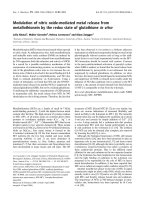
used. CD spectra of the range between 230 and 300 nm are
shown in Fig. 1A. The Cd-S charge transfer band at 260 nm
is reduced clearly after the addition of NO, indicating the
breaking of cadmium-cysteine bonds and therefore release
of cadmium. The presence of GSH reduces the metal release
almost completely, while GSSG even slightly increased the
cadmium release by nitric oxide. The decay occurs in the
first 10 min after the addition of NO as observed by
monitoring the CD at the maximum of the charge transfer
band at 260 nm (Fig. 1B). The lower molar ellipticity at
time 0 in the GSSG/NO treated sample derives from partial
metal release during the period from mixing the solutions
until the start of the data acquisition. While with the CD
measurements nothing can be said about the faith of zinc
bound in Cd
5
Zn
2
-MT2 after the addition of NO, the
amount of cadmium is reduced at this rather extreme NO
Fig. 1. Wavelength and time scans of the CD of MT2 in the presence
and absence of NO and GSH/GSSG. CD spectra of 100 l
M
Cd
5
Zn
2
-
MT2 alone or with 1 m
M
GSH in 20 m
M
potassium phosphate buffer,
pH 7.5 upon the exposure to 1 m
M
DEA/NO are shown in (A). The
molar ellipticity ([h]inkdegÆdmol
)1
Æcm
)2
)atthemaximumoftheCd-S
charge-transfer band at 260 nm as a function of time can be seen in (B).
2410 L. Khatai et al.(Eur. J. Biochem. 271) Ó FEBS 2004
concentration by at most 2 cadmium atoms per MT2
molecule.
SEC–ICPMS
CD spectroscopy enables the monitoring of breaking Cd-S
bonds, but it does not give information about released
metals, because there might be a situation when some metal-
thiolate bonds are broken, but the metal is still held in place
by remaining Cd-S bonds. In addition, no information
about bound zinc in the mixed metal MT2 is obtained. Both
uncertainties can be clarified with SEC–ICPMS [43,44]. The
sample is applied to a gel filtration column, which separates
free from protein-bound metal and subsequently both zinc
and cadmium levels are determined by ICPMS. Stock
solutions of 2 m
M
DEA/NO, 10 m
M
GSH and 5 m
M
GSSG were added to samples of 20 l
M
Cd
5
Zn
2
-MT2 to
give final ratios as indicated at the bottom of Fig. 2. The
normalized amounts of zinc and cadmium in the MT2
fraction, taking into consideration the dilution effects by
adding stock solutions of GSH, GSSG and DEA/NO
(Fig. 2) clearly show that the release of both cadmium and
zinc by NO is suppressed completely by GSH, but not
GSSG. As already suggested in our previous paper [31],
zinc is more readily released than cadmium. Rather high
concentrations of NO are needed to observe significantly
reduced cadmium levels in MT2, which corroborates the
role of MTs in heavy metal detoxification as a result of
rather tight binding of cadmium to MTs [18,19]. The
maximum number of metals released from Cd
5
Zn
2
-MT2 at
the highest NO concentrations used in these ICPMS studies
amount to 1.5 Zn and 3.25 Cd per MT2 molecule, which
shows that in contrast to mouse MT1, [31] significant
amounts of metal are also set free from the a-domain.
The clean separation of MT2 from GSH, GSSG and
DEA/NO can be seen in representative UV traces recorded
after the gel-filtration step. Dilution effects are partly
responsible for slight differences in both these UV traces
as well as
66
Zn and
114
Cd intensities between the pure MT2
sample and mixtures with DEA/NO and GSH (Fig. 3). The
disappearance of small amounts of MT2 dimers by adding
NO may be a result of disrupting S-Cd-S bonds in these
presumably metal-bridged dimers [47,48]. The zinc and
cadmium released from MT2 are not seen in these
chromatograms due to slight binding to the column.
However, they could be detected as very broad peaks in
subsequent runs or be removed from the column with weak
metal chelators, like cysteine (not shown).
A major molecule of NO toxicity under physiological
conditions is ONOO
–
whose function in the stimulation of
guanylyl cyclase requires the presence of reduced gluta-
thione [39]. To elucidate the possible role of peroxynitrite in
the metal release from MTs in the presence of reduced and
oxidized glutathione we carried out SEC–ICPMS measure-
ments on a series of solutions containing a mixture of
Cd
5
Zn
2
-MT2 (20 l
M
stock solution), the peroxynitrite
donor SIN-HCl (2 m
M
stock solution), a freshly prepared
peroxynitrite solution (2 m
M
stock solution) and either
GSH (10 m
M
stock solution) or GSSG (10 m
M
stock
solution) at the ratios shown in Fig. 4. As can be seen, the
metal release by both SIN-HCl and ONOO
–
is not as
pronounced as for NO itself, leading to a maximum of
about 1.2 Zn at far from physiological NO/MT ratios of
100 : 1 and only insignificant amounts of cadmium being
released at the highest ONOO
–
concentration. Even more
interestingly, in contrast to the peroxynitrite-mediated
activation of the guanylyl cyclase the presence of GSH
does not lead to enhanced but lower levels of metal release,
thus pointing to a fundamentally different mode of action.
NMR spectroscopy
To obtain domain-specific structural information about the
metal release from MTs and its regulation by GSH series of
2D TOCSY spectra [45] were acquired. Thereby, we titrated
a solution containing 0.6 m
M
rabbit Cd
5
Zn
2
-MT-2, rabbit
Zn
7
-MT-2 or rabbit Zn
7
-MT-2 + GSH with different
concentrations of DEA/NO 0, 0.2, 0.5, 1, 3 m
M
. After each
addition of DEA/NO, the solution was equilibrated for at
least 20 min and subsequently the 2D spectrum recorded
during 12 h resulting in a total experimental time of
2.5 days for one full titration. In the 2D TOCSY spectra,
Fig. 2. Histograms showing the normalized Zn and Cd contents in the
protein fraction of the SEC–ICPMS chromatograms with relative error
bars in the presence of NO, GSH and/or GSSG. A solution of 20 l
M
Cd
5
Zn
2
-MT2 was diluted with stock solution of 2 m
M
DEA/NO,
10 m
M
GSH and 5 m
M
GSSG to give ratios of these compounds as
indicated at the bottom.
Ó FEBS 2004 NO-mediated metal release from metallothionein (Eur. J. Biochem. 271) 2411
only well-resolved peaks were integrated and their signal
intensities normalized to the intensity in the absence of NO
(I
0
). Representative NO-concentration dependences for all
well-resolved signals from the a- (22 peaks) and b-domain
(31 peaks) were averaged and are shown in Fig. 5. The
reductions in proton signal intensities reflect the increase of
dynamic processes when metal is released and/or the
conformational variety in the disulfide bridged MT2 formed
after NO treatment as described [31] and so it can be used
indirectly to follow metal binding stochiometries. The
addition of NO at these high concentrations leads to signal
reductions both in the a-andtheb-domains of Zn
7
-MT2
and Cd
5
Zn
2
-MT2 with however, larger decays in Zn
7
-MT2.
As expected, based on the observations from CD-spectros-
copy and SEC–ICPMS measurements, GSH led to a
significant reduction in signal losses. A more quantitative
estimate of proton signal reductions as a function of time
can be obtained from the signal reductions in a well-resolved
signal that are shown for the two domains separately in
Fig. 6. In contrast to mouse Cd
7
-MT1 [31], there is a
reduction in signal intensities of a similar magnitude from
both a-andb-domains with NO. Obviously the differences
in metal binding strength between the two separate domains
is more pronounced in MT1 than MT2. This is corrobor-
ated by the higher flexibility observed in the b-domain of
MT1 [8], based on increased NH and cadmium-cadmium
exchange rates and the low number of NOEs observed
in the b-domain of mouse Cd
7
-MT1 compared with the
a-domain. Recently, Maret and coworkers found a large
difference in the amount of metal released by NO in the two
domains of MT3 [49]. Zinc from the b-domain was set free
much easier than from the a-domain. Thus, the already
observed distinctive metal mobilities in b-domains of
MT isoforms 1, 2 and 3, which follow the order
MT3 > MT1 > MT2 [8,50,51] are mirrored in the metal
release upon NO exposure.
Discussion
The presented results show clearly that the metal release
from MT2 by nitric oxide and peroxynitrite is suppressed
by reduced but not oxidized glutathione. Due to different
requirements of sample concentrations in the presented
experiments, the interaction of MT2, NO, ONOO
–
and
GSH has been established for ratios ranging from
1:0.3:1.4upto1:10:100(MT:NO/ONOO
–
:GSH)
with MT2 being between 20 and 600 l
M
. The reason for
NO protection by glutathione could be attributed to its
faster reaction with NO or the reported binding of GSH in
the b-domain of metallothionein [52,53] and thus the
blocking of certain nitrosation sites. Surprisingly, we did
not observe any changes in the TOCSY NMR spectra upon
the addition of GSH (data not shown), which is indicative of
no specific binding under the conditions (buffer system and
pH) used here. Still, the suppression of the NO–MT2
interaction by GSH may be a result of faster reaction with
glutathione [54], the binding of GSH to MT2 or a
combination of both. GSNO which is formed in the
Fig. 3. UV traces and online element-selective detection of the SEC–ICPMS characterization of pure 20 l
M
Cd
5
Zn
2
-MT2 and mixtures of
MT2 + NO (1 +11) and MT2 + GSH + NO (1 +25 +10). The amount of zinc and cadmium is shown as a function of retention time and is
given as counts per second with the higher number for cadmium representing its higher sensitivity on the ICPMS system used. The extinction in the
UV trace is given in mAU.
2412 L. Khatai et al.(Eur. J. Biochem. 271) Ó FEBS 2004
reaction of NO with GSH serves as a carrier for NO in vivo
and acts as an NO-donor that undergoes spontaneous
homolytic release of NO radicals [36,55,56].
Under physiological conditions, concentrations of NO
between 0.1 and 4 l
M
have been described [54]. Considering
that the GSH concentration in mammalian cells varies over
the range of 0.5–10 m
M
[57] our results suggest that the NO
and ONOO
–
-mediated metal release from metallothionein
should play no significant role in living systems. The
amount of NO and oxidized glutathione increases during
inflammation [58,59] but we are not aware of any report of
GSH concentrations low enough to enable metal release
from MT2 upon the exposure to nitric oxide or peroxy-
nitrite. However, a number of reports have been published
demonstrating the physiological significance of the NO–MT
interaction and in particular the metal release in vivo.Using
a fluorescent MT2 fusion protein, a conformational change
in MT2, indicative of metal- release, has been observed by
Pearce et al. [28] after the administration of NO or NO-
stimulating factors in endothelial cells. The metal release
itself has been studied in cultured epithelial cells [26].
Metallothionein has also been shown to protect eukaryotic
cells from the cytotoxic and DNA-damaging effects of nitric
oxide [29]. So, while the binding of NO to GSH in vivo does
not obviously prevent NO from interacting with MT2,
we have shown that in vitro it suppresses the metal release
from metallothioneins. This points to an hitherto unknown
mechanism or compound(s) being involved in this inter-
action in living cells and information about this additional
factor is needed in order to perform physiologically relevant
future in vitro studies and in the interpretation of
results obtained from in vivo experiments on the NO–MT
interaction.
As predicted earlier [31], zinc is more readily released
from MTs than cadmium, which is probably a combination
of tighter binding of cadmium than zinc in metallothioneins
and the preference of zinc in the more flexible b-domain. In
addition to the already described differences in flexibility of
the b-domain in MT isoforms 1 and 2 [8], we found that the
domain specific distinctions upon NO exposure are less
Fig. 4. Dilution-corrected Zn and Cd contents in the MT2 fraction.
Histograms of normalized, dilution-corrected Zn and Cd contents in
the MT2 fraction of SEC–ICPMS chromatograms obtained by
applying mixtures of stock solutions of 20 l
M
Cd
5
Zn
2
-MT2, 2 m
M
SIN-HCl, 2 m
M
ONOO
–
,10m
M
GSH and 5 m
M
GSSG yielding final
ratios as indicated.
Fig. 5. Histograms of NMR proton peak intensity changes of 0.6 m
M
solutions of either Zn
7
-MT2 (+GSH) or Cd
5
Zn
2
-MT2 b-(top) and
a-domain (bottom) exposed to NO in the absence or presence of 5 m
M
GSH. The average intensities of all intense, well-resolved peaks in the
2D TOCSY spectra (22 peaks from the b-domain and 31 from the
a-domain) with 0 and 3 m
M
DEA/NO were used.
Ó FEBS 2004 NO-mediated metal release from metallothionein (Eur. J. Biochem. 271) 2413
significant in MT2 unlike previously found for mouse Cd
7
-
MT1 [31].
In conclusion, we have shown that reduced but not
oxidized glutathione suppresses the NO and ONOO
–
-
mediated metal release from metallothionein in vitro and
that zinc is indeed more readily released under these
conditions as suggested earlier [31]. The millimolar concen-
trations of GSH present in mammalian cells should thus
eliminate any nitric oxide or peroxynitrite mediated metal
release from MTs. However, as such an interaction has been
found in vivo, an unknown mechanism or compound must
also be involved in this interaction. Therefore, we believe
that results from both in vivo and in vitro studies on the
NO–MT interaction should be interpreted with caution for
as long as this discrepancy has not been resolved.
Acknowledgements
This work has been supported by the Austrian Science Foundation
(Project No. P15289 to K. Z.). We would like to thank Monika Oberer
for help in recording the CD spectra and Regina Golser for helpful
discussions.
References
1. Ka
¨
gi,J.H.&Scha
¨
ffer, A. (1988) Biochemistry of metallothionein.
Biochemistry 27, 8509–8515.
2. Vasak, M. & Hasler, D.W. (2000) Metallothioneins: new func-
tional and structural insights. Curr. Opin. Chem. Biol. 4, 177–183.
3. Zangger, K. & Armitage, I.M. (2004) Metallothioneins. In
Handbook of Metallothioneins (Messerschmidt, A., ed.) pp. 353–
364, John Wiley & Sons, Chichester, in press.
4. Otvos, J.D. & Armitage, I.M. (1980) Structure of the metal clus-
ters in rabbit liver metallothionein. Proc. Natl Acad. Sci. USA 77,
7094–7098.
5. Karin, M. & Herschman, H.R. (1980) Characterization of the
metallothioneins induced in HeLa cells by dexamethasone and
zinc. Eur. J. Biochem. 107, 395–401.
6. Bremner, I. & Young, B.W. (1976) Isolation of (copper, zinc) -
thioneins from the livers of copper-injected rats. Biochem. J. 157,
517–520.
7. Ohi, S., Cardenosa, G., Pine, R. & Huang, P.C. (1981) Cadmium-
induced accumulation of metallothionein messenger RNA in rat
liver. J. Biol. Chem. 256, 2180–2184.
8. Zangger, K., O
¨
z, G., Otvos, J.D. & Armitage, I.M. (1999) Three-
dimensional solution structure of mouse [Cd7]-metallothionein-1
by homonuclear and heteronuclear NMR spectroscopy. Protein
Sci. 8, 2630–2638.
9. Arseniev, A., Schultze, P., Wo
¨
rgo
¨
tter,E.,Braun,W.,Wagner,G.,
Vasak, M., Ka
¨
gi,J.H.&Wu
¨
thrich, K. (1988) Three-dimensional
structure of rabbit liver [Cd7]metallothionein-2a in aqueous
solution determined by nuclear magnetic resonance. J. Mol. Biol.
201, 637–657.
10. Messerle, B.A., Scha
¨
ffer,A.,Vasak,M.,Ka
¨
gi,J.H.&Wu
¨
thrich,
K. (1990) Three-dimensional structure of human [113Cd7]metal-
lothionein-2 in solution determined by nuclear magnetic resonance
spectroscopy. J. Mol. Biol. 214, 765–779.
11. Robbins, A.H., McRee, D.E., Williamson, M., Collett, S.A.,
Xuong,N.H.,Furey,W.F.,Wang,B.C.&Stout,C.D.(1991)
Refined crystal structure of Cd, Zn metallothionein at 2.0 A
resolution. J. Mol. Biol. 221, 1269–1293.
12. Schultze, P., Wo
¨
rgo
¨
tter,E.,Braun,W.,Wagner,G.,Vasak,M.,
Ka
¨
gi,J.H.&Wu
¨
thrich, K. (1988) Conformation of [Cd7]-metal-
lothionein-2 from rat liver in aqueous solution determined by
nuclear magnetic resonance spectroscopy. J. Mol. Biol. 203,
251–268.
13. Nettesheim, D.G., Engeseth, H.R. & Otvos, J.D. (1985) Products
of metal exchange reactions of metallothionein. Biochemistry 24,
6744–6751.
14. Briggs, R.W. & Armitage, I.M. (1982) Evidence for site-selective
metal binding in calf liver metallothionein. J. Biol. Chem. 257,
1259–1262.
15. Vasak, M. (1991) Metal removal and substitution in vertebrate
and invertebrate metallothioneins. Methods Enzymol. 205,
452–458.
16. Palmiter, R.D. (1998) The elusive function of metallothioneins.
Proc.NatlAcad.Sci.USA.95, 8428–8430.
17. Li, T.Y., Kraker, A.J., Shaw, C.F.d. & Petering, D.H. (1980)
Ligand substitution reactions of metallothioneins with EDTA and
apo-carbonic anhydrase. Proc.NatlAcad.Sci.USA77, 6334–
6338.
18. Hamer, D.H. (1986) Metallothionein. Annu.Rev.Biochem.55,
913–951.
19. Cherian, M.G., Howell, S.B., Imura, N., Klaassen, C.D.,
Koropatnick, J., Lazo, J.S. & Waalkes, M.P. (1994) Role of
metallothionein in carcinogenesis. Toxicol. Appl. Pharmacol. 126,
1–5.
20. Sato, M. & Bremner, I. (1993) Oxygen free radicals and
metallothionein. Free Radic. Biol. Med. 14, 325–337.
21. Thornalley, P.J. & Vasak, M. (1985) Possible role for
metallothionein in protection against radiation-induced oxidative
stress. Kinetics and mechanism of its reaction with superoxide and
hydroxyl radicals. Biochim. Biophys. Acta. 827, 36–44.
Fig. 6. Changes in proton NMR peak intensity as a function of DEA/
NO concentration are shown for one representative signal of the b-do-
main (Ala8 CH
3
) and one from the a-domain (Ala53 CH
3
). NO-con-
centration dependent intensities of: j,Zn
7
-MT2; m,Cd
5
Zn
2
-MT2 and
s,Zn
7
-MT2 in the presence of up to 5 m
M
GSH. Standard deviations
were extracted from the observed signal to noise ratio.
2414 L. Khatai et al.(Eur. J. Biochem. 271) Ó FEBS 2004
22. Maret, W. (1995) Metallothionein/disulfide interactions, oxidative
stress, and the mobilization of cellular zinc. Neurochem. Int. 27,
111–117.
23. Aravindakumar, C.T., Ceulemans, J. & De Ley, M. (1999) Nitric
oxide induces Zn2+ release from metallothionein by destroying
zinc-sulphur clusters without concomitant formation of S-nitros-
othiol. Biochem. J. 344, 253–258.
24. Kro
¨
ncke, K.D., Fehsel, K., Schmidt, T., Zenke, F.T., Dasting, I.,
Wesener,J.R.,Bettermann,H.,Breunig,K.D.&Kolb-Bachofen,
V. (1994) Nitric oxide destroys zinc-sulfur clusters inducing zinc
release from metallothionein and inhibition of the zinc finger-type
yeast transcription activator LAC9. Biochem. Biophys. Res.
Commun. 200, 1105–1110.
25. Misra,R.R.,Hochadel,J.F.,Smith,G.T.,Cook,J.C.,Waalkes,
M.P. & Wink, D.A. (1996) Evidence that nitric oxide enhances
cadmium toxicity by displacing the metal from metallothionein.
Chem.Res.Toxicol.9, 326–332.
26. Katakai, K., Liu, J., Nakajima, K., Keefer, L.K. & Waalkes, M.P.
(2001) Nitric oxide induces metallothionein (MT) gene expression
apparently by displacing zinc bound to MT. Toxicol. Lett. 119,
103–108.
27. Pearce, L.L., Wasserloos, K., St Croix, C.M., Gandley, R.,
Levitan, E.S. & Pitt, B.R. (2000) Metallothionein, nitric oxide
and zinc homeostasis in vascular endothelial cells. J. Nutr. 130,
1467S–70S.
28. Pearce, L.L., Gandley, R.E., Han, W., Wasserloos, K., Stitt, M.,
Kanai, A.J., McLaughlin, M.K., Pitt, B.R. & Levitan, E.S. (2000)
Role of metallothionein in nitric oxide signaling as revealed by a
green fluorescent fusion protein. Proc. Natl Acad. Sci. USA. 97,
477–482.
29. Schwarz, M.A., Lazo, J.S., Yalowich, J.C., Allen, W.P.,
Whitmore, M., Bergonia, H.A., Tzeng, E., Billiar, T.R., Robbins,
P.D.,Lancaster,J.R.Jr,et al. (1995) Metallothionein protects
against the cytotoxic and DNA-damaging effects of nitric oxide.
Proc.NatlAcad.Sci.USA. 92, 4452–4456.
30. St Croix, C.M., Wasserloos, K.J., Dineley, K.E., Reynolds, I.J.,
Levitan, E.S. & Pitt, B.R. (2002) Nitric oxide-induced
changes in intracellular zinc homeostasis are mediated by
metallothionein/thionein. Am.J.Physiol.LungCellMol.Physiol.
282, L185–L192.
31. Zangger, K., O
¨
z,G.,Haslinger,E.,Kunert,O.&Armitage,I.M.
(2001) Nitric oxide selectively releases metals from the amino-
terminal domain of metallothioneins: potential role at inflam-
matory sites. Faseb J. 15, 1303–1305.
32. Persechini, A., McMillan, K. & Masters, B.S. (1995) Inhibition of
nitric oxide synthase activity by Zn2+ ion. Biochemistry 34,
15091–15095.
33. Abou-Mohamed, G., Papapetropoulos, A., Catravas, J.D. &
Caldwell, R.W. (1998) Zn2+ inhibits nitric oxide formation in
response to lipopolysaccharides: implication in its anti-inflamma-
tory activity. Eur. J. Pharmacol. 341, 265–272.
34. Penkowa, M., Espejo, C., Martinez-Caceres, E.M., Poulsen, C.B.,
Montalban, X. & Hidalgo, J. (2001) Altered inflammatory
response and increased neurodegeneration in metallothionein
I+II deficient mice during experimental autoimmune encephalo-
myelitis. J. Neuroimmunol. 119, 248–260.
35. Schwarz, M.A., Lazo, J.S., Yalowich, J.C., Reynolds, I., Kagan,
V.E., Tyurin, V., Kim, Y.M., Watkins, S.C. & Pitt, B.R. (1994)
Cytoplasmic metallothionein overexpression protects NIH 3T3
cells from tert-butyl hydroperoxide toxicity. J. Biol. Chem. 269,
15238–15243.
36. Jourd’heuil, D., Hallen, K., Feelisch, M. & Grisham, M.B. (2000)
Dynamic state of S-nitrosothiols in human plasma and whole
blood. Free Radic. Biol. Med. 28, 409–417.
37. Jiang, L.J., Maret, W. & Vallee, B.L. (1998) The glutathione redox
couple modulates zinc transfer from metallothionein to zinc-
depleted sorbitol dehydrogenase. Proc. Natl Acad. Sci. USA. 95,
3483–3488.
38. Jacob,C.,Maret,W.&Vallee,B.L.(1998)Controlofzinctransfer
between thionein, metallothionein, and zinc proteins. Proc. Natl
Acad. Sci. USA. 95, 3489–3494.
39. Mayer,B.,Schrammel,A.,Klatt,P.,Koesling,D.&Schmidt,K.
(1995) Peroxynitrite-induced accumulation of cyclic GMP in
endothelial cells and stimulation of purified soluble guanylyl
cyclase. Dependence on glutathione and possible role of
S-nitrosation. J. Biol. Chem. 270, 17355–17360.
40. Garthwaite, J. (1995) Neural nitric oxide signalling. Trends
Neurosci. 18, 51–52.
41. Papee, H.M. & Petriconi, G.L. (1964) Formation and ecomposi-
tion of alkaline pernitrite. Nature 204, 142–144.
42. Willner, H., Vasak, M. & Kagi, J.H. (1987) Cadmium-thiolate
clusters in metallothionein: spectrophotometric and spectro-
polarimetric features. Biochemistry 26, 6287–6292.
43. Richarz, A.N. & Bratter, P. (2002) Speciation analysis of trace
elements in the brains of individuals with Alzheimer’s disease with
special emphasis on metallothioneins. Anal. Bioanal. Chem. 372,
412–417.
44. Lobinski, R., Chassaigne, H. & Szpunar, J. (1998) Analysis
for metallothioneins using coupled techniques. Talanta 46,
271–289.
45. Braunschweiler, L. & Ernst, R.R. (1983) Coherence transfer by
isotropic mixing: application to proton correlation spectroscopy.
J. Magn. Reson. 53, 521–528.
46. Piotto, M., Saudek, V. & Sklenar, V. (1992) Gradient-tailored
excitation for single-quantum NMR spectroscopy of aqueous
solutions. J. Biomol. NMR. 2, 661–665.
47. Otvos,J.D.,Liu,X.,Li,H.,Shen,G.&Basti,M.(1993)Dynamic
aspects of metallothionein structure. In Metallothionein III
(Suzuki,K.T.,Imura,N.&Kimura,M.,eds),pp.57–74.
Birkha
¨
user-Verlag, Basel, Switzerland.
48. Palumaa, P., Mackay, E.A. & Vasak, M. (1992) Nonoxidative
cadmium-dependent dimerization of Cd7-metallothionein from
rabbit liver. Biochemistry 31, 2181–2186.
49.Chen,Y.,Irie,Y.,Keung,W.M.&Maret,W.(2002)
S-nitrosothiols react preferentially with zinc thiolate clusters of
metallothionein III through transnitrosation. Biochemistry 41,
8360–8367.
50. Hasler, D.W., Jensen, L.T., Zerbe, O., Winge, D.R. & Vasak, M.
(2000) Effect of the two conserved prolines of human growth
inhibitory factor (metallothionein-3) on its biological activity and
structure fluctuation: comparison with a mutant protein.
Biochemistry 39, 14567–14575.
51. O
¨
z,G.,Zangger,K.&Armitage,I.M.(2001)Three-dimensional
structure and dynamics of a brain specific growth inhibitory fac-
tor: metallothionein-3. Biochemistry 40, 11433–11441.
52. Afonso, C., Hathout, Y. & Fenselau, C. (2002) Qualitative char-
acterization of biomolecular zinc complexes by collisionally
induced dissociation. J. Mass. Spectrom. 37, 755–759.
53. Brouwer, M., Hoexum-Brouwer, T. & Cashon, R.E. (1993) A
putative glutathione-binding site in CdZn-metallothionein identi-
fied by equilibrium binding and molecular-modelling studies.
Biochem. J. 294, 219–225.
54. Aravindakumar, C.T., De Ley, M. & Ceulemans, J. (2002)
Kinetics of the anaerobis reaction of nitric oxide with
cysteine, glutathione and cysteine-containing proteins: implica-
tions for in vivo S-nitrosation. J. Chem. Soc. Perkin Trans. I. 2,
663–669.
55. Stamler, J.S. (1994) Redox signaling: nitrosylation and related
target interactions of nitric oxide. Cell 78, 931–936.
56. Stamler, J.S., Simon, D.I., Osborne, J.A., Mullins, M.E., Jaraki,
O., Michel, T., Singel, D.J. & Loscalzo, J. (1992) S-nitrosylation
of proteins with nitric oxide: synthesis and characterization of
Ó FEBS 2004 NO-mediated metal release from metallothionein (Eur. J. Biochem. 271) 2415
biologically active compounds. Proc. Natl Acad. Sci. USA. 89,
444–448.
57. Meister, A. & Anderson, M.E. (1983) Glutathione. Annu. Rev.
Biochem. 52, 711–760.
58. Ding, H.Q., Zhou, B.J., Liu, L. & Cheng, S. (2002) Oxidative
stress and metallothionein expression in the liver of rats with
severe thermal injury. Burns 28, 215–221.
59. Rahman, I. & MacNee, W. (2000) Regulation of redox
glutathione levels and gene transcription in lung inflam-
mation: therapeutic approaches. Free Radic. Biol. Med. 28, 1405–
1420.
2416 L. Khatai et al.(Eur. J. Biochem. 271) Ó FEBS 2004