Báo cáo khoa học: 3 Cdt1 and geminin are down-regulated upon cell cycle exit and are over-expressed in cancer-derived cell lines potx
Bạn đang xem bản rút gọn của tài liệu. Xem và tải ngay bản đầy đủ của tài liệu tại đây (428.21 KB, 11 trang )
3
Cdt1 and geminin are down-regulated upon cell cycle exit and
are over-expressed in cancer-derived cell lines
Georgia Xouri
1
, Zoi Lygerou
1
, Hideo Nishitani
2
, Vassilis Pachnis
3
, Paul Nurse
4,
* and Stavros Taraviras
5
1
Laboratory of General Biology, Medical School, University of Patras, Rio, Patras, Greece;
2
Department of Molecular Biology,
Graduate School of Medical Science, Kyushu University, Fukuoka, Japan;
3
Division of Molecular Neurobiology, National Institute for
Medical Research, London, UK;
4
Cell Cycle Laboratory, Cancer Research UK, London Laboratories, London, UK;
5
Laboratory of
Pharmacology, Medical School, University of Patras, Rio, Patras, Greece
Licensing origins for replication up on completion of mitosis
ensures genomic stability in cycling c ells. C dt1 w as recently
discovered as an essential licensing factor, w hich is inhibited
by geminin. Over-expression of Cdt1 was shown to predis-
pose c ells for m alignant transformation. We show here that
Cdt1 is d own-regulated at both t he protein a nd RNA l evel
when p rimary human fibroblasts exit the c ell c ycle into G0,
and its e xpression is induced as cells re-enter the cell cycle,
prior to S phase onset. C dt1’s inhibitor, geminin, is similarly
down-regulated upon cell cycle e xit a t both the protein and
RNA level, and geminin p rotein accumulates with a 3–6 h
delay over Cdt1, following serum re-addition. Similarly,
mouse NIH3T3 cells down-regulate Cdt1 and geminin
mRNA and p rotein when serum starved. Our data suggest a
transcriptional c ontrol over C dt1 a nd geminin at the trans-
ition from quiescence to proliferation. In situ hybridization
and immunohistochemistry localize Cdt1 as well a s geminin
to the p roliferative compartment of the developing mouse
gut epithelium. Cdt1 and geminin levels were compared in
primary cells vs. cancer-derived human cell lines. We show
that Cdt1 is consistently over-expressed in cancer cell line s at
both the protein and RNA level, and that the Cdt1 p rotein
accumulates to higher levels in individual cancer cells.
Geminin i s similarly o ver-expressed i n the majority of cancer
cell lines tested. The re lative ratios of Cdt1 and g eminin differ
significantly amongst cell lines. Our data establish that Cdt1
and geminin are r egulated at cell cycle e xit, and suggest that
the mechanisms controlling Cdt1 an d geminin levels may be
alteredincancercells.
Keywords: c ancer; C dt1; G0; geminin; licensing.
Genomic stability is maintained in proliferating cells
through control mechanisms which ensure that the cell’s
genetic content i s duplicated entirely and only once i n each
cell cycle, a nd is correctly partitioned to the two daughter
cells during mitosis [1]. In eukaryotes, replication starts from
multiple origins along each chromosome during S phase,
and re-firing of t he same origins is inhibited until m itosis is
completed. This is achieved through the replication licensing
system [ 2], a regulatory system conse rved in evolution from
yeast t o humans, which ÔlicensesÕ each origin for a single
round of DNA replication. This license is lost upon origin
firing, and is r eestablished only upon completion of mitosis,
thereby preventing over-replication of the genome.
Recent studies, mostly using yeasts and a Xenop us laevis
in vitro licensing system, have permitted an understanding
of the licensing process at the molecular level (reviewed
in [3–5]). A multisubunit complex is formed on origins of
DNA replication upon completion of mitosis, by the
stepwise association of licensing factors to origin
sequences, which confers to each origin the license to
replicate. Origins are recognized by the s ix-subunit origin
recognition complex (ORC), which, at least in lower
eukaryotes, r emains chromatin associated t hroughout the
cell cycle. T emporal regulation of origin licensing is
achieved through t he action of two loading factors,
Cdc6/18 a nd Cdt1, w hich are required for the chromatin
association of the six subunit mini chromosome main-
tenance (MCM) complex. The MCM complex is believed
to function as the replicative helicase [6], and its
chromatin association confers to origins the license to
replicate.
Cdt1 was recently identified as a factor essential for
the chromatin loading o f MCM proteins upon comple-
tion of mitosis in both lower and higher eukaryotes
[7–11]. The identification of the human homologue of
Cdt1 permitted its analysis during the cell cycle in
cultured human cells [ 12–14]. Cdt1 is tightly regulated so
that its protein accumulates only in G1, when licensing is
legitimate. This regulation is mediated mostly by targeted
proteolysis of C dt1 f rom S phase to m itosis, r ather t han
by transcriptional controls [13]. In addition, Cdt1 bind s
strongly to and i s inhibited by geminin [12,15]. Geminin,
originally identified in Xenopus as an inhibitor of
Correspondence to S. Taraviras, Laboratory of Pharmacology,
Medical School, University of Patras, 26500, Rio, Patras, Greece.
Fax: +30 2610 994720
2
, Tel.: +30 2610 997638,
E-mail: and Z. Lygerou, Laboratory of
Biology, Medical School, University of Patras, 26500, Rio, Patras,
Greece. Fax: +30 2610 991769, Tel.: +30 2610 997621,
E-mail:
Abbreviations: HFF, human foreskin fibroblasts; MCM, mini chro-
mosome maintenance; ORC, origin recognition complex.
*Present address: The Rockefeller University, NY, USA.
(Received 19 April 2004, revised 17 June 2004, accepted 28 June 2004)
Eur. J. Biochem. 271, 3368–3378 (2004) Ó FEBS 2004 doi:10.1111/j.1432-1033.2004.04271.x
licensing specifically degraded at the end of mitosis [16],
is believed to act through binding to Cdt1 [12,15]. Cdt1
and geminin are, however, hardly c oexpressed during the
cell c ycle in cultured human ce lls [13], r aising the
question of when and how geminin exerts its fun ction.
In contrast to other cell cycle inhibitors, geminin was
shown to be a marker of proliferating cells [17].
Cells cease to proliferate a nd exit the cell cycle (G0 phase)
in response to growth arrest and differentiation signals or
when deprived of growth factors. The vast majority of cells
in multicellular o rganisms exist in Ôout of cycleÕ states, either
temporarily resting in G0, from which t hey can respond
to stimuli f or cell cycle re-entry or differentiation, or in
terminally differentiated or arrested (senescent) states.
Defects in the mechanisms that ensure the t imely prolifer-
ation o f human cells are key events in the development o f
neoplasia.
Cells exit the cell cycle from the G1 phase and
previous work has shown t hat licensing is lost when G1
cells exit to G0. Nuclei isolated from early G0 c ells fail
to replicate in a Xenopus in vitro system, similar to G2
nuclei [18–21]. ORC2-5 proteins p ersist on G0 chromatin,
but MCM proteins and Cdc6/18 rapidly dissociate from
chromatin a nd are gradually lost f rom G0 cells [20–22].
When cells re-enter the cell cycle, expression o f Cdc6/18
and MCM proteins is induced [22,23]. Cdc6/18, Orc1
and several of the MCM proteins have been shown t o be
transciptionally regulate d by E2F at the transition f rom
quiescence t o prolifera tion [ 24–28]. M CM prot eins
have been proposed as sensitive proliferation markers
for the detection of premalignant and malignant states
[29–31].
In this study we examine whether Cdt1, a factor essential
for licensing across evolution a nd tightly controlled during
the cell cycle, is negatively regulated in quiescent cells. We
studied Cdt1 and its inhibitor g eminin at the transition from
quiescence to proliferation in c ultured p rimary human cells
and NIH3T3 cells and we compared their expression
patterns in tissue sections and their expression levels in
primary and normal d iploid vs. cancer cell lines. Our data
show a c orrelation of Cdt1 and geminin expression levels
with cell proliferation.
Materials and methods
Cell culture
Human foreskin fibroblast (HFF), HeLa, MDAMB231,
MCF7, HT1080, U2OS, MRC5 and LNCAP cells were
grown in DMEM/high gluco se m edium with 10% (v/v)
fetal bovine serum. NIH3T3 cells were grown in DMEM/
high glucose medium with 10% (v/v) calf serum. Most cell
lines used were provided by the Cancer Research UK cell
line facility. For serum starvation, HF F or NIH3T3 cells
were incubated in the presence of 0.1% (v/v) serum for 48 h.
Cells were then induced to re-enter the cell cycle by addition
of 20% (v/v) serum. For contact inhibition, NIH3T3 cells
were cultured in the p resence of 10% (v/v) c alf serum until
confluent (day 0) and t hen for the indicated number of days
following confluency. To induce cell cycle re-entry following
contact i nhibition, 4 days f ollowing confluency cells were
split 1 : 10.
Plasmids
Mouse Cdt1 and geminin full-length cDNAs were
compiled by combining EST entries in the nucleotide
databases. Based on the deduced sequences, specific
oligonucleotides were designed for P CR cloning the f ull-
length open r eading frames of mouse Cdt1 and mouse
geminin into BamHI/HindIII and EcoRI/BamHI sites of
pBluescript KS, respectively. These were u sed to generate
specific probes for Northern hybridization on total RNA
extracted from NIH3T3 cells and mouse in situ hybrid-
ization.
Antibodies, Western blotting, immunofluorescence
Antibodies against hCdt1 were described previously [13].
Affinity purified anti-hCdt1 Ig was used for all experi-
ments. Anti(h-geminin) serum raised in rabbits against
the C-terminal 94 amino acids o f human gem inin
(expressed as a 6· His fusion protein in Escherichia coli
and purified on an Ni-column) was affinity purified using
the same recombinant f ragment. These affinity purified
antibodies raised against g eminin will be referr ed to
hereafter a s a nti-Gem2. A major b and with the expected
apparent molecular mass for h-geminin (around 30 kDa)
was detected by Western blotting using anti-Gem2 on
HeLa total cell extracts. RNAi directed against human
geminin resulted in complete disappearance of this band
(data not shown), v erifying that it indeed corresponds to
human geminin.
For Western blotting, total cell lysates we re prepared by
lysing c ell pellets directly in SDS/PAGE loading buffer a nd
boiling. Antibodies were used at the following dilutions:
anti-hCdt1, 1 : 500; anti(h-geminin) (Santa Cruz), 1 : 500;
anti-Gem2, 1 : 2000, anti-hCdc6/18 (Upstate Biotechno-
logy), 1 : 1000; anti-cyclin A (Upstate Biotechnology),
1 : 2000, and anti(a-tubulin) (Sigma), 1 : 10 000.
Immunofluorescence on HFF and HeLa cells, using
affinity purified anti-Cdt1 Ig (1 : 200 dilution), or anti-
Gem2 Ig (1 : 1000) was carried out as previously described
[13]. Unrelated rabb it IgG or p re-immune serum was used
as a negative control.
For BrdU s taining, cells were incubated f or 30 min in
the presence of 20 l
M
BrdU (Sigma) added directly to
the culture medium prior to collection. Cells were then
washed twice with ice-cold NaCl/P
i
, fixed in 3 .8% ( v/v)
formaldehyde for 10 m in, washed twice with NaCl/P
i
,
and permeabilized with 0.3% (v/v) T riton X-100 in
NaCl/P
i
. After washing cells three times with NaCl/P
i
and once with double distilled H
2
O, DNA was denatured
by incubation in 2
M
HCl for 1 h at room temperature.
Cells were then washed for 5 min in 0.1
M
Tris/HCl,
pH 8.8, to neutralize the pH, and three times with NaCl/
P
i
containing 0.1% (v/v) Tween. Cells were treated with
blocking buffer [3% (w/v) bovine serum albumin/10%
(v/v)goatseruminNaCl/P
i
] for 30 min and incubated
with anti-BrdU (Sigma B2531, 1 : 150) in blocking
buffer, overnight in a wet chamber. Cells were washed
in NaCl/P
i
containing 0.1% (v/v) Tween three times and
incubated w ith an A lexa 488 c onjugated goat anti-mouse
secondary antibody (Molecular Probes). After washing,
DNA was stained briefly with Hoechst 33258.
Ó FEBS 2004 Cdt1 and geminin down-regulation in quiescence
1
(Eur. J. Biochem. 271) 3369
Quantitation of protein levels in cancer cell lines and
primary cells
To calculate t he number o f hCdt1 and h -geminin molecules
present per HeLa cell, full-length hCdt1 (HisT7Cdt1)
and f ull-length h-geminin (Hisgeminin) w ere e xpressed as
His-tagged proteins in E. coli (using vectors pET28a-Cdt1
and pQE-geminin), purified on Ni-agarose, and protein
amounts of each full-length protein were quantified by
comparison with increasing amounts of bovine serum
albumin on an SDS/PAGE stained with Coomassie brilliant
blue. Increasing a mounts of e ach recombinant pr otein were
then loaded on an SDS/PAG E alongside a total cell extract
corresponding to 1.5 · 10
5
asynchronously growing HeLa
cells and immunoblotted using anti-Cdt1 and anti-geminin
specific antibodies. Comparison of t he Western blot s ignals
showed that approximately 0.4 ng of Cdt1 protein and
0.2 n g of geminin protein are present in 1.5 · 10
5
HeLa
cells. Because the molecular mas s of Cdt1 is nearly twice t hat
of geminin, it was calculated that about 30 000 molecules of
each protein are presen t on average in each HeLa cell. It
should be noted, however, that Cdt1 is present in cells that
are i n G1 w hile geminin i s present in cells in S to M phases.
In order t o quantify the amount of Cdt1 detected by
immunofluorescence in individual HFF and HeLa cells,
indirect immunofluorescence was carried out as described
above and signal intensity was quantified using
IPLAB
software (Scanalytics Inc., F airfax, VA, U SA)
5
.Themean
fluorescence intensity f or 25 high-power fields (40· magni-
fication) fo r HFF cells and 25 h igh-power fields for HeLa
cells, f rom t hree independent experiments, was quantified by
defining the r espective a rea a s a region of interest and after
applying background correction. Western blots were quan-
tified using the
QUANTIFY O NE
6
program (BioRad).
Northern blot analysis and semiquantitative RT-PCR
analysis
Total cell RNA was p repared by the Trizol
TM
method
7
(Invitrogen) and 1 0 lg of total RNA per sample was used
for Northern blot analysis. N orthern blot analysis was as
described [13,32]. P robes specific for the mouse C dt1 and
geminin cDNAs, generated by random priming, were used
for NIH3T3 cells (see above) while probes s pecific for the
human gemin in g ene w ere g enerated by random priming
using the complete open reading frame of the human
geminin cDNA. A probe directed against the actin mRNA
served as a loading control. A blot containing total mRNA
from human tumor a nd normal samples was purchased
from ResGen (Invitrogen Corporation). Northern b lots
were quantified using the
QUANTIFY ONE
program (BioRad).
Semiquantitative RT-PCR analysis was performed to
examine hCdt1 and h-geminin mRNA levels in HFF cells.
Total RNA was isolated f rom cycling, serum d eprived and
re-stimulated HFF cells using the Trizol method (Invitro-
gen). Reverse transcription was performed using 1–5 lg
total R NA and random primers according t o t he manufac-
turer’s protocol (Superscript; Invitrogen). cDNA was
amplified by PCR using specific sets of primers for hCdt1,
h-geminin and h-actin. Primers used w ere: 5¢-AAGGATC
CCGCCTACCAGCGCTTCC-3¢ and 5¢-CCAAGCTTGA
AGGTGGGGACACTG-3¢ for hCdt1 (288 nucleotide
product); 5¢-CTTCTGTCTTCACCATCTACA-3¢ and
5¢-AGTGGAGGTAAACTTCGGCAG-3¢ for h-geminin
(710 nucleotide product) and 5¢-CACCTTCTACAATG
AGCTGC-3¢ and 5 ¢-AGGCAGCTCGTAGCTCTTCT-3¢
for h-actin (437 nucleotide product). PCR was performed
under the following conditions: denaturation at 94 °Cfor
45 s, annealing for 30 s at 65 °C for hCdt1 and 62 °Cfor
h-geminin and h-actin, extension at 72 °Cfor1min;26
cycles were used for the amplification of hCdt1 and
h-geminin cDNAs a nd 20 cycles for the amplification of
h-actin cDNA. T he number o f c ycles was adjusted to ensure
that the reaction was in the linear range. PCR products were
analyzed by agaros e g el electrophoresis. Two PCRs with
twofold dilution of cDNA were performed for each sample,
to show linearity in detection.
In situ
hybridization and immunohistochemistry
Non-radioactive in situ hybridization was performed on
fresh-frozen sections of E17 m ouse embryos
8
. Embryos were
obtained f rom t imed pregnancies of outbred (Parkes) mice.
All animal work was performed according to the Home
Office (UK) and local (NIMR-MRC) Ethical Commitee
guidelines. Frozen sections were postfixed for 10 min at
room temperature with 4% (v/v) paraformaldehyde. Sub-
sequently the slides were pretreated with 0.25% (w/v) acetic
anydride for 10 min and hybridization was c arried out in a
5· NaCl/Cit humidified chamber overnight at 65 °C. The
slides were then washed at high stringency (0.2· NaCl/Cit at
65 °C) for 1 h a nd transferred t o 0.2· NaCl/Cit at room
temperature f or 5 min. S lides were blocked for 2 h at room
temparature, with 10% (v/v) sheep serum in 0.1
M
Tris/HCl
pH 7.5/0.15
M
NaCl and incubated overnight at 4 °C
with anti-DIG Ig (1 : 5000 dilution, Roche) in 0.1
M
Tris/HCl pH 7.5/0.15
M
NaCl. Slid es we re rinsed in 0.1
M
Tris/HCl pH 7.5 /0.15
M
NaCl, equilibrated in 0 .1
M
Tris/
HCl pH 9.5/0.1
M
NaCl/50 m
M
MgCl
2
and incubated
with 262.5 lgÆmL
)1
Nitro Blue tetrazolium
9
(Roche)
and 175 lgÆmL
)1
5-bromo-4-chloroindol-2-yl phosphate
10
(Roche) in 0.1
M
Tris/HCl pH 9.5/0.1
M
NaCl/50 m
M
MgCl
2
for 3–6 h [33]. Antisense r iboprobes were g enerated
using t he full-length open r eading frames of mouse Cdt1
and geminin as templates and the T3 a nd T7 polymerase,
respectively, while sense probes s erved as negative controls
and were generated using T7 and T3 polymerase.
E17 dpc mouse embryos used for i mmunohistochem-
istry experiments were fixed overnight with 4% (v/v)
paraformaldehyde, transferred to a 30% (v/v) sucrose
solution in NaCl/P
i
for24handembeddedinOCT
TM
11
compound (BDH). Immunohistochemistry was performed
on consecutive fresh-frozen sections th at were postfixed i n
4% (v/v) p araformaldehyde in N aCl/P
i
,washedwith
NaCl/P
i
and permeabilized with 0.3% (v/v) Triton X-100
in NaCl/P
i
. Horseradish peroxidase activity was quenched
by a 10 min incubation in 10% methanol/10% H
2
O
2
(v/v). Sections were then blocked in 3% (w/v) bovine
serum albumin, 10% (v/v) g oat serum in NaCl/P
i
for 2 h
and incubated o vernight at 4 °C with a nti-hCdt1 or anti-
geminin Ig (Santa Cruz) at 1 : 100 dilution in blocking
buffer. Secondary antibodies, anti-rabbit or anti-goat
horseradish peroxidase conjugated (Roche), were used.
Incubation with the pre-immune serum or s econdary
3370 G. Xouri et al. (Eur. J. Biochem. 271) Ó FEBS 2004
antibody only was used to determine t he specificity of the
primary a ntibodies used.
Results
Cdt1 protein levels are low in quiescent cells
Our previous work showed that Cdt1, a DNA licensing
factor, is tightly controlled by proteolysis during the cell cycle
in human cells, accumulating only du ring the G1 ph ase,
when licensing is legitimate [13]. When cells exit the cell cycle,
licensing in lost, a nd is est ablished again a s cells prepare t o
reenter the cell cycle. While a previous study did not detect a
significant down-regulation of Cdt1 in serum deprived cells
[14], a different study s howed that cells which express higher
levels of Cdt1 in G0 exhibit a quicker entry into S phase and
are predisposed for malignant transformation [34].
We therefore investigated whether Cdt1 is down-regula-
ted upon cell cycle exit. To this effect, HFF cells were
deprived of serum f or 48 h to induce cell cycle exit. Serum
was th en r e-added a nd samples taken as cells progressed
into the cell cycle. Total Cdt1 levels were measured by
Western blotting ( Fig. 1A). Cyclin A and Cdc6 served as
controls of proteins previously shown to be down-regulated
during G0, while tubulin served as a loading control. Cdt1 is
markedly down-regulated upon cell cycle exit and then
quickly re-accumulates as cells re-enter the cell c ycle.
Antibodies against hCdt1 were used to assess by immu-
nofluorescence the p ercentage of cells expressing Cdt1 in
asynchronously growing HFF cells, and at different time
points during the transition from quiescence to proliferation
(Fig. 2 A, left panels, immunofluorescence images; Fig. 2B,
quantitation). The percent of B rdU positive cells at each
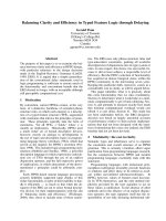
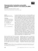
Fig. 1. Cdt1 protein expression in human fibroblasts (HFF) at the
transition from quiescence (G0) to proliferation. Totalcellextractsfrom
HFF c ells were analyzed by Western blotting u sing Cdt1, c yclin A,
Cdc6 and t ubulin specific antibodies. Lane 1 , proliferating HFF cells;
lane 2, HFF cells deprived of serum for 48 h; lanes 3–8, serum deprived
HFF c ells induced to re-enter the cell cycle by a ddition of serum and
collected at 6, 1 2, 15, 18, 21 and 24 h, respectively. The band corres-
ponding to Cd t1 has b een marked b y an a rrow, w hile a c ross-reacting
band ru nning above Cdt1 is i ndicated by an asterisk. Th e position o f
migration of the 66-kDa molecular mass marker ba nd is indicated a t
the l eft of the Cdt1 blot.
A
Pr
0 h
6 h
9 h
12 h
15 h
18 h
21 h
24 h
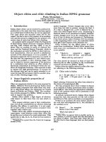
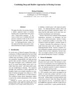
Anti-Cdt1 DAPI DAPIAnti-geminin
B
Fig. 2. Cdt1 and geminin protein levels and localization at the transition
from quiescence to proliferation. Proliferating HFF cells (Pr), HFF cells
deprived of serum f or 48 h (0 h ), or serum deprived c ells induced to re-
enter the cell cycle by s erum re-addition for 6, 9, 12, 1 5, 18, 21 and 24 h
were processed for indirect immunofluorescence using anti-Cdt1 and
anti-geminin (anti-Gem2) Ig. (A) Microscopy ima ges ( reco rde d with
identical exposure settings fo r all tim e points). ( B) Percentage of cells
showing staining for Cdt1 (white bars), BrdU (black bars)
14
or geminin
(hatched bars) in each time point. Over 200 cells were measured for
each time point. In order to assess cell cycle progression, cells were
incubated with B rdU 30 m in prior to fixatio n, and processed f or BrdU
staining as described in M aterials an d m ethods. Percent of B rdU
positive cells in each time point was scored.
Ó FEBS 2004 Cdt1 and geminin down-regulation in quiescence
1
(Eur. J. Biochem. 271) 3371
time-point is also shown in F ig. 2B f or comparison. Cdt1 is
detected in the nucleus of approximately o ne-third of cells
from an asynchronous population but its levels are mark-
edly dec reased in cells cultured in the absence of serum. Cells
staining for Cdt1 appear around 12 h f ollowing serum re-
addition, several hours before the p eak of cells in S phase
(21–24 h). We wished to compare the behavior of Cdt1 to
that of its inhibitory molecule, geminin, during cell cycle exit
and r e- entry. To that effect, w e assessed t he levels of the
geminin protein by immunofluorescence, in proliferating
HFF cells, upon serum withdrawal and upon serum re-
addition, in parallel to C dt1 i mmunofluorescence detection
described above (Fig. 2A, r ight pan els, immunofluorescence
images; F ig. 2 B quantitation) . G eminin was detected in the
nucleus of around one-quarter of asynchronously growing
HFF cells. Upon serum withdrawal geminin levels were
markedly de creased, similar to C dt1. Geminin staining first
re-appeared in a small number of cells around 15 h
following serum readdition, and peaked at 21–24 h together
with the p eak o f cells in S phase, as judged by the percentage
of BrdU positive cells. Geminin appeared in the cell
nucleus significantly later upon serum re-addition than
Cdt1 (a 3–6 h d elay) a nd its accumulation paralleled the
accumulation of BrdU positive cells.
Our data s uggest that both Cdt1 a nd geminin are down-
regulated in quiescent HFF cells. When cells re-enter the cell
cycle, Cdt1 is expressed first, a s cells prepare for a new
round of S phase, while geminin accumulates a s cells enter
S phase.
Geminin, an inhibitor of Cdt1 is severely down-regulated
upon cell cycle exit
Cdt1 is negatively regulated by g eminin [12,15], and, during
the cell cycle, g eminin acc umulates in S phase and G2, when
Cdt1 levels are low, while Cdt1 accumulates in G1, when
geminin is undetectable [12,13,16]. O ur imm unofluorescence
findings, showing that upon serum starvation of human
fibroblasts geminin is down-regulated similar to C dt1, were
somewhat surprising, a s g eminin might have been expected
to be up-regulated upon cell cycle exit. W e therefore wished
to examine t his point more carefully. The low l evels of
geminin protein and mRNA present in H FF cells howe ver
(see below) hampered a detailed analysis in these cells. We
therefore turned to mouse N IH3T3 cells, which express
geminin to l evels similar to HeLa cells (Fig. 3, left: compare
lanes 1 and 2), but can be induced to exit the cell cycle by
serum withdrawal or contact inhibition.
Figure 3 shows the levels of Cdt1 and geminin in
NIH3T3 ce lls, which a re induced to exit the cell cycle either
by serum d eprivation o r c ontact i nhibition. Cdc6/18 a nd
cyclin A protein levels serve as controls for proteins
previously shown t o be down-r egulated upon cell cycle exit,
while tubulin serves as a loading control. As shown above
for HFF cells, Cdt1 protein levels are significantly reduced
in serum starved NIH3T3 cells and re-accumulate upon
addition of serum. Cdt1 protein levels a re much less affected
by contact inhibition (still present 4 days following conflu-
ency). Geminin is s everely down-regulated by both serum
deprivation and contact i nhibition, similar to cyclin A and
more dramatically than Cdt1. For example, geminin protein
levels are undetectable upon serum starvation and are
already reduced from the first day following confluency,
when Cdt1 levels are still unaffecte d.
We conclud e that geminin is dramatically down-regulated
in NIH3T3 cells in G 0, consistent w ith our findings with
HFF human cells.
Cdt1 and geminin mRNAs are down-regulated in G0
During the cell cycle, Cdt1 and geminin mRNA levels are
mostly stable and protein levels are primary controlled by
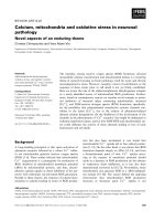
Fig. 3. Expr ession of Cdt1 and geminin proteins in quiescent and proliferating NIH3T3 cells. NIH3T3 cells were in duce d to exit t he cell cy cle by serum
starvation ( –S) or contact inhibition (Ci). Total cell extracts at the conditions i ndicated below w ere prepared a nd Western b lot analysis w as
performed using antibodies that recognize specifically Cdt1, geminin (Santa Cruz ), cyclin A, Cdc6 and t ubulin proteins. Lane 1, proliferating H eLa
cells; lane 2, proliferating N IH3T3 cells; l ane 3, s erum deprived NI H3T3 cells (cultu red for 48 h i n low serum); lane 4 , NIH3T3 c ells induced to
re-enter the cell c ycle upon serum addition for 6 h; lanes 5–8, contact inhibited NIH3T3 cells, 2, 3 and 4 da ys following confluency; lane 9, NIH3T3
cells induced to re-enter cell cycle by splitting 1 : 10, 4 da ys after confluency. The arrow on the cyclin A blot in dicates the band co rrespondin g to
cyclin A w hile the a sterisk marks a c ross-reacting band. Mouse Cdt1 m igrates slower t han human C dt1.
3372 G. Xouri et al. (Eur. J. Biochem. 271) Ó FEBS 2004
proteolysis [13]. Given the down-regulation of both proteins
upon serum deprivation, we wished to examine their
respective mRNA levels.
In Fig. 4A, t he mRNA levels of Cdt1, geminin and actin
(as loading control) are shown in serum deprived NIH3T3
cells, and 6 and 12 h following serum re-addition, and
compared with mRNA levels in proliferating ce lls. Both
Cdt1 and geminin mRNAs are down-regulated upon
serum deprivation and re-accumulate as cells prepare f or
S phase. Densitometry scanning and data normalization
against the actin c ontrol shows that g eminin mRNA is at
background l evels i n serum deprived NIH3T3 cells and at
6 h following serum re-addition, while at 12 h it has
returned to the l evel detected in proliferating c ells. Cdt1
mRNA levels are reduced twofold a nd similarly return t o
the l evels detected i n proliferating c ells by 12 h following
serum readdition.
We then examined how quickly upon serum deprivation
Cdt1 and g eminin mRNA levels are reduced (Fig. 4 B).
Densitometry analysis showed that geminin mRNA levels
appear significantly reduced already a t early time points
(12 h minus serum, twofold r eduction) and are further
reduced when ce lls are c ultu red longer in the absence of
serum ( reaching a sevenfold reduction at 40 h minus serum).
Cdt1 mRNA levels show a twofold reduction 24 h follow-
ing serum deprivation a nd remain at approximately the
same level to the end of the time course.
In order t o r eproduce our findings also in human HFF
cells, and given that Cdt1 and geminin mRNAs were hardly
detectable in these c ells by Northern blotting (see below) we
employed reverse-transcription followed by semiquantita-
tive PCR amplification (RT-PCR) of human Cdt1 and
geminin mRNAs in cycling, serum deprived and re-stimu-
lated H FF cells (Fig. 4C). C onsistent with our finding with
NIH3T3 cells, geminin mRNA levels were dramatically
down-regulated in s erum starved H FF cells and geminin
mRNA accumulated again around 1 8 h following serum
re-addition. Cdt1 mRNA levels were also decreased in
serum starved HFF cells and re-accumulated from 12 h
following serum re-addition. S imilar t o o ur findings with
NIH3T3 cells , mRNA fluctuations upon s erum withdrawal
in HFF c ells appeared less d ramatic for C dt1 than g eminin.
We conclude that Cdt1 and g eminin mRNA levels are
reduced in quiescent cells, suggesting that i n contrast to their
regulation during the cell cycle [13], upon exit and entry to
the cell cycle both genes are controlled, at least in part,
transcriptionally.
Cdt1 and geminin are highly expressed in proliferating
cells
in vivo
The experiments with cultured cells have suggested that
Cdt1 and geminin mRNA and protein are down-regulated
upon cell cycle exit and a re progressively up-regulated upon
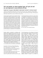
Fig. 4. Trans criptional control of C dt1 and
geminin in quiescent cells. (A) T otal cell RNA
prepared from proliferating NIH3T3 cells
(lane 1), from cells d eprived o f serum f or 48 h
(lane 4) o r from cells first serum deprived for
48 h and t hen cultured f or 6 o r 12 h in t he
presence o f 20% (v/v) serum (lanes 3 and 2,
respectively) was subjected to Northern b lot
analysis using a probe specific for human Cdt1
(upper), h uman g eminin (middle) or actin as a
loading control ( lower). (B) Northern blotting
analysis was performed using t ot al cell RNA
prepared from NIH3T3 cells that were grown
in the p resence of serum (lane 1), in the ab-
sence of serum for 12, 24, 3 2, 40 and 48 h
(lanes 2–6) or for 18 h after re-addition of
serum to c ells which w ere previously s erum
deprived for 48 h (lane 7) and hybridized usin g
specific probes f or Cdt1, geminin and a ctin.
(C) Total RNA extracted from HFF cells was
subjected t o reverse transcription and PCR
amplification with oligonucleotides specific for
the hCdt1 and h-geminin cDNAs. PCR w ith
oligonucleotides specific for actin served as a
loading control. Twofold dilutions of starting
cDNA were used to sh ow linearity (data not
shown). Lane 1 , proliferating H FF cells; l ane
2, HFF c ells deprived of serum for 48 h; lanes
3–7, s erum deprived HFF cells u pon serum
readdition for 6–24 h.
Ó FEBS 2004 Cdt1 and geminin down-regulation in quiescence
1
(Eur. J. Biochem. 271) 3373
re-entry to the cell c ycle. A recent report s howed that
geminin protein levels are positively correlated with cell
proliferation [17]. We investigated the in vivo expression of
Cdt1 and compared it t o that o f geminin in the d eveloping
mouse gut epithelium, a tissue in which a p roliferating and a
differentiating z one can be distinguished histolo gically. G ut
epithelium d iffe rentiation is initiated after E14dpc in mouse
embryos a nd co ntinues postnatally. We determined Cdt1
and g eminin mRNA and p rotein expression using in situ
hybridization and immunohistochemistry, respectively, on
sections from the gastrointestinal tract of an E17dpc mouse
embryo. A t t his stage of development, the g ut epithelium is
organized into villi, wh ich are separated at their bases by a
proliferating compar tment known as the intervillus epithe-
lium.
Cdt1 mRNA is mainly localized at the bases of the
developing intestinal villi (the intervillus e pithelium), where
the proliferating cells of the intestinal epithelium are
localized (Fig. 5A). Geminin mRNA has a distribution
similar to Cdt1 in the small and large intestine. Immuno-
histochemistry using antibodies specific for Cdt1 and
geminin (Fig. 5B) showed t hat both p roteins are detected
in the proliferating cell layer of the developing gut epithe-
lium in a similar expression pattern.
Therefore, Cdt1 and geminin mRNA and protein reveal a
similar distribution a long the gastrointestinal tract, localiz-
ing mainly in the proliferating zone of the g ut epithelium.
Cdt1 and geminin are over-expressed in cancer cells
Given the correlation we observed between Cdt1 and
geminin mRNA and protein levels and proliferation, we
wished to examine the expression levels of these two genes i n
different tumor cell lines and compare them to primary c ells.
Cellular lysates were prepared from human foreskin fibro-
blasts, a primary cell line, an d the tumorigenic cell lines
Saos, MDAMB231, MCF7, HeLa and LNcap. Western
blot analysis using a nti-Cdt1 Ig showed that Cdt1 protein is
detected at much lower levels in the primary HFF cells
compared with all the tumorigenic cell lines tested (Fig. 6A).
Western b lotting with commercial antibodies against gem-
inin showed that geminin protein levels are also increased in
cancer cell l ines, while diffe rent cancer cell lines appear to
over-express geminin to varying degrees. In order to more
carefully compare t he relative levels of Cdt1 and geminin in
different primary and cancer cell lines, we u tilized a more
sensitive a ntibodies against geminin (anti-Gem2) a nd inclu-
ded primary endothelial cells (Huvec), the normal diploid
human cell line MRC5 and two more cancer cell lines
(HT1080 and U2OS), in addition to HFF, HeLa and
MCF7 analyzed in Fig. 6A. A s shown i n Fig. 6 B, cancer
cell lines appear to consistently over-express Cdt1 in
comparison with primary and norma l d iploid c ell lines
(for lanes 5–8, a higher exposure is shown f or the Cdt1
blot, as e vident by the intensity in lanes 2 and 8, which both
correspond to HeLa cell, to permit detection of Cdt1 in t he
normal diploid MRC5 cells). Quantitation of the blots in
Fig. 6A,B showed that the majority of cancer cell lines
express Cdt1 over 10-fold more than primary cell lines.
Geminin i s h ardly d etectable in the primary cell lines, and
accumulates to higher levels in the majority of cancer cell
lines tested. I t i s noteworthy, however, that geminin levels
vary significantly amongst the cancer cell lines tested
(compare, for example, HeLa, lane 2 , t o MCF7, lane 3),
suggesting that the relative amount of Cdt1 and its inhibito r
geminin may differ in different cell lines.
In order to further investigate this point, we estimated how
many molecules o f Cdt1 and geminin are present on average
per cell in an asynchronous population of HeLa cells. To
this end, known amounts o f recombinant full-length
Cdt1 (HisT7-Cdt1) and recombinant full-length geminin
(His-geminin) were loaded o n a n S DS/PAGE g el alongside
total cell extract from 1.5 · 10
5
asynchronously growing
HeLa cells, and Western blotted with anti-Cdt1 a nd anti-
geminin Ig (Fig. 6C). We estimate that approximately 30 000
molecules of Cdt1 and an equal number of geminin
molecules are present on average per HeLa cell ( Materials
and methods), suggesting that rather similar levels of Cdt1
and i ts inhibitor are produced in this cell line, though a t
different cell cycle stages [13]. In contrast, we calculate a r atio
of Cdt1 to geminin of approximately 1 0 : 1 for MCF7 cells.
The difference in Cdt1 p rotein levels which is consistently
observed b etwee n primary a nd cancer ce ll lines teste d could
have been due to the l arger Ôin cycleÕ pool of the c ancer cell
lines. I n order to address t his, we used immunofluorescence
to assess whether Cdt1 is over-expressed in individual cells
Fig. 5. Cdt1 and geminin a re e xpressed i n proliferating cells of the
gastrointestinal tract. In situ hybridization (·5, ·10)
15
(A) an d
immunohistochemical (·5) (B) analysis o f Cdt1 ( left) and geminin
(right) expression on frozen section of an E17 dpc mouse emb ryo. Cdt1
and geminin mRNA and protein expression show a s imilar expression
profile. Consecutive sections of the gastroinestinal tract a re shown.
3374 G. Xouri et al. (Eur. J. Biochem. 271) Ó FEBS 2004
of a tumor cell line population in c omparison w ith primary
cells (Fig. 7). The number of H eLa cells staining positive
for C dt1 was higher (approximately 50% of HeLa cells
in comparison with 35% of H FF cells), consistent with a
higher percentage of HeLa cells act ively cycling. In addition
to that however, the staining observed in individual HeLa
cells was higher than the staining observed in individual
HFF cells (Fig. 7 A). Quantitation of 25 high-power fields
each for HFF and HeLa immunostainings shows that a
range of e xpression levels are observed in both HFF and
HeLa cells, as expected for a protein whose levels fluctuate
during the cell cycle, individual HeLa cells, however, express
on average over twofold higher levels of C dt1 than HFF
cells (Fig. 7B). These data show that Cdt1 is expre ssed to
higher levels in individual cycling cancer cells in comparison
with cycling primary cells.
To address whether Cdt1 and geminin up-regulation in
cancer cell lines would also occur at the mRNA level, we
used Northern blot analysis with total cell RNA extracted
from the primary HFF c ells and d ifferent tumor cell lines.
Similar t o protein levels, both C dt1 a nd geminin m RNA
was markedly increased in all the tumor c ell lines tested
compared with the primary HFF cells (Fig. 8A). We
employed densitometry in order to compare the levels of
hCdt1 and h-geminin m RNA amongst the d ifferent cancer
cell lines teste d. W hen c ompared w ith H eLa c ells,
Fig. 6. Cdt1 and geminin are highly expressed in cancer cells. (A, B) Western blotting an alysis was u sed to determine the expression of Cdt1, geminin
and tubulin as a loading control in cellular extracts from different h uman cell lines. (A) Lanes 1–6, H FF, Saos, MDAMB231, MCF7, HeLa and
LNcap, respectively. ( B) Lanes 1–8 , HFF, HeLa, MCF7, Huvec, HT1080, U20S, MRC5 and HeLa, respe ctively. A commercial anti-geminin I g
(Santa Cruz) was used for (A), while anti-Gem2, which s hows a higher sensitivity, was used for ( B). F or Cdt 1, a higher exposure is shown for lanes
5–8 in respect to lanes 1–4, to allow visualization of Cdt1 in MRC5 cells. For lanes 1–4, a higher exposure of the geminin blot (Gem lo ng) is also
shown a t the bottom of the panel, to pe rmit visualization of geminin in the primary HFF cells. ( C) Estimation of the number of Cdt1 a nd geminin
molecules present in H eLa cells. Western blotting of total cell extract from 1.5 · 10
5
asynchronously growing HeLa cells (marked HeLa) was run
alongside known amounts of recombinant full-length Cdt1 and geminin (HisT7-geminin and His-geminin, amount of recombinant protein run in
each lane in ng is indicated) and immunoblotted with anti-Cdt1 and anti-geminin Ig. See Materials and m ethods for calculations.
Ó FEBS 2004 Cdt1 and geminin down-regulation in quiescence
1
(Eur. J. Biochem. 271) 3375
MDAMB231 cells showed somewhat decreased levels for
both h Cdt1 and h-geminin (approximately threefold reduc-
tion) while MCF7 cells showed increased le vels o f hCdt1
mRNA (twofold) and d ecreased levels o f h-geminin mRNA
(threefold reduction), s upportive of our findings concerning
hCdt1 and h-ge minin protein levels in this cell line.
Therefore, while all c ancer cell lines tested express much
higher levels of hCdt1 and h-geminin mRNAs when
compared with primary cells, cancer cell lines may differ
in the le vels of o ve rexpre ssion o f the se mR NAs.
In Fig. 8B, a Northern blot containing total RNA
extracted from t umorigenic and matched normal specimens
(kidney cancer, liver cancer and lung canc er and respective
normal specimens) was hybridized with an h-geminin
specific probe and actin as a loading control. Densitometry
analysis sh ows that h-geminin mRNA is increased in the
tumor specimens tested (approximately twofold) in com-
parison with t he matched normal specimens, consistent with
our findings with cancer cell lines.
Discussion
In cycling cells, C dt1 i s specifically expressed during the G1
phaseofthecellcycleandisbelievedtoacttogetherwith
Cdc6 to load the MCM protein complex onto chromatin,
thereby licensing DNA for a further round of DNA
replication. When G1 cells exit the cell cycle and enter
quiescence, licensing is gradually lost. W e show her e that
Cdt1 is down-regulated when cells are i nduced to transit
from G1 into G0 by serum deprivation. Cdt1 protein levels
are low in serum deprived human primary fibroblast HFF
cells and NIH3T3 cells. Cdt1 appears again as cells are
induced to re-enter the cell cycle up on serum re-addition,
before a new round o f S phase is initiated, consistent with a
requirement for Cdt1 in re-licensing G0 chromatin for a new
round of DNA replication. Cdt1 protein l evels are not
appreciably affected early upon contact i nhibition, suggest-
ing that the degree of down-regulation of Cdt1 m ay vary
depending on how cells have entered t he quiescent state.
Fig. 7. Qu antitative immunofluorescence to compare Cdt1 protein l evels
in individual HFF a nd HeLa cells. Tr iplicates of HFF and H eLa cells
grown on c overslips were subjected to i mmunofluorescence with an ti-
Cdt1 Ig. (A) Microscopy images recorded under identical conditions
are shown. (B ) A s catter p lot of t he e xpression values o f C dt1 i n 25
different high-p ower fields each f or HFF and H eLa cells is sho wn.
Relative expression values were measured with
IPLAB
software in
arbitrary u nits.
Fig. 8. Cdt1 and geminin mRNA levels in cancer cell lines and tumours.
16
(A) Northern blotting analysis was p erformed to d etermine mRNA
levels of hCdt1, h-gem inin and actin i n different human cell lines.
Lanes 1 –5, HeLa, MDAMB231, LNcap, MCF7 and HFF cells,
respectively. ( B) Northern b lot bearing total mRNA from human
kidney, liver and lung tumors and corresponding normal specimens
was hybridized with a geminin specific probe, and an actin specific
probe as l oa ding control.
3376 G. Xouri et al. (Eur. J. Biochem. 271) Ó FEBS 2004
This would e xplain wh y a significant down-regulation of
Cdt1 in G0 was not previously detected [14]. Cdc6, Cdt1’s
partner f or DNA licensing, has been shown to b e down-
regulated in G0 cells and induced upon cell cycle re-entry
[23] and to be under the transcriptional c ontrol of E2F [24–
26]. We show here that, similar to Cdc6, Cdt1 mRNA levels
are r educed in G0 and r e-accumu late at the G0 to cell c ycle
transition, suggesting that, in this transition, Cdt1 m ay be
controlled transcriptionally. T he presence of predicted E2F
binding sites on the putative Cdt1 promoter (D. Kougiou
and S. Taraviras, unpublished observation) attests to a
possible E 2F mediated regulation of Cdt1. I ndeed E2F was
recently reported to regulate the transcription o f Cdt1 [34].
In contrast, in cycling c ells, C dt1 appears to be controlled
mostly post-transcriptionally [13]. The difference in Cdt1
regulation we observe between serum deprived and contact
inhibited NIH3T3 cells may indicate a regulation of Cdt1 b y
growth factors p resent in the serum. In th at respect it is
interesting to note that consensus sites for myc and TCF-1
A are also present on t he Cdt1 promoter. In addition, we
detected slower migrating forms of Cdt1 in serum d eprived
HFF cells upon longer exposure (data not shown),
suggesting that an additional control of Cdt1 at the post-
translational level may also operate in G0.
Geminin i s b elieved to act as a cell cycle inhibitor, with a
role to prevent untimely licens ing by specifically binding to
Cdt1 and inhibiting its MCM loading function [12,15,16].
During the cell cycle, g eminin is expressed in S and G2
phases, when licensing should b e i nhibited and when the
Cdt1 protein is undetectable [13,16]. Based on these
findings, on e might have expected geminin to be i nduced
when G1 cells exit the cell cycle into G0. In c ontrast,
however, w e s h ow h ere that g eminin levels are e xtre mely
low in quiescent cells, with a decrease even more pro-
nounced than that observed f or Cdt1. For example, geminin
is already undetectable in NIH3T3 cells in early confluency,
when Cdt1 levels are not affected. Geminin levels closely
mirror the levels o f cyclin A in these experiments. Geminin
mRNA levels also appear to be reduced early upon serum
withdrawal and more dramatically than Cdt1 mRNA levels.
These findings are consistent with a recent publication
linking geminin expression to the p roliferating cell [ 17]. An
E2F binding site is als o present on t he predicted geminin
promoter (D. Kougiou and S. Taraviras, unpublished
observations) suggesting that C dt1 and its inhibitor might
be under s imilar transcriptional regulatory mechanisms.
Indeed, while this m anuscript w as under r eview, regulation
of geminin b y E2F and R B was reported [34,35]. Geminin
protein accumulates in HFF cells re-entering the cell cycle
from G0 3–6 h later t han Cdt1, and as cells enter into
S phase (Fig. 2). The accumulation of geminin may i nhibit
further licensing and define the Ôwindow of opportunityÕ for
licensing at the transition f rom quiescence to proliferation.
The distribution of Cdt1 a nd geminin in the gut
epithelium mirrors our findings with cultured cells. We
show by in situ hybridization and imm unohistochemistry
that both Cdt1 and geminin are expressed in the prolifer-
ative compartment of the developing mouse g ut epithelium,
consistent with a down-regulation of these factors upon cell
cycle exit. This is the first report o f t he distribution of C dt1
in a mammalian tissue, while our findings are consistent
with a previous report on geminin’s localization [17].
Cancer cells have defects in the control mechanisms
regulating cell cycle exit and therefore divide uncontrolla-
bly. In addition, cancer cells often exhibit genomic
instability a nd are aneuploid. A recent report showed that
over-expression of Cdt1 can predispose cells to a malignant
transformation [36], thereby identifying Cdt1 as a putative
oncogene. In addition, over-expression of Cdt1 t ogether
with Cdc6 has been shown t o result i n re-replication a nd
genomic instability in both yeast and human ce lls [7,37].
We wished to examine whether Cdt1 is over-expressed i n
cancer cells in culture, in comparison with normal cycling
cells. We show that both Cdt1 protein and mRNA
accumulate to much higher levels in cancer cells. Immuno-
fluorescence experiments showed that Cdt1 is present in
higher levels in individual cancer cells, vs. cycling primary
fibroblasts. Over-expression of Cdt1 in cancer cell lines can
therefore not be solely accounted for by t he larger Ôin cycleÕ
fraction of cancer cells . It would b e interesting to
investigate the mechanism that leads to over-accumulation
of Cdt1 in cancer cells and the functional s ignificance of
this over-expression for malignant transformation. The
presence of increased mRNA levels suggests t hat over-
expression may be at least partly due to increased
transcription or increased gene copy number. Over-expres-
sion of a stable form of geminin was recently s hown to
induce cell cycle arrest or apoptosis in human cell lines
[17,38]. However, endogenous geminin is over-expressed in
the m ajority o f c ancer cells tested, i n comparison with
normal cells. This is obser ved both in cancer cell lines and
human tumors and is consistent with a recent report [17].
The degree of over-expression of the geminin protein
differs between cell line s, r esulting in gross differences in
relative amounts of Cdt1 and its i nhibitor in different cell
lines. I t would b e interesting to investigate w hether geminin
and/or Cdt1 levels or the ratio of Cdt1 to its i nhibitor
geminin may show a correlation with the type, aggressive-
ness or molecular pathology of a given t umor.
Acknowledgements
We would like to t hank A. Pyriohou, D. Kalatzis, M. Iliou and V .
Roukos for assistance with experiments, M. Ohtsubo and N.
Tsopanoglou for cell l ines, t he Ba stiaens laboratory f or help with
quantitations and Profs G. Maniatis, C. Flordellis and A. Athanassia-
dou for their support and critical reading of t he manuscript. Our w ork
is sup ported b y g r ants f rom the As sociation for Intern ational C a ncer
Research, University of Patras-Karatheodori Program, the Emperirikio
Foundation, Human Frontiers Science P rogram, and the Japanese
Ministry of Education, Culture, Sports, S cience and T ech nology.
References
1. Nurse, P. (1994) Ordering S phase and M pha se in t he cell cycle.
Cell 79, 547–550.
2. Blow, J.J. & Laskey, R.A. (1988) A role for t he nuclear envelope in
controlling DNA replication within the cell cycle. Nature 33 2,546–
548.
3. Blow, J.J. & Hodgson, B. (2002) Replic ation licensing – defining
the proliferative s tate? Trends Cell Biol. 12, 72–78.
4. Bell, S.P. & Dutta, A. (2002) DNA replication in eukaryotic cells.
Annu. R ev. Biochem. 71 , 333–374.
5. Nishitani, H. & Lygerou, Z. (2002) C ontrol of D NA r eplication
licensing in a cell cycle. Genes C ells 7, 523–534.
Ó FEBS 2004 Cdt1 and geminin down-regulation in quiescence
1
(Eur. J. Biochem. 271) 3377
6. Labib, K. & Diffley, J.F. (2001) Is the M CM2-7 complex t he
eukaryotic DNA replication fork helicase? Curr. Opin. Genet. D ev.
11, 6 4–70.
7. Nishitani, H., L ygerou, Z., Nishimoto, T. & Nurse, P. (2000) The
Cdt1 protein i s required to lic ense DNA f or replication in fission
yeast. Natur e 404, 625–628.
8. Maiorano, D., Moreau, J. & Mechali, M. (2000) XCDT1 is
required for the assembly of pr e-replicative complexes in Xenopus
laevis. Nature 404, 6 22–625.
9. Whittaker, A.J., R oyzman, I. & Orr-Weaver, T .L. (2000) Dr oso-
phila double parked: a conserved, essential replication p rotein that
colocalizes with the origin recognition co mplex and links DNA
replication with mitosis and the down-regulation of S phase
transcripts. Genes D ev. 14, 1765 –1776.
10. Tanaka, S. & Diffley, J .F. (2002) Interdependent nuclear a ccu-
mulation of budding yeast Cdt1 and Mcm2–7 during G1 ph ase.
Nat. Cell Biol. 4, 1 98–207.
11. Devault, A., Vallen, E.A., Yuan, T., G reen, S., Bensimon, A. &
Schwob, E. ( 2002) Id en tification of Tah11/Sid2 a s t he ortholog of
the r eplication licensing factor Cdt1 in Sa ccha romyces cerevisiae.
Curr. B iol. 12, 689– 694.
12. Wohlschlegel, J.A., Dwyer, B.T., Dhar, S.K., Cvetic, C.,
Walter, J.C. & Dutta, A. (2000) In hibition of e ukaryotic DNA
replication by g eminin binding to Cdt1. Science 290, 2309–2312.
13. Nishitani, H., Taraviras, S., Lygerou, Z. & Nishimoto, T . (2001)
The human licensing factor for DNA r eplication Cdt1 accumu-
lates i n G1 and is destabilized after initiation of S-phase. J. Bio l.
Chem. 276, 449 05–44911.
14. Rialland, M., Sola, F. & Santocanale, C. (2002) Essential role of
human CDT1 in DNA replication and c hroma tin licensing. J. Cell
Sci. 115, 1435–1440.
15. Tada, S., Li, A., Maiorano, D., Mechali, M. & Blow, J.J. (2001)
Repression of origin assembly in metaphase de pends on in hibition
of RLF-B/Cdt1 by geminin. Nat. Cell Biol. 3, 107 –113.
16. McGarry, T.J. & Kirschner, M.W. ( 1998) G eminin, a n i nhibitor
of DNA replication, is degraded during mitosis. Cell 93, 1043–
1053.
17. Wohlschlegel, J.A., K utok, J. L., W eng, A.P. & Dutta, A . (2002)
Expression of geminin as a marker o f cell proliferation in normal
tissues and m alignancies. Am. J. P athol. 161, 267–273.
18. Leno, G.H. & M unshi, R. (1994) Initiation of DNA replication in
nuclei from quiescent cells re quire s permeabilization of the nuclear
membrane. J. Cell Biol. 127, 5–14.
19. Fang, J. & Be nbow, R.M. ( 1996) Nuclear proteins of quiescent
Xenopus laevis cells inhibit DNA replication in intact and per-
meabilized nuclei. J. Cell Bi ol. 133, 955–969.
20. Madine, M.A., Swietlik, M ., Pelizon, C., Romanowski, P., M ills,
A.D. & Laskey, R.A. (2000) The roles of the MCM, ORC, a nd
Cdc6 proteins in determining the replication competen ce of
chromatininquiescentcells.J. Struct. Biol. 129, 198–210.
21. Sun, W., Hola, M ., Pedley, K., Tada, S., B low, J.J., Todorov, I.T.,
Kearsey, S.E. & Brooks, R.F. ( 2000) The replication capacity o f
intact mammalian nuclei in Xenopus egg extracts declines wi th
quiescence, but th e residual DNA syn the sis i s independe nt of
Xenopus MCM proteins. J. Cell Sci. 113, 683–695.
22. Stoeber, K., Tlsty, T .D., H apperfield, L., Thomas, G.A., Roma-
nov, S., Bobrow, L., Williams, E.D. & Williams, G.H. (2001)
DNA replication licensing and human cell proliferation. J. Cell
Sci. 114, 2027–2041.
23. Williams, R.S., Shohet, R.V. & Stillman, B. (1997) A human
protein related to yeast Cdc6p. Proc. N atl Acad. Sci. USA 94 , 142–
147.
24. Yan, Z., DeGregori, J., Shohet, R., Leone, G., Stillman, B.,
Nevins, J.R. & Williams, R.S. (1998) Cdc6 is regulated by E2F and
is essential for DNA r eplication in mammalian cells. Proc. Natl
Acad. Sci. USA 95, 3603–3608.
25. Ohtani, K., Tsujimoto, A., Ikeda, M . & Nakamura, M. (1998)
Regulation of cell growth-dependent expression of mammalian
CDC6 gene by the cell cycle transcription factor E2F. On cogene
17, 1 777–1785.
26. Hateboer,G.,Wobst,A.,Petersen,B.O.,LeCam,L.,Vigo,E.,
Sardet,C.&Helin,K.(1998)Cellcycle-regulatedexpressionof
mammalian CDC6 is depende nt on E2F. Mol. Cell. B io l . 18, 6679–
6697.
27. O htani, K., DeGregori, J., Leone, G., Herendeen, D.R.,
Kelly, T.J. & Nevins, J.R. (1996) Expression of the HsOrc1 gene, a
human O RC1 homolog, is r egulated by cell p roliferation v ia the
E2F transcription factor. Mol. Cell. Biol. 16, 6977–6984.
28. Ohtani, K., Iwanaga, R., Nakamura, M., Ikeda, M., Yabuta, N .,
Tsuruga, H. & Nojima, H. (1999) Cell growth-regulated expres-
sion of mammalian MCM5 a nd MCM6 genes mediated by the
transcription factor E2F. Oncogene 18 , 2299–2309.
29. Williams, G.H., Romanowski, P ., M orris, L., Madine, M.,
Mills, A.D., Stoeber, K., Marr, J ., Laskey, R.A. & Coleman, N.
(1998) Improved cervical smear assessment using antibodies
against proteins that r egulate DNA replication. Proc.NatlAcad.
Sci. USA 95 , 14932–14937.
30. Stoeber, K., Halsall, I., Freeman, A., Swinn, R., Doble, A.,
Morris,L.,Coleman,N.,Bullock,N.,Laskey,R.A.,Hales,C.N.
& Williams, G.H. (1999) Immunoassay for urothelial cancers that
detects DNA replication protein Mcm5 in urine. Lanc et 354,
1524–1525.
31. Freeman, A., Morris, L.S., Mills, A.D., Stoeber, K., Laskey, R.A.,
Williams, G.H. & Coleman, N. (1999) Minichromosome main-
tenance proteins as biological markers of dysplasia and malig-
nancy. Clin. Cancer Res. 5, 2121–2132.
32. Samb rook, J., Fritsch, E.F. & Maniatis, T. (1989) Molecular
Cloning: a Laboratory M anual, 2nd edn. Cold Spring Harbor
Laboratory Press, Cold Spring Harbor, NY.
33. Schaeren-Wiemers, N. & Gerfin-Moser, A. (1993) A single
protocol to detect transcripts of various types and expression
levels in ne ural tissue and cultured c ells: in situ hybridisation using
digoxigenin-labelled c RNA probes. Histochemistry 100, 431–440.
34. Yoshida, K. & Inoue, I. ( 2004) Regulation of Geminin and Cdt1
expression by E2F transcription factors. Oncogene 23, 3802–3812.
35. Markey, M .P., Siddiqui, H. & K n udsen, E.S. (2004) G eminin is
targeted for repression by the RB pathway through intragenic E2F
sites. J. Biol. Chem. 279, 29255–29262.
36.Arentson,E.,Faloon,P.,Seo,J.,Moon,E.,Studts,J.M.,
Fremont, D.H. & Choi, K. (2002) Oncogenic potential of th e DNA
replication licensing protein C DT 1. Oncogene 21 , 1150–1158.
37. Vaziri,C.,Saxena,S.,Jeon,Y.,Lee,C.,Murata,K.,Machida,Y.,
Wagle, N., Hwang, D.S. & Dutta, A. (2003) A p53-dependent
checkpoint pathway p revents rereplication. Mol. Cell 11, 997 –
1008.
38. Shreeram, S., Sparks, A., Lane, D.P. & Blow, J.J. (2002) Cell type-
specific r esponses of human cells to inhibition of replication
licensing. Onc ogene 21, 6624 –6632.
3378 G. Xouri et al. (Eur. J. Biochem. 271) Ó FEBS 2004