Báo cáo khoa học: GlnK effects complex formation between NifA and NifL in Klebsiella pneumoniae docx
Bạn đang xem bản rút gọn của tài liệu. Xem và tải ngay bản đầy đủ của tài liệu tại đây (277.34 KB, 10 trang )
GlnK effects complex formation between NifA and NifL in
Klebsiella
pneumoniae
Jessica Stips, Robert Thummer, Melanie Neumann and Ruth A. Schmitz
Institut fu
¨
r Mikrobiologie und Genetik, Go
¨
ttingen, Germany
In Klebsiella pneum oniae, the nif specific transcriptional
activator NifA is inhibited by NifL in response to molecular
oxygen and a mmonium. H ere, we demonstrate complex
formation between NifL and N ifA (approximately 1 : 1
ratio), when synthesized in the presence of oxygen and/or
ammonium. U nder simultaneous oxygen- and nitrogen-
limitation, significant but fewer NifL–NifA complexes
(approximately 1%) were formed in the cytoplasm a s a
majority of NifL was sequestered to the cytoplasmic mem-
brane. These findings indicate that inhibition of NifA in the
presence of oxygen and/or ammonium occurs via direct
NifL interaction and formation of those inhibitory NifL–
NifA complexes appears to be directly and exclusively
dependent on the localization of N ifL in the cytoplasm. We
further observed e vidence t hat t he nitrogen sensory protein
GlnK forms a trimeric complex w ith N ifL a nd NifA under
nitrogen limitation. Binding of GlnK to NifL–NifA was
specific; however the amount of GlnK within these com-
plexes was small. Finally, two lines of evidence were obtained
that under anaerobic conditions but in the p resence of
ammonium additional N trC-independent GlnK synthesis
inhibited the formation of stable i nhibitory NifL–NifA
complexes. Thus, we propose that the NifL–NifA–GlnK
complex reflects a transitional structure and hypothesize that
under nitrogen-limitation, GlnK interacts with the inhibi-
tory NifL–NifA complex, resulting in its d issociation .
Keywords: Klebsiella pneumoniae; nitrogen fixation; NifL;
NifA; Gln K.
Nitrogen-fixing microorganisms tightly control both syn-
thesis and activity of nitrogenase in response to o xygen and
nitrogen availability, because of the high energy demands of
nitrogen fixation and the oxygen sensitivity of nitrogenase
[1,2]. Transcription of the nitrogen fixation (nif)genesin
diazotrophic bacteria is, in general, mediated by the
activator protein NifA in combination with the alternative
r
54
-RNA polymerase [3,4]. In the free-living Klebsiella
pneumoniae, Azotobacter vinelandii and Azoarcus sp. B H72,
NifA transcriptional a ctivity is r egulated by a second
regulatory protein, NifL, which inhibits NifA in response
to external molecular oxygen and ammonium [5–8]. This
inhibition of NifA activity by NifL apparently occurs via
direct protein–protein interaction, which is i mplied by
evidence from immunological studies in K. pneumoniae [9],
and is consistent with recent studies for A. vinelandii using
the yeast two-hybrid system and in vitro analysis of complex
formation between NifL and NifA [10–14].
Under c onditions of nitrogen limitation, NifL allows
NifA activity only in the absence of oxygen, w hen i ts FAD
cofactor is reduced [6,15,16]. Recently, we have shown t hat
in K. pneumoniae, NifL is membrane-associated under
simultaneous anaerobic and nitrogen-limited conditions,
but is in the cytosolic fraction when in the presence of
oxygen or sufficient nitrogen [ 17]. We further demonstrated
that membrane association o f NifL depends on NifL
reduction at the cytoplasmic membrane by electrons derived
from the reduced quinone pool [18,19]. These findings
indicate that sequestration of NifL to the cytoplasmic
membrane under d erepressing conditions appears to be t he
main mechanism for regulation of cytoplasmic NifA activity
by NifL. R ecent gen etic evidence strongly suggests that the
nitrogen status of the cells is transduced towards the NifL/
NifA regu latory system by the GlnK p rotein, a paralogue
PII-protein [20–24]. Interactions between A. vineland ii
GlnK and NifL w ere recently demonstrated using t he yeast
two-hybrid system, a nd in vitro studies indicated that the
nonuridylylated form of A. vinelandii GlnK activates the
inhibitory function of NifL under n itrogen excess by d irect
protein–protein interaction [25,26]. Under nitrogen limita-
tion, however, the inhibitory activity of A. vinelandii NifL
appears to be relieved by e levated levels of 2-oxoglutarate
[14,24,27]. In contrast t o A. vinelandii,inK. pneumoniae the
relief o f NifL inhibition under n itrogen limitation depends
on GlnK, the uridylylation state of which appears not to be
essential for its nitrogen signaling function [20–23]. We have
recently s hown that i n the absenc e of G lnK, K. pneumoniae
NifL was located in the cytoplasm a nd inhibited NifA
activity under derepressing conditions [17]. However, it is
currently not known how GlnK influences the localization
of NifL in response to the nitrogen status and whether
Correspondence to R. A. Schmitz, Institut fu
¨
r Mikrobiologie und
Genetik, Georg-August Universita
¨
tGo
¨
ttingen, Grisebachstr. 8,
D37077 Go
¨
ttingen, Germany. Fax:+49 551 393808,
Tel.:+49 551 393796, E-mail:
Abbreviation: IPTG, isopropyl thio-b-
D
-galactoside.
Note: J. S tips and R. T h ummer contributed equally to this work and
should both be considered first authors.
(Received 9 May 2004, revised 2 2 June 2 004, accepted 29 June 20 04)
Eur. J. Biochem. 271, 3379–3388 (2004) Ó FEBS 2004 doi:10.1111/j.1432-1033.2004.04272.x
GlnK interacts directly with NifL or NifA, or affects the
NifL–NifA complex formation. In order to address those
questions, we analyzed in vivo complex formation between
the regulatory proteins after coexpression under various
nitrogen and oxygen availabilities. During these studies we
obtained evidence for the presence of an intermediate NifL–
NifA–GlnK complex, which is to our knowledge the first
report f or an in viv o formationofsuchaNifL–NifA–GlnK
complex.
Materials and methods
Bacterial strains
The bacterial strains used i n this wo rk were K. pneumoniae
M5al (wild type) and K. pneumoniae UN4495 [/(nifK-
lacZ)5935 Dlac-4001 hi D4226 Gal
r
] [28]. Plasmid DNA
was transformed into K. pneumoniae cells by electropora-
tion.
Construction of plasmids
Plasmid pRS201 contains the K. pneumoniae nifLA operon,
5¢-fused to the Escherichia coli malE gene in pMAL-c2 (New
England Biolabs) which is under the control of the tac
promoter. The plasmid was constructed as follows: A 3.1 kb
PCR fragment carrying nifLA was generated using chro-
mosomal K. pneumoniae DNA as template and a set of
primers, which were homologous to the nifLA flanking 5¢-
and 3 ¢-regions with a dditional Eco RI and HindIII synthetic
restriction r ecognition sites (underlined) (5¢-CACACA
GGAAACA
GAATTCCCGGG-3¢, sense primer (NifLE-
coRI); 5¢-CAATGTCCTG
AAGCTTACATAAGGCTT
CAC-3¢, antisense primer (NifAHindIII). The 3.1 kb PCR
product was cloned into t he EcoRI and HindIII sites of
pMAL-c2, resulting in malE fused to nifLA with one
additional amino acid (Ala) preced ing the me thionine of
NifL. The correct insertion was analyzed by sequencing.
Plasmids encoding MBP-NifL (pRS180), MBP-NifA
(pRS158), a nd MBP-NifL plus NifA (pRS209), in a ddition
to K. pneumoniae GlnK under the control o f the tac
promoter, were constructed as follows. Plasmids pRS163,
pRS98 and pRS205 were constructed by inserting a
tetracycline-resistance cassette [29] into the HindIII site of
plasmids pJES794, pJES597, and pRS201 encoding MBP-
NifL, MBP-NifA, and MBP-NifL plus NifA, respectively
[30,31, this paper]. An 0.4 kb PCR fragment carrying glnK
under the control o f the tac promoter was g enerated using
pRS155 [32] as template and a set of phosphorylated
primers: sense primer (pKK223–3F, 5¢-GACCACCGCG
CTACTGCC-3¢) and ant isense p rimer (pKK223–3R,
5¢-GATGCCGGCCACGATGCG-3 ¢). This 0.4 kb PCR
fragment was cloned into the ScaI site located inside the
ampicillin re sistance gene (bla) in pRS163, p RS98, and
pRS205 re sulting in pRS180 (MBP-NifL plus GlnK),
pRS158 (MBP-NifA p lus GlnK), a nd pRS209 (MBP-NifL
plus NifA plus GlnK), respectively. pRS192 was constructed
by inserting t he 0.4-kbp PCR fragment c arrying glnK under
the control o f the tac promoter generated as mentioned
above into the SacIandPstI site of pMAL-c2 (New England
Biolabs) and the tetracycline-resistance cassette into the
HindIII site. pRS239 was obtained by inserting the tetra-
cycline-resistance cassette into the HindIII site of pRS155,
encoding glnK under the control of t he ta c promoter.
Growth conditions
K. pneumoniae strains were g rown aerobically or anaer-
obically at 30 °C in minimal medium supplemented with
either 4 m
M
glutamine (nitrogen limitation) or 10 m
M
ammonium (nitrogen sufficiency) as the sole nitrogen
source and 1% (w/v) sucrose as t he sole carbon source
[33]. F or anaer obic g rowth conditions in closed bottles
with molecu lar n itrogen (N
2
) as gas phase, the medium
was supplemented with 0.3 m
M
sulfide and 0.002% (w/v)
resazurin to monitor anaerobiosis. P recultures of the 1 L
anaerobic main cultures were grown overnight in closed
bottles with N
2
as gas phase in the same medium
but lacking sulfide and resazurin. A erobic 1 L cultures
were incubated in 2 L flasks with vigorous shaking
(130 r.p.m).
Cell extracts and purification of proteins
MBP-NifL and MBP-NifA was synthesized at 30 °C
under nitrogen limitation or sufficiency in K. pneumoniae
carrying pJES794 [30] and pJES597 [31], respectively.
Expression of fusion protein w as induced from the tac
promoter for 2 h with 100 l
M
isopropyl t hio-b-
D
-gal-
actoside (IPTG) when cultures reached D
600
¼ 0.6. After
disruption of cells in breakage (B) buffer and centrifu-
gation at 20 000 g, fusion proteins were purified from the
supernatant by amylose affinity chromatography [16].
Expression and purification of K. pneumoniae GlnK and
E. coli GlnDDC w ere c arried out as described recently
[32]. Purified GlnK was modified in vitro by uridylylation
with E. coli GlnDDC and the modification was investi-
gated in nondenaturatin g polyacrylamide gels as recently
described [32,34].
Complex formation assays with purified proteins
To analyze whether purified GlnK interacts with NifL or
NifA, a binding assay using affinity chromatography was
used. Reactions were carried out in B-buffer in a total
volume of 230 lL in the presence or absence of MgATP
(1 l
M
), MgADP (1 l
M
)ora-ketoglutarate (10 l
M
). Purified
MBP-NifL, MBP-NifA, unmodified GlnK and uridylylated
GlnK were generally used at 3 l
M
in the reactions; the
concentration of the GlnK fractions were calculated in
terms of the trimer. A fter preincubation for 10 min at
30 °C, 500 lL of amylose resin (New England Bioloabs)
equilibrated with B-buffer w as added to t he mixtures
followed by an additional incubation for 20 m in at room
temperature. N onbinding protein w as subsequently washed
from the columns with B-buffer and the bound material was
then eluted from the column with B-buffer containing
10 m
M
maltose. Aliquots of the wash and e lution fractions
were separated on a denaturing 12.5% polyacrylamide gel,
which was subsequently stained with silver. The elution
fractions were further analyzed by Western blot analysis
using polyclonal antibodies raised against K. pneumoniae
MBP-NifA, MBP-NifL or GlnK to detect small amounts of
proteins.
3380 J. Stips et al. (Eur. J. Biochem. 271) Ó FEBS 2004
Isolation and characterization of complexes formed
in vivo
by affinity chromatography
Coexpression of malE-nifLA, malE-nifL plus glnK, malE-
nifA plus glnK,andmalE-nifLA plus glnK were induced
with 100 l
M
IPTG at a D
600
between 0.5 and 0.6 in
K. pneumoniae strain M5a1 carrying pRS201, pRS180,
pRS158 and pRS209, respectively. Main cultures (1 L)
were grown under aerobic or anaerobic conditions in the
presence of 10 m
M
ammonium or 4 m
M
glutamine (see
growth conditions). The respective growth and synthesis
conditions were maintained until cell breakage, if not
stated otherwise (e.g., in s hift experimen ts). In g eneral,
purification of complexes subsequently followed d irectly
after cell harvest without any storage at lower temper-
atures. Preparation of cell extracts in B-buffer and all
following purification steps were performed in the
presence of the protease inhibitor cocktail f or bacterial
cell e xtracts (Sigma). Depending on the synthesis condi-
tions, cell extract preparation and purification of the
fusion proteins from the 2 0 000 g supernatant by amylose
affinity chromatography was performed either under
aerobic conditions or under anaerobic conditions inside
an anaerobic c hamber with a nitrogen atmosphere and
using anaerobic buffers supplemented with 2.0 m
M
dithiothreitol [16]. The respective wash and elution
fractions were analyzed by gel electrophoresis an d silver
staining.
Quantification of NifL, NifA and GlnK in isolated
complexes by Western blot analysis
After purification of potential complexes, proteins from the
respective elution fractions were separated on denaturating
polyacrylamide gels and transferred t o nitrocellulose mem-
branes (BioTraceÒNT, Pall Life Science) [35]. Membranes
were exposed to specific polyclonal rabbit antisera directed
against the MBP-NifL, MBP-NifA, GlnB or GlnK protein
of K. pneumoniae. The primary antibodies were used in a
high dilution range, conditions under which cross-reaction
with other proteins are negligible. Protein bands were
detected with secondary antibodies directed against r abbit
immunoglobulin G a nd coupled to horseradish peroxidase
(Bio-Rad Laboratories) and visualized using the ECLplus
system (Amersham Pharmacia) with a fluoroimager ( Storm,
Molecular Dynamics). T he protein b ands of the c omplexes
were quantified for each growth condition from at least
three independent cultures using the ImageQuant v1.2
software (Molecular Dynam ics) and known amounts of the
respective purified control proteins, which were simulta-
neously detected and quantified with the respective complex
fraction on the same m embrane for each experiment.
Quantification of purified proteins MBP-NifL and MBP-
NifA was linear within absolute amounts of 0.06–0.25 lg
per lane a nd GlnK wi thin 0.01–0.14 lg. All quantifications
of proteins were performed w ithin this linear range of the
detection system. The relative amounts of GlnK in
complexes a re in general s tated in terms of the trimeric
GlnK protein (GlnK
3
). Degrada tion of M BP-NifL a nd
MBP-NifA in the elution fraction was frequently observed,
as was the case for purified standard proteins. This
degradation is based upon protein instability even at low
temperature. As other proteins within the isolated com-
plexes were not detected by SDS/PAGE and s ilver staining,
the f usion protein and the major degradation products
detected by the immunoblot were quantified together, if
degradation occurred.
b-Galactosidase assay
NifA-mediated activation of transcription from the
nifHDK promoter in K. pneumoniae UN4495 and
UN4495 carrying pRS239 was monitored by measuring
therateofb-galactosidase synthesis durin g exponential
growth (units per ml p er cell turbidity at 600 nm (D
600
)
[33]). Inhibitory effects of NifL on NifA activity in
response to ammonium were assessed b y virtue of a
decrease in nifH expression.
In vitro
transcription assay
Single cycle transcription assays were performed at 30 °C
with purified r
54
RNAP as desc ribed by N arberhaus et al.
[30] using 1.0 l
M
central domain of NifA (cdNifA),
r
54
RNAP ( 60 n
M
core po lymerase and 100 n
M
r
54
)and
5n
M
pJES128 as template (containing the K. pneumoniae
nifH promoter regulatory region) [36]. When analyzing the
effect o f the inhibitory activity of MBP-NifL synthesized
under a naerobic and nitrogen limited conditions, all the
reaction steps were p erformed under anaerobic conditions
in th e presence of 2 m
M
dithiothreitol and inside an
anaerobic chamber until open complex formation was
completed. Subsequently, synthesis of transcripts was
allowed by the addition of the nucleotide mix (400 l
M
ATP, 400 l
M
GTP, 400 l
M
UTP, 100 l
M
CTP, 200 kBq
[
32
P]CTP[aP], 0.1 mg ÆmL
)1
heparin) and further incubation
for 10 min at 30 °C outside the anaerobic chamber.
[
32
P]CTP[aP]-labeled transcripts were analyzed by electro-
phoresis in denaturing 6% polyacrylamide g els and quan-
tified with a BAS 1500 Image Analyzer (Fuji) or with the
PhospohorImager Storm (Molecular Dynamics).
Membrane preparations
Cytoplasmic and membrane fractions of cell-free cell
extracts were separated by several centrifugation steps
under aerobic or anaerobic conditions as recently des-
cribed by Klopprogge et al. [17] i n the presence of the
protease inhibitor cocktail for bacterial cell e xtracts
(Sigma). The quality of the membrane preparations was
evaluated by determination of the malate dehydrogen ase
activity in both the membrane and the cytoplasmic
fraction, according to Bergmayer [37]. In addition quino-
proteins were specifically detected by a redox-cycle stain
assay to detect leakage of membrane proteins into the
cytoplasmic fraction [ 38]. T he MBP-NifL and GlnK bands
of cytoplas mic and membrane fractions were quantified in
Western b lot analyses using known amounts of purified
proteins as descr ibed above. Quantities of MBP-NifL and
GlnK in the cytoplasmic and membrane fractions were
calculated as relative to total MBP-NifL and GlnK,
respectively, setting the absolute amo unts of the respective
protein in both fractions (cytoplasmic and membrane
fraction) as 100%.
Ó FEBS 2004 Complex formation between NifL, NifA and GlnK (Eur. J. Biochem. 271) 3381
Results and Discussion
We propose that GlnK transduces the nitrogen signal to the
nif-regulatory system in K. pneumoniae by affecting t he
localization of NifL in response to the nitrogen statu s,
possibly b y direct interaction with NifL or the NifL–NifA
complex. We thus examined: (a) the f ormation of complexes
between NifL, NifA and the primary nitrogen sensor GlnK;
and ( b) how GlnK effects N ifL localization in response to
the nitrogen stat us.
NifL and NifA form stoichiometric complexes after
coexpression in
K. pneumoniae
As no protein i nteractions between purified GlnK and
MBP-NifL or MBP-NifA were detectable by cochroma-
tography on amylose resin, we decided to examine c omplex
formation betwe en the three regulatory proteins in vivo.
MBP-fusion proteins of NifL and NifA expressed in
K. pneumoniae have been shown to be functional and
regulated normally in response to environmental changes
[30,39]. Thus, we studied complex formation in vivo between
NifL fused t o t he maltose binding protein (MBP-NifL) a nd
a nontagged NifA version by pull-down experiments using
affinity chromatography on amylose r esin for detecting
complexes. Synthesis of M BP-NifL and NifA was induced
in K. pneumoniae under different nitrogen and oxygen
availabilities to approximately e qual amounts from the
plasmid pRS201, which carries malE fused to the nifLA
operon under the control of the tac promoter. Preparation
of cell extracts an d purifica tion of MBP-NifL by affinity
chromatography was performed under either aerobic o r
anaerobic conditions, respectively, in order not to change
the o xygen conditions during c ell breakage, fractionation
and purification, which may effect th e localization o f MBP-
NifL and/or the i nteraction between M BP-NifL and N ifA.
Analysis of the e lution fractions by SDS/PAGE showed
that purification of MBP-NifL resulted in the isolation of
MBP-NifL–NifA complexes, when synthesis occurred in the
presence of oxygen under either nitrogen sufficiency
(+O
2
,+N) or limitation (+O
2
, )N), or under anaerobic
but nitrogen sufficient growth conditions (–O
2
,+N). The
amounts of NifL and NifA in those complexes were
calculated by quantitative Western blot analysis using
known amounts of purified proteins as standards, which
were simultaneously quantified on the same blot as
described in M aterials and methods (Fig. 1, lanes 1–6).
Independently of the three different growth c onditions, the
overall amounts of purified MBP-NifL–NifA complexes
were comparable and the amount of NifA coeluting with
MBP-NifL was, in general, within the range of 0.9 ± 0.1
NifA per molecule of MBP-NifL. Rechromatography
further showed that up to 90% of the isolated complexes
bound again to amylose resin, indicating that NifL–NifA
complexes f ormed in vivo are stable and do not rapidly
dissociate upon storage at 4 °C. These findings indicate that
stable complexes between K. pneumoniae NifA and NifL
are formed exclusively in vivo under physiological condi-
tions, which is in contrast to A. vinelandii [10,11]. Alter-
natively, for K. pneumoniae bridging proteins might be
necessary for complex formation b etween NifL and NifA,
which are m issing in the in vitro analysis. H owever, we have
not detected other proteins in significant amounts besides
MBP-NifLandNifAinthein vivo formed complexes by
silver staining.
In vivo
complex formation between NifA and
the cytoplasmic NifL fraction occurs independently
of the nitrogen and oxygen status
Unexpectedly, significant but small amounts of MBP-NifL–
NifA complexes were also detected when synthesis occurred
under simultaneous nitr ogen- and oxygen-limitation f ol-
lowed by purification of MBP-NifL under strictly anaerobic
conditions (Fig. 1 , lanes 7–11). The relative amount of these
complexes was % 1% compared to the amounts of com-
plexes seen with growth in the presence o f either o xygen o r
ammonium or both; however, the ratio b etween NifA and
MBP-NifL was in the same range (0.86 ± 0 .1 NifA per
MBP-NifL). As only MBP-NifL, not a ssociated to mem-
brane fragments, can be purified from cell extracts by
affinity chromatography, this finding suggests that under
simultaneous nit rogen- and oxygen-limitation only a small
amount of MBP-NifL stays in the cytoplasm as has been
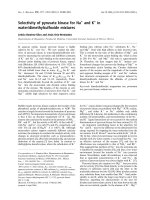
Fig. 1. Coelution of MBP-NifL w ith N ifA u nder various growth conditions after coexpression from pRS201 in K. pneumoniae. MBP-NifL was
purified from cell extracts by affinity chromatography as described in Materials and methods. The elution fractions 2 and 3, eluted in the presence of
10 m
M
maltose in the buffer, were analyzed by SDS/PAGE and subsequent Western blotting using polyclonal antibodies raised against MBP-NifL
(A) and MBP-NifA (B). Known amounts of purified MBP-NifL and MBP-NifA were simultaneously quantified on the same blot for each growth
condition as exemplarily shown in lanes 9–11 for synthesis under derepressing conditions (–O
2
, )N). L anes 1 and 2, 5 lL elution fractions 2 and 3
after synthesis in the presence o f oxygen and 10 m
M
ammonium (+O
2
,+N); lanes 3 and 4, 5 lL elution fractions after synthesis in the presence of
oxygen and 4 m
M
glutamine (+O
2
, )N); lanes 5 and 6, 5 lL elution fractions after anaerobic synthesis in the presence 1 0 m
M
ammonium
(–O
2
,+N); lanes 7 and 8, 30 lL elution fractions after synthesis under nitrogen and oxygen limitation (–O
2
, )N); lanes 9–11, 0.06, 0.13 and 0.25 lg
MBP-NifL, respectively (A) and 0.06, 0.13 and 0.25 lg MBP-NifA, respectively (B). Data are representative of four indep endent pur ifications for
each growth c ond ition.
3382 J. Stips et al. (Eur. J. Biochem. 271) Ó FEBS 2004
shown for chromosomally expressed NifL [17]. T his small
amount of MBP-NifL remaining in the cytoplasm under
derepressing co nditions is apparently still able to interact
and form i nhibitory complexes with NifA i n a stoichiomet-
ric 1 : 1 ratio (Fig. 1, lanes 7 and 8); the majority of NifA,
however, stays free in the cytoplasm and can activate nif
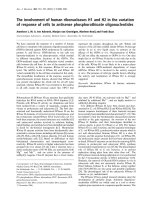
Fig. 2. Effects of MBP-NifL sy nthesized u nder different conditions on transcriptional activation by the central domain of NifA. MBP-NifL was
synthesized and purified (A) under aerobic and n itrogen sufficient conditions (MBP-NifL) or ( B) under simultaneous oxygen- and nitrogen-
limitation [MBP-NifL(–N, )O
2
)]. Activities o f the isolated central domai n of NifA (1 l
M
) were measured in the presence of different amounts of
MBP-NifL in a single cycle transcription assay under aerobic (A) or anaerobic (B) conditions as described in Materials and methods. R adioactivity
in transcripts is plotted as a percentage of the maximum value (100% NifA activity corresponded to approximately 11.2 fmol transcript). The data
presented a re based on at l east th ree i n dependent experiments; the i nsets s how the correspondin g radio active t ranscription bands of one repre-
sentative experiment for A and B in the p resen ce of increasing inh i bitor concentrations.
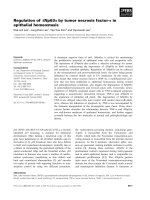
Fig. 3. Coelution of GlnK with NifL and NifA after coexpression in K. pneumoniae und er nitrogen-limiting conditions. (A) MBP-NifL was p urified
from cell extracts by affinity chromatography as described in Materials an d methods. Aliquots of the purified MBP-NifL fractions were analyzed by
SDS/PAGE and subsequent Western blotting using polyclonal antibodies raised against MBP-NifL, MBP-NifA or GlnK. For detecting NifL and
NifA, 2 lL aliquots were applied to t he SDS-containing gel, an d 20 lL aliquots for d etecting GlnK. Left panel, MBP-NifL coexpressed with NifA
from pRS201 and c hromosomally syn the sized GlnK (Gln K
chrom.
); right panel, MBP-NifL coexpressed w ith NifA and GlnK from pRS209 ; data a re
representatives of three independent purific ations. (B) After coexpression with GlnK under nitrogen-limiting growth conditions in K. pne umoniae,
MBP-NifL and MBP-NifA were purified from cell extracts by affinity chromatography, respectively. Aliquots (7.5 lL) of the elution fractions were
analyzed by SDS/PAG E and subsequent W estern blot analysis using polyclonal antibodies raised agai nst NifL, NifA or GlnK as indicated. Left
panel, MBP-NifL coexpressed with GlnK f rom pRS180: lanes 1 and 2, wash fractions; lanes 3–5, elution fractions 1–3. Right panel, MBP-NifA
coexpressed with GlnK from pRS158: lanes 6 and 7, was h fractions; lanes 8 –10, elution fractions 1–3. Data are representative of at least four
independent purifications.
Ó FEBS 2004 Complex formation between NifL, NifA and GlnK (Eur. J. Biochem. 271) 3383
gene transcription. In o rder to examine MBP-NifL local-
ization in response to environmental signals we performed
shift experiments. After synthesis o f MBP-NifL and NifA
under simultaneous nitrogen- a nd oxygen-limitation for 3 h
in a 2 L culture, t he culture was split into three equal parts,
one of which was further i ncubated for 30 min as a control;
the o ther two were shifted to anaerobic growth in the
presence of 10 m
M
ammonium and aerobic nitrogen-limited
growth for 30 min before cell harvest. Quantification of
MBP-NifL in the different cell extract fractions separated
under anaerobic or a erobic conditions, respectively, showed
that under derepressing conditions, % 95 ± 3% of total
MBP-NifL was found in the membrane fraction in four
independent experiments. However, after the shift to
nitrogen or oxygen sufficiency, t he relative amount of total
MBP-NifL in the cytoplasmic f raction increased up to
88 ± 8 and 85 ± 5%, respectively. These data confirm
that under derepressing conditions the majority of M BP-
NifL is membrane-bound, the r elative amount of NifA in
the various cytoplasmic fractions, however, was nearly
identical independent of the growth conditions.
To obtain a dditional evidence t hat NifL remaining in the
cytoplasm under derepressing conditions is still able to
interact with NifA, w e characterized the i nhibitory activity
of anaerobically purified MBP-NifL synthesized und er
simultaneous nitrogen- and oxygen-limitation [MBP-
NifL(–N, )O
2
)]. In a purified in vitro transcription a ssay
performed under anaerobic conditions, MBP-NifL(–N, )O
2
)
clearly inhibited NifA transcriptional activity to approxi-
mately the same degree as aerobically synthesized and
purified MBP-NifL in the p resence of oxygen ( Fig. 2) . T his
indicates a direct protein–protein interaction between MBP-
NifL(–N, )O
2
) and NifA, which is consistent with the
finding of complex formation between cytoplasmic MBP-
NifL and NifA under derepressing conditions. Based on
those findings we conclude that in viv o complex formation
between NifL and NifA i n K. pneumoniae occurs independ-
ently of the nitrogen and oxygen status but is exclusively
dependent on the localization of NifL in the cytoplasm.
Detection of a trimeric complex between NifA, NifL
and GlnK in
K. pneumoniae
A regulatory r ole of G lnK in the modulation of NifA
activity in response to th e nitrogen status of the cell has
previously been shown for several diazotrophic bacteria.
GlnK protein a ppears t o m ediate the nitrogen status of the
cell by direct protein–protein interaction with NifL in
A. vinelandii [25,26]; and in diazotrophs, w hich do not
contain NifL, there is evidence that GlnK or the paralog
GlnB-protein directly modulate the NifA activity in
response to the nitrogen status [40–43]. Th us, we further
analyzed the elution fractions containing the MBP-NifL–
NifA complexes for the presence of c hromosomally
expressed GlnK, using Western blot analysis. I nterestingly,
we could demonstrate the presence of small amounts of
GlnK in the MBP-NifL–NifA complexes purified from cells
grown aerobically under nitrogen limitation for several
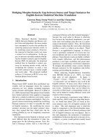
Fig. 4. Effects of additional GlnK synthesis on nif induction in K. pneumoniae UN4495 in the presence of small amounts of ammonium. NifA-mediated
activation of transcription from the nifHDK-promoter in K. pn eumoniae UN4495 was monitored by measuring the b-galactosidase activity during
anaerobic growth at 30 °C in min imal medium with glutamine (4 m
M
) as limiting nitrogen source (A) and with 4 m
M
glutamine but in the presence
of 0.25 m
M
(B), 0.5 m
M
(C) and 1.0 m
M
ammonium (D). NtrC-independent synthesis of GlnK w as induced from p lasmid pRS239 with 0.1 and
1.0 l
M
IPTG. Activities of b-galactosidase were plotted a s a function of D
600
. r, UN4495; j, UN4495/pRS239, 0.1 l
M
IPTG; m, UN4495/
pRS239, 1.0 l
M
IPTG. Data a re representative of t hree independent growth expe riments.
3384 J. Stips et al. (Eur. J. Biochem. 271) Ó FEBS 2004
independent experiments (Fig. 3A, l eft panel). Western blot
analysis using antibodies raised against G lnB verified that it
was GlnK which copurified with the MBP-NifL–NifA
complex and not GlnB. In order to rule out that GlnK binds
nonspecifically to the MalE-fusion protein (MBP) or to the
amylose resin itse lf, w e coexp ressed GlnK and MBP in
K. pneumoniae from the plasmid pRS192, t hat contains
both g enes, malE and glnK, under the control of the tac
promoter, and purified MBP by affinity chromatography.
Western blot analysis s howed that GlnK was not detectable
in the elution fractions containing purified MBP, all
synthesized GlnK was found in the flow-through a nd wash
fractions (data not shown). These findings strongly suggest
that the c hromosomally synthesized GlnK protein detected
within the purified MBP-NifL–NifA complexes was pulled
down from the cytoplasm and copurified with the MBP-
NifL–NifA complexes b ased on specific binding to either
NifL or NifA, or to the NifL–NifA complex.
In order to confirm the in vivo formation of a trimeric
complex between NifL, NifA and GlnK, we coexpressed
MBP-NifL, NifA and GlnK in K. pneumoniae under
aerobic and nitrogen-limited growth conditions. Protein
synthesis of approximately equivalent amounts of all three
proteins was induced from plasmid pRS209, which contains
the operon malE-nifLA and gln K under the control of the
tac promoter. After purification, the complexes formed were
analyzed by SDS/PAGE and silver staining, which showed
that besides MBP-NifL, NifA and GlnK no other poten-
tially brid ging proteins were present in t he elution fractions
in significant amounts ( > 1 % o f GlnK amount). The ratio
between the three regulato ry proteins was determined f rom
five independent purification experiments to be MBP-
NifL/NifA/GlnK
3
¼ 1.0 : 0 .86 ± 0.1 : 0.16 ± 0.015 by
quantitative W estern blot analysis calc ulating GlnK
concentrations as GlnK-trimers (Fig. 3A, right panel).
These findings are the first to indicate that in K. pneumoniae
a NifL–NifA–GlnK complex is formed during the trans-
duction process o f the nitrogen signal to the NifL/NifA
system by GlnK.
The primary nitrogen-sensor protein GlnK interacts
simultaneously with both nif regulatory proteins,
NifA and NifL
The finding that potentially a complex is formed between
GlnK, MBP-NifL a nd NifA raises the question of wh ether
GlnK interacts with NifL or NifA, or perhaps with both
regulatory proteins. In order to answer this question we
coexpressed GlnK with MBP-NifL or MBP-NifA in
K. pneumoniae to approximately e qual a mounts under
aerobic and nitrogen-limited growth conditions from the
plasmids pRS180 and pRS158, which both contain gln K
and either malE-nifL or malE-nifA un der t he co ntrol of the
tac promoter. The respective MBP-fusion proteins were
purified by affinity chromatography and the elution
fractions analyzed for coeluting GlnK. Interestingly, GlnK
coeluted with both, MBP-NifL and MBP-NifA (Fig. 3B),
indicating that GlnK interacts d irectly with both regulatory
proteins as unspecific binding of GlnK to the affinity
chromatography material and the MBP-fusion protein
has been excluded. Quantification analysis of at least five
independent purification experiments showed that
% 0.2 ± 0.02 GlnK
3
coeluted with MBP-NifL, which is in
the range observed for the MBP-NifL–NifA–GlnK
3
com-
plexes, whereas a significant but lower r atio between G lnK
3
and MBP-NifA was observed ( 0.06 ± 0.005 GlnK
3
per
MBP-NifA). This finding strongly indicates that under
conditions of nitrogen limitation the primary nitrogen
sensor GlnK interacts simultaneously with both regulatory
proteins apparently transducing the signal of nitrogen
limitation. The interaction between GlnK with NifL and
NifA, however, appeared to be weak as judged from the
observed G lnK
3
amount within the isolated complexes,
potentially indicating that the GlnK-complexes are not
stable.
GlnK effects stability of NifL–NifA complexes
To address the question of whether interaction with GlnK
leads to dissociation of NifL–NifA complexes we analyzed
the effects of purified GlnK on isolated MBP-NifL–NifA
complexes preformed in vivo. Purified MBP-NifL–NifA
complexes (% 2 n mol) synthesized under ammonium and
oxygen sufficiency were incubated at r oom temperature for
30 min in t he presence of 4 nmol purified GlnK in its
unmodified state (GlnK
3
) or completely uridylylated
[(GlnK-UMP)
3
], or in the absence of GlnK. After repuri-
fication of MBP-NifL–NifA complexes all fractions were
analyzed for the presence of NifL, NifA and GlnK, the
amounts of which w ere quantified by Western blot analysis.
However, no complex dissociation was obtained in the
presence of GlnK; MBP-NifL–NifA complexes were puri-
Fig. 5. Localization analysis of MBP-NifL synthezised under anaerobic
and n itrogen sufficient conditions in the p resence of NtrC-independent
GlnK synthesis. MBP-NifL, N ifA and GlnK were synthesized from
plasmid pRS209 with 100 l
M
IPTG under anaerobic con ditions but in
the presence of 10 m
M
ammonium at 30 °C. Cell extract was prepared
and separated in to membrane an d cytoplasmic frac tions as described
in Materials and methods. Aliquots of t he observed membrane a nd
cytoplasmic fraction were subjected to SDS/PAGE, and subsequently
analyzed by Western blotting. Polyclonal antibodies directed against
MBP-NifL (A) or GlnK (B) were used to detect MBP-NifL an d GlnK
in th e different f ractions and protein amounts were quantified wit h a
fluoroimager (Molecular Dynamics) using purified proteins as des-
cribed in Material and methods. Quantities of NifL a nd GlnK in the
cytoplasmic and membrane fractions were calculated as relative to
total NifL and total GlnK, resp ectively, setting the abs olute amounts
in both fractions (cytoplasmic and membrane fraction) of the
respective protein as 100%. Lanes 1–3, controls for quantification,
0.03, 0.065 a nd 0.13 lg MBP-NifL (A) and 0.028, 0.056 and 0.113 lg
GlnK (B); lane 4, 4 lL of the membrane fraction (0.9 mL); lane 5,
4 lL of the cytoplasmic f raction (4.2 mL). D ata are re presentative of
four ind ependent membrane preparations.
Ó FEBS 2004 Complex formation between NifL, NifA and GlnK (Eur. J. Biochem. 271) 3385
fied to approximately the same amount (1.9 ± 0.1 nmol)
and w ith approximately t he same ratio between MBP-NifL
and NifA ( MBP-NifL/NifA ¼ 1 : 0.92 ± 0.06) independ-
ently of the presence of GlnK.
As no effect of GlnK on NifL–NifA complex stability
was d ete ctable in vitro, we examined the effect of additional
GlnK synthesis on chromosomally (NtrC-dependent)
expressed NifL and NifA in vivo. K. p neumoniae UN4495
containing glnK under the control of t he ta c promoter on a
plasmid (pRS239) was grown under anaerobic conditions
with 4 m
M
glutamine as limiting nitrogen s ource and small
amounts of ammonium. NtrC-independent synthesis of
GlnK was induced with low IPTG concentrations (0.1 or
1.0 l
M
). Monitoring NifA-dependent transcription of the
nifHÕ-lacZ fusion during exponential growth showed
that additional synthesis of GlnK in the absence of
ammonium did not significantly influence nif induction,
which was determined to be in the range of 2500 ± 200
UÆmL
)1
ÆD
À1
600
(Fig. 4 A). In the presence o f small amounts of
ammonium, nif-induction was delayed independently of
additional GlnK synthesis and started at % D
600
¼ 0.37
(0.25 m
M
NH
4
+
), D
600
¼ 0.6 (0.5 m
M
NH
4
+
)andD
600
¼
0.9 (1.0 m
M
NH
4
+
) (Fig. 4B–D). This indicates that at
those c ell d ensities the respective amounts of ammonium
were used up and NtrC-dependent synthesis of NifL and
NifA occurred. However, compared to nitrogen limitation
from the beginning (Fig. 4A; r) the resulting nif induction
in the absence of additional GlnK synthesis was significantly
decreased in cultures initially containing small amounts of
ammonium (Fig. 4 B–D; r). The b-galactosidase synth esis
wasdeterminedtobe1250±150UÆmL
)1
ÆD
À1
600
(0.25 m
M
NH
4
+
cultures; Fig. 4B), 740 ± 40 UÆmL
)1
ÆD
À1
600
(0.5 m
M
NH
4
+
cultures, Fig. 4C), and 500 ± 3 0 U ÆmL
)1
Æ
D
À1
600
(1.0 m
M
NH
4
+
-cultures; Fig. 4D), indicating that
NifL inhibition of NifA was not completely relieved.
Additional GlnK synthesis in those cultures, however,
restored nif induction to wild-type levels under nitrogen
limitation (2500 ± 200 UÆmL
)1
ÆD
À1
600
) (Fig. 4B–D; j, m).
This finding indicates that either additional inhibitory NifL–
NifA complexes dissociated upon interaction with overex-
pressed GlnK or additional GlnK inhibited stable NifL–
NifA complex f ormation, both resulting in NifL se questra-
tion at the cytoplasmic membrane and relief of NifA
inhibition.
To obtain further evidence we analyzed whether
additional synthesis of GlnK effects complex formation
between NifL and NifA under oxygen limitation and
nitrogen sufficiency. Under those g rowth conditions,
Fig. 6. Hypothetical regulation model. The regulatory mechanism is primarily based on changes i n the cellular localization of regulatory proteins in
response to changes in environmental signals. (A) Simultaneous nitrogen- and oxygen limitation (–O
2
, )N). (B) Oxygen limitation but shift to
nitrogen sufficiency (–O
2
,+N›). (C) Aerobic but nitrogen limiting growth conditions (+O
2
, )N). (D) Simultaneous aerobic and nitrogen sufficient
growth conditions (+O
2
,+N›).
3386 J. Stips et al. (Eur. J. Biochem. 271) Ó FEBS 2004
significant amounts of MBP-NifL–NifA complexes were
isolated, when MBP-NifL and NifA synthesis occurred
from plasmid pRS201, in the absence of additional GlnK
synthesis (Fig. 1, lanes 5 and 6). However, when GlnK
was additionally synthesized under oxygen limitation in
the presence of 10 m
M
ammonium, using plasmid pRS209
for NtrC-independent synth esis of MBP-NifL, NifA and
GlnK, the purification under anaerobic conditions did not
result at all in the isolation of MBP-NifL o r a complex
including MBP-NifL. L ocalization analysis of MBP-NifL
in those cells further showed that 95 ± 2% of total
MBP-NifL was f ound in the membrane fraction (Fig. 5),
which is consistent with the finding that no MBP-NifL
was purified from the s oluble cell extract. Interestingly,
70 ± 5% o f t otal GlnK was a lso f ound in the m embrane
fraction. However, at the c urrent experimental status we
do not know, whether the overproduced GlnK binds to
the cytoplasmic membrane in a NifL-dependent manner.
The relative amounts of NifA in the cytoplasm d id not
change upon additional G lnK synthesis. These findings,
which were confirmed by several independent experiments,
again strongly indicate that the additional GlnK s ynthesis
resulted in the dissociation of the inhibitory NifL–NifA
complexes o r inhibited the formation of stable NifL–NifA
complexes. Thus, we conclude that GlnK effects the
cellular localization of NifL in r esponse to the nitrogen
status by influencing the formation of NifL–NifA com-
plexes. This proposed mechanism for nitrogen signal
transduction by GlnK in K. pneumoniae differs signifi-
cantly from the mechanism of nitrogen signal transduction
by GlnK in A. vinelandii [14,24–26].
Hypothetical model for oxygen and nitrogen control
of
nif
regulation in
K. pneumoniae
On the basis of those data and the finding that only small
amounts of G lnK
3
are present in the MBP-NifL–NifA–
GlnK complexes f ormed under nitrogen-limitation, we
hypothesize that the NifL–NifA–GlnK complex reflects a
transitional status within the signal transduction of nitro-
gen-limitation to the NifL/NifA system and propose the
following working model (Fig. 6). Under a naerobic and
nitrogen-limited conditions, the interaction with GlnK
eventually results in unbound NifA and NifL, which is
able to receive electrons at the cytoplasmic m embrane from
the anaerobic quinol pool [19]. Upon reduction NifL is
sequestered to the cytoplasmic membrane and thus allows
NifA to activate nif genes i n the cytoplasm (Fig. 6A). After
a period of oxygen- and nitrogen-limitation, an ammonium-
upshift re sults i n d euridylylation of GlnK and unmodified
GlnK may be sequestered to the cytoplasmic membrane in
an AmtB-dependent manner as has been recently shown for
E. coli and A. vinelandii GlnK [44]. Sequestration of GlnK
to the cytoplasmic membrane would significantly r educe
NifA–NifL complex dissociation by GlnK; consequently,
most of NifL stays i n t he cytoplasm as recently demonstra-
ted [17] and inhibits NifA activity by forming inhibitory
complexes (Fig. 6B). When a shift to oxygen occurs in
addition, NifL is oxidized and upon o xidation the main part
of NifL dissociates from the cytoplasmic membrane a nd
forms inhibitory NifL–NifA complexes in the cytoplasm
(Fig. 6 ,D). This occurs even under nitrogen-limitation in the
presence of GlnK, as membrane-bound reduced NifL is
rapidly oxidized and quickly dissociates i nto the c ytoplasm
resulting in a high NifL–NifA c omplex formation r ate,
which appears to b e much higher than t he GlnK-dependent
dissociation rate (Fig. 6C).
Acknowledgments
We thank Gerhard Gottschalk for generous support, helpful discus-
sions, and lab space, and Andre a Shauger for critical reading of th e
manuscript. T his work was supported by the Deutsche Forschungs-
gemeinschaft (SCHM1052/4–4 and 4–5) and the Fonds der Chemis-
chen Industrie.
References
1. Burgess, B.K. & Lowe, D.J. (1996) Mechanism of molybdenum
nitrogenase. Chem. Rev. 96, 2983–3012.
2. Rees, D.C. & Howard, J .B. (1999) Structural bioenergetics and
energy transduction mechanisms. J. Mol. Biol. 293, 343–350.
3. Hoover, T.R., Santero, E., Porter, S. & Kustu, S. (1990) The
integration host factor stimulates interaction of RN A polymerase
with NIFA, t he transcriptional activator for nitrogen fixation
operons. Cell 63, 11–22.
4. Morett, E. & Buck, M. (1989) In vivo studies on the interaction of
RNA polymerase-sigma 54 with the Klebsiella pneumoniae and
Rhizo bi um melil o ti nifH promoters. J. Mol. Biol. 210 , 65–77.
5. Merrick,M.,Hill,S.,Hennecke,H.,Hahn,M.,Dixon,R.&
Kennedy, C. (1982) Repressor properties of the nifL gene product
in Kle bsiella pneumoniae. Mol. Gen. Genet. 185, 75–8 1.
6. Hill, S., Austin, S., Eydmann, T., Jones, T. & D ixon, R. (1996)
Azotobacter vinelandii NIFL is a flavoprotein that modulates
transcriptional activation of nitrogen-fixation genes via a redox-
sensitive switch. P roc . Natl Acad Sci. U SA 93, 2143 –2148.
7. Hill, S., Kennedy, C., Kavanagh, E., Goldberg, R.B. & H anau,
R. (1981) Nitrogen fixation gene (nifL) involved in oxygen reg-
ulation of nitrogenase synthesis in K. pneumoniae. Nature 290,
424–426.
8. Egener,T.,Sarkar,A.,Martin,D.E.&Reinhold-Hurek,B.(2002)
Identification of a NifL-like protein in a diazotroph of the b eta-
subgroup of the Proteobacteria, Azoarcus sp. strain BH72.
Microbiology 148, 3203–3212.
9. Henderson, N., Austin, S. & Dixon, R . (1989) Role of metal
ions in negative regulation of nitrogen fixation by the nifL gene
product f rom Kle bsiella pneumoniae. Mol. G en. Genet. 216, 484–
491.
10. Money, T., Jones, T., Dixon, R. & Austin, S. (1999) Isolation and
properties of the com plex betwee n the en hancer b inding prote in
NIFA an d the sensor NIFL. J. Bacteriol. 18 1 , 4461–4468.
11. Money, T., Barrett, J., Dixon, R. & Austin, S. (2001) Protein–
protein interactions in the complex between the enhancer binding
protein NIFA an d the sensor NIFL from Azotobacter vinelandii.
J. Ba cteriol. 183 , 1359–1368.
12. Lei, S., P ulakat, L. & Gavini, N. (1999) Genetic analysis of nif
regulatory genes by utilizing the yeast two-hybrid system detected
formation of a NifL-NifA complex that is implicated in regulated
expression of nif genes. J. Bacter iol. 181, 6535–6539.
13. Reyes-Ramirez, F., Little, R. & Dixon, R. (2002) Mutant forms of
the Azotobacter vi nelandii transcriptional activator NifA resistant
to inhibition by the NifL regulatory protein. J. Bacteriol. 184,
6777–6785.
14. Little, R. & Dixon, R. (2003) The amino-terminal GAF domain of
Azotobacter vinelandii NifA binds 2-oxoglutarate to resist inhibi-
tion by NifL under nitrogen-limiting conditions. J. Biol. Chem.
278, 2 8711–28718.
Ó FEBS 2004 Complex formation between NifL, NifA and GlnK (Eur. J. Biochem. 271) 3387
15. Mach eroux, P., Hill, S., A ustin, S., Eydmann , T., Jones, T ., Kim,
S.O., Poole, R. & Dixon, R. (1998) Electron donation to the
flavoprotein NifL, a redox-sensing transcriptional regulator.
Biochem. J. 332, 4 13–419.
16. Schmitz, R.A. (1997) NifL of Klebsiella pneumoniae carries an
N-terminally bound FAD cofactor, which is not directly required
for the inhibitory fu nction o f NifL. FEMS Microb iol. Lett. 157,
313–318.
17. Klopprogge, K., Grabbe, R., Hoppert, M. & S chmitz, R.A. (2002)
Membrane association of Kle bsiella pneumoniae NifL is affected
by molecular oxygen and combined nitrogen. Arch. Microbiol.
177, 2 23–234.
18. Grabbe, R., Klopprogge, K. & Schmitz, R.A. (2001) Fnr Is
required for NifL-dependent oxygen control o f nif gene expression
in Kle bsiella pneumoniae. J. Bacteriol. 183, 1385–1393.
19. Grabbe, R. & Schmitz, R.A. (2003) Oxygen control of nif ge ne
expression in Klebsiella pneumoniae depends on NifL reduction at
the cytoplasmic membrane by electrons derived from the reduced
quinone pool. Eur. J. Biochem. 27 0 , 1555–1566.
20. He, L ., Soupene, E ., N infa, A . & K ustu, S . ( 1998) Physiological
role f or the GlnK protein of enteric bacteria: relief of NifL
inhibition under nitrogen-limiting conditions. J. Bacteriol. 180,
6661–6667.
21. Jack, R., De Zamaroczy, M. & Merrick, M. (1999) The signal
transduction protein G lnK is required for Nif L-dependent nitro-
gen con trol of nif gen e e xp ression in Klebsiella pneumonia.
J. Bacteriol. 181, 1156–1162.
22. Arcondeguy, T ., van He eswijk, W .C. & Merrick, M. (19 99) Stu-
dies on the role s of G lnK and G lnB in regulating Klebsiella
pneumoniae NifL-dependent nitrogen co ntrol. FEMS Microbiol.
Lett. 180 , 263–270.
23. Arcondeguy, T., Lawson, D. & Merrick, M . (2000) T wo residues
in the T-loop of GlnK determ ine NifL-dependent nitro gen control
of nif ge ne expression. J. Biol. Chem. 275, 38452–38456.
24. Little, R. , Reyes-Ramirez, F., Zhang , Y., v an He eswijk, W .C. &
Dixon, R. (2000) Signal transdu ction to the Azotobacter vinelandii
NIFL-NIFA regulatory system is i nfluenced directly by interac-
tion with 2-oxoglutarate and the PII regulatory protein. EMBO
J. 19 , 6041–6050.
25. Rudn ick, P., K unz, C., Gunatilaka, M .K., Hines, E.R. & Ken-
nedy, C. (2002) R o le of GlnK i n NifL-mediate d regulation
of NifA activity in Azotobacter vinelandii. J. Bacteriol. 184, 812–
820.
26. Little, R., Colombo, V., Leech, A. & Dixon, R. (2002) Direct
interaction of t he NifL regulatory protein w ith the GlnK signal
transducer enables the Azotobacter vinelandii NifL-NifA reg-
ulatory system to respond to conditions replete for nitrogen.
J. Bi ol. Chem. 277, 15472–15481.
27. Reyes-Ramirez, F., L ittle, R. & Dixon, R . (2001) Role of
Escherichia coli nitrogen regulatory genes in the nitrogen response
of the Azotobacter vinelandii NifL-NifA complex. J. Bacteriol. 183,
3076–3082.
28. MacNeil, D., Z hu, J. & Brill, W.J. (1981) Regulati on of nitrogen
fixation in Klebsiella pneumoniae: isolation and characterization of
strains with nif-lac fusions. J. Bacteriol. 145, 3 48–357.
29. deLorenzo,V.,Herrero,M.,Jakubzik,U.&Timmis,K.N.(1990)
Mini-Tn5 transposon derivatives for insertion mutagenesis, p ro-
moter probing, and chromosomal insertion of cloned DNA in
gram-negative e ubacteria. J. Bacteriol. 172 , 6568–6572.
30. Narberhaus,F.,Lee,H.S.,Schmitz,R.A.,He,L.&Kustu,S.
(1995) The C-terminal domain of NifL is sufficient to inhibit NifA
activity. J. Bacteriol. 177 , 5078–5087.
31. Lee, H.S., B erger, D.K. & Kustu, S . (1993) Activity of purified
NIFA,atranscriptionalactivator of nitrogen fixation genes. Proc.
NatlAcad.Sci.USA90, 2266–2270.
32. Ehlers, C., Grabbe, R ., Veit, K. & Schmitz, R .A. ( 2002) Char-
acterization of GlnK
1
from Methanosarcina mazei strain Go
¨
1:
complementa tion of an Escherichia coli glnK mutant strain by
GlnK
1
. J. Bacteriol. 184, 102 8–1040.
33. Schmitz, R.A., He, L. & Kustu, S. (1996) Iron is required to relieve
inhibitory effects on NifL on transcriptional a ctivation by NifA in
Klebsiella pne umoniae. J. Bacteriol. 178, 4679–4687.
34. Forchhammer, K. & Hedler, A . (1997) Phosphopr otein PII from
cyanobacteria -analysis of functional conservation with the PII
signal-transduction protein from Escherichia coli. Eur. J. Biochem.
244, 8 69–875.
35. Sambrook, J., E.F.Fritsc h & T.Maniatis. (1989) Molecular Clon-
ing: a Laboratory Manual, 2nd edn. Cold Spring Harbor
Laboratory Press, New York.
36. Berger, D.K., Narberhaus, F. & Kustu, S. (1994) The isolated
catalytic d omain of NIFA, a bacterial enhanc er-bindin g prote in,
activates transcription in vitro:activationisinhibitedbyNIFL.
Proc.NatlAcad.Sci.USA91, 1 03–107.
37. Bergmayer, H. (1983) Methods o f Enzymatic Analy sis in Methods
of Enz ymatic Analysis. Verlag C hemie, Weinheim.
38. Fluckiger, R., Paz, M .A. & Gallop, P.M. (1995) Redox-cycling
detection of dialyzable pyrro loquinoline quinone and quinopro-
teins. Methods Enzymol. 258, 140–149.
39. Berger, D.K., Narberhaus, F., Lee, H.S. & Kustu, S. (1995) I n
vitro s tudies of the domains of the nitrogen fixation regulatory
protein NIFA. J. Bacteriol. 177, 191– 199.
40. Liang, Y.Y., de Zamaroczy, M., Arsene, F., Paquelin, A. &
Elmerich, C. (1992) Regulation of nitrogen fixation in Azospirillum
brasilense Sp7: involvement of nifA, glnA and glnB gene products.
FEMS Microbiol. Lett. 79 , 113–119.
41. Arsene , F., Kaminski, P.A. & Elmerich, C. (1996) Modulation of
NifA activity b y PII in Azospirillum brasilense: evidence for a
regulatory role of the NifA N-terminal domain. J. Bacteriol. 178,
4830–4838.
42. Arsene, F., Kaminski, P.A. & Elmerich, C. (1999) Control of
Azospirillum b rasilense NifA activity by P (II): effect of replacing
Tyr residues o f the N ifA N -term inal domain o n NifA activi ty.
FEMS Microbiol. Lett. 17 9, 339–343.
43. Dreppe r, T ., Gross, S., Yakunin, A.F., Hallenbeck, P.C., Mas-
epohl, B. & Klipp, W. (2003) Role of GlnB and GlnK in ammo-
nium control of both nitrogenase systems in the phototrophic
bacterium Rhodobacter capsulatus. Microbiology 149, 2203–2212.
44. Coutts, G., Thomas, G., Blakey, D. & Merrick, M. (200 2)
Membrane sequestration of the signal transduction protein GlnK
by t he ammonium transporter AmtB. EMBO J. 21, 536– 545.
3388 J. Stips et al. (Eur. J. Biochem. 271) Ó FEBS 2004