Báo cáo khoa học: NMR and molecular dynamics studies of an autoimmune myelin basic protein peptide and its antagonist Structural implications for the MHC II (I-Au)–peptide complex from docking calculations ppt
Bạn đang xem bản rút gọn của tài liệu. Xem và tải ngay bản đầy đủ của tài liệu tại đây (869.05 KB, 15 trang )
NMR and molecular dynamics studies of an autoimmune myelin basic
protein peptide and its antagonist
Structural implications for the MHC II (I-A
u
)–peptide complex from docking
calculations
Andreas G. Tzakos
1
, Patrick Fuchs
2
, Nico A. J. van Nuland
2
, Anastasios Troganis
3
, Theodore Tselios
4
,
Spyros Deraos
4
, John Matsoukas
4
, Ioannis P. Gerothanassis
1
and Alexandre M. J. J. Bonvin
2
1
Department of Chemistry, Section of Organic Chemistry and Biochemistry, University of Ioannina, Greece;
2
Bijvoet Center for
Biomolecular Research, Department of NMR Spectroscopy, Utrecht, the Netherlands;
3
Department of Biological Applications and
Technologies, University of Ioannina, Greece;
4
Department of Chemistry, University of Patras, Greece
Experimental autoimmune encephalomyelitis can be
induced in susceptible animals by immunodominant deter-
minants o f myelin basic protein (MBP). To cha racterize the
molecular features of antigenic sites important for designing
experimental autoimmune encephalomyelitis suppressing
molecules, we report structural studies, based on NMR
experimental data in conjunction with molecular dynamic
simulations, of the potent linear dodecapeptide epitope of
guinea pig MBP, Gln74-Lys75-Ser76-Gln77-Arg78-Ser79-
Gln80-Asp81-Glu82-Asn83-Pro84-Val85 [MBP(74–85)],
and its antagonist analogue Ala81MBP(74–85). The two
peptides were studied in both w ater and Me
2
SO in order t o
mimic solvent-dependent structural changes in MBP. The
agonist MBP(74–85) adopts a compact conformation
because of electrostatic interactions of Arg78 with the side
chains of Asp81 and Glu82. Arg78 is ÔlockedÕ in a well-
defined conformation, perpendicular to the peptide back-
bone which is practically solvent independent. These
electrostatic interactions are, however, absent from the
antagonist Ala81MBP(74–85), resulting in great flexibility
of the side chain of Arg78. Sequence alignment of the two
analogues w ith several species of MBP suggests a critical role
for the positively charged residue Arg78, firstly, in the sta-
bilization of the local microdomains (epitope s) of the integral
protein, and secondly, in a number o f post-translational
modifications relevant to multiple s clerosis, s uch a s t he
conversion of charged arginine residues to uncharged cit-
rullines. F lexible dock ing calculations on the binding of the
MBP(74–85) antigen to the MHC class II receptor s ite I-A
u
using
HADDOCK
indicate that Gln74, Ser76 and Ser79 are
MHC II anchor residues. Lys75, Arg78 and Asp81 are
prominent, solvent-exposed residues and, thus, may be of
importance in the formation of the trimolecular T-cell
receptor–MBP(74–85)–MHC II complex.
Keywords: conformation; docking; major histocompatibility
complex; molecular dynamics; mye lin basic epitope.
Multiple sclerosis is a chronic i nflammatory demyelinating
disease of the central nervous system, which is believed to be
mediated by autoreactive T cells [1–3]. The a ctivation of
resting T cells reacting with antigens of the central nervous
system, specifically with the major histocompatibility
(MHC)–antigen complex, is thought to be the primary
autoimmune event in m ultiple sclerosis. Myelin basic protein
(MBP) represents 5–15% of the peripheral nervous system
myelin protein [4] and plays an integral role in t he structure
and function of the myelin sheath [5,6]. It was the first agent
in brain or spinal cord homogenates found to be responsible
for e xperimental allergic encephalomyelitis (an animal
model for human multiple sclerosis) [7–9]. Some o f the most
important functions of MBP are stimulation of phospho-
lipase C activity [10], actin polymerization in conjunction
with Ca
2+
–calmodulin [11], tubulin stabilization [12], and
potential regulatory roles as t ranscription factors [13].
The d etailed high-resolution tertiary structure of MBP
is not known [14]. The main structural m odels of th is
protein d ate from the 1980s a nd represent the abstract
combination of biochemical data and secondary-structure
prediction algorithms [15–18]. The c onformation of the
first 14 residues of the acetylated N-terminus [19] and the
last 17 residues of the MBP have been investigated by
NMR [20]. The most sophisticated structural models of
the integral protein are those of Stoner [17] and Marten-
son [18], base d on e xtensive biochemical and secondary-
structure data and the recently determined 3D structure
by single-particle electron crystallography [21,22]. It was
shown that MBP is a C-shaped molecule when adsorbed
into a lipid monolayer, comprising five b-sheets and a
large proportion of irregular coil.
Correspondence to I. P. Gerothanassis, Department of Chemistry,
Section of Organic Chemistry and Biochemistry, University of
Ioannina, Ioannina GR-45110, Greece. Fax: + 302651098799,
Tel.: + 3026510983397, E-mail:
Abbreviations:Me
2
SO, dimethyl sulfoxide; MBP, myelin basic protein;
MD, molecular dynamics; MHC, major histocompatibility complex;
TCR, T-cell receptor.
(Received 2 8 February 2004, revised 3 0 June 2 004,
accepted1July2004)
Eur. J. Biochem. 271, 3399–3413 (2004) Ó FEBS 2004 doi:10.1111/j.1432-1033.2004.04274.x
The lack of a high-resolution structure of MBP means
that it is important to investigate the structure of its
epitopes, found in segments 1–14, 22–34, 43–68, 67–75,
75–82, 83–96, 90–99, 114–121, 118–131, 125–131, 130–137
and 131–140 [23–26], which have antigenic properties.
Characterization of the molecular features of these antigenic
sites may provide insights into their immunogenic properties .
This would be useful i n the design of synthetic p eptides and
nonpeptide mimetics that c an act as vaccines o r artificial
regulators of the immune response. Linear and c yclic
analogues of several MBP epitopes have been synthesized
to identify pharmacophoric groups and develop a molecular
model, which may be useful in drug design [27–29].
We have focused our studies on the 74–85 segment of
guinea pig MBP, Gln74-Lys75-Ser76-Gln77-Arg78-Ser79-
Gln80-Asp81-Glu82-Asn83-Pro84-Val85 [MBP(74–85)].
Arg78 is proximal to a triproline Pro99-Pro100-Pro101
segment, which h as been suggest ed to have potential
synergestic e ffects on the e ntire structure [30]. Furthermore,
this dodecapeptide epitope of MBP is a target of the
peptidylarginine deiminase action on Arg78, leading to
demyelinaton and thus chemical pathogenesis of multiple
sclerosis. We examined the structural features of the
encephalitogenic agonist epitope MBP(74–85) and the
antagonist analogue Ala81MBP(74–85), using detailed
NMR a nd molecular dynamic studies. It is known f rom
spectroscopic studies that MBP is more extensively folded in
the presence o f lipids or detergents [31–35] t han in aqueous
solution [31,32]. We therefore investigated solvent-induced
structural changes of the peptides in water and Me
2
SO,
which might be related to the solvent-dependent structural
changes i n integral g uinea pig MBP. Sequence alignment of
the two analogues with several MBP species and docking
calculations with respect to the M HC II (I-A
u
) receptor s ite
are also reported in an effort to elucidate the role of the
positively charged residue Arg78 and the effect of the
reduction of cationicity of MBP in the triggering of multiple
sclerosis. Flexible docking calculatio ns of the MBP(74–85)
epitope with the MHC II–I-A
u
recognition site are also
reported to explore MHC II anchor residues and solvent-
exposed residues that may be important for t he interaction
of the T-cell receptor (TCR) with the b imolecular complex
MHC II–antigen [MBP(74–85)].
Materials and methods
Synthesis of peptide analogues of MBP(74–85)
The linear MBP analo gues Gln-Lys-Ser-Gln-Arg-Ser-Gln-
X-Glu-Asn-Pro-Val, wh ere X ¼ Asp (agonist) or Ala
(antagonist), were synthesized using Fmoc/tBu methodo-
logy. 2-Chlorotrityl chloride resin and N
a
-Fmoc amino
acids w ere used for the synthesis as described previously
[27–29a]. Peptide purity was assessed by analytical HPLC
(Nucleosil-120 C18; reversed phase; 250 · 4.0 mm), MS
(fast-atom bombardment, electrospray ionization) and
amino-acid analysis [29].
NMR spectroscopy
Preliminary NMR spectra were acquire d at 400 MHz u sing
a Bruker AMX-400 spectrometer (NMR Centre, University
of Ioannina, Greece). High-field NMR spectra were
acquired a t 750 MHz using a Bruker Avance 750 spectro-
meter (Bijvoet Center for Biomolecular R esearch, Utrecht,
the Netherlands). For water suppression, excitation sculp-
ting with gradients was used [36]. Samples of the MBP-
(74–85) and Ala81MBP(74–85) analogues were dissolved
in Me
2
SO-d
6
at 2 m
M
concentration, and the spectra were
recorded at 300 K. Chemical shifts were reported with
respect to the resonance of the solvent. The samples in
aqueous solution (90%
1
H
2
O/10%
2
H
2
O, v/v) were pre-
pared for NMR spectroscopy by dissolving the peptide in
0.01
M
potassium phosphate buffer (pH ¼ 5.7), containing
0.02
M
KCl a nd 1 m
M
2,2-dimethyl-2-silapentanesulfonate
as an internal chemical-shift reference. Peptide concentra-
tion was usually 4 m
M
, and the spectra were recorded
at 277 K. Trace amounts of N aN
3
were added a s a
preservative.
NOESY experiments – determination of distance
restraints
2D Spectra were acquired using the S tates-TPPI method for
quadrature d etection, with 2K · 512 complex data points,
16 scans per increment for 2D TOCSY, and 64 scans for 2D
NOESY experiments. T he mixing time for the TOCSY
spectra was 80 m s. The mixing times for NOESY experi-
ments w ere 100, 200, 300 and 400 ms. D ata were z ero-filled
in t
1
to giv e 2K · 2K real data poin ts. A 60 ° phas e-shifted
square sine-bell window function was applied in both
dimensions using the
NMRPIPE
software [37].
Interproton distances were derived by measuring cross-
peak intensities in the NOESY spectra. Intensities were
calibrated to give a set of distance constraints using the
NMRVIEW
software package [38].
Structure calculations
Structure calculations were performed with
CNS
[39] using
the
ARIA
setup and protocols [40,41], a s described in Bonvin
et al . [42] C ovalent interactions were calculated with the 5.3
version of the
PARALLHDG
parameter file [43] based on the
CSDX
parameter set [44]. Nonbonded interactions were
calculated with the repel function, using the
PROLSQ
parameters [45] as implemented in the new
PARALLHDG
parameter file. The
OPLS
nonbonded parameters [46] were
used for the final explicit solvent refinement (water or
Me
2
SO) including full van der Waals and electrostatic
energy terms.
A simulated annealing protocol in Cartesian space was
used starting from an extended conformation consisting of
four stages: (a) high-temperature SA stage (10 000 steps,
2000 K); (b) a first cooling phase from 2000 to 1000 K in
5000 st eps; (c) a second cooling phase from 1000 t o 50 K in
2000 steps; (d) 200 steps of energy minimization. The time
step for the integration was set to 0.003 ps.
The s tructures were s ubjected to a final refinement
protocol with the explicit solvent by solvating them with
either a 8 A
˚
layer of TIP3P water m olecules [46] or a 12 A
˚
layer of Me
2
SO molecules [43]. The resulting structures were
energy-minimized with 100 step s of P owell steepest d escent
minimization, and t he stereochemical quality was evaluated
with
PROCHECK
[47].
3400 A. G. Tzakos et al.(Eur. J. Biochem. 271) Ó FEBS 2004
Molecular dynamics (MD) simulations
Simulations were performed with
GROMACS
3.1 [48,49],
using the
GROMOS
96 43A1 f orce field [50]. The simulations
were run for 10 ns at 300 K starting from the l owest-energy
NMR structures, in either explicit water, using the SPC
model[51],orMe
2
SO, using the model of Liu et al.[52]
(Table 1). A nalysis of the trajectories was performed using
the programs included in the
GROMACS
package.
The peptides were solvated in a cubic box of explicit water
or Me
2
SO with a minimum distance solute–box of 14 A
˚
.
The various systems comprised 3899 and 4516 SPC
molecules for the agonist and the antagonist in water,
respectively, and 817 and 927 Me
2
SO molecules for the
agonist and the antagonist, respectively, corresponding to a
total number of atoms of 11 797, 13 673, 3398 and 3832,
respectively. Periodic boundary conditions were applied.
Each system was fi rst energy-minimized using 2000 steps of
steepest descent algorithm. For the antagonist, t he system
was neutralized by replacing a w ater molecule (with the
highest electrostatic potential energy) with a Cl
–
counter ion,
and then energy-minimized with 2000 steps of steepest
descent.
Each system was e quilibrated in five 20 ps phases, during
which the force constant of the position restraints term for
the solute was decreased from 1000 to 0 kJÆmol
)1
Ænm
)2
(1000, 1000, 100, 10, 0). T he initial velocities w ere generated
at 300 K following a M axwellian distribution. The simula-
tions were performed at constant pr essure (101 kPa) and
temperature (300 K ) by w eakly coupling the system to
external temperature and pressure baths [53], except for the
first 20 ps equilibration part which was performed at
constant volume. All bonds were constrained by u sing the
LINCS
algorithm [ 54], and the w ater molecules were kept
rigid u sing the
SETTLE
algorithm [55]. The p eptide and the
solvent (as well as the counter ion in the case of the
antagonist simulations) were coupled separately to a
temperature bath with a time constant of 0.1 ps. The
pressure was coupled to an external bath at 100 kPa
with a time constant of 0.5 ps and a compressibility of
4.5 · 10
)3
kPa
)1
. Periodic boundary conditions were
applied all along the simulation. A twin-range cut-off of
0.8 and 1.4 nm was used for the nonbonded interactions. I n
water, the generalized reaction field [56] was u sed with a
dielectric constant of 54 beyond the 1.4 nm cut-off, whereas
in Me
2
SO a classical shifting functio n was used with a cut-
off of 1.4 nm. A 2 f s time step was used for the leapfrog
algorithm integration.
All simulations were performed in parallel on t wo
processors on a
LINUX
cluster (1.3 MHz Athlon processors)
using the parallel version of
GROMACS
. As a cost per unit
cost indication, 1 ns took about 2.5 h for the simulations in
Me
2
SO and 14 h for t hose in water. The average solvent-
accessible s urface area was calculated f rom frames taken
every 100 ps using t he program
NACCESS
[57].
Sequence alignment
Sequence alignment of the different MBP families for the
fragment 74(3))85(7) was performed with
CLUSTALW
[58].
Docking calculations
The docking calculations were perfor med with
HADDOCK
1.2 [59] ( using the
standard protocols. The a mbiguous interaction r estraints
for docking calculations were defined for the P4, P6 and P9
pockets of the peptide-binding groove of the MHC II
(I-A
u
), based on the interactions derived from the X-ray
crystallographic structures of several MHC II–MBP epi-
tope complexes (se e discussion below). A total of 1000 rigid-
body docking solutions were generated. In addition, for
each of the starting conformations, 10 rigid-body trials were
performed, and only the best solution based on the
intermolecular energy was kept, bringing the total effective
docking trials to 10 000. The best 500 solutions sorted
according to the intermolecular energy (sum of van der
Waals, electrostatic, and ambiguous interaction restraints
energy terms) were further subjected to the semi-flexible
simulated annealing and Me
2
SO refinement as described
previously [59]. The solutions were clustered u sing a 1.0 A
˚
rmsd cut-off criterion and ranked according to their average
interaction energies (sum of E
elec
, E
vdw
, E
ACS
)andtheir
average buried surface area.
Results and Discussion
NMR studies
Amino-acid sp in syste ms w ere identified by locating
networks of characteristic connectivities i n t he 2D TOCSY
and NOESY spectra [60].
Qualitative results on the conformational properties of
the two peptides can b e extracted from the d ifference of
the amide protons (NH) ch emical-shift temperature coeffi-
cients (Dd/DT) b etween agonist and antagonist. Exposed
NHs typically have coefficients i n the range )6.0 t o
)8.5 p.p.b.ÆK
)1
, a nd hydrogen-bonded o r protected NHs
typically have Dd/DT of )2.0 to +1.4 p.p.b.ÆK
)1
[61]. In
Me
2
SO solution, only t he NH of Lys75 h as a Dd /DT va lue
characteristic of solvent shielding, while the remaining NH
groups have Dd /DT values < )4.5 p.p.b.ÆK
)1
, indicating
their exposure to solvent. From the comparison of Dd/DT
values of agonist and antagonist, i t can be concluded that
the agonist has a more compact co nformation in Me
2
SO.
Dd/DT values in aqueous solution are not reported because
of the large overlap for the NH resonances.
Table 1. Summary of the various MD simulations at 300 K. The
simulated t ime in e ach case w as 10 ns.
Code Peptide Solvent Starting structure
(1) Agonist H
2
O Lowest energy NMR
structure in H
2
O
(2) Antagonist H
2
O Lowest energy NMR
structure in H
2
O
(3) Agonist Me
2
SO Lowest energy NMR
structure in Me
2
SO
(4) Antagonist Me
2
SO Lowest energy NMR
structure in Me
2
SO
(5) Agonist H
2
O Lowest energy NMR
structure in Me
2
SO
Ó FEBS 2004 An autoimmune MBP peptide and its antagonist (Eur. J. Biochem. 271) 3401
Chemical-shift differences between MBP(74–85) and
Ala81MBP(74–85) of 0.02 < Dd < 0.04 p .p.m. were
found for Ser76, Gln77, Arg78, Ser79, Asn83 i n M e
2
SO
and aqueous solutions. Larger differences (> 0.05 p.p.m)
were observed for Gln80 a nd Glu82, which a re neighbours
to the variant position 81. The l arge deviations for the
C-terminal residues A sn83 and Val85 observed in aqueous
solution, compared with Me
2
SO, are possibly due to
electrostatic interactions promoted in this solvent ( see
discussion below). A comparison of the chemical-shift data
of the two peptides suggests that the backbone of the two
molecules should exhibit different structural features in both
solvents.
Structure determination of MBP(74–85) and
Ala81MBP(74–85)
The primary NMR data used in the structure calculation s
were sequential (|i–j|<1), medium-range (1<|i–j|<4) and
long-range (|i–j|>4) NOEs, obtained from
1
Hto
1
H2D
NOESY experiments. Several NOE connectivities indicative
of a folded conformation were observed for the two
analogues. For the structure calculations of MBP
(74–85) in Me
2
SO, 98 sequential a nd medium-range NOEs
and two long-range NOEs were used as distance restraints,
whereas, in aqueous solution, 116 sequential and medium-
range NOEs and two long-range NOEs were considered.
For the structure calculations of the Ala81MBP(74–85)
analogue in Me
2
SO, 6 4 s equential a nd medium-range
NOEs were used as distance restraints, and in the case of
the aqueous solution, 83 sequential and medium-range
NOEs were considered. NOE cross-peaks were separated
into three distance categories according to their intensity.
Strong NOEs were given an upper distance restraint of
3.0 A
˚
, medium NOEs of 4.0 A
˚
, and weak NOEs of 5.5 A
˚
.
The lower distance limits were s et to 1.8 A
˚
.Noother
restraints were applied. Structure c alculations were per-
formed using a simulated annealing protocol, following the
ARIA
/
CNS
setup [40–42] (see Materials and methods).
Structure in aqueous solution. A family of 200 structures
was c alculated for both analogues in aqueous solution. The
20 structures with the lowest total energy and NOE
violations smaller than 0.25 A
˚
were selected after the final
refinement in explicit water. Both the N-terminal and
C-terminal regions exhibit significant c onformational het-
erogeneity, w hereas the Lys75–Glu82 segment m aintains a
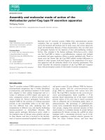
more consistent conformation (Fig. 1 ).
The MBP(74–85) epitope a dopts a compact, S-shaped,
conformation in aqueous solution, with rmsd values fro m
the m ean s tructure for the Lys7 5–Glu82 fragment of
0.90 ± 0.25 A
˚
for the backbone N, C
a
,C¢ atom s and
2.05 ± 0.55 A
˚
for a ll heavy atoms. A characteristic feature
of this ensemble of structures is the presence of two
conformational f amilies with different orientations of the
side chain of Glu82. We termed these two families of
conformers 1 (#1) and 2 (#2) (Fig. 1A, black and grey
backbones, respectively). I n family 1 (#1) the side chain of
Glu82#1 is i n close proximity to Gln77, whereas in family 2
(#2, Glu82#2) it approaches the side chain of Lys75 [this is
consistent with the observed long-range NOE c ross-peak
between Lys75 (Ha) and Arg78 (HN)]. Furthermore, in
both families, Glu82 is in close proximity to the side c hain of
Arg78, forcing it into a perpendicular position relative to the
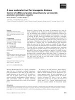
Fig. 1. Ensembles of 3D structures of the agonist (A) and antagonist (B) linear analogues of MBP(74–85) in aqueous solution. The lower thicker trace
corresponds to a represe ntative conformer, the s tructure of which i s closest to the average structure o f the ensemble.
3402 A. G. Tzakos et al.(Eur. J. Biochem. 271) Ó FEBS 2004
plane defined by the backbone of the peptide [this is
consistent with the observed two long-range NOE cross-
peaks between the side chain of Arg78 (H
e
) and Glu82
(H
c1
and H
b1
)]. The side-chain carboxylate g roup of Asp81,
is well defined, and it interacts with the side-chain amide
proton (He) of the neigh bouring residue Gln80.
The Ala81MBP(74–85) variant seems to adopt a more
open U-shaped loop conformation of residues Arg78–Ala81
(Fig. 1B). The rmsd values from t he mean structure for the
Lys75–Glu82 fragment are 0.95 ± 0.40 A
˚
and 2.65 ±
0.75 A
˚
for b ackbone N, C
a
,C¢ atoms and all heavy atom s,
respectively. A h igh degree of c onformational heterogeneity
can be observed f or the side chain of Arg78, in contrast with
the native MBP
74)85
epitope. This can be attributed to the
absence of the negative charge at position 8 1. This is an
important structural feature that discriminates the agonist
encephalitogenic MBP(74–85) epitope from the antagonist
analogue Ala81MBP(74–85) (see discussion below).
Structure in Me
2
SO solution. Aswasthecaseforthe
aqueous solution, the 20 structures o f the two peptid es with
the lowest total energy and N OE violations smaller than
0.25 A
˚
were selected after the final refinement in Me
2
SO.
The N-terminal and C-terminal r egions exhibit c onforma-
tional h eterogeneity (especially in the case o f the antagonist
analogue), whereas the Lys75–Glu82 s egment maintains a
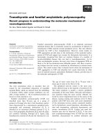
more consistent conformation.
The MBP(74–85) epitope adopts a quite compact
conformation (Fig. 2A), with rmsd values from the mean
structure for the Lys75–Glu82 fragment of 1.05 ± 0.40 A
˚
and 2 .05 ± 0.60 A
˚
for backbone N, C
a
,C¢ atoms and all
heavy atoms, respectively. As was the case in water, the k ey
characteristic of the structure of the native epitope in
Me
2
SO is the presence of a large number of electrostatic
interactions, especially in the central region of the peptide.
The side chain of Arg78 is locked into a conformation
resembling its conformation in aqueous solution, i.e.
perpendicular t o the plane defined by the backbone of the
peptide. The origin of this structural orientation in Me
2
SO is
also the strong electrostatic interactions of the side c hain of
Arg78 with the side chains of Asp81 and G lu82 [this is
consistent wi th the observed long-range NOE cross-peak
between the side chain of Arg78 (H
g
) and Glu82 (H
c1
)and
the medium-range NOE between the side chains of Asp81
(H
b
) and Arg78 (H
g
and H
c
)]. In addition, Glu82 i nteracts
withthesidechainofLys75.
The Ala81MBP(74–85) antagonist in Me
2
SO adopts a
less compact conformation than the MBP(74–85) epitope
(Fig. 2 B). The rmsd values from the m ean structure for
the Lys75–Glu82 fragment are 1.90 ± 0.55 A
˚
and
3.40 ± 0.80 A
˚
for backbone N, C
a
,C¢ atoms and all heavy
atoms, respectively. The C-terminal part (Ala81–Val85)
appears to be much more flexible, with Ala81 far distant
from the side chain of Arg78. The side chain of Arg78 is less
well defined, as in the case of aqueous solution, because of
the absence of interactions with the s ide chains of A sp81 (in
the agonist analogue) and Glu82.
MD simulations of MBP(74–85), and Ala81MBP(74–85)
in water and Me
2
SO
To further assess the structural origin of the difference in
activity between the agonist and antagonist, their dynamic
behaviour was investigated in detail by MD simulations in a
specific solvent (Me
2
SO and water) [ 62]. Five 10 ns MD
simulations starting from various structures in either
Me
2
SO or water were performed (Table 1). The evolution
of the r adius o f g yration, which reflects the c ompactness of
Fig. 2. Ensembles o f 3D structures of t he agonist ( A) and antagonist (B) linear analogues o f MBP(74–85) in Me
2
SO-d
6
solution. The lower thicker
trace c orresponds t o a r epresentative c onformer, t he structure of which is closest t o the average structure of t he ensemble.
Ó FEBS 2004 An autoimmune MBP peptide and its antagonist (Eur. J. Biochem. 271) 3403
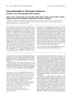
the molecule, is presented as a function of time in Fig. 3.
The peptides adopt more extended conformations in
Me
2
SO than in water. The a gonist in Me
2
SO is more
compact than the antagonist, in agreement with the NMR
data. In water, however, no conclusion can be drawn
because we cannot distinguish any statistically relevant
differences, and longer simulation times would be required
for comparison w ith NMR data. Interestingly, various turn
structures (type I ¢ b, t ype I I a nd one turn of an a-helix) are
observed in various simulations for the segment Arg78-
Ser79-Gln80-Asp81.
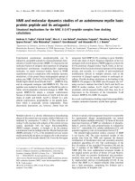
The cross rmsd matrix in Fig. 4 allows us to compare the
different trajectories. The pairwise backbone rmsd values
are colour-coded from 0.15 nm (blue) to 0.88 nm (red). As
already revealed by the gyration radius (Fig. 3 ), the cross
rmsd matrix clearly shows that t he structures in Me
2
SO and
water are different (yellow–orange off-diagonal blocks
between the simulations in water and Me
2
SO). Further,
the differences between the a gonist and the antagonist are
somewhat larger in water than i n Me
2
SO. A t 8 ns in water,
the antagonist seems to move t owards a conformation that
is closer to the conformation of the agonist. In M e
2
SO, the
differences are less important. Trajectory 5 shows the case of
the agonist in water s tarting from the NMR structure in
Me
2
SO; it can be seen that the starting structure disrupts
very quickly and moves towards the conformation of the
agonist in water [blue off-diagonal blocks b etween simula-
tions (1) and (5)].
As mentioned previously, Arg78 is of particu lar interest
as it is the target o f various post-translational modifica-
tions that might lead to demyelination. Theref ore, parti-
cular attention was paid to the dynamic behaviour of its
side chain, as well as to its interactions with other charged
groups. Figure 5 illustrates the evolution of some relevant
distances between charged groups in water. In the agonist,
the side chain of Arg78 is almost always less than 4 A
˚
from a negatively charged group, mainly Asp81 and
Glu82, but also the COO
–
terminal of Val85. This is
definitely not the c ase for the a ntagonist, a s Arg78 forms a
significantly smaller number of i nteractions. I n Me
2
SO,
the situatio n is even more striking; in the agonist, the side
chain of Arg78 ÔsticksÕ tightly t o Glu82 during the entire
trajectory, and it interacts with Asp81 between 0 and 5 ns.
This is further confirmed b y the average number o f
hydrogen bonds of the side c hain of Arg78 for the
simulations in Me
2
SO; the agonist forms a significantly
higher number of interactions than the antagonist. For the
simulations in water, the differences are not significant
enough to draw any conclusion. We can conclude that
Arg78 adopts a predetermined geometry in the case of the
agonist, which makes it somewhat more accessible than
the antagonist, as suggested by the solvent-accessible
surface area. These data clearly demonstrate the structural
importance of the nature o f the amino acid at position 81
of the encephalitogenic sequence 74–85 of guinea MBP:
replacement of Asp81 with an alanine seems to break a
chain of electrostatic interactions, especially between the
side chains of Arg78 and Glu82/Asp81 i ndependently of
the nature of the solvent (protic or nonprotic). This might
come from the peptide conformation, which drives the
orientation of these two side chains and lets them part
preventing any interaction.
Fig. 3. Evolution o f the rad ius of gyration of t he MBP(74–85) and Ala81MBP(74–85) pe ptides for t he five M D simulations of Table 1.
3404 A. G. Tzakos et al.(Eur. J. Biochem. 271) Ó FEBS 2004
Sequence alignment of the MBP(74–85) epitope
with the same region of various MBP species
The sequences ofseveral forms of MBP from different species
are known [7,8]. The relationship between the amino-acid
sequence and immune response has been extensively i nves-
tigated [2,5]. Differences in the amino-acid sequence of MBP
from various animal species h ave a significant effect on the
encephalitogenicity of different determinants from MBP [5].
Sequence alignment of the 74–85 sequence of guinea pig with
the same r egion of M BPs from o ther species is illustrated in
Fig. 6, and reveals that Gln74, Lys75, Ser76, Arg 78, Asp81,
Glu82, Asn83, Pro84 and Val85 (numbered a ccording to the
guinea pig species) a re highly conserved. Structure–activity
studies have shown that the MBP( 74–85) peptide analogue
induces experimental autoimmune encephalomyelitis in
Lewis rats and that single alan ine-substituted peptide
analogues at positions Lys75, Ser76, Arg78, Gln80, Asp81,
Glu82, and Pro84 resulted in significant reduction of the
proliferative responses of a T-cell line specific for the
MBP(74–85) peptide [63]. The studied segment of guinea
pig MBP(74–85), which lacks the His77–Gly78 segment
present in bovine MBP, h as been reported to be much more
encephalitogenic [5].
The sequence a lignment illustrated i n Fig. 6 reveals t hat
Arg78 and Asp81 are present in all forms of MBP and are
thus probably essential for the structure and fu nction of
MBP. As previously reported, aspartic acid at position 82
(81 according to the sequence numbering followed here)
may be a critical TCR contact residue for the Vb8.2
+
encephalitogenic T cells that predominate in the response of
LEW rats to the MBP(74–85) epitope [64]. This may explain
the antagonistic properties of the Ala81MBP(74–85) pep-
tide. Interestingly Glu82, which is also a conserved residue,
shows s ome i nteractions with Arg78 i n our NMR a nd MD
simulations. Glu82 may therefore act in the same way as
Asp81 to stabilize the specific conformation of Arg78 via an
electrostatic interaction.
One of the basic characteristics of MBP is its strong
positive net charge, which may have a critical role in the
Fig. 4. Backbone cross RMSD matrix for the comparison of the various conformers of the five MD simulations of Table 1. The x and y axes
correspond to the simulation time of the various systems (10 ns each). The rmsd values are co lour-coded accordingly to the sc ale given at the
bottom.
Ó FEBS 2004 An autoimmune MBP peptide and its antagonist (Eur. J. Biochem. 271) 3405
compaction of myelin, via electrostatic interactions with the
cell membrane [65]. It has been suggested [22,25] that the
reduction in charge density o n citrullinization o f arginines
(as occurs in multiple sclerosis) diminishes the interaction
with negatively charged lipids in the myelin membrane,
accounting for a c ertain amount of destabilization. The r ole
of this strong positive net charge is also important in the
MBP e pitope st udied ( Gln74, Lys7 5, Gln77, Arg78, Gln80,
Asn85) in comparison with the more positively charged
Ala81 variant. From our studies of the epitope of MBP
therefore we may conclude that the conformation of
epitopes of the integral protein must be affected by post-
translational modifications.
Docking calculations of the MBP(74–85) antigen
to the MHC class II receptor site I-A
u
– implications
for structure–activity relationships
The activation of CD4
+
T cells by peptide–MHC com-
plexes is a key event in the induction of autoimmune
diseases, such as multiple sclerosis. For a better under-
standing of the molecular basis of the MBP(74–85) antigen–
MHC II recognition, a model for the 3D structure of the
MBP(74–85) antigen–MHC II complex is r equired. The
only available structural data are the X-ray crystallographic
structures of the bimolecular complexes of the epitopes
1–11, 85–99 and 86–105 with the M HC class I I (pdbids:
1fv1, 1bx2 and 1k2d) [66–68]. Superimposition of t he above
MBP peptides on MHC class II is illustrated in Fig. 7A,
and was carried out by superimposing the a1/b1 domains of
the MHC class II molecules of the complexes. The main
MHC II peptide binding-groove anchor residues o f the
MBP peptides (P4, P6, P9), as well as important TCR
contact residues that are solvent-exposed (P5, P8),
superimpose quite well. In addition, the superimposed
MBP peptides shown in Fig. 7 are bound in MHC class II
molecules of different subclasses a nd expected to be highly
polymorphic in the relevant antigen-binding grooves.
Fig. 5. Evolution o f selected distances between c harged groups during the five MD simulations.
Fig. 6. Sequence alignment of the 74(3)-85(7 ) segment of
MBP_CAVPO [Cavia p orcellus (guinea pig)], w ith the sam e region of
MBP_CHICK ( chicken), MBP_PIG [ Sus scrofa (pig)], M BP_BOVIN
[Bos taurus (bovine)], MBP_RABIT [Oryctolagus cuniculus (rabbit)],
MBP_PANTR [Pan troglodytes (chimpanzee)], MB P_RAT [ Rattus
norvegicus (rat)], MBP_HUMAN [Homo sapiens (human)],
MBP_MOUSE [Mus musculus (mouse)]. The standard colour
parameter file of
CLUSTALX
was used. Ô*Õ Indicates positions that have a
single, fully co nserved r esidue; Ô:Õ indicates t hat one of the HRK (his-
tidine-arginine-lysine) groups is fully conserved; Ô.Õ indic ates that one of
the STP (serin e-threon ine- proline) groups is fully cons erved.
3406 A. G. Tzakos et al.(Eur. J. Biochem. 271) Ó FEBS 2004
Nevertheless, highly conserved MHC residues exist in the
P4, P 6 a nd P9 anchoring p ockets [N62a/N62a,Y13b/F11b
(F11b points towards Y13b superimposed on DR2 and
I-A
u
)andY26b/Y26b form the P 4 pockets; N62a/N62a,
V65a/T65a and Y 13b/F11b form the P6 pock ets; N69a/
N69a,I72a/V72a,D57b/D57b and Y60b/Y60b form the P9
pockets of DR2/I-A
u
]. Sequence alignment of the three
antigens derived f rom the structure alignment and sequence
alignment of M BP(74–85) on MHC II b inding t o t he t hree
antigens is illustrated i n Fig. 7B ( residues conserved am ong
the p eptide antigens are indicated i n r ed). The alignment of
MBP(74–85) was performed based on the highest aligned
score with t he three antigens, on the basis of the amino-acid
preference fo r the P4, P6 and P9 MHC I I anchor residues
and biological experiments in the lite rature [64]. Interest-
ingly, a s tatistically significant number of positively charged
residues (His, Arg and Lys) were found to project outside
the MHC binding groove in the X-ray structures of
MHC II–peptide complexes and, thus, may be readily
accessible for TCR recognition (Table 2 ) [66–72].
The sequence of peptide-binding motifs of several MBP
peptides and t heir capacity to bind to MHC class II are
illustrated in Table 3. Evidently, there i s a preference for
serine or threonine as an anchor point for position P9 of t he
MBP peptide-binding grooves. I nterestingly, truncation o f
the C-terminus of the MBP(86–105) peptide to P9 Thr
greatly diminished b inding, whereas truncation to P10 Pro
had little effect [73,74]. Biological data for the I-A
u
–MBP
(1–11) complex further support this hypothesis, as delet ion
of Ser7 of MBP (from the P9 pocket) greatly reduces the
affinity of I-A
u
for the MBP(1–6) epitope, but substitution
with Thr maintains the interaction [68]. This is in accord-
ance with the high conservation of the P9 pocket in the DR2
and I-A
u
MHC II molecules, as reported above. The
general peptide-binding motifs proposed for MHC class II
molecules define amino-acid preferences at positions P1, P4,
P6 and P 9 [75]. Vogt et al. [73] f ound that MHC II requires
an aliphatic residue (valine, isoleucine, methionine or
glutamine) at P4 as a main anchor point. As shown in
Fig. 7, the sequence alignment of MBP(74–85) with the
three antigens fulfil the a bove amino-acid preferences for
the MHC II anchor positions P4, P6 and P9 and show the
highest alignment similarity to the MBP(85–106) antigen.
Moreover, previous studies have shown that MHC mole-
cules can impose different alignments and conformations on
the same bound peptide, as a consequence of topological
differences in their peptide-binding sites [67]. Flexible
docking calculations using
HADDOCK
[59] were performed
for t he docking of the MBP(74–85) epitope to the I-A
u
-
binding pocket of the immunodominant MBP(1–11) self-
peptide [Protein Data B ank (PDB) number 1 k2d] [68]. We
used the specific MHC, as it has been shown that cryptic
epitopes [76,77] within the MBP sequence could, in princi-
ple, compete with the immunodominant Ac1–11 epitope for
binding to I-A
u
[77]. The docking studies were focused on
Table 2. Positively charged residues of bound peptides which are ex-
posed from the peptide-binding groove of MHC II of X-ray structures of
MHC II–peptide complexes. Single lette r c ode i s used for amino acids.
Peptide bound to MHC II
Exposed
residues
Brookhaven
PDB code
Human myelin basic protein
epitope MBP(85–99) [66]
H90, K93, R99 1BX2
Human myelin basic protein
epitope MBP(86–105) [67]
H90, K93, R99 1FV1
Influenza virus haemagglutinin
peptide HA(126–138) [69]
H126, H137 2IAD
CLIP fragment (87–101) [70] K90, R92 1A6A
Endogenous peptide
A2(103–117) [71]
R108, R111,
H114
1AQD
Ovalbumin peptide OVA
(323–339) [72]
H328, H331 1IAO
Fig. 7. (A) Superimposition of the MBP (85–99) pep tide (red) bound to
HLA-DR2b (PDB number 1bx2 [66]) o n the MBP(8 6–105) peptide
(blue) boun d to H LA-DR2a (PDB n umber: 1fv1 [ 67]) and th e MBP(1–
11) peptide (green) bound to I-A
u
(PDB number 1k2d [68]) and (B)
sequence alig nment of the three antigens derived from the structure
alignment. (A) Superimposition was carried out using th e a1andb1
domains of MHC class II molecules. (B) P5 an d P8 are prominent,
solvent-exposed TCR contact residues of the M BP peptides, and P4,
P6 and P9 are MHC II anchor residues of the MBP peptides. Sequence
alignment of MBP (74–85) on MHC II binding is also illustrated.
Residues conserved among the peptide an tige ns are indicated i n red.
Table 3. Sequence p eptide-binding motifs of several MBP peptides a nd
their binding capacity to the MHC class II receptor site (I-A
u
). On ly the
amino acids of the relevant epitopes involved i n the binding of the P1
to P10 MHC pockets are shown. MBP 18 and MBP 21 are derivatives
of MBP and defined as described by Garcia et al.[68].ND,Not
determined. P4, P6 and P9 (indicated in bold) represent M HCII
anchor residues.
P1 P2 P3 P4 P5 P6 P7 P8 P9 P10
Binding
capacity
(IC
50
n
M
)
MBP(1–11)
[67]
A S Q K RP S Q70
MBP21 [67] G A S Q Y RP S Q 8.3
MBP18 [67] R S H G K Y LAT A15
MBP(85–99) V H F F K N IVT PND
MBP(86–105) F K N I V T PRT PND
MBP(74–85) Q K S QRS QND
Ó FEBS 2004 An autoimmune MBP peptide and its antagonist (Eur. J. Biochem. 271) 3407
the binding grooves P4, P6 and P9, which were used as
restraints, so as to examine the possible structural rear-
rangement of the residues at positions 78 (lysine) and 81
(arginine), which are highly c onserved in all species for the
relevant MBP fragment (Fig. 6). A ll the restraints used in
the docking calculations are shown in Table 4.
The molecular model of t he I-A
u
–MBP(74–85) complex
[MBP(74–85) shown in orange] generated wit h
HADDOCK
superimposed on the X-ray crystallographic s tructure of the
I-A
u
–MBP(1–11) complex [ MBP(1–11) shown in blue]
is shown in Fig. 8. The peptide groups of MBP(74–85)
occupying pockets P4–P10 of I-A
u
superimposed quite well
on the relevant peptide groups of the MBP(1–11) epitope.
Compared with other peptides bound to class II molecules,
the C-terminal part of MBP(74–85) is positioned higher in
the I-A
u
binding groove, like t he C-terminus of MBP(85–99)
in the HLA-DR2a complex and the MBP(86–105) in the
HLA-DR2b complex [66,67]. The MBP(74–85) peptide is
bound to I-A
u
in an extended, type II polyproline confor-
mation, as previously observed in other class II structures.
The mode of binding of the MBP(74–85) pep tide t o I -A
u
is
determined by the occupied MHC anchor positions at P4,
P6, and P9. In
HADDOCK
, electrostatics is used during the
docking and in the scoring. No explicit hydrogen-bonding
potential is used because the electrostatics will take care of
proper hydrogen bonding. As a result, several hydrogen
Table 4. Residues used in the definition of the ambiguous interaction
restraints for the flexible docking calculations for I-A
u
and MBP(74–85)
epitope using
HADDOCK
[59]. The ambiguous interaction restraints are
defined between any atom of the MBP(74–85) listed residue and a ny
atom of the c orresponding listed I-A
u
residues. The effective distance is
calculated by sum averaging over a ll individual d istances ( see [59] for
details). Single l etter code is used for amino acids.
MBP(74–85) I-A
u
Q74 Y9a, F11b, P13b, Y62b
S76 N62a, T65a, F11b, Y30b
S79 H68a, N69a, V72a, D57b, Y61b
Fig. 8. Superimposition of (A) the X-ray
structure o f the MBP(1 –11) (blue)–I-A
u
com-
plex on the model of the MBP(74–85) antig en
(orange) complexed to I-A
u
obtained by flexible
docking with
HADDOCK
[59] an d (B) MBP
(1–11) on the MBP(74–85) antigen. Lys75 and
Arg78 o f MBP(74–85) are prominent, solvent-
exposed T CR contact r esidues. Arg78 and
Asp81 ( yellow) are probably involved in
electrostatic interactions.
3408 A. G. Tzakos et al.(Eur. J. Biochem. 271) Ó FEBS 2004
bonds are formed, which are presented in Table 5. More
explicitly, Gln74 occupies the P4 pocket, with a h ydrogen
bond to Ty r9a N(Gln74Oe1). The chemical environment
of the P6 pocket is a combination of a hydrophobic neck
and a moderately hydrophilic base, which favours accom-
modation of a large, bulky residue in the bound peptide
[Tyr4 in the case of the MBP(1–11)]. In the MBP(74–85)
epitope, this pocket could a ccommodate S er76 which could
form a hydrogen bond with Asn62a Od1 ( Ser76 N) and is in
close proximity to residues forming the hydrophobic neck of
this pocket (Table 5 ). The P9 pocket of I-A
u
is partially
filled b y Ser79 similarly to Ser7 of the MBP(1–11) epitope
(Fig. 8 A,B). This l oose fit is com pensated f or, in p art, by a
hydrogen bond between Ser79 N and Asn69a Od1and
Ser79 Oc and Asn69a Od1. This hydrogen bond is
important for the overall I-A
u
–MBP stabilization, because
deletion of Ser7 of MBP(1–11) greatly reduces the I-A
u
affinity for MBP(1–6), but substitution with Thr maintains
the interaction [68].
The exposed regions [P5 (Lys75) an d P8 (Arg78)] of the
peptide point outward and c omprise TCR contact residues.
In position P8 of the MBP(85–106) and MBP(74–85)
epitopes, there i s an a rginine ( Fig. 7A), which in the HLA-
DR2a–MBP(85–106) complex is exposed and is a potent
TCR contact residue. The relevant arginine (Arg78) of the
I-A
u
–MBP(74–85) complex was also found to be exposed,
in agreement with the NMR and MD structural studies. I n
addition, Asp81 is also found in an exposed region that is
consistent with previou s biological experiments, suggesting
that this specific residue is a TCR contact residue [64].
A vast body of structural and experimental data [78–80]
demonstrates the importance of the P1 anchor for M HC II
(I-A
u
,I-A
d
,I-A
k
). We speculate therefore that an ext ension
of the MBP(74–85) epitope in the N-terminus could increase
the b inding and affinity for I-A
u
because o f occupation of
the P1 pocket.
Recent thermodynamic a nd kinetic studies of the b inding
of TCRs to peptide–MHC ligands suggested that the low
affinity of the TCR–peptide–MHC complexis a consequence
not of insufficient c ontacts at the interface but, rather, of the
entropic penalty associated with the conformational a djust-
ment required for binding [81]. Our s tudies indicate that the
predetermined geometry of Arg78, reduced mobility, and
slightly increased average accessible surface area (Fig. 7A) in
the c ase of the agonist peptide result in lower activation
energy barriers a nd smaller c onformational a djustments
during the TCR–peptide–MHC recognition process.
There is accumulating evidence that epitopes that are
similar in sequence and present on viruses and normal
human tissue give rise to multiple sclerosis, in w hich the
immune response (T c ells or antibodies), directed primarily
against the virus, cross-reacts with the human tissue to cause
autoimmune disease. An interesting observation is the
structural organization of the fragment 76–84 of the Vb
chain of t he TCR with specific ity for a latent antigen of
Table 5. Intermolecular contacts between I-A
u
and MBP(74–85)
peptide. Single letter code is used for amino acids.
MBP(74–85) I-A
u
Hydrogen bonds
Q74 Oe1Y9a N
K75 N E74b Oe2
S76 N N62a Od1
Q77 O Y61b OH
R78 O Y66b OH
S79 N N69a Od1
S79 Oc N69a Od1
van der Waals contacts (< 4.5 A
˚
cut-off)
Q74 Y9a, F11b, P13b, T28b, Y62b, E74b
K75 F11b, E74b
S76 N62a, T65a, F11b, Y30b
Q77 T65a, N69a, Y30b, Y61 b, Y67b
R78 T65a, N69a, H68a, Y61b, Y67b
S79 H68a, N69a, V72a, D57b, Y60b, Y61b
Q80 P56b, Y60b
Fig. 9. Structure and sequence a lignments.
(A) Structure of the 76–82 fragment of the
TCR (pdbid: 1kgc) [82] and (B) sequence
alignmentoftheVb chain TCR(79–83),
MBP(74–85) and MBP(95–103). P5 a nd P8
are p rominent, s olvent-exposed TCR contact
residues of the MBP(86–105) peptide, and P4,
P6 and P 9 are MHC II anchor residues of the
MBP(86–105) e pitope. Residues cons erved
among the peptid e antigens are indicated in
red. (C) Superimposition of the MBP(97–101)
peptide (blue) bound by HLA-DR2b (PDB
number 1 fv1 [67]) on the TCR (79–83) frag-
ment ( blue). ( D) Sequence al ignment o f the
TCR(79–83) fr agment and the MBP(97–101)
peptide. R esidues co nserved among the pep-
tide antigens are indicated by a red square.
Ó FEBS 2004 An autoimmune MBP peptide and its antagonist (Eur. J. Biochem. 271) 3409
Epstein–Barr virus [82]. As shown i n F ig. 9B, the sequence
of the 76–84 fragment of this TCR aligns well with the
sequence of both MBP(74–82) (56%) and MBP(95–103)
(45%). Interestingly, the structural organization of the
76–84 fragment of this TCR meets the requ irements for
peptide–MHC II binding, a s follows from the conforma-
tional reorientation for residues P5 and P8 (exposed) and
P4, P6 and P9 (buried ) according to the X-ray structure
of the MBP(86–102) epitope complexed to M HC II.
Figure 9C,D illustrates the structure a nd sequence a lign-
ment of the MBP epitope 97–101 complexed to MHC II
and T CR(79–83), focusing on the c ritical residues arginine
and threonine (rmsd 0.27 A
˚
). This provides further evidence
for the proposed model of the MBP(74–85) epitope
complexed to MHC II.
Conclusions
These NMR, MD and sequence alignment conservation
data were obtained to try to identify the microdomain
structural organization of critical re sidues of guinea pig
MBP(74–85), which may be involved in triggering mult iple
sclerosis or affected by post-translational modifications.
This local microdomain structural organization may be
conserved when the antigen is bound to the MHC II
molecule. We focused o n Arg78 (a poten t target for post-
translational modifications) and Asp81 ( a T CR contact
residue [64]) and found that, in solution, the epitopes studied
adopt compact conformations, with a pred etermined
geometry for the critical residues, i.e. the formation of an
Arg78-Asp81 salt bridge in the case of the agonist, which
makes it more solvent accessible than the Ala81MBP
(74–85) antagonist. Interestingly, this interaction was also
found in the MBP(74–85) antigen when bound to MHC II,
which adopts an extended, type II polyproline conforma-
tion, as revealed by flexible docking calculations. According
to biological experiments [64], Asp81 is probably exposed
when the MBP(74–85) epitope is bound to I-A
u
allowing
TCR r ecognition. As we have identified a direct interaction
of this residue with Arg78, it can be expected that this
residue is also exposed allowing TCR recognition [P8
MHC II binding pocket i n the modelled MBP(74–85)–I-A
u
complex].
Our research has shed light on the conformational
properties of the guinea pig encephalitogenic epitope
MBP(74–85) and the antagonist Ala81MBP(74–85) ana-
logue both in water and Me
2
SO solution. Specifically, the
study indicates that MBP(74–85) has a compact confor-
mation, with the side chain of Arg78 positioned in a
well-defined, predetermined conformation and thus read-
ily accessible for post-tran slational modifications, which is
relevant to multiple sclerosis. This phenomenon is due to
the development, in the case o f the native epitope, o f a
network of electrostatic interactions among Lys75,
Arg78, Asp81 and Glu82, which results in a bioactive
conformation. Although differences were observed in the
backbone conformations of the epitope in Me
2
SO and
aqueous solutions, the conformation of Arg78 is practi-
cally solvent independent. Substitution of Asp81 by
Ala81 results in high side-chain mobility of the key
amino acid Arg78 in both solvents because of the
absence of t he above i nteractions.
Sequence alignment of MBP(74–85) with several s pecies of
MBP indicates the important role of Arg78, firstly in the
stabilization of local microdomains (e pitopes) of the integral
protein and in a number of post-translational modification s
relevant to multiple sclerosis, such as the reduction in
cationicity of MBP, especially due to conversion of positively
charged arginine residues into uncharged citrulline . In
addition, an aspartate residue (81 in the MBP epitope of
the guinea pig) is h ighly conserved in all MBP species,
implying a critical f unctional role previously rationalized to
be an important TCR contact residue [64]. T he construction
of the molecular model of the I-A
u
–MBP(74–85) complex
through flexible docking calculations and comparison with
the HLA-DR2a–MBP(85–106) complex indicate the vital
role of Lys75, Arg78 and Asp81 as TCR contact residues.
These results should provide new insights into the
molecular mechanism of T-cell activation and be of value
in designing experimental autoimmune encephalomyelitis
suppressing mimetic analogues with improved pharmaco-
logical profile and receptor selectivity.
Acknowledgements
Financial s upp ort from the Greek General Secretary o f Research and
Technology (EPET II 15, PENED 1999) is gratefully acknowledged.
The 750 MHz spectra were recorded at the SONNMR Large Scale
Facility in Utrecht, which is funded by the ÔAccess to Research
Infrastructures P rogramme of t he European U nionÕ (HPR1-CT -1999-
00005). We also thank the SONNMR Large Scale Facility for the
use of th e co mputational f acilities. Professor H. Kalbacher and
Dr V. Apostolopoulos are thanked f or useful comments and sugges-
tions. An anonymous referee is greatly acknowledged for his critical
comments, which sign ificantly improved t he paper.
References
1. Martin, R., McFarland, H.F. & McFarlin, D.E. (1992)
Immunological aspects of demyelinating diseases. Annu. Rev.
Immunol. 10 , 153–187.
2. Steinman, L . (1996) Multip le sclerosis: a coordinated i mmuno lo-
gical attack against m yelin in the c entral nervous system. Cell 85,
299–302.
3. Ota,K.,Matsui,M.,Milford,E.L.,Mackin,G.A.,Weiner,H.L.
& Hafler, D.A. ( 1990) T-cell rec ognition of an immunodominant
myelin basic protein e pitope i n m ultiple scle rosis. Na tur e
(London) 346 , 183–187.
4. Kursula, P. (2001) The current status of structural studies on
proteins of the myelin sheath. Int. J. Mol. Med. 8, 475–479.
5. Deber, C.M. & Reynolds, S.J. (1991) Central nervous system
myelin: structure, function, and pathology. Clin. Biochem. 24,
113–134.
6. Kirschner, D.A., Inouye, H., Ganser, A.L. & Mann, V. (1989)
Myelin membrane structure and compos ition correlated: a
phylogenetic study. J. N eurochem. 53, 1599–1609.
7. Carnegie, P.R. (1971) Amino acid sequence of the encephalito-
genic basic protein from human myelin. Biochem. J. 123,
57–67.
8. Eylar,E.H.,Brostoff,S.,Hashim,G.,Caccam,J.&Burnett,P.
(1971) Basic A1 protein of the myelin membrane. The complete
amino a cid sequence. J. Biol. Chem. 246, 5770–5784.
9. Tompkins, T.A. & Moscarello, M.A. (1993) Stimulation of bovine
brain p hospholip ase C ac tivity by myelin basic protein requires
arginyl residues in p eptide linkage. Arch. B iochem. Biophys. 302,
476–483.
3410 A. G. Tzakos et al.(Eur. J. Biochem. 271) Ó FEBS 2004
10. Tompkins, T.A. & Moscarello, M.A. (1994) The m echanism of
stimulation of brain ph ospholipase C-alpha by my elin basic p ro-
tein involves specific interactions. Biochim. Biophys. Acta 1206,
208–214.
11. Pirollet,F.,Derancourt,J.,Haiech,J.,Job,D.&Margolis,R.L.
(1992) Ca(2+)-calmodulin regulated effectors of microtubule
stability in bovine b rain. Biochemistry 31, 8849–8855.
12. Staugaitis, S.M., Colman, D.R. & Pedraza, L. (1996) Membrane
adhesion and o ther functions for the myelin basic prot eins.
Bioessays 18, 13–18.
13. Sires, L.R., Hruby, S ., Alvord, E.C. J r, Hells trom, I., Hellstrom,
K.E.,Kies,M.W.,Martemspm,R.,Deibler,G.E.,Beckman,E.D.
& Casnellie, J.E. (1981) Species restriction of a monoclonal anti-
body reacting with residues 130–137 in encephalitogenic myelin
basic protein. Science 214 , 87–89.
14. Sedzik, J . & Kirschn er, D.A. (1992) Is myelin basic p rotein crys-
tallizable? N eurochem. Res. 17, 157–166.
15. Golubovich, V.P., Kirnarskii, L.I. & Galaktionov, S.G. (1989)
Theoretical conformation al a nalysis of a encephalitogenic peptide
molecule and a study of the structure-activity relationship in a
series of its analogs. Bioph ysics 34, 368–371.
16. Inouye, H. & Kirschner, D.A. (1991) Folding and function of the
myelin proteins from primary sequence data. J. Neurosci. Res. 28,
1–17.
17. Stoner, G.L. (1990) Conservation throughout vertebrate evolution
of the predicted beta-strands in myelin basic protein. J. Neuro-
chem. 55, 1 404–1411.
18. Martenson, R.E. (1981) Prediction of t he secondary s tructure of
myelin basic protein. J. Neur och em. 36, 1 543–1560.
19. Mendz, G.L., Barden, J.A. & Martenson, R.E. (1995) Con-
formation of a tetradecapeptid e epitope of myelin basic p rotein.
Eur. J. Biochem. 231, 6 59–666.
20. Price, W.S., Mendz, G.L. & Martenson, R.E. (1988) Conforma-
tion of a heptadecapeptide co mprising t he segment enceph-
alitogenic in rhesus monkey. Biochemistry 27, 899 0–8999.
21. Beniac, D.R., Luckevich, M.D., Czarnota, G.J., Tompkins, T.A.,
Ridsdale, R .A., Ottensmeyer, F. P., Moscarello, M.A. & H arauz,
G. (1997) Three-dimensional structure of myelin basic protein. I.
Reconstruction via angu lar reconstitution of randomly oriented
single particles. J. Biol. Chem. 272, 4261–4268.
22. Ridsdale, R.A., Beniac, D.R., Tompkins, T.A., Moscarello, M .A.
& Harauz, G. (1997) Three-dimensional structure o f myelin basic
protein. II. Molecular modeling and consideratio ns of predicte d
structures in multiple sclerosis. J. Biol. C hem. 272, 4269–4275.
23. Fritz, R.B. & Ch ou, C.H . (1983) Epitopes of peptid e 43–88 of
guinea pig myelin basic protein: localiz ation with monoclonal
antibodies. J. Immu nol. 130, 2180–2182.
24. Carnegie, P.R., Dowse, C.A. & Linthicum, D.S. (1983) Antigenic
determinant recognized by a monoclonal antibody to human
myelin basic protein. J. Neuroimmunol. 5, 125–134.
25. Hruby, S., Alvord, E.C. Jr, Martenson, R.E., Deibler, G.E.,
Hickey, W.F. & Gonatas, N.K. (1985) Sites in myelin basic protein
that react with monoclonal antibodies. J. Neurochem. 44, 637–650.
26. Hruby, S., Alvord, E.C. Jr, Groome, N.P., Dawkes, A. & Mar-
tenson, R.E. (1987) Monoclonal an tibodies reactive with myelin
basic protein. Mol. Immu nol. 24, 1359–1364.
27. Tselios, T., Daliani, I., Probert, L., Deraos, S., Matsoukas, E .,
Roy, S., Pires, J., Moore, G. & Matsoukas, J. (2000) Treatment of
experimental allergic encephalomyelitis (EAE) induced by g uin ea
pig myelin basic protein epitope 72–85 with a human MBP (87–99)
analogue and effects of cyclic peptides. Bioorg.Med.Chem.8,
1903–1909.
28. Tselios, T., Daliani, I., Deraos, S., Thymianou, S., Matsouka, E.,
Troganis, A., Gerotha nassis, I. , M ouzaki, A., Mav romoustakos,
T., Probert, L. & Matsoukas, J. (2000) Treatment of experimental
allergic encephalomyelitis (EAE) by a rationally designed cyclic
analogue of myelin basic protein (MBP) epitope 72–85. Bioorg.
Med. Chem. L ett. 10, 271 3–2717.
29. Tselios, T., Probert, L., Daliani, I ., Matsoukas, E ., Troganis, A.,
Gerothanassis, I.P., Mavromoustakos, T., Moore, G.J. & Mat-
soukas, J.M. ( 1999) D esign and synthesis of a p otent c yclic ana-
logue of the myelin basic p rotein epitope MBP72-85: importance
of the Ala81 carboxyl group and of a cyclic conformation for
induction of experimental allergic encephalomyelitis. J. Med.
Chem. 42 , 1170–1177.
29a. Tselios, T., Apostolopoulos, V., Daliani , I., Deraos, S., Grda-
dolnik, S., Mavromoustakos, T., Melachrinou, M., Thymianou,
S., Probert, L., Mouzaki, A., Matsoukas, J. (2002) Antagonistic
effects of human c yclic MBP
87-99
altered peptide ligands in
experimental allergic encephalomyelitis and human T-cell prolif-
eration. J. Med. Chem. 45 , 275–283.
30. Lees, M.B. & Brostoff, S.W. (1984) Proteins of myelin. In Myelin,
2nd edn (Morell, P., ed.), pp. 197–224. Plenum Press, New York.
31. Smith, R. (1992) The basic protein of CNS myelin: its structure
and ligand binding. J. Ne urochem. 59, 1 589–1608.
32. Stuart, B.H. (1996) A Fourier transform infrared s pectroscopic
study of the secondary structure of myelin basic p rotein in
reconstituted myelin. Bioc hem. Mol. Biol. Int. 38, 839–845.
33. Deibler, G.E., S tone, A.L. & Kies, M.W. ( 1990) Role of phos-
phorylation in co nformational adaptability of bovine myelin basic
protein. Proteins 7, 32–40.
34.Ramwani,J.J.,Epand,R.M.&Moscarello,M.A.(1989)
Secondary structure of charge isomers of myelin basic protein
before and a fter phosphorylation. Biochemi stry 28, 6538–6543.
35. Keniry, M.A. & S mith, R. (1981) Dependence on lipid structure of
the coil-to-helix transition of bovine myelin basic protein. Biochim.
Biophys. A cta 668, 107–118.
36. Hwang, T.L. & Shaka, A.J. (1995) Water suppression that works.
Excitation sc ulpting using arbitrary waveforms and pulsed fi eld
gradients. J . Magn. R eson. A 112, 275–279.
37. Delaglio, F., Grzesiek, S., Vuister, G.W., Zhu, G., Pfeifer,
J. & Bax, A. (1995) NMRPipe: a multidimensional spectral
processing system based o n UNIX pipes. J. Biomol. NMR 6, 277–
293.
38. Johnson, B.A. & Blevins, R.A. ( 1994) Nmrview: a computer-
program f or the visualization and analysis of NMR data. J. Bio-
mol. NMR 4, 603–614.
39. Brunger, A.T., Adams, P.D., Clore, G .M., DeLano, W.L., G ros,
P., Grosse-Kunstleve, R.W., Jiang, J.S., Kuszewski, J., Nilges, M.,
Pannu,N.S.,Read,R.J.,Rice,L.M.,Simonson,T.&Warren,
G.L. (1998) Crystallography & NMR system: a new software suite
for macromolecu lar s tructure determination. Acta Crystallogr.
D 54, 905–921.
40. Linge, J.P. & Nilges, M. ( 1999) Influence of n on-bonde d p ara-
meters on the quality of NMR structures: a new force field for
NMR structure c alculatio n. J. Bio mol. NMR 13, 51–59.
41. Nilges,M.&O’Donoghue,S.I.(Year?)AmbiguousNOEsand
automated NOE assignment. Prog. Nucl. Magn. Reson. Spectrosc.
32, 1 07–139.
42. Bonvin, A.M., Houben, K., Guenneugues, M., Kaptein, R. &
Boelens, R. (2001) Rapid protein fold determination u sing s ec-
ondary c hemical shifts and c ross-hydrogen b ond
15
N–
13
C¢ scalar
couplings ( 3hbJNC¢). J. Bio mol. NMR 21 , 221–233.
43. Linge, J.P., Williams, M.A., Spronk, C.A., Bonvin, A.M. &
Nilges, M . (2003) Refinement of protein structures in explicit
solvent. Pr oteins Struct. Funct. Genet 50, 496–506.
44. Engh, R .A. & H uber, R. (1991) Accurate bond and angle
parameters for X -ray protein-structure r efi nement. Acta Crystal-
logr. A 47, 392–400.
45. Hendrickson, W.A. (1985) Stereochemically restrained refine-
ment of macromolecular st ructures. Methods Enzymol. 115, 252–
270.
Ó FEBS 2004 An autoimmune MBP peptide and its antagonist (Eur. J. Biochem. 271) 3411
46. Jorgensen, W .L. & Tirado-Rives, J. (1988) The O PLS force field
for p roteins. Energy minimizatio ns f or crystals of cyclic peptides
and crambin. J. Am. Chem. So c. 110, 1657 –1666.
47. Laskowski, R.A., Macarthur, M.W., Moss, D.S. & Thornton, J.M.
(1993) Procheck: A program to check the stereochemical quality of
protein structures. J. Appl. Crystallogr. 26 , 283–291.
48. Berendsen, H.J.C., van der Spoel, D. & van Drunen, R. (1995)
GROMACS: a message-passing paralle l mo lecu lar d ynamics
implementation. Comp. Phys. Commun. 91, 43–56.
49. Lindahl, E., Hess, B. & van der Spoel, D. (2001) GROMACS 3.0:
a package for molecular simulation and trajectory analysis. J. Mol.
Model. 7, 306–317.
50. Daura, X., Mark, A.E. & Van Gunsteren, W.F. (1998) Para-
meterization of aliphatic CHn united atoms of GROMOS96 force
field. J. Comp. Chem. 19, 535–547.
51. Berendsen, H.J.C., P ostma, J.P.M., v an Gunsteren, W.F. &
Hermans, J. (1981) Interaction models for water in relation to
protein hydration. In Intermolecular Forces (Pullman, B., ed.), pp.
331–342. R eidel, Dordrecht.
52. Liu, H., Muller-Plathe, F. & van Gunsteren, W.F. (1 995) A force
field for liquid dimethyl sulfoxide and physical properties of liquid
dimethyl sulfoxide calculated using molecular d ynamics simula-
tion. J. Am. Chem. Soc . 117, 4363–4366.
53. Berendsen, H.J.C., Postma, J.P.M., DiNola, A. & Haak, J.R.
(1984) Molecular dynamics with coupling to an external bath.
J. Che m. Phys. 81 , 3684–3690.
54. Hess, B., Bekker, H., Berendsen, H.J.C. & Fraaije, J.G.E.M.
(1997) LINCS: a linear constraint solver for molecular simula-
tions. J. Comp. Chem. 18 , 1463–1472.
55. Miyamoto, S. & Kollman, P.A. (1992) SETTLE: an analytical
version of the SHAKE and RATTLE algorithms for rigid w ater
models. J. Comp. Chem. 13 , 952–962.
56. Tironi,I.G.,Sperb,R.,Smith,P.E.&vanGunsteren,W.F.(1995)
Generalized re action field me tho d for molecular dynamics simu -
lations. J. Ch em. Phys. 102, 5451–5459.
57. Hubbard, S.J., Thornton, J.M. NACCESS: Computer Program.
Department of Biochemistry and Molecular Biology, Unive rsity
College Lo ndon.
58. Thompson, J.D., Higgins, D.G. & Gibson, T.J. (1994) CLUSTAL
W: improving the sensitivity of progressive multiple sequence
alignment throu gh sequence weighting, position s-sp ecific gap
penalties a nd weight matrix choice. Nucleic Acids Res. 22, 4673 –
4680.
59. Dominguez, C., Boelens, R . & Bonvin, A.M. ( 2003) HADDOCK:
a protein-prote in do cking a pproac h based o n bioc he mical or
biophysical information. J. Am. Chem. Soc. 125 , 1731–1737.
60. Wu
¨
thrich, K. (1986) NMR of Proteins and Nucleic Acids.John
Wiley and So ns, Inc, New York.
61. Andersen, N.H., Neidigh, J.W., Harris, S.M., Lee, G.M., Liu,
Z.H. & Tong, H. (1997) Extracting information from the tem-
perature gradients o f polypeptide NH ch emical shifts.1. The
importance of co nformational averaging. J. Am. Chem. Soc. 119,
8547–8561.
62. Grdadolnik, S.G., Mierke, D.F., Byk, G., Zeltser, I., Gilon, C. &
Kessler, H. (1994) Comparison of the conformation o f active and
nonactive backbone cyclic analogs of substance P as a tool to
elucidate features of the bioactive conformation: NMR a nd
molecular dynamics in DMSO and water. J. Med. Chem. 37,
2145–2152.
63. Wauben, M.H., Boog, C.J., van der Zee, R., Joosten, I., Schlief, A.
& van Eden, W. (1992) Disease inhibition by major
histocompatibility complex binding peptide analogues of
disease-associated epitopes: more than blocking alone. J. Exp.
Med. 176, 6 67–677.
64. Smeltz, R.B., Wauben, M.H., Wolf, N.A. & Swanborg, R.H.
(1999) Critical requirement for aspartic acid at position 82 of
myelin basic protein 73–86 for recruitment of V beta 8.2+ T cells
and encephalitogenicity in the Lewis rat. J. Immunol. 162, 829–
836.
65. Moscarello, M.A. ( 1997) Myelin basic p rotein, t he ÔexecutiveÕ
molecule of the myelin membrane. In Cell Biology and Pathology
of Myelin: E volving Biological C oncepts and Therapeutic Approa-
ches (Jurlink, B.H.J., Devon, R.M., Doucette, J.R., Nazarali, A.J.,
Scheyer, D.J. & Verge, V.M.K., eds), pp. 13–26. Plenum Press,
New York.
66. Smith, K.J., P yrdol, J., Gauthier, L., Wiley, D.C. & Wucherp-
fennig, K.W. (1998) Crystal structure of HLA-DR2 (DRA*0101,
DRB1*1501) complexed with a peptide from human myelin basic
protein. J. Exp. Med. 188 , 1511–1520.
67. Li,Y.,Li,H.,Martin,R.&Mariuzza,R.A.(2000)Structuralbasis
for the binding of an immunodominant peptide fro m myelin basic
protein i n d iffere nt r egisters by two H LA-DR2 p rot eins. J. Mol.
Biol. 304 , 177–188.
68. Xiao-Iin H e, R adu, C., Sidney, J., Sette, A., Ward, S. & Garcia,
K.C. ( 2002) Structural snapshot o f aberrant a ntigen presentation
linked to autoimmunity: th e immunodomin ant epitope of MBP
complexed with I-A
u
. Immunity 17 , 83–94.
69. Hennecke, J., Carfi, A. & Wiley, D.C. (2000) Structure of a
covalently stabilized complex of a human Ab-T cell r eceptor,
influenza Ha peptide and MHC c lass II molecule, Hla-DR1.
EMBO J. 19 , 5611–5624.
70. Ghosh, P., Amaya, M., Mellins, E. & Wiley, D.C. (1995) The
structure of an intermediate in class II MHC maturation: CLIP
bound to H LA-DR3. Nature ( London) 378, 457–462.
71. Murthy, V.L. & Stern, L.J. (1997) The class II MHC protein
HLA-DR1 in c omple x with an endogeno us peptide: implicat ions
for the structural basis of the specificity of peptide binding.
Structure 5, 1385–1396.
72. Scott, C.A., P eterson, P.A., Teyton & Wilson, I.A. (1998) Crystal
structures of two I-A
d
-peptide co mplexes reveal t hat high a ffinity
can be achieved w ithout large anchor r esidues. Immunity 8, 319–
329.
73. Vogt, A.B., Kropshofer, H., Kalbacher, H., K albus, M., Ram-
mensee, H .G., Coligan, J.E. & Martin, R. (1994) Ligand motifs of
HLA-DRB5*0101 and DRB1*1501 molecu les delineated from
self-peptides. J . Immunol. 153, 1665–1673.
74. Wucherpfennig, K.W., S ette, A ., S outhwood, S., Oseroff, C.,
Matsui, M., Strominger, J.L. & Hafler, D.A. (1994) Structural
requirements for binding of an immunodominant myelin basic
protein peptide to DR2 isotypes and for its recognition by human
Tcellclones.J. Exp. M e d. 179, 2 79–290.
75. Madden, D. (1995) The three-dimensional structure of peptide-
MHC complexes. Annu.Rev.Immunol.13, 5 87–622.
76. Kumar, V. & Sercarz, E.E. (1993) The involvement of T c ell
receptor peptide-spec ific regulatory CD4+ T cells in recovery
from antigen-induced autoimmune disease. J. Exp. Med. 178, 909–
916.
77. Fairchild, P.J., Pope, H. & Wraith, D.C. (1996) The nature of
cryptic epitopes within th e self-antigen myelin basic protein.
Int. Imm unol. 8, 1035–1043.
78. Lee, C., Liang, M.N., Tate, K.M., Rabinowitz, J.D., Beeson, C.,
Jones, P.P. & McCon nell, H.M. (1998) Eviden ce that the auto-
immune antigen myelin basic protein (MBP) Ac1–9 binds towards
one end of the major histocompatibility complex (MHC) cleft.
J. Exp. Med. 187, 1505–1516.
79. Fremont, D.H., Monnaie, D., Nelson, C.A., Hendrickson, W.A.
& Unanue, E.R. (1998) Crystal structure of I-Ak in complex with
a dominant epitope o f lysozyme. Immunity 8, 305– 317.
80. Nelson, C.A., Viner, N.J., Yo ung, S .P., Petzold, S.J. & Unanue,
E.R. (1996) A negat ive ly charged an chor residue promotes high
affinity bin ding to the MHC c lass II molecule I-Ak. J. Immunol.
157, 7 55–762.
3412 A. G. Tzakos et al.(Eur. J. Biochem. 271) Ó FEBS 2004
81. Willcox, B.E., Gao, F.G., Wyer, J.R., Ladbury, J.E., Bell, J.I.,
Jakobsen, B.K. & van der Merwe, P.A. ( 1999) TCR binding to
peptide-MHC stabilizes a flexible recognition interface. Immunity
10, 357–365.
82. Kjer-Nielsen, L., Clements, C.S., Brooks, A.G., Purcell, A.W.,
McCluskey, J. & Rossjohn, J. (2002) The 1.5 A
˚
crystal structure
of a highly selected antiviral T cell receptor provides evidence
for a structural basis of immunodominance. Structure 10, 1521–
1532.
Supplementary material
The follow ing material is available from http://www.
blackwellpublishing.com/products/journals/suppmat/EJB/
EJB4274/EJB4274sm.htm.
Appendix S1. Four Tables with the full assignment (
1
H
and
13
C) of the two peptides in both Me
2
SO and aqueous
solution (Tables S1, S2, S3, S4) and Table S5 with the buried
residues for three complex MHC I I peptides.
Fig. S1. (A) Plot of temperature coefficients Dd/DT vs. the
amino-acid residues of the MBP(74–85) agonist and the
Ala81MBP(74–85) antagonist. Differences in experimental
HA and HN chemical-shift values (in p.p.m.) between the
proton resonances of MBP(74–85) and Ala81MBP(74–85)
in aqueous (B) and Me2SO solution ( C).
Fig. S2. Oc currences of a type II b turn on the tetrapeptide
RSQD segment.
Fig. S3. (A) Average number of hydrogen bonds of the s ide
chain of Arg78 for the agonist and t he antagonist in water
and Me2SO solutions. (B) Average solvent-accessible sur-
face area (ASA).
Fig. S4. Superimposition o f MHC II molecules (A) and
MHC II-bound peptides (B). DR2 (PDB number 1fv1)/
[MBP(86–105)], I-Au (PDB number 1k2d)/[MBP(1–11)]
and DR2 (PDB number 1bx2)/[MBP(85–99)]. Superimpo-
sitions were carried out using the a1andb1 domains of
MHC class II molecules.
Ó FEBS 2004 An autoimmune MBP peptide and its antagonist (Eur. J. Biochem. 271) 3413