Báo cáo khoa học: Differential effects of RU486 reveal distinct mechanisms for glucocorticoid repression of prostaglandin E2 release docx
Bạn đang xem bản rút gọn của tài liệu. Xem và tải ngay bản đầy đủ của tài liệu tại đây (667.12 KB, 11 trang )
Differential effects of RU486 reveal distinct mechanisms for
glucocorticoid repression of prostaglandin E
2
release
Joanna E. Chivers
1
, Lisa M. Cambridge
1
, Matthew C. Catley
1
, Judith C. Mak
1
, Louise E. Donnelly
1
,
Peter J. Barnes
1
and Robert Newton
2
1
Department of Thoracic Medicine, National Heart and Lung Institute, Imperial College London, Faculty of Medicine, London, UK;
2
Department of Biological Sciences, University of Warwick, Coventry, UK
In A549 pulmonary cells, the dexamethasone- and budeso-
nide-dependent repression of interleukin-1b-induced pros-
taglandin E
2
release was mimicked by the s teroid antagonist,
RU486. Conversely, whereas dexamethasone and budeso-
nide were highly effective inhibitors of interleukin-1b-
induced cyclooxygenase (COX)/prostaglandin E synthase
(PGES) activity and COX-2 expression, RU486 (< 1 l
M
)
was a poor inhibitor, but was able to efficiently antagonize
the effects of dexamethasone and budesonide. In addition,
both dexamethasone and RU486 repressed [
3
H]arachido-
nate release, which is consistent with an effect a t the level
of phospholipase A
2
activity. By contrast, glucocorticoid
response element-dependent transcription was unaffected by
RU486 but induced by dexamethasone and budesonide,
whilst dexamethasone- and budesonide-dependent repres-
sion of nuclear factor-jB-dependent transcription was
maximally 30–40% and RU486 (< 1 l
M
) was without
significant effect. Thus, two pharmacologically distinct
mechanisms of glucocorticoid-dependent repression of
prostaglandin E
2
release are revealed. First, glucocorticoid-
dependent repression of arachidonic acid is mimicked by
RU486 and, second, repression of COX/PGES is antagon-
ized by RU486. Finally, whilst all compounds induced
glucocorticoid receptor translocation, no role for glucocor-
ticoid response element-dependent transcription is suppor-
ted in these inhibitory processes and only a limited role for
glucocorticoid-dependent inhibition of nuclear factor-jBin
the repression of COX-2 is indicated.
Keywords: corticosteroid; cyclooxygenase; epithelial cell;
glucocorticoid receptor; prostaglandin E
2
.
Synthetic glucocorticoids are potent repressors of inflam-
mation and are a first-line therapy for i nflammatory diseases
[1]. However, their clinical usage is limited by immunosup-
pression as well as by metabolic effects, including increased
gluconeogenesis, i ncreased blood glucose, amino and fatty
acid mobilization, and loss of bone [2]. In addition,
endogenous glucocorticoids p articipate in feedback inhibi-
tion of the hypothalamo-pituitary-adrenal axis, and long-
term high-dose synthetic glucocorticoid usage may cause
hypothalamo-pituitary-adrenal insufficiency and glucocor-
ticoid dependency.
Glucocorticoids are believed to act primarily via the
glucocorticoid receptor (GR), which is maintained as an
inactive cytoplasmic c omplex with heat shock proteins (hsp)
and immunophilins [3]. Following ligand binding and
complex dissociation, the GR translocates to the nucleus
where it binds glucocorticoid response elemen ts (GREs), as
a dimer, to promote the transcription of responsive genes
[2]. However, the GR may also act a s a monomer to inhibit
key inflammatory transcription factors, such as nuclear
factor-jB(NF-jB) and activator protein-1, by direct
interaction, competition for cofactors or by modifying the
chromatin structure to prevent the expression of inflamma-
tory genes [1,2].
Inflammatory prostaglandins, produced by the arachi-
donic acid cascade, play a pathophysiological role in
edema, bronchoconstriction, fever and hyperalgesia [4].
Arachidonic acid, released from cell membranes by
phospholipase A
2
(PLA
2
), is converted to prostaglandin
H
2
(PGH
2
) by c yclooxygenase enzymes ( COX), and
further modified by specific isomerases and reductases to
produce b iologically relevant prostaglandins, including
prostaglandin E
2
(PGE
2
), which is the major prostaglan-
din product of both airway epithelial and A549 cells [5]. In
inflammation, the inducible COX, COX-2, is normally
up-regulated a nd accounts for the elevated levels of
prostaglandins [4]. Conversely, COX-2 expression is highly
sensitive to glucocorticoid inhibition, suggesting that
inhibition of COX-2 is critical in t he repression of
prostaglandins by glucocorticoids. As cytokine-induced
COX-2 and PGE
2
release are highly NF-jB-dependent in
A549 cells [6], and treatment with dexamethasone pro-
foundly represses PGE
2
release a nd COX-2 expression [7],
Correspondence t o R. Newton, Department of Biological Sciences,
University of Warwick, Coven try CV4 7AL, UK.
Fax: +44 2476 523701; Tel.: +44 2476 574187;
E-mail:
Abbreviations: C OX, cyclooxygenase; CRE, cyclic A MP response
element; DAPI , 4¢,6¢-diamidino-2-phenylinole dihydrochloric hydrate;
EGF, ep idermal growth f actor; GR, glucocorticoid receptor;
GRE, glucocorticoid response e lement; hsp, heat shock pr otein;
IL,interleukin;NF-jB, nuclear factor-jB; PGE
2
, p rostaglandin E
2
;
PGES, prostaglandin E synthase; PLA
2
, phosph olipase A
2
;
SFM, serum-free media.
(Received 13 January 2 004, revised 1 6 A ugust 2 004,
accepted 23 August 2004)
Eur. J. Biochem. 271, 4042–4052 (2004) Ó FEBS 2004 doi:10.1111/j.1432-1033.2004.04342.x
we have used this system to further explore the mecha-
nisms of glucocorticoid action.
Materials and methods
Cell culture
A549 cells were cultured to confluence, as described
previously [7]. Following overnight incubation in serum-
free media (SFM), d rugs (dexamethasone, budesonide,
ionomycin, R U486) were a dded 1 h before stimulation with
interleukin-1b (IL-1b) (R & D Systems, Oxon, UK).
Dexamethasone and budesonide (both Sigma, Poole, UK)
were dissolved in Hank’s balanced salt solution (Sigma).
Ionomycin and RU486 (both Sigma) were dissolved in
ethanol. Final concentrations of ethanol were less than
0.1% (v/v).
PGE
2
release, COX/prostaglandin E synthase (PGES)
activity and COX-2 expression
PGE
2
released into the medium was measured using a
commercially available PGE
2
antibody (Sigma) [5,8]. For
the assay of combined COX/PGES activity, cells were
rinsed with SFM prior to incubation at 37 °Cfor10minin
SFM supplemented with 3 0 l
M
arachidonic acid, and
released PGE
2
was taken as a index of COX/PGES a ctivity
[5,8]. Northern and Western blot analyses were performed
as described previously [7].
Reporter cell lines and luciferase assay
A549 cells containing the N F-jB-dependent r eporter,
6jBtkluc, have been described previously [9]. The 1·GRE-
dependent and 2·GRE-dependent reporters, pGL3.neo
TATA.GRE and pGL3.neo.TATA.2GRE, respectively,
were based on the parent vector pGL3.neo.TATA, which
contains a modified minimal b-globin promoter, as pre-
viously described [10]. This was digested at the SmaI
site, upstream of t he minimal promoter, and double-
stranded oligonucleotides (sense strand: 5¢-GC
TGTACAG
GATGTTCTAG-3¢ and 5¢-GCTGTACAGGATGTTC
TAGGCTGTACAGGATGTTCTAG-3¢), containing one
or two copies of a consensus GRE site (underlined) [11],
were inserted to produce pGL3.neo.TATA.GRE and
pGL3.neo.TATA.2GRE, respectively. A 2·GRE(mut )
reporter was generated as described above, but using a
mutated 2·GRE oligonucleotide (sense strand
5¢-GC
TcaACAGGATcaTCTAGGCTcaACAGGATcaT
CTAG-3¢) (mutated bases in lower case). The cyclic AMP
response element (CRE)-dependent reporter, which con-
tains six CRE s ites, was as previously described [ 12]. A549
cells, stably harboring the luciferase reporters, were gener-
ated as previously described [9]. Prior to experiments,
confluent plates of reporter cells were incubated overnight in
serum-free, G-418-free, media. Cells were subsequently
harvestedin1· reporter lysis buffer (200 ll) (Promega) 6 h
after treatment for luciferase activity assay (Promega).
As each well is confluent and a ll the cells contain the
reporter construct, we find reporter activity to be highly
reproducible, and normalization to a second reporter is
unnecessary [9].
[
3
H]Arachidonic acid release
As previously described [8], cells were incubated overnight
in 0.5 mL of S FM supplemented with 0.125 lCi
[5,6,8,9,11,12,14,15-
3
H]arachidonic acid (Amersham Phar-
macia). Cells were washed twice prior to treatment with
dexamethasone or RU486. After 1 h, supernatants were
changed to fresh SFM containing 2 mgÆmL
)1
fatty acid-free
BSA (Sigma) plus drugs prior to stimulation. Supernatants
were collected and cells washed prior to harvesting in
1% (w/v ) S DS. Relea se of [
3
H]arachidonic acid, or its
metabolites, was expressed as a percentage of the total
incorporated.
Ligand binding
At 80% c onfluence, A549 cells cultured in T175 flasks w ere
transferred to SFM and harvested the following day in
cell dissociation solution (C-5789; Sigma). Cells
(1.5–4 · 10
6
cells per mL) were incubated overnight at
4 °C with increasing concentrations of [
3
H]dexamethasone,
in the p re sence of 10 l
M
dexamethasone, to determine
nonspecific binding. F ree radioligand was removed by t he
rapid filtration of cells through glass-fibre filters (GF/B)
presoaked i n NaCl/P
i
(PBS), 0.1% ( v/v) polyethylenimine,
using a cell harvester [M-24R Brandel, SEMAT Technical
(UK) Ltd, St. Albans, Hertfordshire, UK]. Filters were
combined with Filtron-X scintillant (National D iagnostics,
Atlanta, GA, USA) and radioactivity was measured using a
beta counter (2200CA Tri-carb Liquid Scintillation Ana-
lyser; Canberra Packard, Berks., UK). K
d
and B
max.
values
were determined using saturation binding isotherms and
Scatchard analysis, [Bound]/[Free] vs. [Bound], where the
x-intercept ¼ B
max.
and the gradient ¼ ) 1/K
d
(Fig. 1 A)
(
PRISM
3; GraphPad, San Diego, CA, USA). Relative
binding affinity was assessed by incubating cells with an
increasing concentration of unlabelled steroid and 4 n
M
[
3
H]dexamethasone overnight at 4°C. Bound and free
radioligand were separated as d escribed above. Specific
binding was c alculated by s ubtraction of nonspecific from
total binding, and Cheng–Prusoff analysis was performed to
determine the K
i
value: K
i
¼ IC
50
/{1 + ([Free Count]/K
d
)},
where IC
50
is the concentration that results i n 50% inhibi-
tion (Table 1) [13].
Immunocytochemistry
Cells grown on coverslips were transferred at 70% conflu-
ence to SFM f or 24 h. After incubation with steroid for the
indicated times, cells were washed with NaCl/P
i
(PBS) and
fixed with 4% (w/v) paraformaldehyde before successive
incubations in 0.5% (v/v) Nonidet P-40 and 100 m
M
glycine. Coverslips were blocked in NaCl/P
i
(PBS) contain-
ing 0 .1% ( v/v) Tween- 20, 0.1% (w/v) BSA and 10% (v/v)
human serum prior to incubation for 1 h in 5 lgÆmL
)1
rabbit anti-human GR (PA1–511A; Affinity Bioreagents
Inc., Golden, CO, USA) or rabbit isotype con trol (Dako,
Glostrup, Denmark). After washing with NaCl/P
i
(PBS)
containing 0.1% (v/v) Tween and incubation with biotin-
ylated anti-rabbit immunoglobulins (Dako) for 1 h, cells
were incubated with fluorescein isothiocyanate (FITC)-
linked s treptavidin (Dako) for 1 h. Nuclei were then stained
Ó FEBS 2004 Glucocorticoid repression: differential mechanisms (Eur. J. Biochem. 271) 4043
with 1 l
M
4¢,6¢-diamidino-2-phenylinole dihydrochloric
hydrate (DAPI) (Sigma) and coverslips were mounted on
glass slides using Citifluor mounting fluid (Citifluor Ltd,
London, UK), prior to analysis using a Leica TCS 4D
confocal microscope (Leica Microsystems, M ilton Keynes,
UK) equipped with argon, krypton, and ultraviolet lasers.
Confocal images w ere acquired at ·40 magnification using
TCS NT
software (Leica Microsystems).
Statistical analysis
Statistical a nalysis was perf ormed u sing analysis of variance
(
ANOVA
) w ith a Dunn’s post-test, unless specifically stated
otherwise in the figure legends. Significance was taken at
P-values of < 0.05 (*), < 0.01 (**) and < 0 .001 (***).
Results
Repression of PGE
2
release, COX/PGES activity
and COX-2 expression
As reported previously [7,14], untreated A549 cells released
low levels of PGE
2
(1.2 ± 0.2 ngÆmL
)1
) and showed low
levels of combined COX/PGES activity (3.1 ± 0 .6 ng Æ
mL
)1
Æmi n
)1
), which were both increased upon stimulation
with IL-1b (1 ngÆmL
)1
) (22.6 ± 3.7 ngÆmL
)1
and
0
50
100
-7-6 -5 -10 -9 -8 -7 -6 -5
NS
IL-1
Log [Bud] (M)Log
[RU486]
(M)
0
50
100
-7-6 -5 -10 -9 -8 -7 -6 -5
NS
IL-1
Log [Bud] (M)Log
[RU486]
(M)
PGE
2
release
(% IL-1
)
0
50
100
-10 -9 -8 -7 -6 -5
Log [Steroid] (M)
NS
IL-1
0
50
100
0
50
100
Log [RU486] (M)
-10 -9 -8 -7 -6 -5
NS
IL-1
IL+Dex
Log [RU486] (M)
-10
-9 -8 -7 -6 -5
NS
IL-1
IL+Dex
0
50
100
0
50
100
-7-6 -5 -10 -9 -8 -7 -6 -5
NS
IL-1
Log [Dex] (M)Log
[RU486]
(M)
-7-6 -5 -10 -9 -8 -7 -6 -5
NS
IL-1
Log [Dex] (M)Log
[RU486]
(M)
PGE
2
release
(% IL-1
)
COX/PGES activity
(% IL-1
)
-11
COX/PGES activity
(% IL-1
)
0
50
100
-10 -9 -8 -7 -6 -5
Log [Steroid] (M)
NS
IL-1
-11
IL+Bud
IL+Bud
ABC
D
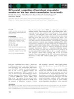
Fig. 1. The e ffect of dexamethasone, budesonide and R U486 on inte rleukin-1b (IL-1 b)-dependent prostaglandin E
2
(PGE
2
) release and cyclooxygenase
(COX)/prostaglandin E synthase (PGES) activity. (A) A549 cells were cultured with various concentrations of dexamethasone (j), budesonide (h)
or RU486 (.) for 1 h prior to stimulation with IL-1b (1 ngÆmL
)1
) or no stimulation (NS). (B) Cells were treated with dexamethasone (Dex)
(0.1 l
M
)(j) or b udesonide (0.1 l
M
)(h), in th e presence of increasing c oncen trations of RU486, for 1 h prior to stimulation w ith IL-1b (1 ngÆmL
)1
)
or no stimulation (NS). (C and D ) Cells were treated w ith various c onc entrations of d examet hasone (C) or budesonide (D) in the abse nce (j)or
presence of RU486 at 0 .1 l
M
(h), 1.0 l
M
(d)or10.0l
M
(s) fo r 1 h prior to stimulation with IL-1b (1 n gÆmL
)1
) or no stimulation (NS). In a ll
cases, PGE
2
release ( upper panels of A, B an d C and t he left panel o f D) and COX/ PGES activity (lower panels of A, B and C a nd the right p anel of
D) were analy zed a fter 24 h. Data ( A a nd B, n ¼ 5–7; C, n ¼ 4; D, n ¼ 4) are e xpressed a s a percentage of the response to IL-1b an d p lotted as mean
± SEM. T he following levels of significanc e w ere e stablished , e xpresse d a s P-values of < 0.05 (* ), < 0 .01 (**) and < 0.001 (***). (A) (upper p anel)
Budesonide at 10
)8
(**), 10
)7
M
(***) a nd 10
)6
M
(***); dexamethaso ne a t 1 0
)8
M
(**), 10
)7
M
(***), 10
)6
M
(***) and 10
)5
M
(***); and RU486 at
10
)6
(*) and 10
)5
(***). (A) (lower panel) Budesonide and dexamethasone at 10
)7
(***), 10
)6
(***) and de xamethasone at 10
)5
M
(***). (B) (lower
panel) Bud esonide + RU486 at 10
)6
M
and 10
)5
M
(both ***); and dexamethasone + R U486 a t 10
)6
M
and 1 0
)5
M
(both ***).
4044 J. E. Chivers et al. (Eur. J. Biochem. 271) Ó FEBS 2004
32.8 ± 2.0 ng ÆmL
)1
Æmin
)1
, respectively). In each case the
IL-1b-induced release of PGE
2
and combined COX/PGES
activity were repressed in a concentration-dependent man-
ner to near-basal levels by dexamethasone [50% effective
concentration (EC
50
) values of 1.9 n
M
and 3.2 n
M
, respect-
ively) and budesonide (EC
50
values of 2.6 n
M
and 7.8 n
M
,
respectively) (Fig. 1A, upper and lower panels). Similarly,
RU486 produced a concentration-dependent repression of
IL-1b-induced PGE
2
release (EC
50
¼ 33.1 n
M
) (Fig. 1A,
upper panel), yet was considerably less effective against
combined COX/PGES activity, with concentrations of less
than 1 l
M
being without significant effect (EC
50
¼ 5 l
M
)
(Fig. 1 A, lower panel).
This effect was even more apparent when RU486 was
used to antagonize the responses to dexamethasone and
budesonide. Thus, whereas the glucocorticoid-depend ent
inhibition of IL-1b-induced PGE
2
release was not antag-
onized (Fig. 1B, upper p anel), the i nhibition of COX/PGES
activity was effectively antagonized by RU486 (Fig. 1B,
lower panel). The abilities of dexamethasone and budeso-
nide to inhibit both PGE
2
release and COX/PGES activity
were further t ested in t he presence of various concentrations
Table 1. K
i
and functional properties of steroid ligands in A549 cells. Cheng–Prusoff analy sis wa s pe rformed using 50% inhibitory concentration
(IC
50
) v alues and glu cocortic oid receptor (GR) number gen erated by s aturation and c ompetition-bindin g studies (see Fig. 6). Data (n ¼ 3–5) are
presented as m e an ± SEM. S ee the text for a full description of o utpu t measurements. COX, c ycloo xygenase; EC
50
, 50% effective concentratio n;
GRE, glucocorticoid response element; NF-jB, n uclear f actor-jB; PGE2, p rostaglandin E
2
; PGES, prostaglandin E syn thase.
Steroid
ligands
Radioligand
binding EC
50
(n
M
) for steroid effect on various functional outputs
K
i
(n
M
)
PGE
2
release
COX/PGES
activity
GRE
(23GRE)
NF-jB
(6jBtk)
Dexamethasone 4.9 ± 1.3 1.9 ± 1.4 3.2 ± 1.3 54.5 ± 23.6 3.2 ± 2.2
Budesonide 1.2 ± 0.4 2.6 ± 1.6 7.8 ± 1.9 65.3 ± 29.7 6.6 ± 3.5
RU486 0.5 ± 0.2 33.1 ± 2.4 4995 ± 23 – 1434 ± 837
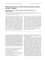
A
B
C
D
Fig. 2. Effect of d examethasone and RU48 6 on cyclooxygenase-2 ( COX-2) expression. (A) Cells were either not stimulated ( NS) or pretreated w ith
various concentrations of dexam ethasone (Dex) or RU 486 for 1 h prior to stimulation with interleukin-1b (IL -1b)(1ngÆmL
)1
). Cells were
harvested a t 6 h for RNA, and N orthern blot (NB) a nalysis was performed f or COX-2 and glyc eraldehyde-3-phosphate dehydrogenase (GAPDH).
Cells harvested at 24 h were subject to Western blot (WB) analy sis for COX-2. (B) Cells were pretreat ed for 1 h with dexamethasone (0.1 l
M
)(Dex)
in the presence of various concentrations of RU486. Cells were harvested as described in (A) f or Northern and Western blot analyses. In each case,
blots representative of t hree or m ore su ch experimen ts a re shown. (C) Following densitometric analysis, data (n ¼ 4–6) (upper panels, Western
blots; lo wer p an els, N orthern blots) from th e e xperiments in (A) were expressed as a percentage of IL -1b, treated and plotted as m ean ± SEM.
(j), De xametha sone; (.), RU486. (D ) D ata ( n ¼ 4–5) f rom the experiments i n ( B) were plotted a s d escribed in (C).
Ó FEBS 2004 Glucocorticoid repression: differential mechanisms (Eur. J. Biochem. 271) 4045
of RU486 (Fig. 1C,D). As shown by the rightwards shift
and the reduced apparent efficacy o f the inhibition curves
described for both dexamethasone and budesonide, the
glucocorticoid-dependent repression of COX/PGES activity
was clearly antagonized by increasing the concentration of
RU486. However, in marked contrast, RU486 primarily
resulted in an increased overall inhibition of the response
curves described for dexamethasone and budesonide on
PGE
2
release, as shown by the progressive flattening of the
respective lines (Fig. 1C,D). These data are t herefore
indicative of a primary inhibitory effect of RU486 on
PGE
2
release, but not on combined COX/PGES activity.
Analysis of COX-2 mRNA and protein expression,
which is responsible for the inflammatory release of PGE
2
from A549 cells [5,15], often revealed basal l evels of
expression, as reported previously [16]. However, in each
case, and as previously shown, COX-2 expression was
dramatically in creased by treatment with IL-1b [7,14].
Consistent with the combined COX/PGES data, the
analysis of COX-2 mRNA and protein expression revealed
a concentration-dependent inhibition of COX-2 expression
by dexamethasone, whereas RU486 showed little effect
except at h igh doses (Fig. 2A,B). Consistent with Fig. 1B,
0.1 l
M
dexamethasone a lmost t otally repressed both
mRNA and protein expression of COX-2, and this effect
was efficiently antagonized by RU486 (Fig. 2B).
Effect of dexamethasone and RU486 on arachidonic
acid release
To investigate the possibility of an effect of steroids
upstream of COX-2, ce lls were loaded with [
3
H]arachidonic
acid prior to stimulation in the presence of dexamethasone
or RU486. As I L-1b alone is a poor activator of arachidonic
acid release [8], cells were also treated with ionomycin or
with IL-1b + ionomycin, w hich provides a Ca
2+
stimulus,
causing translocation and membrane association of cyto-
solic (c)PLA
2
to markedly enhance cPLA
2
activity [8 ,17,18].
IL-1b, ionomycin and IL-1b + ionomycin increased
[
3
H]arachidonic acid release by 1.6-fold, 3.2-fold and 7.2-
fold, respectively (Fig. 3A). In each case, dexamethasone
produced repressions of 50, 61 and 68%, whilst RU486
resulted in rep ressions of 58, 5 3 and 63%, respectively. To
further characterize this inhibition, cells were treated with
various concentrations of either dexamethasone or RU486
prior t o s timulation with IL-1b + i onomycin. I n each case,
a concentration-dependent inhibition of [
3
H]arachidonic
acid release (EC
50
¼ 1 8.7 ± 10.6 and 26.2 ± 11.6 n
M
,
respectively) was observed, thereby confirming the inde-
pendent inhibitory effect of RU486 acting at the level of
arachidonic acid release (Fig. 3B).
Transactivation and transrepression by glucocorticoids
and RU486
The effect of dexamethasone and R U486 was a nalyzed on
GRE-dependent and N F-jB-dependent transcription.
From the 1·GRE r eporter, pGL3.neo.TATA.GRE,
GRE-dependent transcription was increased by 4.5-fold
(EC
50
¼ 46 .7 ± 17.7) by dexamethasone and fivefold
(EC
50
¼ 53.5 ± 20.8 n
M
, r espectively) by budesonide
(Fig. 4 A). Similarly the 2·GRE-driven reporter,
pGL3.neo.TATA.2GRE, gave r ise t o o ver a 15-fold
(EC
50
¼ 54.5) induction by dexamethasone and a 20-fold
induction (EC
50
¼ 65.3 n
M
) by budesonide (Fig. 4B). No
response was observed with reporters containing either
mutated GRE elements (pGL3.neo.TATA.2GREmut) or
no GRE s ites (pGL3.neo.TATA) ( data not shown), w hich
confirms the specificity of these reporter systems for the
presence of GRE sites. In e ach case, RU486 showe d little
or no ability to activate GRE-dependent transcription
(Fig. 4 A,B), but demonstrated a profound ability to antag-
onize both 1·GRE and 2·GRE reporter activity induced by
0.1 l
M
of either dexamethasone or bu desonide (Fig. 4C,D).
Analysis of IL-1b-induced NF-jB-dependent transcrip-
tion revealed a modest 30–40% inhibition (EC
50
¼
3
H arachidonic acid
release (% of total
incorporated)
NS
IL-1
Iono IL+Iono
Dex
Ru486
0
5
10
*
*
*
**
**
**
***
***
**
**
**
*
0
50
100
3
H arachidonic acid
release (% IL-1
+
ionomycin)
-10 -9 -8 -7 -6 -5
Log [Steroid] (M)
NS
IL+Iono
A
B
Fig. 3. Inhibition of arachidonic acid release by dexamethasone and
RU486. ( A) Following loading with [
3
H]arachidonic acid, c ells were
either not treated or pretreated with dexameth asone (1 l
M
)(Dex)or
RU486 (1 l
M
) for 1 h. Cells were then either not stimulated (NS) or
stimulated wi th interleukin-1b (IL-1b)(1ngÆmL
)1
), ion om ycin (3 l
M
)
(Iono) or both together (IL + Iono), and the supe rnantants and cells
were harvested after 1 h for liqui d s cintillation counting. Data (n ¼ 4
or 5) are shown as arachidonate release expressed as a percentage of
the total incorporated ± SEM. Significance was assessed using the
Student’s t-test. *P <0.05,**P < 0 .01. (B) Cells were treated as in (A)
except that various concentrations of either dexamethasone (j)or
RU486 (.) were added p rior t o t he IL-1b (1 ngÆmL
)1
)+ionomycin
(3 l
M
) stimulus. After harvesting, 1 h fo llowing stimulation, arachi-
donate re lease as a fraction of the total incorporated was expressed as a
percentage of the IL-1b + ionomyc in stimulus and plotted as
mean ± SEM. Significance was assessed using analysis of va riance
(
ANOVA
) with a Dunn’s post-test. **P < 0.01, ***P <0.001.
4046 J. E. Chivers et al. (Eur. J. Biochem. 271) Ó FEBS 2004
3.2 ± 1.3 a nd 7.8 ± 1 .9 n
M
) by d examethasone and
budesonide, respectively, and just over a 50% inhibition
by 10 l
M
RU486 (Fig. 5A). RU486 was without effect at
0.1 l
M
and required to be present at concentrations of
100-fold higher than either dexamethasone or budesonide
to achieve similar levels (30–40%) of inhibition. It is worth
noting that the inhibition of NF-jB by RU486 correlates
very closely with the effects o bserved on COX activity and
COX-2 expression (Figs 1 and 2 ). In ad dition, the ability of
RU486 to antagonize the repressive effects of 0.1 l
M
dexamethasone or budesonide was examined. In each case,
a concentration-dependent antagonism was observed up to
a maximum of 0.1 l
M
RU486 (Fig. 5B). Above this
concentration, increasing levels of inhibition were observed
owing to the r epressive effect of RU486 acting alone (data
not shown and see Fig. 5 A).
The expression of COX-2 m ay also depen d on activatin g
transcription factors (ATFs) and activator protein-1 (AP-1)-
like factors acting at a CRE site located in the proximal
region of the COX-2 promoter [19–21]. Consistent with this,
we have previously found that a CRE-driven reporter
construct was unresponsive to cAMP in A549 cells, but
responded t o IL-1b [10]. This was not believed to reflect a
general problem with this reporter, as strong cAMP-indu-
cibility has be en d emonstrated in other experimental systems
[12]. Consistent with these earlier fin dings, IL-1b was s hown
to induce r eporter activity twofold (Fig. 5C). In each c ase,
both dexamethasone (0.1 l
M
) and RU486 (10 l
M
)were
found to produce marked repressive effects (Fig. 5C).
Binding affinity of steroid ligands and effect
on GR translocation
Saturation binding studies using [
3
H]dexamethasone dem-
onstrated one-site binding in A549 cells and revealed
16 500 ± 2700 GR/cell w ith an affinity of 1.36 ±
0.10 n
M
, which is consistent with other reports, including
primary epithelial cells, indicating an affinity in the low n
M
range (Fig. 6A) [22–24]. Competitive binding studies were
performed to examine the relative GR-binding affinity of
these steroid ligands, and the following rank order of
affinity was observed : RU486 > budesonide > dexameth-
asone (Fig. 6B). The appropriate K
i
values are given in
Table 1.
0
1
2
3
4
5
6
-11 -10 -9 -8 -7 -6 -5
0
5
10
15
20
-11 -10 -9 -8 -7 -6 -5
NS
NS
Log [Steroid] (M)Log [Steroid] (M)
Fold activation
Fold activation
0
20
40
60
80
100
120
-10 -9 -8 -7 -6 -5
0
20
40
60
80
100
120
-10 -9 -8 -7 -6 -5
Log [RU486] (M)Log [RU486] (M)
Luciferase activity
(% Dex)
Luciferase activity
(% Dex)
NS
Dex
Bud
RU486
NS
Dex
Bud
RU486
***
***
***
***
***
***
1
GRE
1
GRE
2
GRE
2
GRE
AB
DC
Fig. 4. Effect o f d examethasone, budesonide and RU486 on glucocorticoid response element (GRE)-dependent tr anscription. (A) 1·GRE o r ( B)
2·GRE A549 reporter cells were either not stimulated (NS) or treated with various concentrations of dexamethasone (j), budesonide (h)or
RU486 (.). After 6 h, cells were harvested f or luciferase assay. Data ( n ¼ 6–10), expressed as f old induction, are plotted as m eans ± SEM.
(C) 1·GRE and (D) 2·GRE A549 reporter cells were activated by dexamethasone (0.1 l
M
)(j) or budesonide (0.1 l
M
)(h) in the presence of
various concentrations of RU486. Cells were harvested as described above, and luciferase activity, expressed as a percentage of the activity induced
by dexamethasone (0.1 l
M
), was plotted a s mean ± SEM. The effect of no stimulation (NS), or of stimulation with dexamethasone (0.1 l
M
)(Dex),
budesonide (0.1 l
M
) ( Bud) or RU486 (10 l
M
) a lone, is also shown. All data are n ¼ 6–10. In (A) and (B), the indicated levels of significance apply to
both budesonide and dexamethasone. In addition, the followi ng levels of significance were established, expressed as P-values of < 0.05 (*), < 0.01
(**) and < 0.001 (*** ). (B) B udesonide at 10
)8
M
(**) and dexamethasone at 10
)8
M
(*). (C) B udeson ide + RU486 at 1 0
)7
M
(**), 10
)6
M
(**) and
10
)5
M
(***); dexamethasone + RU486 at 10
)7
M
(**), 10
)6
M
and 10
)5
M
(***). (D) Budesonide + RU486 at 10
)6
M
(**), and 10
)5
M
(**);
dexamethas one + RU486 a t 10
)7
M
(*), 10
)6
M
(**) and 10
)5
M
(**).
Ó FEBS 2004 Glucocorticoid repression: differential mechanisms (Eur. J. Biochem. 271) 4047
Nuclear translocation of GR by dexamethasone
and RU486
Dexamethasone induced a rapid (within 15 min) transloca-
tion of GR from the c ytoplasm to the nuclear compartment,
with complete translocation observed by 1 h ( Fig. 7A ).
Similarly, and as e xpected, nuclear translocation of GR was
also induced by budesonide (Fig. 7B). In addition, RU486
was also efficient at inducing GR nuclear translocation,
indicating that binding of the antagonist can result in
dissociation of the cytoplasmic hsp–GR complex (Fig. 7B).
Analysis of an isotype-control a ntibody revealed no s igni-
ficant i mmunoreactivity, suggesting that the observed signal
was GR-specific (Fig. 7C).
Discussion
In the above studies, dexamethasone and budesonide
produced a near-total inhibition of both PGE
2
and COX/
PGES activity, and acted with similar efficacies (Table 1)
and potencies. However, whilst the steroid receptor antag-
onist, RU486, showed reversal of both C OX-2 expression
and COX/PGES a ctivity, which is c onsistent with a
GR-dependent mechanism, RU486 was incapable of ant-
agonizing the repression of IL-1b-induced PGE
2
release
produced by either dexamethasone or budesonide. I n fact,
RU486 resulted in the progressive repression of PGE
2
release at increasing concentrations. Analysis of RU486
alone on IL-1b-induced PGE
2
release revealed a concentra-
tion-dependent inhibition of PGE
2
release, yet s howed little
or no effect on COX/PGES activity or COX-2 expression
until RU486 concentrations of 1 l
M
were reached. This
clear discrepancy stro ngly suggests that RU486 may exert
an inhibitory effect upstream o f COX-2, possibly at the level
of PLA
2
and arachidonic acid release.
This proposal was confirmed by the analysis of
[
3
H]arachidonate release, which revealed concentration-
dependent inhib ition by both dexamethasone and RU486.
Interestingly, the EC
50
values for repression of PGE
2
release, and the repression of arachidonic acid release by
RU486 (33.1 and 26.2 n
M
, respectively), correlate closely
and therefore support the suggestion of a mechanistically
distinct action for RU486 at the level of arachidonic acid
release. We therefore conclude that these data docu ment the
existence o f at least two f unctionally distinct processes for
the inhibition of inflammatory PGE
2
release by ste roid s.
In the fi rst mechanism, glucoco rticoids, such as dexameth-
asone or budesonide, inhibit the expression of COX-2, and
this response is antagonized efficiently by RU486. This
contrasts w ith a second, and p harmacologically distinct
mechanism, which occurs at the level of arachidonic acid
release, in which the actions of glucocorticoids are mimicked
by RU486.
Previous reports have also documented the inhibition of
arachidonic acid release in A549 cells by dexamethasone
[25]. H owever, t hese authors did not report any inhibition
by RU486 (10 n
M
) alone [26], and showed a 50% antag-
onism of the dexamethasone-dependent repression when
using RU486 at 10 l
M
[25]. In an attempt to reconcile the
apparent differences between the r esults of these reports and
those of the present study, it is noticeable that different
mechanisms of stimulation w ere u sed in each of the studies,
and this alone could account for any differen ces. Further-
more, inspection of our current data on the repression of
both PGE
2
release and arachidonic acid release, suggests
that the effects of 10 n
M
RU486 could be at the margins of
experimentally discernable r epression (see Figs 1A and 3B).
We also note that Croxtall et al. did not seemingly test
higher concentrations of RU486 acting alone for an
inhibitory effect o n epidermal g rowth factor (EGF)-stimu-
lated arachidonic acid release [25]. This therefore leaves
open the possibility that the incomplete antagonism of
RU486 observed on dexamethasone-dependent repression
of EGF-stimulated arachidonic acid release is, in fact,
Luciferase activity
(% IL-1
)
Dex
RU486
80
70
60
-10 -9 -8 -7
Dex
Bud
IL-1
Log [Steroid] (M)
NS
IL-1
-10 -9 -8 -7 -6 -5
0
100
50
Luciferase activity
(% of IL-1
)
IL-1
NS
Fold activation
0
1
2
3
*
**
NF- B
NF-
B
CRE
100
90
Log [RU486] (M)
A
B
C
Fig. 5. Transrepression by dexamethasone, budesonide and RU486. (A) 6jBtk reporter cells were either not treated or were treated with various
concentrations of dexamethasone (j), budesonide (h)orRU486(.) for 1 h, prior to stimulation with IL-1b (1 ngÆmL
)1
) or no stimulation (NS).
After 6 h, cells were harvested for analysis in the luciferase a ssay. Data ( n ¼ 8), expressed a s percentage of the r esponse to IL-1 b stimulation, are
plotted as mean ± SEM. Significance was established, expressed as P-values of < 0.05 (*), < 0.01 (**) and < 0.001 (***), for: budesonide at
10
)8
M
(*), 10
)7
M
(**) and 10
)6
M
(**); dexamethasone at 10
)7
M
(**), 10
)6
M
(***) and 10
)5
M
(***); and RU486 at 10
)5
M
(**). (B) 6jBtk
reporter cells, were treated with dexamethasone (0.1 l
M
)(j) or budesonide (0.1 l
M
)(h) in the presence of various concentrations of RU486.
Luciferase assay data (n ¼ 7–9), expressed as a percentage of the response to IL-1b, are plotted as mean ± SEM. The effect of IL-1b +
dexamethas one (0.1 l
M
)(Dex)andIL-1b + bud esonide (0.1 l
M
) ( Bud) alone are sh own. ( C) C RE re porter c ells w ere e ither n ot t reated o r were
treated for 1 h with dexamethasone (0.1 l
M
)orRU486(10l
M
) prior to no stimulation (NS) or s timu lation with IL-1b (1 ngÆmL
)1
), as in dicated.
Cells were harvested after 6 h for analysis in the luciferase assay, as described above. Data (n ¼ 6), expressed as fold activation, are plotted as
mean ± SEM. *P < 0.05, **P <0.01.
4048 J. E. Chivers et al. (Eur. J. Biochem. 271) Ó FEBS 2004
attributable to a partial agonistic effect of RU486 acting
alone [25].
It is well established that glucocorticoids can repress
the transcription of inflammatory genes via transcription
factors such as NF-jB [1,2]. However, whilst s ome
degree (30–40% inhibition) of glucocorticoid-dependent
inhibition of NF-jB-dependent transcription was
observed in response to both dexamethasone and budes-
onide, this effect is clearly insufficient to account for the
near-complete repression of COX-2 expression or PGE
2
release observed with each of these compounds. As
PGE
2
release and COX-2 expression in A549 cells is
highly NF-jB-dependent, and this level of inhibition of
NF-jB-dependent transcription correlates very well with
our previous observation that the IL-1b-induced COX-2
transcription rate was inhibited by 40 % by dexameth-
asone, we are compelled to suggest that additional
mechanisms of glucocorticoid-dependent repression of
COX-2 must also e xist [6,7]. Similarly, whilst G RE-
dependent transcription was robustly increased following
dexamethasone and budesonide treatment, this mechan-
ism is unlikely t o account for the repression of COX-2 o r
COX/PGES activity, as the EC
50
for this effect is greater
than 10-fold more than that required for the inhibition
of PG E
2
release o r COX/PGES a ctivity (Tab le 1 ).
Interestingly, this shift in the concentration–response
curve for transactivation effects at GREs (EC
50
values
of 54.5 and 65.3 n
M
for dexamethasone and budesonide,
respectively) when compared with transrepression, for
example of NF-jB(EC
50
values of 3.2 and 7.8 n
M
for
dexamethasone and budesonide, respectively), has been
previously reported , although t he exact mechanistic
explanation is currently lacking [27]. Therefore, in respect
of COX-2, these data suggest that other, non-NF-jB-
mediated and probably non-GRE-mediated, mechanisms
of dexamethas one-dependent inhibition must be in
operation to account for the full repression of COX-2
and COX/PGES activities in these cells.
By contrast, the inhibition of NF-jB-dependent tran-
scription by high c oncentrations of RU486 correlated v ery
closely, in terms of both apparent efficacy a nd potency, with
the inhibition of COX/PGES activity, thereby providing
further strength to the argument that additional mecha-
nisms, other than the inhibition of NF-jB, account for the
inhibition by dexamethasone. However, the basis of this
inhibition by RU486 is currently unclear to us because these
levels of steroid are vastly in excess of that necessary to
saturate GR, as suggested by our own, and previously
reported [24,28], ligand-binding studies (Fig. 6). It is pos-
sible that at these high concentrations RU486 is acting in a
GR-independent manner. Notwithstanding the inhibition at
high doses, it is clear that at concentrations of 1 or 0.1 l
M
,
RU486 sho ws a limited or no effect on NF-jB-dependent
transcription, yet is effective at inhibiting both PGE
2
and
arachidonic acid release, suggesting that the inhibition of
NF-jB plays no role in this response.
Previous studies have suggested that, relative to
dexamethasone, RU486 is a poor inducer of glucocorti-
coid-dependent transcription [29–35]. Similarly, in the
present study, RU486-induced GRE-dependent transcrip-
tion from either a 1·GRE or a 2·GRE reporter was
virtually absent, and this is consistent with data from
primary human bronchial epithelial cells [24]. These data
therefore raise the po ssibility that R U486 inhibits
arachidonic acid release via a mechanism that is
independent of transcription. Indeed, the rapid dexa-
methasone-dependent repression of EGF-induced release
of arachidonic acid was previously shown to be actino-
mycin D insensitive and therefore independent of tran-
scription [25]. In this respect, RU486 has previously been
shown to mimic other nongenomic glucocorticoid
responses, including the down-regulation of GR itself
[36,37]. Certainly, our data indicate that RU486, can, like
dexamethasone and budesonide, bind to and induce the
nuclear translocation of GR. We therefore speculate that
binding of ligand, including antagonists such as RU486,
to GR, and complex dissociation, may be sufficient for
the inhibition of arachidonic acid release and that this
represents a mechanistically distinct event from the
inhibition of inflammatory gene express ion . In this
context i t is notable that various nongenomic actions
of steroid hormones have been id entified [38,39], which
raises the possibility o f ligand-dependent nongenomic
anti-inflammatory functions for GR or for GR-associated
-11 -10 -9 -8 -7 -6 -5
0
25
50
75
100
Log [Steroid] (M)
Specific binding
(%)
0 10 20 30
0
2.5
5.0
7.5
10
Log [Dex] (nM)
Specific Binding
x 10
3
(dpm)
Specific Binding x10
2
(dpm)
0
5
0
7
5
1
0
0
0
1
2
3
4
Bound/Free x 10
3
2
5
A
B
Fig. 6. Analysis of glucocorticoid receptor (GR) number and relative
affinity o f ligands. (A) A typical saturation–binding isotherm, showing
specific GR binding using 2.4 · 10
6
cells and resulting Scatchard
analysis (inset), wh ere the ratio o f free t o bound radioligand is p lotted
against log [steroid ] to give a straight line with a gradient equal to
)1/K
d
and and an x intercept that equals B
max
. (B) Competition
binding curves showing relative affinity in A549 cells, where dexa-
methasone ( j), bu desonide ( h), RU24858 ( d)orRU486(.)compete
with 4 n
M
[
3
H]dexamethasone to b ind the GR . D ata are pre sented a s
mean ± SEM for n ¼ 3–5 observations.
Ó FEBS 2004 Glucocorticoid repression: differential mechanisms (Eur. J. Biochem. 271) 4049
proteins present in the GR–hsp complex. Finally, we
should point out that a number of effects of glucocor-
ticoids, which are independent of the classical GR, are
also reported to occur a nd these could help to e xplain
our results [39]. Thus, the mineralocorticoid receptor may
mediate glucocorticoid responsiveness in the brains of
GR knockout mice [40]. In addition, a pharmacologically
distinct pool of membrane-localized glucocorticoid recep-
tors have been identified by various authors [39]. For
example, a membrane glucocorticoid receptor has been
biochemically identified in amphibians [41]. However, it
is currently unclear whether this represents a version of
the classical GR [42] or P-glycoprotein/multiple drug
resistance gene, a member of the ATP-binding cassette
(ABC) transporters [43,44], or some other receptor [45].
In this context, P-glycoprotein is of interest as it actively
exports certain steroids, and blocking its function has
been shown to promote glucocorticoid actions [46,47].
In conclusion, we present data w hich further confirm t hat
the inhibition of NF-jB-dependent transcription cannot
account for all the repressive effects of glucocorticoids on
inflammatory genes such as COX-2. Furthermore, we
present e vidence t hat glucocorticoids and RU486 also inhibit
the r elease of arachidonic acid via a process that does not
involve either inhibition of NF-jB or the activation of
GRE-mediated transcription and which is mechanistically
distinct from the inhibition of COX-2. Taken together, these
data indicate the existence of pharmacologically distinct
processes that are collectively responsible for the repression
of inflammatory P GE
2
release b y g lucocorticoids.
Acknowledgements
J.E.C. and M.C.C. were collaborative students with the BBSRC and
the MRC, respectively, an d both were supported by Aventis Pharm a-
ceuticals.
References
1. Barnes, P.J. (1999) Therapeutic strategies for allergic diseases.
Nature 402 (Suppl.), B 31–B38.
2. Newton, R . (2000) Molecular mechanisms o f glucocorticoid
action: w hat is important? Thorax 55, 6 03–613.
3. Schaaf, M.J. & Cidlowski, J.A. (2002) Molecular mechanisms of
glucocorticoid action and resistance. J. Steroid Biochem. Mol.
Biol. 83, 37–48.
4. Funk, C.D. (2001) Prostaglandins and leukotrienes: advances in
eicosanoid biology. Science 294, 1871–1875.
5. Mitchell, J.A., Belvisi, M.G., Akarasereenont, P., Robbins, R .A.,
Kwon, O.J., Croxtall, J., Barnes, P.J. & Vane, J.R. (1994)
Induction o f c yclo-oxyge nase-2 by cytokines in human pulmonary
epithelial cells: regulation by dexamethasone. Br.J.Pharmacol.
113, 1008–1014.
6. Catley, M .C. , Chivers, J.E., Camb r idg e, L.M., Hol d en, N.,
Slater, D.M., Staples, K.J., Bergmann, M.W., Loser, P., Barnes,
P.J. & Newton, R. (2003) IL-1beta-dependent activation of
NF-kappaB mediates PGE
2
release via the expression of
A
BC
Fig. 7. Nuclear translocation o f th e glucocorticoid r ecepto r ( GR). (A) Cells were either not treated or were treated w ith dexamethasone (1 l
M
)(Dex)
for 15, 30 or 60 min and then probed with a fluorescein i sothiocyanate (FITC)-conjugated GR immunoglobulin (green) prior to imaging by
confocal m icroscopy. N uclei are indicated by 4 ¢,6¢-d iamidino-2-phenylinole dihydrochloric hydrate (DAPI) staining of chromatin ( blue). (B) Cells
were treated with d exam ethasone (1 l
M
) (Dex), budesonide ( 1 l
M
)orRU486(1l
M
) for 1 h prior to probing and analysis as described above. (C)
Cellsweretreatedasin(A)andthenprobedwithanisotypecontrolfortheGRantibodyusedin(A)and(B).Allimages are representative of three
experiments.
4050 J. E. Chivers et al. (Eur. J. Biochem. 271) Ó FEBS 2004
cyclooxygenase-2 and microsomal prostaglandin E synthase.
FEBS Lett . 547 , 7 5–79.
7. Newton, R., Seybold, J., Kuitert, L.M.E., Bergmann, M. &
Barnes, P.J. (1998) Repression of cyclooxygenase-2 and pros-
taglandin E
2
release by dexamethasone occurs by transcriptional
and post-transcriptional mechanisms in volving loss of poly-
adenylated mRNA. J. Biol. Chem. 273 , 32312–32321.
8. Newton, R., Cambridge, L., Hart, L.A., Stevens, D.A., Lindsay,
M.A. & Barnes, P.J. (2000) The MAP kinase inhibitors,
PD098059, UO126 and SB203580, inhibit IL-1beta-dependent
PGE(2) release via mechanistically distinct processes. Br. J.
Pharmacol. 130, 135 3–1361.
9. Newton, R., Hart, L.A., Stevens, D.A., Bergmann, M., Donnelly,
L.E., Adcock, I.M. & B arnes, P.J. (1998) E ffec t of dexame thasone
on interleukin-1beta (IL-1beta) induced nuclear factor-kappaB
(NF-kappaB) and kappaB-de pendent transcript ion in epithelial
cells. Eur. J. Biochem. 254, 81–89.
10. Catley, M.C., Cambridge, L.M., Nasuhara, Y., Ito, K., Chivers,
J.E., Beaton, A., Holden, N.S., B ergmann, M.W., B arnes, P.J. &
Newton, R. (2004) I nhibitors o f prot ein kinase C ( PKC) preven t
activated transcription: role of even ts downstream of N F -kappaB
DNA b indin g. J. Biol. Chem. 279, 18457–18466.
11. Strahle, U., Sch mid, W. & Schutz, G. (1988) Synergistic action of
the glucoco rticoid r ec eptor w ith t rans cription fa ctors. EMBO J. 7,
3389–3395.
12. Meja, K.K., Catley, M.C., Cambridge, L.M., Barnes, P.J., Lum,
H., Newton, R. & Giembycz, M.A. (2004) Adenovirus-mediated
delivery and expression of a cAMP-de pendent protein kinase
inhibitor g en e to BEAS-2B epithelial cells abolishes th e anti-in-
flammatory e ffect s o f rolipram, salbutam ol, a nd prostaglandin E2:
a com parison w ith H-89. J. Pharmacol. Exp. Ther. 30 9, 8 33–844.
13. Cheng, Y. & Prusoff, W.H. (1973) Relationship between the
inhibition constant ( K1) and the concentration o f inhibitor wh ich
causes 50 per cent inhibition (I50) of an enzymatic reaction.
Biochem. Pharmacol. 22, 3 099–3108.
14. Newton, R., Kuitert, L.M., Bergmann, M., Adcock, I.M. &
Barnes, P.J. (1997) Eviden ce for involve ment of NF-jBinthe
transcriptional control of COX-2 gene expression by IL-1b. Bio-
chem. Biophys. Res. Commun. 237, 2 8–32.
15. Newton, R., Eddleston, J., Haddad, E., Hawisa, S ., Mak, J., Lim,
S., Fox, A.J., Donnelly, L .E. & Chung, K.F. (2002) Regulation of
kinin r eceptors in airway epithelia l c ells by inflammatory cytokines
and d ex ameth aso ne. Eu r. J. Pharmacol. 441, 193–202.
16. Watkins, D.N., P eroni, D.J., L enzo, J.C., Knight, D.A., Garlepp,
M.J. & Thompson, P.J. (1999) Expression and localization of
COX-2 in h uman ai rways and cultured airway epithelial cells. Eur.
Respir. J. 13 , 999–1007.
17. Schievella, A.R., Regier, M .K., Smith, W.L. & Lin, L.L. (1995)
Calcium-mediated translocation of cytosolic phospholipase A2 to
the nuclear envelope and endoplasmic reticulum. J. Biol. C hem.
270, 30749–30754
18. Leslie, C.C. (1997) Properties and regu lation of cytosolic phos-
pholipase A 2. J. Biol. Chem. 27 2 , 16709–16712
19. Xie, W., Fletcher, B.S., Andersen, R.D. & Herschman, H.R.
(1994) v-src induction o f the T IS10/PGS2 prostaglandin synt hase
gene is mediated by an ATF/CRE transc ription resp onse eleme nt.
Mol. Cell Biol. 14, 6531–6539.
20. Inoue, H., Yokoyama, C., Hara, S., T one, Y. & T anabe, T. (19 95)
Transcriptional r egulation of human prostaglandin-endoperoxide
synthase-2 gene by lipopolysaccharide and phorbol ester in
vascular endothelial cells. I nvolvement of both nu clear f ac tor f or
interleukin-6 expression site and cAMP re sponse element. J. Biol.
Chem. 270, 24965–24971.
21. Xie, W. & Herschman, H .R. ( 1996) Transcriptional r egulation o f
prostaglandin synthase 2 gene expression by platelet-derived
growth facto r an d s erum. J. Biol. Chem. 271, 31742–31748.
22. Hammer, S., Spika, I ., Sippl, W ., Jessen, G., Kleuser, B., Holtje,
H.D. & Schafe r-Ko rting, M. (2003) G lu cocortic oid receptor
interactions with gluc oco rticoids: evaluation by molecular m od-
eling and functional analysis of glucocorticoid receptor mutants.
Steroids 68, 329–339.
23. Robin-Jagerschmidt, C., Wurtz, J.M., Guillot, B., Gofflo, D.,
Benhamou,B.,Vergezac,A.,Ossart,C.,Moras,D.&Philibert,D.
(2000) Residues in the ligand binding dom ain that confer progestin
or glucocorticoid specificity and modulate the receptor transacti-
vation capacity. Mol. Endocrinol. 14, 1 028–1037.
24. LeVan, T.D., Babin, E.A., Yamamura,H.I.&Bloom,J.W.(1999)
Pharmacological characte rization of glucocortic oid receptors in
primary human bronchial epithelial c ells. Biochem. Pharmacol. 57 ,
1003–1009.
25. Croxtall, J.D., Choudhury, Q. & Flower, R.J. (2000) Gluco-
corticoids act within minutes to inhibit recruitment of signalling
factors to activated EGF receptors through a receptor-depen dent,
transcription-independent mechanism. Br.J.Pharmacol.130,
289–298.
26. Croxtall, J.D., Paul-Clark, M. & van Hal, P .T. (2003) Differential
modulation of glucocorticoid action by FK506 in A549 cells.
Biochem. J. 376, 2 85–290.
27. Jonat, C., Rahmsdorf, H.J., Park, K.K., Cato, A.C., Gebel, S.,
Ponta, H. & Herrlich, P. (1990) Antitumor promotion and anti-
inflammation: down-modulation of AP-1 (Fos/Jun) activity by
glucocorticoid hormone. Cell 62, 1189–1204.
28. Gagne, D., Pons, M. & Philibert, D. (1985) RU 38486: a potent
antiglucocorticoid in vitro and in vivo. J. Steroid Biochem. 23,
247–251.
29. Vayssiere,B.M.,Dupont,S.,Choquart,A.,Petit,F.,Garcia,T.,
Marchandeau, C., Gronemeyer, H. & Resche Rigon, M. (1 997)
Synthetic glucocortic oids that disso ciate transactivation and AP-1
transrepression exhibit antiinflammatory activity in vivo. Mol.
Endocrinol. 11, 1245–1255.
30. Heck, S., Bender, K., Kullman, M., Gottlicher, M., Herrlich,
P. & Cato, A.C.B. (1997) IkBa-independent downregulation o f
NF-kB a ctivity by g lucocorticoid r eceptor. EMBO J. 16, 4698–
4707.
31. Wissink, S., van-Heerde, E.C., vand-der, B.B. & van-der-Saag,
P.T. (1998) A dual mechanism mediates repression of NF-kappaB
activity by glucocorticoids. Mol. Endocr inol. 12, 355–363.
32. Liden, J., Delaunay, F., Rafter, I., Gustafsson, J. & Okret, S.
(1997) A new function for the C-terminal zinc fin ger of the glu-
cocorticoid r ecepto r. R epressio n o f R elA tra nsactiva tion. J. Biol.
Chem. 272, 21467–21472.
33. Fryer, C.J., Kinyamu, H.K., Rogatsky, I ., Garabedian, M.J. &
Archer, T.K. (2000) Selective activation of the glucocorticoid
receptor by steroid antagonists in human breast canc er and os-
teosarcoma cells. J. Biol. Chem. 275, 17771–17777.
34. Rogatsky, I., Hittelman, A.B., Pearce, D. & Garabedian, M.J.
(1999) Distinct glucocorticoid receptor transcriptional regulatory
surfaces mediate the cytotoxic and cytostatic effects of gluco-
corticoid s. Mol. Cell Biol. 19, 5 036–5049.
35. Pariante, C.M., Pearce, B.D., Pisell, T.L., Su, C. & Miller, A.H.
(2001) The steroid receptor antagonists RU40555 and RU486
activate glucocortico id rece ptor translocation and are not exc rete d
by the steroid hormones transporter in L929 cells. J. Endocrinol.
169, 309–320.
36. Hoeck, W., R usconi, S . & Groner, B. (1989) Down-re gulation a nd
phosphorylation of glucocorticoid receptors in cultured
cells. Investigations with a monospecific antiserum against a bac-
terially ex pressed receptor fragment. J. Biol. Chem. 26 4, 14396–
14402.
37. Burnstein, K.L., Jewell, C.M., Sar, M. & Cidlowski, J.A. (1994)
Intragenic sequences of the human glucocorticoid receptor com-
plementary DNA mediate hormone-inducible receptor messenger
Ó FEBS 2004 Glucocorticoid repression: differential mechanisms (Eur. J. Biochem. 271) 4051
RNA down-regulation through multiple mechanisms. Mol.
Endocrinol. 8, 1 764–1773.
38. Losel, R. & Wehling, M. (2003) Nongenomic ac tions of steroid
hormones. Nat. Rev. Mol. Cell Biol. 4, 4 6–56.
39. Losel, R.M., Falkenstein, E ., Fe uring, M., Schultz, A., Tillmann,
H.C., Rossol-Haseroth, K . & Wehling, M. (2003) Nongenomic
steroid action: controv ersies, questions, and answers. Physiol. Rev.
83, 965–1016.
40. Meijer, O.C. & De Kloet, E.R. (1995) A role for the miner-
alocorticoid receptor in a rapid and transient suppression of
hippocampal 5 -HT1A r ecep tor mRNA by c orticosteron e.
J. Neuroendocrinol. 7, 6 53–657.
41. Evans, S.J., Murray, T.F. & M oore, F.L. (2000) P artial purifica-
tion and b iochemical c haracterization of a m embrane glucocorti-
coid rec eptor from an amphibian brain. J. Steroid B ioc hem. Mol.
Biol. 72, 209–221.
42. Gametchu, B., Watson, C.S. & Wu, S. (1993) Use of receptor
antibodies to demonstrate membrane g luc ocorticoid receptor i n
cells from human leukemic patients. FASEB J . 7, 1283–1292.
43. Meijer, O.C., de Lange, E.C., Breimer, D.D., de Boer, A.G.,
Workel, J.O. & De Kloet, E.R. (1998) Penetration of dexa-
methasone into brain glucocorticoid targets is enhanced in mdr1A
P-glycoprotein knockou t m ice. Endocrinology 139, 1789–1793.
44. Kralli, A. & Yamamoto, K.R. (1996) An FK506-sensitive trans-
porter selectively decreases intracellular levels and potency of
steroid horm ones. J. Biol. Chem. 27 1, 17152–17156.
45. Evans, S.J., Searcy, B.T. & Moore, F .L. (2000) A s ubset of ka ppa
opioid ligands bind to the membrane g luco corticoid receptor in a n
amphibian brain. Endo crinology 14 1, 2 294–2300.
46. Pariante, C.M., Makoff, A., Lovestone, S., Feroli, S., Heyden, A.,
Miller, A.H. & Kerwin, R.W. (2001) Antidepressants enhance
glucocorticoid receptor function in vitro by modulating the
membrane steroid transporters. Br. J. Pharmacol. 134,
1335–1343.
47. Pariante, C.M., Kim, R.B., Makoff, A. & Kerwin, R.W. (2003)
Antidepressant fluoxetine enhances gluco corticoid receptor func -
tion in vitro by modulating me mbrane s te roid transporters. Br. J.
Pharmacol. 139, 1111 –1118.
4052 J. E. Chivers et al. (Eur. J. Biochem. 271) Ó FEBS 2004