Báo cáo khoa học: The role of helix 8 and of the cytosolic C-termini in the internalization and signal transduction of B1 and B2 bradykinin receptors potx
Bạn đang xem bản rút gọn của tài liệu. Xem và tải ngay bản đầy đủ của tài liệu tại đây (636.57 KB, 12 trang )
The role of helix 8 and of the cytosolic C-termini in
the internalization and signal transduction of B
1
and B
2
bradykinin receptors
Alexander Faussner
1
, Alexandra Bauer
1
, Irina Kalatskaya
1
, Steffen Schu
¨
ssler
1
, Cornelia Seidl
1
,
David Proud
2
and Marianne Jochum
1
1 Ludwig-Maximilians-Universita
¨
t, Abteilung fu
¨
r Klinische Chemie und Klinische Biochemie, Mu
¨
nchen, Germany
2 Department of Physiology & Biophysics, University of Calgary, Alberta, Canada
G protein-coupled receptors (GPCRs) form a vast and
diverse superfamily of proteins with seven transmem-
brane-spanning domains. They transduce specific exter-
nal stimuli to intracellular second messenger-dependent
effector cascades via recruitment and activation of
heterotrimeric G proteins [1]. To protect cells from
chronic overstimulation, desensitization processes such
as the rapid attenuation of receptor responsiveness and
Keywords
G protein-coupled receptor, helix 8
modeling, internalization, receptor chimera
Correspondence
A. Faussner, Ludwig-Maximilians-
Universitaet Muenchen, Abt. Klinische
Chemie und Klinische Biochemie,
Nussbaumstr. 20, D-80336 Muenchen,
Germany
Tel: +49 89 51602602
Fax: +49 89 51604740
E-mail: alexander.faussner@
med.uni-muenchen.de
(Received 21 July 2004, revised 14 September
2004, accepted 15 September 2004)
doi:10.1111/j.1432-1033.2004.04390.x
Determinants for desensitization and sequestration of G protein-coupled
receptors often contain serine or threonine residues located in their C-ter-
mini. The sequence context, however, in which these residues have to
appear, and the receptor specificity of these motifs are largely unknown.
Mutagenesis studies with the B
2
bradykinin receptor (B
2
wt), stably
expressed in HEK 293 cells, identified a sequence distal to N338
(NSMGTLRTSI, including I347 but not the basally phosphorylated S348)
and in particular the TSI sequence therein, as a major determinant for
rapid agonist-inducible internalization and the prevention of receptor
hypersensitivity. Chimeras of the noninternalizing B
1
bradykinin receptor
(B
1
wt) containing these B
2
wt sequences sequestered poorly, however, sug-
gesting that additional motifs more proximal to N338 are required. In fact,
further substitution of the B
1
wt C-terminus with corresponding B
2
wt
regions either at C330(7.71) following putative helix 8 (B
1
CB
2
) or at the
preceding Y312(7.53) in the NPXXY sequence (B
1
YB
2
) resulted in chi-
meras displaying rapid internalization. Intriguingly, however, exchange
performed at K322(7.63) within putative helix 8 generated a slowly inter-
nalizing chimera (B
1
KB
2
). Detailed mutagenesis analysis generating addi-
tional chimeras identified the change of V323 in B
1
wt to serine (as in B
2
wt)
as being responsible for this effect. The slowly internalizing chimera as well
as a B
1
wt point-mutant V323S displayed significantly reduced inositol
phosphate accumulation as compared to B
1
wt or the other chimeras. The
slow internalization of B
1
KB
2
was also accompanied by a lack of agonist-
induced phosphorylation, that in contrast was observed for B
1
YB
2
and
B
1
CB
2
, suggesting that putative helix 8 is either directly or indirectly
(e.g. via G protein activation) involved in the interaction between the
receptor and receptor kinases.
Abbreviations
BK, bradykinin; B
x
wt, wild-type B
x
bradykinin receptor; DAK, desArg10kallidin; GPCR, G protein-coupled receptor; GRK, G protein-coupled
receptor kinase; HEK, human embryonic kidney; IP, inositol phosphate.
FEBS Journal 272 (2005) 129–140 ª 2004 FEBS 129
G protein uncoupling are essential. Some of these
desensitization mechanisms involve the translocation
of the stimulated receptor to distinct compartments
and endocytosis after phosphorylation of serine ⁄ threo-
nine residues mostly located in the receptor C-termini
(reviewed in [2]). Little is known so far about the
sequence context in which these residues have to
appear to become phosphorylated by kinases and to
be recognized by the internalization machinery. In par-
ticular, the receptor specificity of these motifs is not
completely understood.
The B
1
bradykinin receptor (B
1
wt) is one of the
few receptors belonging to the class A family of rho-
dopsin-like ⁄ b2-adrenergic-like GPCRs that does not
get internalized, i.e. sequestered to intracellular com-
partments upon agonist stimulation [3]. It does, how-
ever, respond with translocation to caveolae but these
remain essentially on the cell surface [4,5]. No phos-
phorylation of B
1
wt either under basal conditions or
after stimulation has been detected [6]. The B
2
brady-
kinin receptor (B
2
wt), by contrast, is a more typical
GPCR that gets internalized rapidly following activa-
tion. Phosphorylation of several serine⁄ threonine resi-
dues in the C-terminus of this receptor, and the
importance of these events for receptor sequestration,
have been described in detail [7]. Whether coupling of
b-arrestin(s) then follows this, and whether internal-
ization occurs via clathrin-coated pits, caveolae or
other less well-defined mechanisms is still a topic of
debate [5,7,8].
The two bradykinin (BK) receptor subtypes exhibit
a relatively low overall amino acid identity of about
36% [9,10], most of it located in the transmem-
brane regions. Both receptors stimulate phospholipase
C
b
-mediated inositol phosphate (IP) release leading to
an elevation of intracellular [Ca
2+
] levels, primarily via
coupling to G protein G
q ⁄ 11
[3,10,11].
They become activated by the kinins, small pro-
inflammatory peptides with great vasoactive potential
implicated as mediators of inflammation, pain and
hyperalgesia [12,13]. The nonapeptide BK and Lys-BK
(kallidin) bind with high affinity to B
2
wt but not B
1
wt.
Removal of the C-terminal arginine through carboxy-
peptidases generates desArg9-bradykinin and desArg10-
kallidin (DAK), two peptides that now bind exclusively
to the B
1
wt [14].
In this study we wanted to exploit the fact that the
B
1
wt does not internalize as part of a gain-of-function
approach to provide insight into the receptor speci-
ficity of the B
2
wt internalization motif. The resulting
data also hint at a receptor specific role of the putative
helix 8 in G protein activation and interaction with
receptor kinases.
Results
Construction of truncated and point mutated
B
2
wts and B
1
⁄ B
2
receptor chimeras
Several studies with truncations of, and deletions in,
the C-terminal part of B
2
wt have demonstrated that
this part plays a central role in the internalization of
this receptor [7,15,16]. A similar function of the C-ter-
minus was also observed in other GPCRs with short
third intracellular loops [2]. In particular, several serine
or threonine residues that become phosphorylated by
protein kinase C and ⁄ or by GPCR kinases (GRKs)
following receptor activation are absolutely required
for rapid B
2
wt sequestration [17].
To determine the C-terminal sequence(s) of the B
2
wt
minimally required for internalization we created two
new B
2
wt truncations, I347* and N338* (Fig. 1). The
former removed the C-terminus including residue
S348, which has been shown to be responsible for the
basal phosphorylation of the B
2
wt, while the latter
truncation deleted all serine and threonine residues
(S339, T342, T345, S346) shown to be phosphorylated
following stimulation of the receptor [17]. In addition,
a triple alanine replacement of
T345-S346-I347(S348)
(mutated residues are underlined) was made, as this
sequence strongly resembles the C-terminal STLS-
motif in the AT
1A
angiotensin II receptor, where a tri-
ple alanine substitution of STL almost completely
abolished receptor sequestration [18].
All of these B
2
receptor constructs were highly
expressed (Table 1). We took care therefore to use
[
3
H]BK concentrations below 1.5 nm, as we have
shown that receptor internalization rates are independ-
ent of agonist concentration in this range [19]. The
truncation I347* internalized as rapidly as B
2
wt
(Fig. 2A) demonstrating that the distal C-terminus,
and in particular S348 and its basal phosphorylation,
do not play a decisive role in the sequestration process.
This notion was further supported by results obtained
with a point mutation of S348 to alanine that exhibited
an almost identical internalization rate as the B
2
wt ([7]
and data not shown).
In contrast, deletion of all phosphorylation sites in
N338* led to an extremely diminished [
3
H]BK internal-
ization (Fig. 2A). Indeed, even the internalization cal-
culated for each time point is an overestimate because
a shift to lower affinity at 37 °C by the receptors
remaining on the cell surface can be assumed, as there
was a clear drop in surface binding that could not be
accounted for by the amount of internalized agonist
[20]. As the internalization is expressed in percentage
of total binding, decreasing the binding affinity of the
Role of helix 8 and C-termini in bradykinin receptors A. Faussner et al.
130 FEBS Journal 272 (2005) 129–140 ª 2004 FEBS
surface receptors simulates an apparent increase in
internalization over time. Although it internalized
[
3
H]BK much slower than the B
2
wt, N338* neverthe-
less was able to induce an accumulation of total IPs
identical to that observed for the B
2
wt (Table 1). This
truncated receptor even became hypersensitive, as its
EC
50
for the IP response was 10-fold lower than
that of B
2
wt (0.072 ± 0.038 nm vs. 0.79 ± 0.34 nm;
Table 1). Most interestingly, the effects of a truncation
at N338 could also be achieved in part by the triple
mutation TSIfiAAA as this construct displayed sim-
ilar properties to truncation N338*. It exhibited a
markedly reduced capacity to internalize [
3
H]BK albeit
not as diminished as truncation N338* and was at
least as hypersensitive with an EC
50
¼ 0.058 ±
0.06 nm (Table 1). This sequence obviously contributes
significantly to agonist internalization and signaling of
B
2
wt.
However, transfer of the B
2
wt C-terminus starting
with this sequence, to the C-terminus of the intact
noninternalizing B
1
wt (B
1
RB
2
; Fig. 1), conferred very
little capability to internalize its agonist to the B
1
wt
(Fig. 2B). The chimera B
1
NB
2
containing all serine
and threonine residues critical for B
2
wt sequestration,
in contrast, was able to internalize [
3
H]DAK at a rate
approximately half of the maximal rate (40% after
10 min) seen for the B
2
wt with [
3
H]BK (Fig. 2B).
As it was obviously not sufficient to simply add the
B
2
wt phosphorylation sites to the B
1
wt to gain full
receptor sequestration as observed in the B
2
wt, we fur-
ther substituted the C-termini of the B
2
wt into the
B
1
wt at two residues conserved in both receptor sub-
types (Fig. 1); specifically at the conserved cysteine
[Cys330(7.71) in B
1
wt, Cys324(7.72) in B
2
wt] that in
the B
2
wt is palmitoylated (chimera B
1
CB
2
) and at
Y7.53 within the NPXXY sequence (chimera B
1
YB
2
)
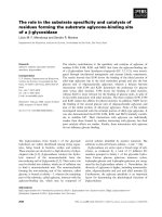
Fig. 1. Schematic representation of the
C-terminal B
1
wt and B
2
wt sequences and
chimera thereof. The C-terminal sequences
beginning at transmembrane domain 7 are
shown. B
1
wt parts are indicated in filled
circles, B
2
wt portions in unfilled ones. The
phosphorylation sites in B
2
wt are highlighted
in light grey, and the position number is indi-
cated. The grey box outside the membrane
indicates the region of the putative cytosolic
helix 8 as found in the crystal structure of
bovine rhodopsin [24]. The assumed palmi-
toylation of B
1
wt and B
2
wt is indicated.
A. Faussner et al. Role of helix 8 and C-termini in bradykinin receptors
FEBS Journal 272 (2005) 129–140 ª 2004 FEBS 131
at the end of the seventh transmembrane domain. We
have shown previously that a B
1
CB
2
chimera stably
expressed in Chinese hamster ovary cells was seques-
tered rapidly upon activation [16]. This was confirmed
in human embryonic kidney (HEK) 293 cells (Fig. 2B).
As the chimera B
1
YB
2
exhibited a slightly attenuated
internalization compared to B
1
CB
2
(Fig. 2B), and the
latter apparently did not gain the full internalization
capability of the B
2
wt, we next tested the possibility
that there is an optimum site for creating rapidly inter-
nalizing chimeras at K7.63 between these two residues
and generated the chimera B
1
KB
2
(Fig. 1). Surpris-
ingly, B
1
KB
2
showed poor ability to internalize
[
3
H]DAK (30% after 10 min), with an internalization
far below those seen for B
1
CB
2
and B
1
YB
2
(Fig. 2B).
Agonist-induced internalization of modified
B
1
KB
2
constructs
The segment between the NPXXY motif and the con-
served cysteine represents one of the regions with the
highest sequence identity between B
1
wt and B
2
wt. The
different internalization of B
1
KB
2
and B
1
CB
2
was
therefore even more surprising given that these two
chimeras have only minor sequence differences
(Fig. 3A). Therefore we considered three possibilities
to explain the cause of this drop in the internalization
of B
1
KB
2
as compared to B
1
CB
2
. First, that the two
residues (KQ) preceding the cysteine were pivotal; sec-
ond, that the cysteine itself needs to be at a specific
position in the C-terminus; or third, that the B
1
residue
V323 instead of the serine is essential in this posi-
tion. Thus, we created three additional chimeras to
test these possibilities: (a) B
1
KB
2
⁄ QGVfiKQ; (b)
B
1
KB
2
⁄ VCfiCV; and (c) B
1
KB
2
⁄ SfiV (Fig. 3A).
Substituting KQ for QGV in B
1
KB
2
led to distinctly
increased agonist internalization as compared with
B
1
KB
2
. This increase was not due to a corrected posi-
tion of the cysteine, as it was not observed with
B
1
KB
2
⁄ VCfiCV (Fig. 3B).
A major effect, however, was seen with the change
of the polar serine (back) to the nonpolar valine
(B
1
KB
2
⁄ SfiV), the amino acid that is normally found
in this position in the B
1
wt. This replacement led to a
chimera exhibiting rapid internalization (60% after
10 min) that was comparable to that of B
1
CB
2
and
B
1
YB
2
(Fig. 2B).
Phosphorylation patterns of B
2
wt and of B
1
⁄ B
2
chimeras reflect their agonist-inducible
internalization
Agonist-induced phosphorylation of serine and threo-
nine residues in the C-terminus has been shown to be
a prerequisite for internalization of B
2
wt and other
receptors [17,21]. B
2
wt in HEK 293 cells displayed a
distinct phosphorylation even in the absence of an
agonist (Fig. 4), as reported recently [22]. When stimu-
lated for 5 min with a saturating concentration of 1 lm
BK at 37 °C, however, B
2
wt responded with a marked
increase (2.50 ± 0.15-fold over basal) in phosphoryla-
tion. The chimera on the other hand displayed little
basal phosphorylation in the absence of their agonist
DAK, although this may, in part, be a sensitivity prob-
lem due to their lower expression levels. Nevertheless,
the rapidly internalizing chimeras B
1
YB
2
and B
1
CB
2
Table 1. Receptor density (B
max
), receptor affinity (K
d
), basal and stimulated total IP accumulation, and EC
50
of B
2
wt, B
1
wt and B
1
⁄ B
2
receptor chimera. ND, not determined.
Receptor construct
B
max
a
(fmolÆmg protein
)1
)
K
d
(nM)
IP accumulation
EC
50
± SEM
(n
M)Unstimulated (30 minÆbasal
)1
)
B
2
wt 10400 ± 600 3.91 ± 1.06 1.93 ± 0.17 12.86 ± 1.37 (n ¼ 7) 0.79 ± 0.34 (n ¼ 4)
I347* 5020 ± 900 3.76 ± 1.61 ND ND 1.13 ± 0.47 (n ¼ 4)
TSIfiAAA 5298 ± 1080 2.82 ± 0.92 2.29 ± 0.77 10.68 ± 2.25 (n ¼ 3) 0.058 ± 0.006 (n ¼ 3)
N338* 3832 ± 290 4.03 ± 0.80 2.54 ± 0.38 13.57 ± 1.76 (n ¼ 3) 0.072 ± 0.038 (n ¼ 3)
B
1
wt 625 ± 24 1.11 ± 0.12 1.6 ± 0.2 8.41 ± 0.52 (n ¼ 7) 0.37 ± 0.06 (n ¼ 7)
B
1
RB
2
127 ± 24 1.09 ± 0.11 ND ND ND
B
1
NB
2
511 ± 160 ND ND ND 0.28 ± 0.1 (n ¼ 3)
B
1
CB
2
1701 ± 503 1.48 ± 0.17 1.53 ± 0.14 7.5 ± 0.6 (n ¼ 7) 1.0 ± 0.08 (n ¼ 3)
B
1
KB
2
1823 ± 664 ND 1.84 ± 0.25 4.1 ± 0.2
b
(n ¼ 5) 0.7 ± 0.3 (n ¼ 3)
B
1
KB
2
⁄ SfiV 1758 ± 150 1.59 ± 0.44 1.31 ± 0.12 7.2 ± 0.8 (n ¼ 3) 1.7 ± 0.2 (n ¼ 3)
B
1
KB
2
⁄ QGVfiKQ 2142 ± 623 ND 1.33 ± 0.12 4.6 ± 0.9
b
(n ¼ 3) 2.0 ± 0.2 (n ¼ 3)
B
1
KB
2
⁄ VCfiCV 1786 ± 320 ND 1.42 ± 0.06 4.3 ± 0.1
b
(n ¼ 3) 0.8 ± 0.1 (n ¼ 3)
B
1
YB
2
2957 ± 1041 1.85 ± 1.4 1.50 ± 0.15 8.7 ± 0.8 (n ¼ 6) 2.2 ± 0.2 (n ¼ 3)
B
1
V323S 846 ± 128 ND 1.44 ± 0.08 4.59 ± 0.84
b
(n ¼ 3) 0.35 ⁄ 0.28
a
Estimated with 10 nM [
3
H]DAK.
b
P < 0.001 vs. B
1
wt.
Role of helix 8 and C-termini in bradykinin receptors A. Faussner et al.
132 FEBS Journal 272 (2005) 129–140 ª 2004 FEBS
responded to stimulation with 1 lm DAK with a dis-
tinct increase in phosphorylation. The slowly internaliz-
ing B
1
KB
2
, in contrast, exhibited no significant
phosphorylation even when challenged with DAK.
Total IP accumulation of B
1
wt and B
1
⁄ B
2
chimeras parallels their agonist-inducible
internalization
The IP release was expressed as unstimulated or DAK-
stimulated accumulation of total IPs for 30 min at
37 °C compared to the IP content of control cells that
had remained at 4 °C. There was a clear correlation
between the agonist-inducible internalization and the
IP accumulation it could induce when stimulated
(Fig. 5). All chimeric constructs displaying rapid agon-
ist-inducible internalization (B
1
CB
2
,B
1
YB
2
,B
1
KB
2
⁄
SfiV) showed an IP response similar to that seen for
B
1
wt (8.41 ± 0.52 fold for B
1
wt and 7.2–8.7-fold for
the chimera). In contrast, the chimera that internalized
poorly (B
1
KB
2
,B
1
KB
2
⁄ QGVfiKQ, B
1
KB
2
⁄ VCfiCV)
showed a significantly reduced IP signal (4.1–4.6-fold)
despite the fact that they were expressed at similar
levels to the chimeras that became rapidly internalized
(Table 1). These results suggested that V323 might
play a role in the activation of phospholipase C
through B
1
wt. Indeed, exchange of V323 for a serine
in B
1
wt (construct B
1
V323S) resulted in a clearly
reduced IP response (5.28 ± 0.91 vs. 8.41 ± 0.52 for
B
1
wt; Table 1 and Fig. 5).
Discussion
Phosphorylation of serine or threonine residues in the
C-terminus of GPCRs by second messenger kinases or
specific GRKs is a requirement for receptor sequestra-
tion [23]. However, the context in which these residues
have to appear, or the receptor specificity of their
function is not very well understood.
0 5 10 15 20 25 30
0
20
40
60
80
100
Internalization [% of total]Internalization [% of total]
N338*
TSI->AAA
B
2
wt
I347*
A
Time [min]
0 5 10 15 20 25 30
0
20
40
60
80
100
B
1
RB
2
B
1
KB
2
B
1
CB
2
B
1
wt
B
1
NB
2
B
1
YB
2
B
Time [min]
Fig. 2. Internalization of [
3
H]agonist by wild-type bradykinin recep-
tors, truncations and chimera. HEK 293 cells expressing the wild-
type receptors B
1
wt or B
2
wt, chimera thereof, or B
2
wt truncations
or mutations were preincubated with the appropriate[
3
H] agonist:
(A) < 1.5 n
M [
3
H]BK; (B) 2 nM[
3
H]DAK) for 90 min on ice. Internal-
ization was started by placing the cells in a 37 °C water bath and
stopped at the indicated times. Surface-bound and internalized
agonist were determined as described in Material and methods.
Agonist internalization was expressed as percentage of total bound
agonist. Results are given as mean ± SEM of at least three inde-
pendent experiments performed in triplicate.
Fig. 3. [
3
H]DAK internalization of B
1
KB
2
derived constructs. (A) Align-
ment of the relevant sequences of the B
1
CB
2
and B
1
KB
2
-derived
chimera compared to wild-type bradykinin receptor subtypes. Resi-
dues found in B
1
wt are in capital letters; those found in B
2
wt are in
lowercase. Amino acids identical to the B
1
wt sequence are indica-
ted by dashes. The residues mutated in B
1
KB
2
are in bold. To allow
comparison the sequence of rhodopsin is also shown. (B) Internal-
ization of [
3
H]DAK was performed as described in the legend to
Fig. 2. Each time point represents the mean ± SEM of at least
three different experiments done in triplicate.
A. Faussner et al. Role of helix 8 and C-termini in bradykinin receptors
FEBS Journal 272 (2005) 129–140 ª 2004 FEBS 133
The bradykinin receptor subtypes are an excellent
tool to address this issue using both loss- and gain-of-
function approaches, as B
2
wt gets internalized rapidly
following stimulation whereas B
1
wt does not become
sequestered [3]. As both receptors couple preferentially
to the same Ga subunit (G
q ⁄ 11
) differential signaling is
less likely to explain differences in internalization than
in two receptors signaling through different G proteins.
Internalization patterns of truncations I347* and
N338*, and the triple point mutant TSIfiAAA more
closely defined the sequence necessary for the internal-
ization of B
2
wt. Because I347* was internalized as rap-
idly as B
2
wt, while the TSIfiAAA mutant showed
reduced internalization and N338* almost none, the
nine residues from S339 to I347 (SMGTLRTSI) must
play a key role in B
2
wt sequestration. The following
results from our gain-of-function approach, however,
led us to conclude that additional motifs in the more
proximal portion of the C-terminus also play a role
in receptor internalization. First, transfer of the B
2
wt
C-terminus starting with the nine residues containing
all known B
2
wt phosphorylation sites did not permit
maximal internalization of [
3
H]DAK, indicating that
this nine residue sequence is either receptor specific or
that other motifs must contribute to B
2
wt sequestra-
tion. Second, faster internalization was obtained when
more extended parts of the C-terminus beginning
either at conserved C7.71(B1) ⁄ 7.72(B2) (B
1
CB
2
)or
conserved Y7.53 (B
1
YB
2
) were transferred, indicating
sequestration motifs in the region between the palmito-
ylated C324 and N338. Candidates would include
G328-C329 and ⁄ or the negatively charged residues
E332 and E337, as they are highly conserved in B
2
wt
among species.
The chimera B
1
YB
2
showed a slightly lower internal-
ization compared to B
1
CB
2
. We therefore tested whe-
ther there was an optimum chimeric exchange point
between these two mutation sites. Intriguingly,
exchange at a conserved lysine (K7.68) between these
two sites resulted in a poorly internalizing chimera
(B
1
KB
2
, Fig. 2B). The crystal structure of inactive
bovine rhodopsin [24] suggested an explanation for this
result by revealing an additional helix 8 close to the
seventh transmembrane domain with a cytosolic local-
ization parallel to the cell membrane. Structure predic-
tion programs [25] indicated that both B
1
wt and B
2
wt
may also contain a helix 8. Our results show that chi-
control
Mr
75
50
300
250
200
150
100
50
BK (DAK)
-
+++++
% of Basal
Phosphorylation
B
2
wt
B
1
YB
2
B
1
KB
2
B
1
CB
2
Fig. 4. Agonist-induced phosphorylation of B
2
wt and B
1
⁄ B
2
-chi-
mera. Upper panel: HEK293 cells expressing B
2
wt, B
1
YB
2
,B
1
KB
2
,
or B
1
CB
2
were labeled for 10 h with [
32
P]orthophosphate before sti-
mulation with 1 l
M BK and 1 lM DAK, respectively, for 5 min. Cells
were lysed and proteins were solubilized, immunoprecipitated and
visualized by autoradiography. Molecular size markers are indicated
to the left. Lower panel: protein phosphorylation, given as optical
densities of the bands in the area between 50 and 85 kDa, is pre-
sented as mean ± SD from three independent experiments; un-
stimulated B
2
wt was set as 100%.
0
B
1
wt
B
1
CB
2
B
1
YB
2
B
1
KB
2
B
1
KB
2
/QGV
B
1
KB
2
/VC
CV
B
1
KB
2
/S
V
B
1
V323S
KQ
2
4
6
8
10
12
Total inositol phosphate
release/control
unstimulated
stimulated
***
***
***
***
Fig. 5. Total IP accumulation of B
1
wt and chimera. HEK293 cells
expressing the indicated receptor constructs were preincubated
with 50 m
M LiCl, and then with (stimulated) or without (unstimula-
ted) 1 l
M DAK for 90 min on ice. IP accumulation was started in a
water bath at 37 °C and stopped after 30 min as described in
Materials and methods. The basal IP accumulation level was deter-
mined on ice. The results are expressed as fold total IP accumula-
tion above basal and given as mean ± SEM of at least three
different experiments performed in triplicate.
Role of helix 8 and C-termini in bradykinin receptors A. Faussner et al.
134 FEBS Journal 272 (2005) 129–140 ª 2004 FEBS
meric receptors with a helix 8 derived either completely
from B
1
wt (B
1
CB
2
)orB
2
wt (B
1
YB
2
) were internalized
rapidly whereas a receptor with a chimeric helix 8
(B
1
KB
2
) was internalized slowly. As the latter dis-
played no agonist-induced phosphorylation – in con-
trast to chimera B
1
YB
2
– this is probably caused by an
impaired interaction with, or activation of, receptor
kinases resulting in the observed slow internalization.
Further examination of helix 8 revealed that S316 in
the B
2
wt sequence of B
1
KB
2
is responsible for the slow
sequestration of this chimera (Fig. 3B). Helices 8 of
the two receptor subtypes show different charge distri-
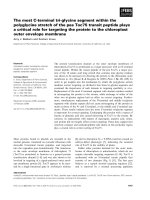
butions despite their high sequence identity (Fig. 6).
The B
2
wt exhibits a highly charged N-terminal half
(two arginines and three lysines) but due to S316 does
not display a clear amphipathic structure. The N-ter-
minal half of B
1
wt, by contrast, is less positively
charged (two arginines and one lysine) but B
1
wt has
a strict amphipathic arrangement of the amino acid
residues. This arrangement is probably important in
receptor signaling because: (a) interruption of the
amphipathic structure of B
1
wt helix 8 in B
1
wt and
B
1
KB
2
through the presence of a serine in position of
V323 leads to a strong attenuation of the IP signal;
(b) substitution of this serine with valine in B
1
KB
2
fully recovers the IP signal; and (c) sequence alignment
of all known B
1
bradykinin receptors shows that
hydrophobicity in this position is absolutely conserved,
while this is not the case for the residues further down-
stream. It has been reported recently that truncation
of the B
1
wt C-terminus at T327 resulted in an 85%
reduction of IP generation, whereas further stepwise
truncation up to R320 – thus including removal of
V323 – did not lead to any further decrease [26]. Thus,
it appears that the presence of a hydrophobic residue
at position 323 in B
1
wt is not necessary for G
q ⁄ 11
activa-
tion but rather that a polar serine there interferes with
this process. This group also described a strongly
increased basal activity for a B
2
receptor construct
where several C-terminal serine and threonine residues
Fig. 6. Structural comparison of helix 8 in B
1
wt, B
2
wt and chimera. Helix 8 (N-terminus on the left hand side) from both bradykinin receptor
subtypes was modeled along the structure of bovine rhodopsin by means of
DEEPVIEW ⁄ Swiss-PdbViewer v3.7 [34]. The dark green ribbon
presentation belongs to B
1
wt, light green ribbon-parts to B
2
wt. The residues different in B
1
wt and B
2
wt are indicated in larger bold labels.
Basic amino acid residues are in blue, acidic residues in red, polar residues are yellow, and unpolar residues are colored in grey. The black
lines in B
1
KB
2
and B
1
KB
2
⁄ SfiV show the transition between B
1
wt and B
2
wt in the chimera.
A. Faussner et al. Role of helix 8 and C-termini in bradykinin receptors
FEBS Journal 272 (2005) 129–140 ª 2004 FEBS 135
were substituted by alanines [27]. We also observed a
tendency to increased basal activity in B
2
constructs that
were lacking all or some of these residues, i.e.
TSIfiAAA and N338*, which was significant only for
the latter (P<0.006) when compared to B
2
wt
(Table 1). Much more apparent, however, was that, in
our hands, these two constructs were hypersensitive, dis-
playing an EC
50
value that was more than 10-fold lower
than that observed for B
2
wt (Table 1). As their K
d
val-
ues were not significantly different this indicates that
apparently relatively few receptors have to be occupied
to achieve half-maximal stimulation. It is important, of
course, to keep in mind that the K
d
was determined at
4 °C where coupling to G proteins does not play a role,
whereas the EC
50
was obtained by determining the IP
accumulation after 30 min at 37 °C. Nevertheless, it is
likely that this hypersensitivity is related to the fact that
the mutated residues play an important role in the inter-
nalization (Fig. 2A) and in the desensitization of B
2
wt
[17]. Much lower BK concentrations than with B
2
wt
may therefore be sufficient to activate enough receptors
for half-maximal IP accumulation.
In our experiments, we did not observe a strong
constitutive B
1
wt signaling activity as compared to
B
2
wt, nor any significant differences between the B
1
wt
and B
1
⁄ B
2
chimera in terms of basal activity (Table 1)
as was reported recently [26]. This discrepancy may be
due to different cell culture conditions (e.g. use of
horse serum vs. fetal bovine serum), or to their tran-
sient low expression vs. our stably high expression and
renders difficult the comparability of our data.
Several reports indicate that a fourth cytoplasmic
loop, formed by membrane insertion of a conserved
palmitoylated cysteine, and in particular the part com-
prising putative helix 8, may be involved in the inter-
action of GPCRs with cognate G proteins.
Synthetic peptides from the C-terminus of the G
a
subunit G
t
and of the Gc subunit of transducin inter-
acted with rhodopsin and kept it in an activated state
[28]. This interaction, however, was abolished in
mutants with replacements in helix 8, suggesting that
G protein subunits interact directly or indirectly with
helix 8. In other experiments, peptides with the
sequence of helix 8 of rhodopsin inhibited activation
of G
t
by rhodopsin [29]. In the angiotensin II receptor
AT
1A
point mutations in the region of putative helix 8
abolished release of inositol trisphosphates and the
GTP-inducible shift in receptor affinity. In addition,
peptides based on its helix 8 sequence stimulated bind-
ing of GTPcStoG
q ⁄ 11
[30]. All of these data point to
an involvement of putative helix 8 in the interaction
with cognate G proteins. As both bradykinin receptors
coupled to the same G
a
subunit G
q ⁄ 11
the different IP
responses obtained with the wild-type receptors and
the chimera let us speculate that each wild-type helix 8
may be specific either for selected bc subunits or for
either G
q
or G
11
. Additional experimental work will be
necessary to test this hypothesis, particularly as the
two receptors, while both coupling to G
q ⁄ 11
(and G
i
)
may very well differ in their capability to activate
other additional signaling pathways. These potential
differences in, for example, the transactivation of
growth hormone receptors and in the activation
of MAPK cascades, as well as different localizations of
the receptor constructs before and after activation may
also contribute to the observed results.
Although helix 8 initially was found in the crystal
structure of inactive bovine rhodopsin [24], prior stud-
ies using NMR and circular dichroism of peptides
taken from the fourth cytoplasmic loop of the angio-
tensin II AT
1A
receptor also indicated that, under cer-
tain experimental conditions, an amphipathic a-helix
was formed in this region [31]. By contrast, NMR
studies of peptides representing the same region of
rhodopsin in membrane and detergent-free solutions
displayed a different structure, with transmembrane
domain 7 being extended and the C-terminus up to the
cysteine existing as a loop [32]. Krishna et al. [33]
demonstrated that the environment in which the pep-
tide exists determines its structure, and suggested that
this region serves as a membrane recognition site
because the presence of detergent or membrane lipids
influences the formation of a helical structure. These
authors proposed that activation of the receptor, and
subsequently of the G protein, leads to a change in the
environment of helix 8 resulting in the loss of the heli-
cal structure. Mutation of specific residues in their
model led to a strongly reduced propensity for helical
formation with the N-terminus of helix 8 being more
influential than the C-terminal portion. Based on this
model, we could speculate that the two bradykinin
receptor subtypes, and those chimeras with an
intact ⁄ homogenous helix 8, are able to appropriately
switch conformation, whereas the receptors with a chi-
meric helix 8 have lost this capacity.
Taken together, our results demonstrate that almost
full capability for receptor internalization can be con-
ferred to the normally noninternalizing B
1
wt, via trans-
fer of the C-terminus of B
2
wt, provided that the new
chimeric receptors have an intact ⁄ homogeneous helix 8
either from B
2
wt or B
1
wt or a chimeric B
1
⁄ B
2
helix
with a conserved V323. Chimeric receptors with a het-
erogeneous helix 8 exhibited an identical effect on sign-
aling as well as on internalization, i.e. poor signaling
was accompanied by reduced internalization. We sug-
gest therefore that helix 8 is directly or indirectly
Role of helix 8 and C-termini in bradykinin receptors A. Faussner et al.
136 FEBS Journal 272 (2005) 129–140 ª 2004 FEBS
involved in the interaction with receptor kinases and in
receptor specific G protein activation.
Materials and methods
Materials
Flp-In T-REx (HEK 293) cells were purchased from Invitro-
gen (Groningen, the Netherlands) and [2,3-prolyl-3,4–
3
H]bradykinin (108 Ci mmol
)1
), [3,4-prolyl-3,4-
3
H]desArg10-
kallidin (80 CiÆmmol
)1
) and myo-[2-
3
H]inositol (21 CiÆ
mmol
)1
) were from PerkinElmer Life Science (Boston,
MA, USA). J. F. Hess (Merck, West Point, PA, USA)
kindly provided us with a vector harboring the sequence of
the human B
1
wt. The antibody AS346 [6] was a generous
gift from W. Mu
¨
ller-Esterl (University of Frankfurt,
Germany). Unlabeled peptides were bought from Bachem
(Heidelberg, Germany). The primers were synthesized by
Invitrogen and delivered desalted and lyophilized. Pfu DNA
polymerase was obtained from Stratagene Europe (Heidel-
berg, Germany). Fetal bovine serum, culture media, and
penicillin ⁄ streptomycin were purchased from PAA Labor-
atories (Co
¨
lbe, Germany). Fugene 6 was from Roche
(Mannheim, Germany) and Invitrogen supplied hygromycin
B and blasticidin. Poly(lysine), captopril, 1.10-phenanthro-
line and bacitracin were purchased from Aldrich (Taufkir-
chen, Germany). Ion exchange columns AG 1 · 8 (formiate
form) were bought from Bio-Rad (Munich, Germany). All
other reagents were of analytical grade and are commer-
cially available.
Cell culture
HEK 293 cells, host cells harboring an Flp recombinant
target (FRT) site in their genome, were cultivated in Dul-
becco’s modified Eagle’s medium (DMEM) with high glu-
cose, 10% (v ⁄ v) fetal bovine serum and 100 UÆmL
)1
⁄
100 lgÆmL
)1
penicillin ⁄ streptomycin. For binding studies or
the measurement of total inositol phosphate accumulation
cells were seeded on cell culture dishes pretreated with
0.01% (w ⁄ v) poly(lysine) in NaCl ⁄ P
i
(phosphate buffered
saline, PBS) to enhance their adherence.
Expression vectors
The sequence of the human B
2
wt starting with the third enco-
ded methionine [9], the sequence of the human B
1
wt, trunca-
tions and chimeras of both were cloned into the BamHI and
the XhoI sites of the pcDNA5 ⁄ FRT vector from Invitrogen.
Each receptor sequence was preceded at the N-terminus by
either a single hemagglutinin-tag (MGYPYDVPDYAGSA)
or a double-tag (MGRSHHHHHH-GYPYDVPDYAGSA)
cloned into the HindIII and BamHI site of the vector. For
comparison of analog positions in both receptors we used the
numbering scheme of Ballesteros & Weinstein [35], where the
most conserved residue in a transmembrane segment is given
the number of the helix followed by the number 50. Residues
proximal to this reference residue are obtained by counting
down, those distal by counting up from 50. The highest con-
served residue in helix 7, the proline within the NPXXY
motif, is therefore named P7.50 and the tyrosine of this
sequence is identified as Y7.53.
Construction of mutated B
1
wt, B
2
wt and
of the B
1
⁄ B
2
receptor chimera
Standard PCR techniques using either receptor-specific or
chimeric primers with the B
1
wt and B
2
wt genes as templates
were applied to generate truncated or point-mutated ver-
sions of the B
1
wt, B
2
wt and several B
1
⁄ B
2
chimeras. All
PCR products were ligated between the BamHI and XhoI
sites of the pcDNA5 ⁄ FRT vector. Cells were transfected
using Fugene 6 following the manufacturer’s instructions,
i.e. 2 l g plasmids (0.4 lg gene of interest in pcDNA5 ⁄ FRT
plus 1.6 lg pOG44-vector) and 5 lL Fugene 6 per six-well
dish. Stably transfected clones were obtained after selection
with 250 lgÆmL
)1
hygromycin B.
[
3
H]Agonist binding studies
For the determination of dissociation constant K
d
and
receptor number B
max
, confluent monolayers on 24-well
plates (B
2
wt) ⁄ 12-well plates (B
1
wt) were washed three
times with ice-cold PBS and incubated on ice with 0.15 or
0.3 mL of ice-cold incubation buffer [40 mm Pipes,
109 mm NaCl, 5 mm KCl, 0.1% (v ⁄ v) glucose, 0.05%
(v ⁄ v) BSA, 2 mm CaCl
2
, pH 7.4; degradation inhibitors
for B
2
wt: 2 mm bacitracin, 0.8 mm 1.10-phenanthroline
and 100 lm captopril; degradation inhibitors for B
1
wt:
0.5 mm bacitracin, 0.02 mm 1.10-phenanthroline and
100 lm captopril] containing increasing concentrations of
[
3
H]BK (10 concentrations ranging from 0.01 to 40 nm)
or [
3
H]DAK (0.01–10 nm) for at least 90 min. The incuba-
tion was stopped by rinsing the monolayers three times
with ice-cold PBS and lysing the monolayers by addition
of 0.2 mL of 0.3 m NaOH. The bound radioactivity was
transferred quantitatively into scintillation vials with
another 0.2 mL of water and measured in a b-counter
after addition of scintillation fluid. Nonspecific binding
was determined in the presence of 5 lm unlabeled agonist
and subtracted from the total binding to calculate the spe-
cific binding.
Internalization of [
3
H]BK and [
3
H]DAK
To determine the internalization of receptor-bound agonist,
cell monolayers on 12-well plates were rinsed three times
with ice-cold PBS (pH 7.2) and incubated with the indi-
A. Faussner et al. Role of helix 8 and C-termini in bradykinin receptors
FEBS Journal 272 (2005) 129–140 ª 2004 FEBS 137
cated concentration of [
3
H]agonist in 0.3 mL incubation
buffer on ice to reach equilibrium binding. To start the
internalization of [
3
H]agonist the plates were transferred to
a water bath at 37 °C. The internalization process was
stopped by placing the trays on ice at the indicated times.
Cells were washed three times with PBS and the remaining
surface-bound [
3
H]agonist was removed by treating the cell
monolayer with 0.2 mL of an ice-cold dissociation solution
(0.2 m acetic acid ⁄ 0.5 m NaCl, pH 2.7) for 10 min. The dis-
sociation solution containing the surface-bound [
3
H]agonist
was quantitatively transferred into scintillation vials by
rinsing the cell monolayer with another 0.2 mL of PBS.
The remaining internalized [
3
H]agonist was subsequently
transferred to scintillation vials by lysing the cells with
0.2 mL of 0.3 m NaOH and rinsing the wells with addi-
tional 0.2 mL of water. The radioactivity of both samples
was determined in a b-counter after addition of scintillation
fluid. Nonreceptor-mediated [
3
H]agonist internalization
was determined in the presence of 5 lm unlabeled agonist
and subtracted from the total binding to obtain the specific
values.
Stimulation of total IP release
Cell monolayers (80% confluent) in 12-well dishes were labe-
led for 18–24 h with 0.5 lCi of myo-[
3
H]inositol in 0.5 mL
DMEM with fetal bovine serum and 100 UÆmL
)1
⁄ 100 lgÆ
mL
)1
penicillin ⁄ streptomycin. The monolayers were then
placed on ice, rinsed three times with ice-cold PBS (pH 7.2)
and incubated with or without the appropriate agonist in
incubation buffer containing 50 mm LiCl. Basal and stimu-
lated IP accumulation was started by placing the tray in a
water bath at 37 °C for 30 min. It was stopped by exchan-
ging the buffer with 0.75 mL of ice-cold 20 mm formic acid
and by transferring the tray onto ice for additional 30 min.
As a baseline control one tray was left on ice with LiCl incu-
bation buffer without agonist. The EC
50
was determined by
adding escalating concentrations of agonist (10
)12
to 10
)6
m)
for 30 min at 37 °C. The supernatant was then applied
together with another 0.75 mL of 20 mm formic acid and
0.2 mL of a 3% (w ⁄ v) ammonium hydroxide solution to AG
1-X8 anion exchange columns, followed by 1 mL 1.8% (w ⁄ v)
ammonium hydroxide solution, 9 mL of 60 mm sodium
formiate, 5 mm sodium tetraborate buffer and 0.5 mL of 4 m
ammonium formate ⁄ 0.2 m formic acid solution. The total
inositol phosphates were eluted by addition of 2.5 mL of the
latter solution. The radioactivity was determined in a b-coun-
ter after the addition of scintillation fluid. All data (basal and
stimulated) at 37 °C are given in fold of the amount of total
IP determined in the baseline control on ice.
Immunoprecipitation and Western blotting
Cells were washed once with PBS and solubilized in RIPA
buffer [50 mm Tris ⁄ HCl, 150 mm NaCl, 1% (v ⁄ v) NP-40,
0.5% (w ⁄ v) sodium deoxycholate, 0.1% (w ⁄ v) SDS, 2 mm
EDTA, pH 7.5] supplemented with 0.5 mm Pefabloc SC
and 10 lm each of 1.10-phenanthroline, aprotinin, leupep-
tin and pepstatin A for 45 min at 4 °C with gentle rock-
ing. The sample was centrifuged at 6240 g for 20 min at
4 °C and the supernatant (0.5 mL with 1.5 mg of total
protein) incubated with 35 lL protein G ⁄ agarose and
2.5 lL of antiserum AS346 for 3 h at 4 °C. The mixture
was then washed twice with RIPA buffer and once with
distilled water, resuspended in 30 lL of Laemmli buffer
and incubated for 6 min at 95 °C. After separation by
electrophoresis on 10% (w ⁄ v) SDS polyacrylamide gels,
the proteins were transferred onto 0.45 lm nitrocellulose
membranes. After blocking the membranes overnight with
blocking buffer [0.25% (w ⁄ v) gelatin in 50 mm Tris ⁄ HCl,
150 mm NaCl, 5 mm EDTA, 0.05% (v ⁄ v) Triton X-100,
pH 7.5] primary high affinity anti-HA Ig (0.1 lgÆmL
)1
)
was added in fresh blocking buffer for 2 h at room tem-
perature. The membranes were washed twice for 10 min in
Tris-buffered saline with 0.1% (v ⁄ v) Tween 20 (TBST) fol-
lowed by addition of the corresponding secondary peroxi-
dase-labeled rabbit anti-rat Ig (1 : 1000) for 1 h. After
washing in TBST three times each for 15 min antibody
binding was detected using the Western Blot Chemolumi-
nescence Reagent Plus.
Receptor phosphorylation
Confluent cells on 6-well plates were washed twice with
phosphate-free DMEM, incubated for 3 h at 37 °C in the
same medium, and labeled with 0.2 mCiÆmL
)1
[
32
P]ortho-
phosphate for 10–12 h. After exposure to 1 lm BK or
DAK for 5 min at 37 °C, monolayers were scraped into
0.5 mL of RIPA buffer containing protease inhibitors (see
above) and phosphatase inhibitors (25 mm NaF, 1 mm
sodium orthovanadate, 0.3 lm okadaic acid). Immunopre-
cipitation and separation on a 10% (w ⁄ v) SDS polyacryl-
amide gel were carried out as described previously. The
proteins of interest were electroblotted onto nitrocellulose
membranes and identified by autoradiography.
Protein determination
Total protein per well was quantified by lysing the cells
with 0.3 mL of 0.3 m NaOH. The protein content of this
solution was determined with the Micro BCA Protein assay
reagent from Pierce (Rockford, IL, USA) using bovine
serum albumin as standard.
Data analysis
All data analysis was performed using graphpad prism for
Macintosh, Version 3.0a (GraphPad Software, Inc., San
Diego, CA, USA).
Role of helix 8 and C-termini in bradykinin receptors A. Faussner et al.
138 FEBS Journal 272 (2005) 129–140 ª 2004 FEBS
Acknowledgements
This work was supported by a grant from the Deutsche
Forschungsgemeinschaft to A. Faussner (FA 288 ⁄ 3–1).
References
1 Gether U (2000) Uncovering molecular mechanisms
involved in activation of G protein-coupled receptors.
Endocrine Rev 21, 90–113.
2 Ferguson SS (2001) Evolving concepts in G protein-cou-
pled receptor endocytosis: the role in receptor desensiti-
zation and signalling. Pharmacol Rev 53, 1–24.
3 Austin CE, Faussner A, Robinson HE, Chakravarty S,
Kyle DJ, Bathon JM & Proud D (1997) Stable expres-
sion of the human kinin B
1
receptor in Chinese hamster
ovary cells. Characterization of ligand binding and
effector pathways. J Biol Chem 272, 11420–11425.
4 Sabourin T, Bastian L, Bachvarov DR & Marceau F
(2002) Agonist-induced translocation of the kinin B
1
receptor to caveolae-related rafts. Mol Pharmacol 61,
546–553.
5 Lamb ME, Zhang C, Shea T, Kyle DJ & Leeb-
Lundberg LMF (2002) Human B
1
and B
2
bradykinin
receptors and their agonists target caveolae-related lipid
rafts to different degrees in HEK 293 cells. Biochem 41,
14340–14347.
6 Blaukat A, Herzer K, Schroeder G, Bachmann M, Nash
N&Mu
¨
ller-Esterl W (1999) Overexpression and func-
tional characterization of kinin receptors reveal subtype-
specific phosphorylation. Biochem 38, 1300–1309.
7 Pizard A, Blaukat A, Mu
¨
ller-Esterl W, Alhenc-Gelas F
& Rajerison RM (1999) Bradykinin-induced internaliza-
tion of the human B
2
receptor requires phosphorylation
of three serine and two threonine residues at its
carboxyl tail. J Biol Chem 274, 12738–12747.
8 Ostrom RS (2002) New determinants of receptor-
effector coupling: trafficking and compartmentation
in membrane microdomains. Mol Pharmacol 61,
473–476.
9 Hess JF, Borkowski JA, Young GS, Strader CD &
Ransom RW (1992) Cloning and pharmacological char-
acterization of a human bradykinin (BK-2) receptor.
Biochem Biophys Res Commun 184, 260–268.
10 Menke JG, Borkowski JA, Bierillo KK, MacNeil T,
Derrick AW, Schneck KA, Ransom RW, Strader CD,
Linemeyer DL & Hess JF (1994) Expression cloning of
a human B
1
bradykinin receptor. J Biol Chem 269,
21583–21586.
11 Gutowski S, Smrcka A, Nowak L, Wu DG, Simon M
& Sternweis PC (1991) Antibodies to the alpha q sub-
family of guanine nucleotide-binding regulatory protein
alpha subunits attenuate activation of phosphatidylino-
sitol 4,5-bisphosphate hydrolysis by hormones. J Biol
Chem 266, 20519–20524.
12 Marceau, F., Hess, J.F. & Bachvarov, D.R. (1998) The
B
1
receptors for kinins. Pharmacol Rev 50, 358–386.
13 Proud D & Kaplan AP (1988) Kinin formation: mech-
anism and role in inflammatory disorders. Annu Rev
Immunol 6, 49–83.
14 Farmer SG & Burch RM (1992) Biochemical and mole-
cular pharmacology of kinin receptors. Annu Rev Phar-
macol Toxicol 32, 511–536.
15 Prado GN, Taylor L & Polgar P (1997) Effects of intra-
cellular tryosine residue mutation and carboxyl terminus
truncation on signal transduction and internalization of
the rat bradykinin B
2
receptor. J Biol Chem 272, 14638–
14642.
16 Faussner A, Proud D, Towns M & Bathon JM (1998)
Influence of the cytosolic carboxyl termini of human B
1
and B
2
kinin receptors on receptor sequestration, ligand
internalization, and signal transduction. J Biol Chem
273, 2617–2623.
17 Blaukat A, Pizard A, Breit A, Wernstedt C, Alhenc-
Gelas F, Mu
¨
ller-Esterl W & Dikic I (2001) Determina-
tion of bradykinin B
2
receptor in vivo phosphorylation
sites and their role in receptor function. J Biol Chem
276, 40431–40440.
18 Hunyady L, Bor M, Balla T & Cat KJ (1994) Identifica-
tion of a cytoplasmic Ser-Thr-Leu motif that determines
agonist-induced internalization of the AT1 angiotensin
receptor. J Biol Chem 269, 31378–31382.
19 Faussner A, Bauer A, Kalatskaya I, Jochum J & Fritz
H (2003) Expression levels strongly affect ligand-
induced sequestration of B
2
bradykinin receptors in
transfected cells. Am J Physiol Heart Circ Physiol 284,
H1892–H1898.
20 Faussner A, Schu
¨
ssler S, Seidl C & Jochum M (2004)
Inhibition of B
2
Bradykinin receptor sequestration by
phenylarsine oxide or sucrose allows determination of
receptor affinity shift and ligand dissociation in intact
cells. Biol Chem 385, 835–843.
21 Marchese A, Chen C, Kim YM & Benovic JL (2003)
The ins and outs of G protein-coupled receptor traffick-
ing. Trends Biochem Sci 28, 369–376.
22 Kalatskaya I, Schu
¨
ssler S, Blaukat A, Mu
¨
ller-Esterl W,
Jochum M, Proud D & Faussner A (2004) Mutation of
tyrosine in conserved NPXXY-sequence leads to consti-
tutive phosphorylation and internalization, but not sig-
naling of the human B
2
bradykinin receptor. J Biol
Chem 279, 31268–31276.
23 Perry SJ & Lefkowitz, R.J. (2002) Arresting develop-
ments in heptahelical receptor signaling and regulation.
Trends Cell Biol 12, 130–138.
24 Palczewski K, Kumasaka T, Hori T, Behnke CA,
Motoshima H, Fox BA, Le Trong I, Teller DC, Okada
T, Stenkamp RE, et al. (2000) Crystal structure of
rhodopsin: a G protein-coupled receptor. Science 289,
739–745.
A. Faussner et al. Role of helix 8 and C-termini in bradykinin receptors
FEBS Journal 272 (2005) 129–140 ª 2004 FEBS 139
25 Jones DT (1999) Protein secondary structure prediction
based on position-specific scoring matrices. J Mol Biol
292, 195–202.
26 Kang DS & Leeb-Lundberg LMF (2002) Negative and
positive regulatory epitopes in the C-terminal domains
of the human B1 and B2 bradykinin receptor sub-
types determine receptor coupling efficacy to G
q ⁄ 11
-
mediated phospholipase Cb activity. Mol Pharmacol
62, 281–288.
27 Fathy DB, Leeb T, Mathis SA & Leeb-Lundberg LMF
(1999) Spontaneous human B2 bradykinin receptor
activity determines the action of partial agonists as ago-
nists or inverse agonists. J Biol Chem 274, 29603–29606.
28 Ernst OP, Meyer CK, Marin EP, Henklein P, Fu WY,
Sakmar TP & Hofmann KP (2000) Mutation of the
fourth cytoplasmic loop of rhodopsin affects binding of
transducin and peptides derived from the carboxyl-term-
inal sequences of transducin alpha and gamma subunits.
J Biol Chem 275, 1937–1943.
29 Marin EP, Krishna AG, Zvyaga TA, Isele J, Siebert
F & Sakmar TP (2000) The amino terminus of the
fourth cytoplasmic loop of rhodopsin modulates
rhodopsin–transducin interaction. J Biol Chem 275,
1930–1936.
30 Sano T, Ohyama K, Yamano Y, Nakagomi Y,
Nakazawa S, Kikyo M, Shirai H, Blank JS, Exton JH
& Inagami T. (1997) A domain for G protein coupling
in carboxyl-terminal tail of rat angiotensin II receptor
type 1A. J Biol Chem 272, 23631–23636.
31 Franzoni L, Nicastro G, Pertinhez TA, Tato
`
M, Nakaie
CR, Paiva ACM, Schreier S & Spisni A (1997) Struc-
ture of the C-terminal fragment 300–320 of the rat
angiotensin II AT1a receptor and its relevance with
respect to G-protein coupling. J Biol Chem 272,
9734–9741.
32 Yeagle PL, Choi G & Albert AD (2001) Studies on
the structure of the G-protein-coupled receptor rhodop-
sin including the putative G-protein binding site in
unactivated and activated forms. Biochem 40, 11932–
11937.
33 Krishna AG, Menon ST, Terry TJ & Sakmar TP (2002)
Evidence that helix 8 of rhodopsin acts as a membrane-
dependent conformational switch. Biochem 41, 8298–
8309.
34 Guex N & Peitsch MC (1997) SWISS-MODEL and the
Swiss-PdbViewer: An environment for comparative pro-
tein modeling. Electrophoresis 18, 2714–2723.
35 Ballesteros JA & Weinstein H (1995) Integrated meth-
ods for the construction of three-dimensional models
and computational probing of structure-function rela-
tions in G protein coupled receptors. Methods Neursci
25, 366–428.
Role of helix 8 and C-termini in bradykinin receptors A. Faussner et al.
140 FEBS Journal 272 (2005) 129–140 ª 2004 FEBS