Báo cáo khoa học: Context-dependent effects of proline residues on the stability and folding pathway of ubiquitin docx
Bạn đang xem bản rút gọn của tài liệu. Xem và tải ngay bản đầy đủ của tài liệu tại đây (305.22 KB, 11 trang )
Context-dependent effects of proline residues on the stability
and folding pathway of ubiquitin
Maria D. Crespo, Geoffrey W. Platt, Roger Bofill and Mark S. Searle
School of Chemistry, Centre for Biomolecular Sciences, University Park, Nottingham, UK
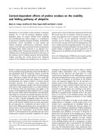
Substitution of trans-proline at three positions in ubiquitin
(residues 19, 37 and 38) produces significant context-
dependent effects on protein stability (both stabilizing
and destabilizing) that reflect changes to a combination
of parameters including backbone flexibility, hydrophobic
interactions, solvent accessibility to polar groups and
intrinsic backbone conformational preferences. Kinetic
analysis of the wild-type yeast protein reveals a predominant
fast-folding phase which c onforms to an apparent two-
state f olding model. Temperature-dependent studies of the
refolding rate reveal thermodynamic details of the nature of
the transition s tate fo r f olding consistent with hydrophobic
collapse providing the overall driving force. Brønsted
analysis of the refolding and unfolding rates of a family of
mutants w ith a variety o f side c hain substitutions for P 37 and
P38 reveals that the two prolines, which are located in a
surface l oop adjacent to the C terminus of the m ain a-helix
(residues 24–33), are not significantly structured in the
transition state for folding and appear to be consolidated
into the native structure only late in the folding process. We
draw a similar conclusion regarding position 19 in the loop
connecting the N-terminal b-hairpin to t he main a-helix. T he
proline residues of ubiquitin are passive spectators in the
folding process, but influence protein stability in a variety of
ways.
Keywords: folding kinetics; NMR structural analysis; proline
mutations; p rotein folding pathway; protein stability.
Proline is unique amongst the natural amino a cid residues;
the five-membered ring significantly reduces the flexibility of
the polypeptide chain by restricting r otation around the
N-Ca bond to a relatively small region of conformational
space. This factor, coupled with the lack of an amide NH
hydrogen bond donor means that proline i s not readily
accommodated into r egular (a-helical or b-sheet) protein
secondary structure. It is, however, more abundant in
connecting loops playing a specific role in b-turn sequences
[1,2], and as a helix capp ing residue or as a helix terminator
[3–5]. Prolines confer pre-organization and rigidity in the
context of small peptide protease inhibitors [6,7], a c oncept
that has been widely used in biomolecular and supramole-
cular design to overcome t he potential energetic cost of loss
of conformational entropy when dynamic molecules asso-
ciate, or when a fl exible polypeptide chain folds. In the
context of protein folding, the observation that cis and trans
forms of the Xaa-Pro peptide bond are n early isoenergetic
[8], and separated by a significant activation barrier, can
lead to slow-folding kinetic phases due to the population of
the non-native cis-form in the unfolded state, where the rate
limiting step is the isomerization of the Xaa-Pro peptide
bond [9–12]. Heterogeneity in the unfolded state due to slow
isomerization reactions potentially complicates the kinetic
elucidation of folding pathways and t he ability to ide ntify
partially folded intermediate states or parallel folding
pathways [13–21]. However, the observation of a wide
variation in the amplitude of slow folding phases associated
with prolyl isomerizatio n (in many cases less than expected
on the basis of frequency of occurrence in the primary
amino acid sequence) suggests that not all non-native cis
prolines result in slow folding p hases, and that cis–trans
isomerization in some structural contexts need not be rate
limiting [22–29]. More recent studies demonstrate that
nonprolyl cis-peptide bonds also contribute to the hetero-
geneous pool of unfolded molecules [18,30]. Although
individual cis-peptide bonds contribute little to the popu-
lation (% 0.15–0.5%) in the unfolded protein, their large
number generates a significant proportion of slow folding
molecules [18,30,31].
We report on the effects of proline on the stability and
folding kinetics of ubiquitin, a small model system of 76
residues that is uncomplicated by disulphide bonds and
bound cofactors [32]. Ubiquitin has been the subject of a
number of investigatio ns regarding i ts folding m echanism.
Early studies had suggested that the protein populates an
intermediate state identified on the basis of deviations of
kinetic data from linearity in the refolding arm of chevron
plots at low denaturant concentrations [33]. M ore recent
studies [13,14,34,35] report apparent two-state kinetics
under similar conditions, suggesting that t he roll-over effect
in the r efolding kinetics may b e a consequence o f either
transient aggregation that is exacerbated by the stabilizing
effects of inorganic salts [15,35], or due to data fitting at
rates near the instrumental limits where interference from
slower phases can decrease apparent folding rates resulting
Correspondence to M. S. Searle, School of Chemistry, Centre for
Biomolecular Sciences, University Park, Nottingham NG7 2RD, UK.
Tel.: +44 115 9513567, E-mail:
Abbreviations: TSE, transition state ensemble; GdmCl, guanidinium
chloride.
(Received 8 July 2004, accepted 30 September 2004)
Eur. J. Biochem. 271, 4474–4484 (2004) Ó FEBS 2004 doi:10.1111/j.1432-1033.2004.04392.x
in chevron rollover effects [13,14]. HX labelling studies and
stopped-flow CD similarly found no evidence for an early
intermediate in the first 2 m s of folding [36].
We show that proline s ubstitutions in yeast ubiquitin
at positions 19, 37 and 38 produce context-depend ent
effects on stability with r emoval of proline at specific
sites having the effect of either significantly increasing
stability (P38A) or destabilizing the protein (P19S and
P37A). A full kinetic analysis of the major fast folding
phase of wild-type yeast ubiquitin (WT*) and of a
number of nondisruptive single-point Ala mutants and
several double mutants, using F-value analysis and
Brønsted plots, s hows t hat t he transition state ensemble
(TSE) is tolerant to proline substitutions at positions 19,
37 and 38, and that these residues are not well structured
in the t ransition state for folding.
Materials and methods
Protein expression
A pKK223-3 plasmid construct containing the yeast
ubiquitin gene was used to express the wild-type protein in
Escherichia coli strain BL21(DE3) under the control o f the
isopropyl thio-b-
D
-galactoside (IPTG)-inducible tac pro-
moter. The F45W m utant gene was cloned b y overlap PCR
methodology using the wild-type y east ubiquitin g ene in
pKK223-3 (Pharmacia Biotech) as a template. The mutated
cassette was inserted between the Eco RI and HindIII
restriction sites of pKK223-3, and the mutation confirmed
by DNA sequencing. Competent E. coli cells were trans-
formed with this construct. Expression and purification
were as described for the wild-type yielding typically
10–15 mgÆL
)1
of ubiquitin, as previously described [34].
NMR structural analysis
All NMR experiments were performed on a Bruker
Avance600 spectrometer. TOCSY and NOESY experi-
ments were used as p reviously described [34] on 1 -m
M
protein samples at pH 5.5. Spectra were referenced to
internal trimethylsilylpropionate. D ata were processed and
assigned using Bruker
XWINNMR
and
ANSIG
software [37].
Structural models were visualized using
MOLMOL
[38].
Equilibrium stability measurements
Protein stability was determined by fluorescence measure-
ments on 1 .5 l
M
solutions of protein i n 2 5 m
M
acetate
buffer at pH 5.0 and 298 K. The change in fluorescence at
358 nm was monitored as a function of guanidinium
chloride (GdmCl) concentration. The linear extrapolation
method was used [39–42] assuming that the stability v aries
with the c oncentration of denaturant [D], according to the
expression DG
D
¼ DG
eq
+ m [D], where DG
D
is the
stability at a given [D], m is the constant of p roportionality,
and DG
eq
is the stability in water alone. The fraction of
folded protein F
f
is derived from fluorescence measurements
according to F
f
¼ (f
D
) f
U
)/(f
N
) f
U
), where f
D
is the
measured fluorescence at a g iven [D] a nd f
U
and f
N
are
the limiting values for the unfolded and native states,
respectively. The mid-point of the unfolding transition
[D]
50%
for each mutant was determined by nonlinear least
squares fitting to the expression:
F
f
¼ exp½mð½DÀ½D
50%
Þ=RT=ð1 þ exp½mð½D
À½D
50%
Þ=RTÞ ð1Þ
The e quilibrium stability DG
eq
was d etermined from the
expression DG
eq
¼ –m[D]
50%
,wherem forasetofmutants
is assumed constant (10.9 ± 0.23 kJÆmol
)1
Æ
M
)1
) [34,43].
This approach is justified by the NMR analysis which shows
that all of the mutants fold to a native-like structure with
only minor localized chemical shift pertu rbations. Thus,
mutations are n ot producing s ignificant c hanges in the
hydrophobic surface area buried, justifying the use of the
same m-value for stability measurements. Additional cor-
rections were used to allow for a small linear denaturant
dependence o f t he fluorescence of both the folded and the
unfolded state [39].
Kinetics experiments
Fluorescence-detected kinetic unfolding and refolding
measurements were performed using an Applied Photo-
physics Pi-star 1 80 spectrophotometer. T emperature was
regulated using a Neslab RTE-300 circulating program-
mable water bath. All kinetics experiments were per-
formed in 25 m
M
acetate buffer pH 5.0 at 298 K.
Refolding experiments were performed by 1 : 10 dilution
of unfolded protein (15 l
M
in 7
M
GdmCl) into buffered
solutions of different GdmCl concentrations yielding a
final protein concentration of 1.36 l
M
. For unfolding
experiments, a buffered solution of native protein was
unfolded by a 1 : 10 dilution to yield final concentrations
of GdmCl near or above the midpoint of the equilibrium
unfolding transition (concentrations of GdmCl in the
range 3.7–7.3
M
). Kinetic measurements for both unfold-
ing and refolding reactions were averaged four to six
times at each GdmCl concentration. In all cases, the
GdmCl c oncentration w as determined using a refracto-
meter [ 40].
Analysis of kinetic data
The kinetic traces were analysed using a multiexponential
fitting procedure (two o r three components). The kinetic
data wer e analysed assuming an apparent two-state
model using standard equations described in detail by
others [41,43,44]. T he observed rate constant k
obs
is the
sum of t he folding and unfolding rates, k
obs
¼ k
fold
+
k
unfold
where k
obs
is dependent on [D] a ccording to t he
expression:
lnk
obs
¼ ln½k
unfold
expðm
unfold
½D=RTÞ
þ k
fold
expðm
fold
½D=RTÞ ð2Þ
The dependence of lnk
obs
on [D] gives extrapolated values
for k
unfold
and k
fold
in water a lone, together w ith t he slopes
of the f olding and unfolding components m
unfold
and m
fold
.
The temperature dependence of the refolding rate w as
examined at a denaturant concentration of 0.4
M
GdmCl
and 1.81 l
M
protein and the data fitted according to the
following expressions [30]:
Ó FEBS 2004 Proline residues in ubiquitin stability and folding (Eur. J. Biochem. 271) 4475
lnk
obs
¼ lnk
o
À DGz =RT ð3Þ
where k
o
is the t emperature independent pre-exponential
factor (% 10
8
), and the temperature dependence of the
activation free energy DGà is given by:
DGz¼DHzþDC
p
zðT À 298Þ
À T½DSzþDC
p
z lnðT=298Þ ð4Þ
with DHà, DC
p
à and DSà representing the change in
activation enthalpy, heat capacity and entropy of formation
of the TSE for folding (U-à). Reported errors reflect the
quality of th e nonlinear l east squares fi t to t he experimental
data.
Results
Context-dependent effects of proline substitutions
on protein stability
We have used the F 45W mutant of yeast ubiquitin as our
Ôwild-typeÕ protein (WT*) for mutational and biophysical
studies. The partially buried indole side chain (Fig. 1)
undergoes a significant (fourfold) quenching of fluorescence
on folding but has previously been shown to have only a
relatively small effect on the stability (DDG % 1kJÆmol
)1
)
and structure of human ubiquitin [45]. Our own structural
analysis of F45W mutants of the yeast protein confirms this.
We have explored the context-dependent effects of proline
on ubiquitin stability by introducing a number of substitu-
tions at positions P37 a nd P38. The equilibrium stability of
the mutants was determined from the change in fluorescence
at 358 nm as a function of GdmCl concentration. The data
show that in each case the fraction unfolded fits well to a
two-state t ransition with t he observation of a r ange of mid-
point denaturant concentrations, [D]
50%
values, indicating
significant context-dependent effects of the mutations on
protein stability (Fig. 2; Table 1). The P37A mutation
produces a large shift in the transition mid-point for
denaturation from 2.62
M
GdmCl (WT*) to 2.18
M
GdmCl.
This equates to a reduction in stability o f 4 .5 ± 0.6 kJÆ
mol
)1
. In contrast, the P38A mutation re sults in a significant
increase in stability of )4.6±0.6kJÆmol
)1
.TheA37A38
double mutant is slightly less stable than WT* ( 1 . 1 ± 0. 6 k J Æ
mol
)1
), showing that the contributions from P37A and
P38A are approximately additive.
We also examined the effects of substituting a proline
residue at position 19 in the loop region connecting the
N-terminal b-hairpin to t he main a-helix (Fig. 1). Proline is
highly conserved at t his site in many s pecies; however,
in yeast ubiquitin residue 19 is serine. The mutation
S19P produces a significant increase in stability o f
)5.3 ± 0.7 kJÆmol
)1
. Thus, the P19S, P37A a nd P38A
mutations produce contrasting effects that do not appear to
simply relate to entropic factors concerning changes in
backbone flexibility.
Structural analysis of the proline mutants by NMR
NMR structural analysis was used to establish whether the
substitutions of P37 and P38 are substantially perturbing
the conformation and dynamics in this region of the protein,
or more specifically, whether st ructural effects are transmit-
ted t o the C terminus of the a djacent main a-helix (residues
24–33). We have completed an NMR backbone assignment
of WT* for comparison with P37A, P38A and the A37A38
double m utant a nd have examined chemical shift p erturba-
tions and the pattern of NOEs in the vicinity of the
mutation sites. Deviations of Ha signals from random coil
chemical shifts pro vide a sensitive probe of local perturba-
tions to secondary structure [ 46,47]. We find that perturba-
tions are largely confined to the residues immediately
adjacent to the m utation s ite, in particular Ile36 (Fig. 3). In
the case of the P37A mutant, some small (< 0.1 p.p.m.)
longer range effects are observed involving residues on the
Pro37 f ace of the main a-helix (namely, Asp24, Ser28 and
Gln31). The characteristic pattern of NH–NH sequential
NOEs enables us to map the e xtent of structure formation
within the main a-helix (residues 24–33), and examine the
integrity o f the helix C-capping motif and of the s hort helix
(residues 3 8–40). In ubiquitin, the C-capping motif involves
a hydrogen bond between Gly35 NH and the backbone
carbonyl of G ln31. This i nteraction positions Ile36 to form
hydrophobic contacts to Ile30 and results in strong NH-NH
sequential NOEs between Gln34 « Gly35 « Ile36. Fur-
ther, the NH signal of Ile36 is > 1 p.p.m. upfield shifted b y
these interactions. These NOEs are clearly evident in the
NOESY data for WT*, P37A, P38A and A37A38. Further,
Ile36 NH has the characteristic upfield shift that confirms
that the C terminus of the helix and the C-capping motif are
not disrupted by the proline mutations. Extending the
analysis to the short helix (residues 38–40), the strong
sequential NH–NH NOEs from D39 through to Q41 are
preserved in all mutants. The P38A mutation appears to
extend the helical turn by one residue with Ala38 having a
3
J
NH–Ha
value < 6 Hz with evidence of i,i+3 NOEs to
Gln41. NOE contacts from Ala38 protons to the side chains
of Lys27 and Gln31 in the main a-helix are also evident and
confirm t hat the Ala38 methyl g roup occupies the same
hydrophobic pocket as the side chain of Pro38. Mod elling
the structure with Ala substitutions imposed on the
backbone conformation of WT* shows that the pattern of
P37
P38
P19
W45
Fig. 1. Ribbon structure modelled on the X-ray structure of human
ubiquitin [32]. The position and orientation of the side chains of Pro19,
Pro37 and Pro38 are highlighted along with the F45W mutation
(drawn using
MOLMOL
[38]). The sequences o f human and yeast
ubiquitin differ at the f ollo wing positions: P19S, E24D and A28S.
4476 M. D. Crespo et al.(Eur. J. Biochem. 271) Ó FEBS 2004
NOEs is entire ly consistent with native-like /,w angles.
Thus, w e conclude that the Pro to Ala substitutions are not
significantly perturbing the backbone conformation and
dynamics of the protein around the mutation sites and in the
adjacent a-helix. Analogous NMR s tudies of the S19P
mutant (data not shown) also establish that chemical shift
perturbations are entirely l ocalized to the mutation site and
immediately flanking residues.
Kinetic analysis of ubiquitin folding
The folding kinetics of WT* have been analysed from
refolding and unfolding stopped-flow exper iments in
GdmCl at 298 K a nd pH 5.0 in 2 5 m
M
acetate buff er.
The refolding traces for WT* in the range 0–2.5
M
GdmCl
are best analysed in terms of a multiexponential fit reflecting
at least three resolved folding phases. The fast phase, which
accounts for % 87% of the amplitude of the fluorescence
change, has an extrapolated folding rate in water of 303 s
)1
,
while seve ral minor slower folding phases are also evi-
dent with extrapolated rate constan ts k
2
¼ 34 s
)1
and
Fig. 2. Equilibrium denatura tion curves for
yeast ubiquitin (WT*) a nd various mutan ts.
Fraction unfolded is p lotted against c oncen-
tration of GdmCl at pH 5.0 in 25 m
M
acetate
buffer at 298 K and was mo nitored by
tryptophan fluorescence. Stability d ata are
shown in T able 1.
Table 1. Equilibrium stability dat a for ubiquitin mutants (pH 5. 0,
25 m
M
acetate b uffer, 298 K) determined by GdmCl denaturation
monitored b y changes i n tryptophan fluorescence.
Mutant
m
eq
a
(kJÆmol
)1
Æ
M
)1
) [D]
50%
b
DG
eq
c
(kJÆmol
)1
)
WT* 11.3 2.62 )28.6 (± 0.6)
P37A 11.9 2.21 )24.1 (± 0.5)
P38A 10.2 3.05 )33.2 (± 0.7)
SQ 10.8 2.21 )24.1 (± 0.5)
QL 10.4 2.39 )26.0 (± 0.5)
AA 11.2 2.52 )27.5 (± 0.5)
VV 11.2 2.22 )24.2 (± 0.5)
S19P 10.0 3.11 )33.9 (± 0.7)
a
Errors in m
eq
are less than ± 0.35.
b
Denaturant concentration
at the mid-point of the folding/unfolding transition; fitting errors
are less than ± 0.008.
c
Equilibrium stability determined from
the [D]
50%
value assuming a mean m-value (± SE) of 10.9 ±
0.23 kJÆmol
)1
Æ
M
)1
.
Fig. 3. Ha chemical shift analysis of residues
22–46 of the yeast ubiquitin mutants P37A,
P38A and the double mutant A37A38. These
residues span the main a-helical region (res i-
dues 21–35) N terminal to the X37 and X38
mutation sites, and t he sequence of the short
helix (residues 38–40) and fourth strand of
b-sheet (re sidues 4 2–46) on the C-terminal side
of the mutation sites (Fig. 1). Differences in
chemical shifts with respect to r ando m coil
values [46,47] are plotte d against sequence
position.
Ó FEBS 2004 Proline residues in ubiquitin stability and folding (Eur. J. Biochem. 271) 4477
k
3
¼ 0.14 s
)1
, and relative amplitudes o f 1 1% and 2 %,
respectively. The k
2
and k
3
processes, also identified for
human ubiquitin [13,33], have previously been attributed to
slow rate-limiting cis–trans prolyl isomerization reactions.
However, we have shown using double-jump (interrupted
unfolding) experiments (data not shown) that k
2
is a direct
refolding event whose amplitude is unaffected by the
equilibration time of the dou ble-jump experiment. I n an
isomerization-limited process, the pop ulation of t he non-
native cis-isomer w ould be expected to build up only slowly
in the unfolded state (rate constant < 2 s
)1
[30]). While k
2
does not show these c haracteristics, the slowest phase (k
3
)is
consistent with a cis–trans rate-limiting event, s howing a
significant reduction in amplitude at short aging times.
We concentrate h ere on t he major fast f olding p hase
which yields a chevron plot with both the folding and
unfolding arms varying linearly with the concentration of
denaturant. Linearity is clearly observed when either
GdmCl or urea are used as denaturants ( Fig. 4A). The
kinetic stability calculated from the folding and unfolding
rate constants are in good agreement with those e stimated
from the equilibrium denaturation measurements. F urther,
as can be seen in Fig. 4A, the linear refolding and
unfolding arms of the chevron plots in GdmCl and urea
extrapolate to v ery similar ln k
obs
values at [D] ¼ 0, and
give closely similar stability estimates, consistent with two-
state folding under these different conditions. A ddition-
ally, we see no evidence for a burst-phase in fluorescence
amplitude in the refolding exp eriment at low denaturant
concentrations (Fig. 5A). Only when refolding experi-
ments a re conducted in moderate concentrations of
stabilizing salts, such as 0 .4
M
Na
2
SO
4
,doweseeany
evidence for deviations from a two-state model. Under
these conditions rollover effects are now apparent in the
refolding data at low denaturant concentrations (Fig. 4B),
together with burst-phase changes in the fluorescence
intensity (Fig. 5B) [33,35]. We conclude that the data
collected for yeast ubiquitin at protein concentrations
<2 l
M
are adequately described in terms of a two-state
folding model in concurrence with recent detailed studies
of human ubiquitin [13,14,35].
Kinetic experiments on the Pro mutants reveal that the
changes in protein stability associated with the P ro substi-
tutions are largely manifested in effects on the unfolding
rather than refolding kinetics (Table 2). The chevron plot
analysis shown in Fig. 6 reveals little change in the m-values
for either the refold ing or unfolding phas es, indicating that
the TSE is not significantly perturbed by the mutations, nor
do we see any evidence for deviation from the two-state
folding model using the criteria described above.
Tolerance to substitutions at the P37P38 site
Kinetic studies with other systems, aimed at probing the
nature of the TSE for folding, have focused primarily on
nondisruptive Ala or Gly s ubstitutions, a rguing that more
sterically demanding substitutions have the potential to
shift the position of the TSE along the folding pathway or
even stabilize intermediate s tates [48,49]. We have exam-
ined the robustness of the TSE for folding in the current
context by also introducing more polar or sterically more
diverse mutations in place of P37 and P38. We have
considered three double mutants with a combination of
polar, nonpolar and b-branched side chains: SQ, QL
and VV, in addition to the Ala substitutions already
described.
Equilibrium denaturation experiments monitored by
fluorescence show that these double mutations have a
modest destabilizing effect of < 5 kJÆmol
)1
(Fig. 2;
Table 1 ), suggesting that their loca tion close to the surface
Fig. 4. Chevron plot analysis of the logarithm of the refolding and
unfolding r ates vs. c oncentration of denaturant (GdmCl). (A) WT* in
GdmCl and ure a (29 8 K in 25 m
M
acetate buffer, pH 5.0). D otted
lines extend the unfolding arms to the y-axis to determine the unfolding
rate constan ts i n b uffer a lon e, [d en aturant] ¼ 0. The estimated sta-
bility constants from DG ¼ –RT ln(k
fold
/k
unfold
)are)25.5 kJÆmol
)1
(GdmCl) and )25.8 kJÆmol
)1
(urea); m-values are estimated as
follows in urea, m
fold
¼ 1604 ± 88 JÆmol
)1
Æ
M
)1
and m
unfold
¼
2919 ± 4 3 J mol
)1
Æ
M
)1
. (B) Refolding and unfolding data f or WT* as
in (A) and in the presence of 0.4
M
Na
2
SO
4
. The data for the latter
were fitted to a three-state on-pathway model (U«I«N) in which the
intermediate state is significantly populated with an equilib-
rium constant K
UI
¼ 204. Rate constants and m-values are as
follows: m
UI
¼6992 ± 250 JÆmol
)1
Æ
M
)1
, k
IN
¼ 468 ± 70 s
)1
, m
IN
¼
1001 ± 3 78 J Æmo l
)1
Æ
M
)1
, k
NI
¼ 0.0034 ± 0.0011 s
)1
and m
NI
¼
3103 ± 1 68 J Æmo l
)1
Æ
M
)1
.
4478 M. D. Crespo et al.(Eur. J. Biochem. 271) Ó FEBS 2004
of the protein may allow some fl exibility in accommodating
these side chains. NMR analysis o f H a chemical shifts for
the SQ and QL mutants, in line with structural studies
described above, confirms that only relatively small local
perturbations to the structure have taken place. Detailed
kinetic analysis shows that the reduction in stability of these
mutants is largely manifested in perturbations to the
unfolding rates with the degree of compactness of the
TSE (a
D
) and linearity of the chevron plots very similar to
WT* (Fig. 6).
The analysis o f m ultiple mutations at a common site
(P37/P38) is conveniently expressed in terms of a Brønsted
plot, allowing the r elationship to be e xamined between the
logarithm o f the re folding and unfolding r ates and the
effect on protein stability [50]. Such a relationship should
enable us to assess the extent to which P37 and P38 are
involved in native-like contacts in the TSE. Linear
Brønsted p lots have been interpreted as indicating that
the r esidues a t the mutation site give rise to the same
degree of partial structure in the transition s tate as in WT*,
and that the substitutions are not significantly perturbing
the position o f the TS E along the folding pathway [51,52].
We have con sidered th e P37/P38 mutations simultaneously
and constructed the Brønsted plot shown in Fig. 7 on the
basis of the following:
lnk
fold
¼ lnk
fold
À b
f
DDG=RT ð6Þ
lnk
unfold
¼ lnk
unfold
þð1 À b
f
ÞDDG=RT ð7Þ
where k
fold
° and k
unfold
° are the rate constants for folding
and unfolding of WT*, k
fold
and k
unfold
are the folding and
unfolding rates of the mutants derived from the chevron
plot analysis, a nd b
f
is a constant describing the degree of
native-like structure formation in the TSE at the P37/P38
site. The plots of k
fold
and k
unfold
vs. DDG/RT (both
DDG
eq
/RT and DDG
kin
/RT; Fig. 7) are linear demonstra-
ting that all mutants show the same degree of structure
formation in the TSE, which appears to be tolerant to the
variety o f changes introdu ced. Values of b
f
¼ 1 have been
interpreted as evidence that residues at the mutation site
occupy a highly native-like environment in the TSE,
whereas much smaller values (close to zero) suggest that
these residues are largely unstructured in the rate-limiting
step for folding. The linear plots in Fig. 7 indicate a b
f
-value
of 0.09 supporting the latter model. We see that the proline
mutations produce very small effects on the folding rate of
ubiqutin with only a two-fold difference between the fastest
and slowest folding mutants. In contrast, we see a 26-fold
range in the rate of unfolding.
This trend i s also r eflected i n t he effects o f t he S19P
mutation on the kinetics. The significant stabilizing effect
of this mutat ion ()5.3 kJÆmol
)1
) is also manifested largely
in a deceleration of the unfolding rate. By a nalogy with the
above analysis, F-values provide an estimate, on the scale
of 0–1, of the extent to which a s ide chain interact ion
formed in the native state, and which is deleted through
mutation, is present (F ¼ 1) or absent (F ¼ 0) in the
TSE for folding [53,54]. Formerly, the F-value was
calculated as:
U ¼ÀRT lnðk
fold
WTÃ
=k
fold
mut
Þ=DDG
eq
ð8Þ
where k
fold
WT
*andk
fold
mut
are the folding r ates for t he
WT* and mutant protein, and DDG
eq
is the difference in
equilibrium stability between mutant and WT*. The single
point S19P mutation leads to a F ¼ 0.37, which points to
the stabilizing effect of this mutation not being realized in
the folding TSE, indicative of the loop between the
N-terminal b-hairpin and the main a-helix remaining
flexible in the TSE, with native-like contacts and back-
bone F,w angles becoming consolidated at a late stage in
the folding process.
Fig. 5. Amplitude of the raw fluorescence signal for the refolding of
WT* ubiquitin. In the absence (A) and presence of 0.4
M
Na
2
SO
4
(B) a t
298 K in 25 m
M
acetate buffer, pH 5 .0. The b lac k dots and solid line
are the fit to the refolding data enabling a two-state equilibrium
unfolding curve to be constructed. The dashed line (circles) is a linear
fit in (A) t o the denaturant dependence of the fluorescence signal of the
unfolded state. In (B), in the presence of stabilizing salt, the fluores-
cence s ignal of th e unfolded state (dashed line, circles) shows deviations
from a linear extrapolation, providing evidence for a burst phase
around 1
M
GdmCl w here the fluorescence intensity increases signifi-
cantly as the collapsed state is destabilized by t he denaturant. This is
consistent w ith the curvature observed in the corresponding chevron
plot in Fig. 4B and formation of an intermediate co llap sed state at low
denaturant concentrations.
Ó FEBS 2004 Proline residues in ubiquitin stability and folding (Eur. J. Biochem. 271) 4479
Activation parameters for folding
The temperature-dependence of the refolding kinetics were
examined in detail for WT* and the A37A38 double mutant
under fixed refolding conditions (0.4
M
GdmCl) to deter-
mine thermodynamic parameters for formation of the
folding TSE. Because formation of the TSE buries a
significant hydrophobic surface area (a
D
values 0.66–0.71),
the temperature dependence of t he refolding rate should be
associated with a nonzero change in heat capacity [8,30].
The experimental data show a pronounced curvature,
consistent with the large a
D
values observed (Fig. 8)
1
.The
data were fitted to Eqn (3) over the temperature range 283–
310 K to give DC
p
à values of )2.1 (± 0 .3) and )2.4 (± 0.5)
kJÆK
)1
Æmol
)1
, respectively. The activation enthalpy and
entropy t erms for folding are also very similar for the two
proteins. The positive entropy change (25 ± 4 and
28 ± 6 JÆK
)1
Æmol
)1
, respectively) reflects a small f avour-
able stabilization of the TS, however, t he enthalpy te rm
(66 ± 2 and 67 ± 2 kJÆmol
)1
, respectively) is highly unfa-
vourable to folding and dominates the size of t he activation
barrier, DGà [9].
Discussion
Context-dependent effects of proline residues
on protein stability
Ubiquitin i s highly conserved across species with the y east
and human forms differing in on ly three r esidues (S19P,
E24D and A28S). The first of these is located in a loop
region which connects the N-terminal b-hairpin sequence
(residues 1–17) to the main a-helix (residues 24–33) (Fig. 1).
The E24D and A28S substitutions lie within the main
a-h elix. Both structures have conserved prolines (P37 and
P38) in adjacent po sitions at the N terminus of a short
a-h elix (residues 38–40) in an otherwise extended loop
region connecting the C terminus of the main a-helix to
subsequent strands of b-sheet (Fig. 1). We have investigated
the context-dependent effects of mutations at these sites on
Fig. 6. Chevron plot analysis of the logarithm
of the r efolding and unfolding rates v s. concen-
tration of denaturant (GdmCl). Da ta shown for
WT* and all ubiquitin m utants studied ( 298 K
in 25 m
M
acetate buffer, pH 5.0). Refolding
and unfolding were monitored by changes in
tryptophan fluorescence at 358 nm. Kinetic
data were determined by fi tting to Eqn (2);
results are shown in Table 2.
Table 2. Kinetic data for the refolding (U fi N)/unfolding (N fi U) of ubiquitin mutants (298K, pH 5.0 in 25 m
M
acetate buffer) monitored by
changes i n tryptophan fluorescence using GdmCl denaturant. a
D
-values determined from m
UN
/(m
UN
+ m
NU
).
Mutant k
NfiU
(s
)1
)
m
N fi U
(JÆmol
)1
Æ
M
)1
)k
U fi N
(s
)1
)
m
U fi N
(JÆmol
)1
Æ
M
)1
) a
D
WT* 0.0090 (± 0.0008) 2876 (± 44) 304 (± 11) 5934 (± 58) 0.67
P38A 0.0036 (± 0.0009) 2992 (± 113) 243 (± 15) 5236 (± 94) 0.64
P37A 0.042 (± 0.004) 2383 (± 51) 161 (± 11) 5881 (± 125) 0.71
AA 0.0204 (± 0.002) 2614 (± 54) 250 (± 13) 5725 (± 89) 0.69
SQ 0.066 (± 0.007) 2370 (± 61) 142 (± 16) 5904 (± 205) 0.71
QL 0.092 (± 0.008) 2555 (± 47) 271 (± 27) 6000 (± 184) 0.70
VV 0.060 (± 0.003) 2428 (± 31) 228 (± 11) 6133 (± 93) 0.71
S19P 0.0038 (± 0.0005) 2953 (± 67) 501 (± 22) 5761 (± 62) 0.66
4480 M. D. Crespo et al.(Eur. J. Biochem. 271) Ó FEBS 2004
protein stability, and their involvement in the folding
pathway from studies of refolding/unfolding kinetics. While
the single point mutation P37A is destabilizing b y
4.5 kJÆmol
)1
, in contrast the P38A mutation produces
an equal and opposite en hancement of stability of
)4.6 kJÆmol
)1
. T he reduction in stability of t he A37A38
double mutant approximates to the additive effects of the
single point mutations (1.1 kJÆmol
)1
). Thus, the observed
changes in stability cannot be inte rpreted purely in terms of
entropic effects on the flexibility of the polypeptide back-
bone si nce i n one case removal of proline leads to an
enhancement of stability. The side chain solvent accessibility
of P37 and P38 is quite similar in t he native protein (53%
and 46%, respectively). Truncation of the P37 side chain
removes van der Waals c ontacts with the side c hain of Q40,
and these may account for some loss of stability. In contrast,
structural analysis suggests that removal of the P38
side chain, which substantially enhances stability b y
)4.6 kJÆmol
)1
, favours greater solvent accessibility o f the
partially buried Q41 side chain and this may be a
contributing factor to the stability changes. Further, proline
is a g ood helix capping residue and P38 is found to N-cap
the short three-residue helix spanning residues 38–40. The
S19P mutation produces a substantial increase in stability
()5.3 kJÆmol
)1
) which we can also attempt to rationalize on
the basis of the X-ray structure of human ubiquitin which
already has Pro at this position. The structure shows t hat
the Pro19 side chain forms significant hydrophobic contacts
with the side chain of Met1, which becomes more solvent
accessible when P ro is replaced with Ser. There may also be
solvation implications for the Ser hydroxyl group, which
may also contribute a small destabilizing effect. The
contrasting effects of the S19P, P37A and P38A mutations
on stability appear to reflect a c omplex balance between
entropic factors relating to changes in backbone flexibility,
changes in hydrophobic surface burial, effects on solvent
accessibility t o other polar group s and changes in intrinsic
backbone conformational preferences. These observations
are consistent with those of others that proline residues play
a variety of context-dependent roles in modulating protein
stability [10–12,16,19].
Apparent two-state model for folding of ubiquitin
There have been conflicting reports as to whether ubiquitin
folds via an apparent two-state model o r via a m ore
complex process involving a significantly populated inter-
mediate, which forms rapidly in t he dead-time of the
stopped-flow experiment [13,14,33]. In the case of the yeast
protein d escribed here, the linear dependence o f the folding
and unfolding rates on denaturant concentration ( both
GdmCl and urea), and the lack of a burst phase change in
fluorescence intensity at low denaturant concentrations, is
indicative of an apparent two-state model in which any
intermediate state is too high in energy to be significantly
populated [34,35]. However, k inetic experiments a t low
temperature, using multiple probes including CD and
SAXS, suggest rapid formation of a c ompact ensemble
which is invisible by fluorescence [55]. All of the mutants
studied here by fluorescence conform to the t wo-state
model. Only in the presence of stabilizing inorganic salts
(0.4
M
Na
2
SO
4
) do we see any evidence for nonlinear effects
consistent with rapid collapse t o a compact intermediate
[15,33,35]. Recent results describing folding studies of
human ubiquitin have established that transient aggregation
effects are an important factor in accoun ting for nonlinear
effects on refolding rates [35]. Possible errors in determining
rate constants near the limit of detection, further compli-
cated by slow isomerization-limited phases, have also been
proposed to result in roll-over effects in chevron-plot
analysis [13,14].
Fig. 8. Temperature dependence of the refolding rate for WT* yeast
ubiquitin and the proline-free A37A38 m utant. Data collected in 0 .4
M
GdmCl at pH 5.0 in 25 m
M
acetate buffer. The logarithm of the
observed rate constant vs. 1/T sh ow s distinc t curvatu re refl ecting a
significant change in heat ca pacity associa ted with TS formation. Solid
lines represent the b est fit t o E qn (3) f rom w hich activation p arameters
(DHà, DSà and DC
p
à) have been determined.
Fig. 7. Brønsted plot showing logarithm of the observed r ate (refolding
and unfolding) v s. change in stability (DDG/RT) for th e family of P 37 /
P38 mutants. DDG values were estimated from both equilibrium (cir-
cles) and kinetic data (squares). D ata were fitted t o the lin ear corre-
lations represented by equations 6 an d 7. A b
f
value of 0.09 indicates
that the loop r egion containing the two adjacent proline residues is
largely u nstructured in the rate-limiting s tep for folding.
Ó FEBS 2004 Proline residues in ubiquitin stability and folding (Eur. J. Biochem. 271) 4481
A description of the TSE for f olding of ubiquitin, at the
level of a detailed F-value analysis to map out interactions
present in the TSE, has not yet been reported. However,
human ubiquitin has been studied by Krantz et al .[56]using
a combination of w-value analysis and protein engineering
methods to introduce bis-His metal coordination sites to
identify native noncovalent interactions involved in the
folding TSE [57]. This approach, through metal complex-
ation, enables the degree of partial structure formation at
specific sites to be continuously varied over a wide range o f
relative populations such that the effects on the rate-limiting
step can be determined. The conclusions of this novel
approach are that ubiquitin folds through a native-like TSE
with a common nucleus but with heterogeneous structural
features populated according to their relative stability. A
broad TSE, a nd pathway diversity, reflects the variable
degrees of structure formation which appears to b e formed
around a common folding nucleus consisting of part of the
major helix docked against native-like b-strand structure.
Previously, HX exchange studies have suggested that the
formation of hydrogen bonded structure (and hence pro-
tection against NH/ND exchange) occurs in a sin gle co-
operative event from which all of the major secondary
structure emerges [36], suggesting a loose TSE driven by
hydrophobic c ollapse in which secondary structure is y et to
be consolidated.
Analysis of the kinetic data for t he single and double
P37P38 mutants using the Brønsted analysis [48,50] dem-
onstrates that all mutants show the same degree of structure
formation in the transition state, with a b-value close to zero
(%0.09). The data indicate that these residues are largely
unstructured in the rate-limiting step for folding, forming
native like contacts at a late s tage along the folding
co-ordinate. We draw a similar conclusion from the S19P
single point mutation where we obtain an estimated F-value
of 0.37 [53,54]. Although the w-value analysis described by
Krantz et al. has implicated the N-terminal b-hairpin
sequence (residues 1–17) and part of the main a-helix
(Fig. 1) in the folding nucleus, t he loop connecting the two
elements of secondary structure does not appear to be
significantly ordered. Similarly, P37 a nd P38 i n adjacent
positions at the N terminus of a s hort a-helix (residues
38–40) in an otherwise extended loop region connecting the
C terminus of the main a-helix to subsequent strands of
b-sheet (Fig. 1 ), also appears to play a passive role in the
rate-limiting step for folding.
Activation parameters for folding and formation
of a compact transition state
The temperature-dependence of the refolding rate provides
thermodynamic insights into the nature of the TSE for
folding. Curvature in the plot of 1/T vs. ln k
fold
is
characteristic of a change in heat capacity associated with
burial of hydrophobic surface area. The a
D
values d erived
from the denaturant dependence of k
fold
and k
unfold
,
namely from the m
fold
and m
unfold
values, a re consistent
with a compact TSE (a
D
in the range 0.66–0.71). T he
temperature dependence of the refolding r ate enables us to
estimate a DC
p
à of )2.1 (± 0 .3) to )2.4 (± 0.5) k JÆK
)1
Æ
mol
)1
for W T and the A37A38 double mutant. Despite
the small fitting errors, the estimated DC
p
à values are
subject to the uncertainties of having measured the
refolding rates over a relatively narrow range (283–
310 K) w here the total curvature of the plot is small.
Literature estima tes o f DC
p
UN
for the full U–N folding
transition from DSC and van’t Hoff analysis are close to
%5000 JÆK
)1
Æmol
)1
[58,59]. It is not entirely clear whether
burial of 66–71% of the hydrophobic surface area of the
native state should account for all of the observed DC
p
à
for folding, and how other factors relating to desolvation
of polar groups, conformational dynamics and hydrogen
bonding also contribute [60]. T he observation of a positive
entropy o f a ctivation (DSà) s uggests that the favourable
entropic contribution from r elease of ordered water
associated with the hydrophobic effect is able to overcome
the conformational entropy term associated with ordering
the flexible polypeptide chain in TSE formation. The large
positive enthalpy of activation also attributed to the
thermodynamic consequences of the hydrophobic effect
[9], dominates DGà for TSE formation. Thus, a positive
DSà, a positive DHà, a significant negative DC
p
à and large
a
D
are all consistent with hydrophobic surface burial
driving t he folding polypeptide chain o ver the transition
state energy b arrier. We have shown that the proline
residues play a passive role in the apparent two-state
folding of ubiquitin, forming native-like contacts at a late
stage in the folding process, despite the observation that
mutations produce significant and highly context-depend-
ent effects on protein stability.
Acknowledgements
MDC thanks the University of Nottin gham, Astex Technology Ltd.
and Roche Products Ltd. for funding, GWP thanks the EPSRC and
GlaxoSmithKline for financial support, and RB acknowledges the EU
for a Ma rie-Curie individual research f ellowship.
References
1. Ge llman, S.H. (1998) Minimal mod el sy stems fo r b-sheet
secondary structure in proteins. Curr. Opin. Chem. Biol. 2, 717–
725.
2. Pal, D. & Chakrabarti, P. (1999) Cis-peptide bonds in proteins:
residues involved, their conformations, interactions and locations.
J. Mol. Biol. 294, 271–288.
3. Forood, B., Feliciano, E.J. & Nambiar, K.P. ( 1993) Stabilisation
of a-helical structure in short peptides by end capping. Proc. Natl.
Acad. Sci. USA 90, 838–842.
4. Prieto, J . & Serrano, L. (1997) C-capping and helix stability: the
Pro C-capping motif. J. M ol. Biol. 274, 276–288.
5. Gunasekaran, K., N agarajaram, H.A., Ram akrishnan, C. &
Balaram, P . (1998) Stereochemical punctuation marks in protein
structures: glycine a nd proline containing helix stop signal s.
J. Mol. Biol. 275, 917–932.
6. Korsinczky, M.L.J., Schirra, H.J., Rosengr en, K.J., West, J.,
Condie, B.A., Anderson, M.A. & Craik, D.J. (2001) Solution
structures by
1
H NMR of the novel cyclic trypsin i nhibitor SFTI-1
from sunflower seeds and an acyclic permutant. J. Mol. Biol. 311,
579–591.
7. Descours,A.,Moehle,K.,Renard,A.&Robinson,J.A.(2002)A
new f amily of b-hairpin mimetics based on a tryp sin inhibitor from
sun flower seeds. Chem.Bio.Chem.3, 318–323.
8. Grathwohl, C. & Wuthrich, K. (1976) The X-Pro peptide bond as
an NMR p robe for conformational studies o f flexible linear pep-
tides. Biopolymers 15 , 2025–2041.
4482 M. D. Crespo et al.(Eur. J. Biochem. 271) Ó FEBS 2004
9. Jackson, S.E. & Fersht, A.R. (1991) Folding of chymotrypsin
inhibitor-2. 2. Influenc e of prolin e i somersiation o n the folding
kinetics and t hermodynamic characterisation of the t ransition
state of folding. Biochemistry 30, 10436–10443.
10. Eyles, S.J. & Gierasch, L.M. (2000) Multiple roles of proline r e-
sidues in structure a nd folding. J. Mol. Biol. 301, 737–747.
11. Maki, K., Ikura, T., Hayano, T., Takahashi, N. & Kuwajima, K.
(1999) Effects of proline residues on the folding of Staphylococcal
nuclease. Biochemistry 38 , 2213–2223.
12. Wedemeyer, W.J., W elker, E. & Scheraga, H.A. ( 2002) Proline cis-
trans isomerisation and protein folding. Biochemistry 41 , 1 4637–
14644.
13. Krantz, B.A. & Sosnick, T.R., (2000) Distinguishing between two-
state and three-state models for ub iquitin folding. Biochemistry 39 ,
11696–11701.
14. Krantz, B.A., Mayne, L., Rumbley, J., Englander, S.W. & S os-
nick, T.R. (2002) Fast and slow intermediate accumulation and
the initial barrier mechanism in protein folding. J. Mol. Biol. 324,
359–371.
15. Silow, M. & Oliveberg, M. (1997) Transient aggregates in protein
folding are easily mistaken for fo lding i nterm ediates. Proc. Natl.
Acad. Sc i. USA 94 , 6084–6086.
16. Kamagata, K., Sawano, Y., T anokura, M. & Kuwajima, K .
(2003) Multiple parallel-pathway folding of proline-free staphy-
loccal nuclease. J. Mol. Biol. 33 2, 1143–1153.
17. Osvath, S. & Gruebele, M. (2003) Proline can have opposite effects
on fast and slow p rotein foldin g phases. Biophys. J. 85, 1215–1222.
18. Odefey, C., Mayr, L.M. & Schmid, F.X. (1995) Non-prolyl cis-
trans peptide bond isomerisation as a rate-determining step in
protein unfolding and refolding. J. M ol. Biol. 245, 69–78.
19. Agah, S., Larson, J.D. & Henzl, M.T. (2003) Impact of proline
residues on p arvalbumin stability. Biochemistry 42, 10886–10895.
20. Sanchez, I.E. & Kiefhaber, T. (2003) Origin of u nusual /-values in
protein folding: evidence against specific nucleation evidents.
J. Mol. Biol. 33 4, 1077–1085.
21. Kiefhaber, T. (1995) Kinetic traps i n lysozyme folding. Proc. Natl.
Acad. Sc i. USA 92 , 9029–9033.
22. Levitt, M. (1981) Effect of proline residues on protein folding.
J. Mol. Biol. 14 5, 251–263.
23. Jullien, M. & Baldwin, R.L. (1981)Theroleofprolineresiduesin
the folding kinetics of bovine pancreatic t rypsin i nhibitor deriva-
tive RCAM (14–38). J. Mol. Biol. 145, 2 65–280.
24. Kreb s, H., Schmid, F.X. & J a enicke, R. (1983) Folding of
homologous proteins: the refolding of different ribonucleases is
independent of seq uence variations, prolin e content and glycosy-
lation. J. Mol. B iol. 169, 619–635.
25. Kiefhabe r, T., Quaas, R ., Hahn, U. & Schmid, F.X. (1990)
Folding of ribonuclease T
1
. 2. Kinetic models for t he folding and
unfolding reactions. Bioc hemistry 29, 3061–3071.
26. Pappenberger, G., Bachmann, A., Muller, R., Aygun, H., Engels,
J.W. & Kiefhaber, T. (2003) Kinetic mechanism and catalysis
of a native-state prolyl isomerisation reaction. J. M ol. Bio l. 326,
235–246.
27. Schindler, T ., Herrler, M., Marahiel, M .A. & Sc hmid, F.X. (1995)
Extremely rapid protein folding in the absence of intermediates.
Nat. Struct. B iol. 2, 663–673.
28. Plaxco, K.W., Guijarro, J.L., Morton, C.J., Pitkeathly, M.,
Campbell, I.D. & D obson, C.M. (1998) The folding kinetics and
thermodynamics of the Fyn-SH3 domain. Biochemistry 37, 2529–
2537.
29. Cook, K .H., Sc hmid, F.X. & Baldwin, R.L. (1979) Role of proline
isomerisation in the folding of ribonuclease A at low temperatures.
Proc. Natl. Acad. Sci. USA 76, 6157–6161.
30. Pappenb erger, G., Aygun, H ., Engels, J .W., Reimer, U., Fischer,
G. & Kiefhaber, T . (2001) Nonprolyl c is peptide bonds in
unfolded proteins cause complex folding kinetics. Nat. Struct.
Biol. 8, 452–458.
31. Reimer, U., Scherer, G., D rewello, M., Kruber, S., Schutkowski,
M. & Fischer, G. (1998) S ide- chain effects on p eptidyl-pro lyl cis/
trans isomerisation. J. Mol. Biol. 279, 449–460.
32. Vijay-Kumar, S., Bugg, C.E., Wilkinson, K.D., Vierstra, R.D.,
Hatfield, P.M. & Cook, W.J. (1987) Comparison of the three-
dimensional structures of human, yeast and oat ubiquitin. J. Biol.
Chem. 262, 6396–6399.
33. Khorasanizadeh, S., Peters, I.D. & Roder, H. (1996) Evidence for
a three state-model of protein folding from kinetic analysis of
ubiquitin variants with altered core residues. Nat. Struct. Biol. 3,
193–205.
34. Platt, G .W., Simpson, S.A., Layfield, R. & Searle, M.S. (2003)
Stability and folding kinetics o f a ubiquitin mutant with a strong
propensity for non-native b-hairpin conformation in the unfolded
state. Biochemistry 42, 13762–13771.
35. Went, H.M., Benitez-Cardoza, C.G. & Jackson, S.E. (2004) Is an
intermediate state populated on the folding pathway of ubiquitin?
FEBS Lett. 567, 333–338.
36. Gladwin, S.T. & Evans, P.A. (1996) Structure of the very early
protein folding intermediates: new insights through a variant of
hydrogen exchange labelling. Fold. Des. 1, 407–417.
37. Kraulis, P.J. (1989) ANSIG: a program for the assignment of
protein
1
H 2D NMR spectra by interactive computer graphics.
J. Magn. Res. 84, 627–633.
38. Koradi,R.,Billeter,M.&Wu
¨
thrich, K. (1996) MOLMOL: a
program for display and analysis of macromolecular structures.
J. Mol. Graph. 14, 51–55.
39. Santoro, M.M. & Bolen, D .W. (1988) Unfolding free e ne rgy
changes determined by the linear extrapolation method. 1.
Unfolding of phenylmethane sulfonyl a-chymotrypsin using
different denaturants. Biochemistry 27, 8063–8068.
40. Pace, N.C. & Scholtz, J.M. (1997) Protein Structure-a Practical
Approach (Creighton, T .E., ed.), 2nd edn. IRL Press, New
York.
41. Ferguson, N., Capaldi, A.P., James, R., Kleanthous, C . & Rad-
ford, S.E . ( 1999) Rap id folding with and without po pulated
intermediates i n t he hom olo gous f our- helix prote ins Im7 and Im9.
J. Mol. Biol. 286, 1597–1608.
42. Myers, J.K., Pace, C.N. & Scholtz, J.M. (1995) Denaturant m
values and heat capacity changes: relation to changes in accessible
surface areas of protein folding. Pro tein Sci. 4, 2138–2148.
43. Main, E.R.G. & Jackson, S.E. (1999) Folding of FKBP12: path-
way of folding and characterisation of the transition state. J. Mol.
Biol. 291, 429–444.
44. Jackson, S.E. & Fersht, A.R. (1991) Folding of chymotrypsin
inhibitor 2: Evidence for a two-state transition state. Biochemistry
30, 10428–10435.
45. Khorasanizadeh, S., Peters, I.D. & Roder, H. (1993) Folding and
stability of a tryptophan containing mutant of ubiquitin. Bio-
chemistry 32, 7054 –7063.
46. Wishart, D.S., Sykes, B.D. & Richards, F.M. (1992) The chemical
shift index – a fast and s imple method f or the assignment
of protein secondary structure through NMR-spectroscopy.
Biochemistry 31 , 1647–1651.
47. Wu
¨
thrich, K. ( 1986) NMR o f P roteins and Nucleic Acids. Wiley,
NY, USA
48. Northey, J.G.B., Maxwell, K.L. & Davidson, A.R. (2002) Protein
folding kinetics beyond the /-value: using multiple amino acid
substitutions to investigate the struc ture o f the SH3 do main
folding transition state. J. Mol. Biol. 320, 389– 402.
49. Matouschek, A. & Fersht, A.R. (1991) Protein engineering in
analysis of protein folding pathways and stability. Methods
Enzymol. 202, 8 2–112.
Ó FEBS 2004 Proline residues in ubiquitin stability and folding (Eur. J. Biochem. 271) 4483
50. Fersht, A.R., Itzhaki, L.S., el Masry, N.F., Matthews, J.M. &
Otzen, D.E. (1994) Single versus parallel pathways of protein
folding and fractional formation of structure in the transition
state. Proc. Natl A cad. Sci. USA 91, 10426–10429.
51. Villegas, V., Martinez, J.C., Aviles, F.X. & Serrano, L. (1998)
Structure of the transition state i n t he foldin g p roce ss o f hum an
procarboxypeptidase A2 activation domain. J. Mol. Biol. 283,
1027–1036.
52. Milla, M.E., Brown, B.M., Waldburger, C.D. & Sauer, R.T.
(1995) P22 Arc repressor: transition state properties inferred from
mutational effects on the rates of protein unfolding and refolding.
Biochemistry 34 , 13914–13919.
53. Matthews, C.R. (1987) Effe cts of point mutations on t he folding of
globular proteins. Methods Enzymol. 154, 498 –511.
54. Fersht, A.R., Matouschek, A. & Serrano, L. (1992) The folding of
an enzyme. I . T heory o f p rotein en gin eering a nalysis o f s tability
and pathway of protein folding. J. Mol. Biol. 224, 771–782.
55.Larios,E.,Li,J.S.,Schulten,K.,Kihara,H.&Gruebele,M.
(2004) Multiple probes reveal a native-like intermediate during
low temperature refo lding of u biquitin. J. Mo l. Bio l. 340, 115–
125.
56. Krantz, B.A., Dothager, R.S. & Sosnick, T.R. (2004) Discerning
the s tructure and energy of multiple t ransition states in protein
folding using w-analysis. J. Mol. Biol. 337, 463–475.
57. Krantz, B.A. & Sosnick, T.R. (2001) Engineered metal binding
sites map the heterogeneous folding landscape of a coiled-coil.
Nat. Struct. B iol. 8, 1042–1047.
58. Wintrode, P .L., Makhatadze, G.I. & Privalov, P.L. ( 1994) The r-
modynamics of ub iqu itin unfo lding. Proteins: Struct. Funct.
Genet. 18, 246–253.
59. Jourdan, M. & Searle, M.S. (2001) Insights into the stability of
native and partially folded states of ubiq uitin: effects of c osolvents
and d enaturan ts o n the thermodynamics of protein foldin g. Bio-
chemistry 40, 10317–10325.
60. Privalov, P.L. & Makhatadze, G.I. (1992) Contribution of
hydration a n d non–covalent in tera ctions to the heat capacity effect
on protein unfolding. J. Mol. Biol. 224, 715–723.
4484 M. D. Crespo et al.(Eur. J. Biochem. 271) Ó FEBS 2004