Báo cáo khoa học: Functional implications of pigments bound to a cyanobacterial cytochrome b6f complex potx
Bạn đang xem bản rút gọn của tài liệu. Xem và tải ngay bản đầy đủ của tài liệu tại đây (256.07 KB, 11 trang )
Functional implications of pigments bound to a
cyanobacterial cytochrome b
6
f complex
Stephan-Olav Wenk
1
, Dirk Schneider
1,5
, Ute Boronowsky
1
, Cornelia Ja
¨
ger
1
, Christof Klughammer
2
,
Frank L. de Weerd
3
, Henny van Roon
3
, Wim F. J. Vermaas
4
, Jan P. Dekker
3
and Matthias Ro
¨
gner
1
1 Plant Biochemistry, Faculty for Biology, Ruhr-University Bochum, Germany
2 Institute for Botany, University of Wu
¨
rzburg, Germany
3 Department of Physics and Astronomy, Vrije Universiteit, Amsterdam, the Netherlands
4 School of Life Sciences, Arizona State University, Tempe, AZ, USA
5 Department of Biochemistry, Albert-Ludwigs-University Freiburg, Freiburg, Germany
The cytochrome b
6
f (cyt b
6
f) complex is one of the
three integral membrane protein complexes in the pho-
tosynthetic electron transport chain. It functions as a
plastoquinol-plastocyanin oxidoreductase and mediates
the electron flow between photosystem II and photo-
system I [1,2], thereby contributing to building up a
proton gradient across the thylakoid membrane that is
used for the generation of ATP [3]. In cyanobacteria,
this complex is involved both in the photosynthetic
and in the respiratory electron transport chain and is
therefore indispensable for growth [4].
The cyt b
6
f complex consists of four main subunits,
cyt f (apparent moleculare mass of 29 kDa), cyt b
6
(24 kDa), the Rieske iron sulfur protein (22 kDa), and
subunit IV (18 kDa), encoded by the genes
2
petA, petB,
petC, and petD, respectively [4]. With exception of sub-
unit IV, all subunits bind redox-active cofactors: cyt f
contains one c-type heme, cyt b
6
two b-type hemes and
Keywords
carotenoid; chlorophyll; linear dichroism;
pigment analysis; Synechocystis PCC 6803
Correspondence
M. Ro
¨
gner, Ruhr-Universita
¨
t Bochum,
Lehrstuhl fu
¨
r Biochemie der Pflanzen, Geb.
ND3 ⁄ 126, Universita
¨
tsstraße 150, D-44780
Bochum, Germany
Fax: +49 2343214322
1
E-mail:
(Received 7 October 2004, revised 20
November 2004, accepted 25 November
2004)
doi:10.1111/j.1742-4658.2004.04501.x
A highly purified cytochrome b
6
f complex from the cyanobacterium Syn-
echocystis sp. PCC 6803 selectively binds one chlorophyll a and one caro-
tenoid in analogy to the recent published structure from two other b
6
f
complexes. The unknown function of these pigments was elucidated by
spectroscopy and site-directed mutagenesis. Low-temperature redox differ-
ence spectroscopy showed red shifts in the chlorophyll and carotenoid spec-
tra upon reduction of cytochrome b
6
, which indicates coupling of these
pigments with the heme groups and thereby with the electron transport.
This is supported by the correlated kinetics of these redox reactions and
also by the distinct orientation of the chlorophyll molecule with respect to
the heme cofactors as shown by linear dichroism spectroscopy. The specific
role of the carotenoid echinenone for the cytochrome b
6
f complex of Syn-
echocystis 6803 was elucidated by a mutant lacking the last step of echine-
none biosynthesis. The isolated mutant complex preferentially contained a
carotenoid with 0, 1 or 2 hydroxyl groups (most likely 9-cis isomers of
b-carotene, a monohydroxy carotenoid and zeaxanthin, respectively)
instead. This indicates a substantial role of the carotenoid – possibly for
strucure and assembly – and a specificity of its binding site which is differ-
ent from those in most other oxygenic photosynthetic organisms. In sum-
mary, both pigments are probably involved in the structure, but may also
contribute to the dynamics of the cytochrome b
6
f complex.
Abbreviations
Chl, chlorophyll; cyt, cytochrome; b-DM, b-dodecyl maltoside; LD, linear dichroism; PS1, photosystem I.
582 FEBS Journal 272 (2005) 582–592 ª 2005 FEBS
one recently discovered new heme named ‘heme x’ [5],
and the Rieske protein one [2Fe-2S]-cluster. For higher
plants and green algae, up to five additional smaller
subunits of the cyt b
6
f complex have been identified
(PetG, L, M, N, O). The deletion of petG [6] or petL
[7] in Chlamydomonas reinhardtii resulted in a greatly
decreased content of the cyt b
6
f complex in the thyla-
koid membrane. PetN is essential for the chloroplast
cyt b
6
f complex [8], and PetL was suggested to stabilize
the complex [7]. PetO apparently is involved in state
transitions [9]. In cyanobacterial cyt b
6
f complex, the
small-subunit composition seems to be different: while
the petO gene is missing, the petN gene is present in
the Synechocystis genome [8], but the corresponding
protein has not yet been detected in this organism.
Subunits PetG, PetL and PetM have been shown to be
part of the cyanobacterial cyt b
6
f complex [10,11], of
which at least PetM does not seem to be essential [12].
In cyt b
6
f preparations of both pro- and eukaryotic
origin [13–16], one chlorophyll a (Chl a) molecule per
monomeric unit was shown to bind to the complex. In
addition, the cyt b
6
f complex appeared to bind a caro-
tenoid as well [14,16]. The existence of both pigments
in a 1 : 1 stoichiometry per monomeric complex could
recently be confirmed by X-ray structural analysis of a
prokaryotic [5] and an eukaryotic [17] cyt b
6
f complex:
both in the case of the cyanobacterial complex (Masti-
gocladus laminosus) and the green algal complex
(Chlamydomonas reinhardtii) the carotene was assigned
as 9-cis b-carotene. This is in agreement with the caro-
tene reported before for the cyt b
6
f complex from spin-
ach. In contrast, the carotene in Synechocystis sp. PCC
6803 was shown to be echinenone [18].
Despite the structural data that are now available,
the function of both the chlorophyll and the caroten-
oid in the cyt b
6
f complex remains unclear. These pig-
ments conceivably could have a structural role as has
been shown for the formation of thylakoids [19,
3
20]
and for the stable assembly of pigment–protein com-
plexes in photosynthetic organisms [21–25]. Besides the
presence of the carotenoid echinenone, Synechocystis
offers the well-established possibility to manipulate
biochemical pathways and individual proteins by direc-
ted mutagenesis [26].
In this report we present an in-depth characterization
of the chlorophyll and echinenone pigments that are
bound to the isolated cyt b
6
f complex of Synechocystis
sp. PCC 6803. Chemical and physical comparison of
the wild type complex with that of targeted mutants
has provided new information on their potential role
within the cyt b
6
f complex beyond the information that
has been derived from the X-ray analysis of another
cyanobacterium with a different carotene [5].
Results
Spectroscopic characterization of the cyt b
6
f
complex
Hemes and chlorophyll
Figure 1 shows the 4 K absorbance spectrum of the
dithionite-reduced, purified cyt b
6
f complex from the
Synechocystis sp. PCC 6803 strain lacking photosystem
I (PS1-less) (solid line). The two main peaks at 422 nm
and 430 nm correspond to the Soret bands of cyt f
and cyt b
6
, respectively. The b-bands of cyt f and
cyt b
6
are observed at 530 and 531 nm, respectively,
while the X- and Y-transitions of the a-band of cyt f
occur at 548 and 555 nm, respectively, and those of
cyt b
6
at 556 and 562 nm, respectively ([27] and refer-
ences therein for definitions and orientations of the
various transitions). An additional peak in the 4 K
absorption spectrum at 671 nm in combination with a
shoulder at about 437 nm suggested the presence of
Chl a [15], which was confirmed by reversed-phase
HPLC. Integration of the chlorophyll peak area and
comparison with defined chlorophyll standard amounts
yielded the chlorophyll content of the samples. These
chlorophyll amounts were related to the cyt f content
determined at room temperature of the respective sam-
ples, and a ratio of about one chlorophyll molecule
(1.0 ± 0.06) per cyt b
6
f was calculated. In addition,
the 4 K absorption spectrum revealed a shoulder
between 450 and 520 nm, suggesting the presence of a
carotenoid (see below).
The reduction of the cyt b
6
f complex with dithionite
caused a 1 nm shift in the absorbance spectrum of the
Fig. 1. Absorbance spectra of cyt b
6
f complexes isolated from vari-
ous Synechocystis 6803 mutant strains. Absorbance spectra of
cyt b
6
f from the PS1-less strain (solid line) and the PS1-less ⁄ CrtO-
less mutant (dashed line) at 4 K. Both samples were reduced with
Na-dithionite. Inset: difference spectra of cyt f (ascorbate-reduced
minus ferricyanide-oxidized, solid line) and cyt b
6
(dithionite-reduced
minus ascorbate-reduced, dashed line) recorded at 4 K using the
complex isolated from the PS1-less mutant.
S O. Wenk et al. Pigments in b
6
f complex
FEBS Journal 272 (2005) 582–592 ª 2005 FEBS 583
chlorophyll molecule to longer wavelengths (Fig. 2A).
This shift was not observed upon reduction with ascor-
bate, which reduces cyt f but not cyt b
6
([13] for redox
potentials). This strongly suggests a position of chloro-
phyll within the range of a possible charge interaction
with one or both of the b hemes. As both available
cyt b
6
f structures [5,17] show that the Chl a and the
heme b
n
planes are parallel and about 1.6 nm apart, it
is very likely that the shift is caused by heme b
n
. Fig-
ure 2B shows the kinetics of the chlorophyll absorb-
ance shift in comparison with the kinetics of the cyt b
6
redox change. Both kinetics were recorded at the wave-
length of maximal difference of absorbance changes
(665 nm minus 676 nm for chlorophyll and 575 nm
minus 564 nm for cyt b) and start after full reduction
of the sample with dithionite, followed by reoxidation
by air. Cyt b oxidation and the Chl a bandshift occur
in parallel, yielding a linear relationship when plotted
against each other (Fig. 2C). This supports a direct
correlation between the absorption spectrum of chloro-
phyll and the redox state of a b-type cytochrome.
To determine the orientations of the various cofac-
tors with respect to the long axis of the cyt b
6
f particle,
linear dichroism (LD) spectroscopy was performed.
Figure 3 (solid line) shows the 77 K LD spectrum of
the ascorbate-reduced cyt b
6
f complex obtained from
the echinenone-deficient mutant. The spectrum
obtained from the wild type cytochrome b
6
f complex
was virtually identical (data not shown). The spectrum
showed a distinct negative signal at 671 nm with a very
similar spectral shape and peak wavelength as the Q
y
(0–0) peak of the absorption spectrum (dashed line).
In addition, the LD spectrum shows small positive and
negative features around 630 and 620 nm, respectively,
as well as a sharp negative feature at 555 nm and pos-
itive features near 548 and 530 nm. These data indicate
negative LD values for the Q
y
transitions of chloro-
phyll (around 670 and 620 nm) and the Y transition of
the a-band of cyt f, as well as positive LD values for
the Q
x
transition of chlorophyll (which dominates the
Chl absorption around 630 nm and between about 570
and 600 nm [28]), the X transition of the a-band of
cyt f and of the b-band of cyt f.
Apart from the cyt b
6
contribution, the spectrum is
virtually identical to that of the complex from
Chlamydomonas reinhardtii recorded by Schoepp et al.
[27]. The dithionite-reduced and ferricyanide-oxidized
LD spectra of our Synechocystis preparation appeared
very similar to those reported in Chlamydomonas (not
shown, [27]). This indicates that the chlorophyll and
Fig. 2. Spectroscopic characterization of cyt b
6
f isolated from the PS1-less mutant strain. (A) 4 K absorbance spectrum of chlorophyll associ-
ated with the isolated cyt b
6
f complex. Solid line, recorded after oxidation by 100 lM ferricyanide, followed by reduction of cyt f with 2 mM
ascorbate. Dashed line, chlorophyll peak after the reduction of cyt b
6
by dithionite. Dotted line, difference spectrum of the solid and dashed
lines. (B) Kinetics of the reoxidation of cytochrome b
6
by air and of the absorbance shift of chlorophyll after reduction of the sample with
0.5 m
M dithionite at room temperature (buffer: 20 mM Mes, pH 6.5, 10 mM CaCl
2
,10mM MgCl
2
,0.5M mannitol, 0.02% b-DM). Cytochrome
and chlorophyll absorbance differences were recorded simultaneously at their respective maxima of absorbance change with a time resolu-
tion of 80 ms. (C) Plot of the kinetics of the cyt b redox changes vs. the Chl a absorbance shift using the data shown in Fig. 4B.
Fig. 3. Comparison of absorbance and LD spectrum. The absorb-
ance spectrum in the chlorophyll region (upper half, dashed line)
and the LD spectrum (lower half, solid line) of the ascorbate-
reduced, isolated b
6
f complex from the CrtO-less mutant at 77 K
are compared. The LD spectrum was recorded using b
6
f complexes
oriented in a two-dimensionally squeezed gelatin gel. The values on
the y-axis represent the absolute absorbance and LD values.
Pigments in b
6
f complex S O. Wenk et al.
584 FEBS Journal 272 (2005) 582–592 ª 2005 FEBS
cyt f molecules adopt very similar orientations in
Chlamydomonas and Synechocystis and suggests that
the chlorophyll molecule binds at a very similar posi-
tion in the cyt b
6
f complex from the two organisms.
Carotenoids
Reversed-phase HPLC pigment analysis of the purified
cyt b
6
f confirmed the presence of both Chl a and a
carotenoid (Fig. 4A); the carotenoid was identified as
the ketocarotenoid echinenone (Fig. 4B), one of the
four common carotenoids in Synechocystis sp. PCC
6803 that makes up 15–20% of the total carotenoid
content of the cell [29]. The absence of other carote-
noids in the preparation suggested the selective binding
of echinenone to the complex. To analyze whether
echinenone had a specific role in the cyt b
6
f complex,
we deleted crtO, the gene coding for b-carotene keto-
lase, from the PS1-less mutant. CrtO is required for
echinenone synthesis [30]. Introduction of this muta-
tion did not affect growth kinetics, and the cyt b
6
f
complex purified from this mutant was normal in
terms of heme content and redox properties, indicating
the absence of major structural or functional changes
in the complex.
Pigment analysis of the cyt b
6
f complex from the
CrtO-less mutant showed that echinenone had been
replaced by three other carotenoids (Fig. 4C). Two of
these carotenoids appear to be b-carotene and zeaxan-
thin, two other major carotenoids in Synechocystis sp.
PCC 6803. However, the HPLC properties of the third
and major carotenoid in the cyt b
6
f complex of the
echinenone-less mutant does not correspond to one of
the four major carotenoids in Synechocystis, and
appears to be a mono-hydroxy-b-carotene instead. All
three carotenoids in the echinenone-minus mutant are
9-cis isomers, showing a characteristic 4–5 nm blue
shift of the main absorption bands, increased absorp-
tion at 340 nm and decreased absorption at 280 nm in
a very similar way to that shown for the 9-cis isomer
of b-carotene [31]. In whole cell extracts the content of
9-cis isomers is less than 1% of the total carotenoid
content (data not shown). All-trans forms prevail.
Based on the absorption characteristics at 340 and
280 nm of echinenone in the cyt b
6
f complex isolated
from strains retaining CrtO, this carotene appears to
be in the all-trans form.
A characteristic difference in the carotenoid content
of the PS1-less mutant and the derived strain lacking
echinenone was also suggested by the 4 K absorbance
spectrum of the cyt b
6
f complex isolated from this
mutant (Fig. 1, dotted curve): while there is no differ-
ence in the cyt f and cyt b
6
peaks, the mutant lacking
echinenone shows two peaks at about 462 nm and
496 nm. At room temperature, the red-most transition
displayed a well-resolved peak at 490 nm, while the
second transition revealed a shoulder near 460 nm (not
shown). Both maxima are about 5 nm red-shifted com-
pared to those of b-carotene in the cyt b
6
f complexes
from spinach [16] and Chlamydomonas reinhardtii [14].
The red shift of the red-most transition of the caroten-
oid in the Synechocystis cyt b
6
f complex upon cooling
to 4 K (about 6 nm or 250 cm
)1
) is similar to that of
b-carotene in CP47 and considerably larger than that
of b-carotene in polymer matrices [32]. The large
temperature effect in CP47 was explained by a phase
transition of the protein [32]. The similarly large
temperature effect of the carotenoid in cyt b
6
f from
Synechocystis is compatible with this view and confirms
the notion that this molecule is buried in the protein.
Figure 5 shows the absorption spectrum of the
cyt b
6
f complex from the CrtO-less strain in the region
of the main absorption bands of the hemes and carote-
noids; reduction of cyt b
6
was found to induce a red
shift of about 1.5 nm of the carotenoid absorption
bands at 496 and 462 nm, whereas reduction of cyt f
Fig. 4. Pigment analysis by reversed phase chromatography (Spher-
isorb ODS 2). The pigments were eluted by three successive linear
gradients, with increasing hydrophobicity (increased ethylacetate
percentage: 0 fi 20%, 20 fi 50%, 50 fi 100%), at room tempera-
ture and at an average flow rate of 0.7 mLÆmin
)1
. (A) Acetone
extract of purified cyt b
6
f of the PS1-less mutant. (B) Absorbance
spectrum of echinenone. (C) Acetone extract of purified cyt b
6
f of
the PS1-less ⁄ CrtO-less mutant. (D0 Absorbance spectrum of the
mono-hydroxy-b-carotene observed in the cyt b
6
f complex.
S O. Wenk et al. Pigments in b
6
f complex
FEBS Journal 272 (2005) 582–592 ª 2005 FEBS 585
does not induce a carotenoid bandshift. A 1.5 nm shift
upon cyt b
6
reduction was also observed in the second-
derivative spectra and at room temperature (not
shown). Carotenoid bandshifts could not be observed
in the cyt b
6
f complex prepared from the PS1-less strain
retaining echinenone, probably due to the structureless
absorption spectrum of echinenone (Fig. 1, solid line).
The occurrence and extent of the carotenoid bandshift
resembles that of the chlorophyll molecule (Fig. 2A)
and strongly suggests a charge interaction between the
carotenoid molecule and the b
6
subunit.
In our cyt b
6
f complex preparation, the molecular
stoichiometry of carotenoids appears to be less than
that of chlorophyll. Because pure echinenone was not
available as pigment standard, its relative content in
the purified cyt b
6
f complex was estimated by compar-
ing with the respective peak area of b-carotene. The
integration of the respective peak areas yields
0.6 ± 0.15 echinenone per cyt b
6
f complex in the PS1-
less strain and 0.65 ± 0.15 carotenoids per cyt b
6
f
complex (sum of all three species of Fig. 4C) in the
CrtO-minus strain. As the published X-ray data suggest
a fixed position of one carotenoid per complex, our
quantification implies that some carotenoid may be
washed out during preparation in part of the centers.
Discussion
Two recently published cyt b
6
f complex structures – of
the cyanobacterium Mastigocladus laminosus [5] and of
the green algae Chlamydomonas [17] – showed the
presence of one chlorophyll molecule and one caroten-
oid per monomeric complex, confirming previous
reports on the presence of pigments in pro- and euk-
aryotic cyt b
6
f complexes [13,15,16,33]. In both cases
the carotenoid was assigned as 9-cis b-carotene. By
comparison with the X-ray structure of cyt bc
1
com-
plexes [17], a structural role of these pigments in
cyt b
6
f is apparent from a different packing and a
modified architecture of subunits involved in their
binding. By analogy, a similar arrangement of both
pigments can be expected in the cyanobacterium Syn-
echocystis sp. PCC 6803. However, in this case the
carotenoid is echinenone, which is suggested to be an
efficient UV-B photoprotector in various cyanobacteria
[34]. As the specific function of these pigments in
cyt b
6
f complexes in general and of echinenone in Syn-
echocystis cyt b
6
f in particular is still unknown, we
applied a targeted mutagenesis approach to probe for
the exclusiveness of echinenone and for potential func-
tional implications of both pigments with their envi-
ronment.
Apart from the presence of echinenone, the isolated
cyt b
6
f complex from Synechocystis sp. PCC 6803 had
several interesting spectroscopic properties: the peak
wavelengths of the a-bands of cyt f occur at consider-
ably longer wavelength than those in Chlamydomonas
reinhardtii (about 551 and 547 nm [27]), whereas those
of cyt b
6
occur at about the same position in both
organisms. Ponamarev et al. [35] showed that if posi-
tion 4 of PetA is occupied by a Trp residue (as in Syn-
echocystis sp. PCC 6803 and other cyanobacteria), the
a-band of cyt f at room temperature is shifted 1–2 nm
to the red than if position 4 is occupied by Phe or Tyr
(as in most eukaryotic organisms). The red-shift of the
peak maximum of the a-band may be related to an
increased splitting between the X and Y transitions at
4 K, which is probably caused by asymmetry in the
heme pocket of the protein [36]. This splitting is relat-
ively large (7 nm, or 230 cm
)1
) in cyt f of Synechocys-
tis PCC 6803 compared to most other c-type
cytochromes [36].
The LD-signals from the two types of cyt b
6
f com-
plexes – i.e. from Chlamydomonas and Synechocystis –
orient in a similar way. In the case of disc-shaped
particles (as is usually assumed for membrane-bound
particles [37]) and two-dimensional squeezing, a posit-
ive LD implies a larger angle between the transition
dipole and the normal of the disc than the magic angle
(55 degrees), whereas a negative LD implies a shorter
angle than the magic angle [38]. If the plane of the disc
equals the plane of the particle in the membrane, posit-
ive and negative LD values indicate a small and large
angle, respectively, between the transition dipole and
Fig. 5. Absorbance spectra of the cyt b
6
f complex from the CrtO-
less mutant at 4 K. The spectra were recorded in the presence of
100 l
M ferricyanide (solid line), 20 mM ascorbate (dashed line), or
after addition of a few grains of dithionite (dotted line). The caroten-
oid absorption bands peaking near 496 and 462 nm shift to the red
upon reduction of cyt b
6
.
Pigments in b
6
f complex S O. Wenk et al.
586 FEBS Journal 272 (2005) 582–592 ª 2005 FEBS
the plane of the membrane. Thus, the X transitions of
chlorophyll and cyt f are probably at smaller angles
with the plane of the membrane than the magic angle
(35 degrees), whereas the Y transitions are at larger
angles. For the chlorophyll molecule this orientation
differs from most antenna chlorophylls, for which the
average Q
y
transitions are at small angles relative to
the plane of the membrane [37]. In conclusion, our
LD-data indicate a similar orientation of the chlorophyll
in the Synechocystis b
6
f complex as the chlorophyll
in the crystal structure of Mastigocladus laminosus [5],
with the X-axis approximately parallel and the Y-axis
about perpendicular to the membrane plane.
The spectroscopic data presented in this paper indi-
cate an interaction of chlorophyll with the cyt b
6
sub-
unit and ⁄ or its redox components, i.e. the heme
groups. This is in line with earlier observations that
indicated a structural proximity of chlorophyll and the
native cyt b
6
subunit by copurification, with the chlo-
rophyll being retained even upon partial denaturation
[18]; a binding of chlorophyll to cyt b
6
was also sug-
gested by Poggese using native polyacrylamide gel elec-
trophoresis [39]. Both crystal structures show that the
tetrapyrrole ring of chlorophyll is bound primarily by
subunit IV, while the phytol chain extends towards the
third transmembrane helix of the b
6
subunit and may
be the main reason for the copurification with this sub-
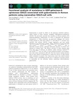
unit due to hydrophobic interactions (Fig. 6).
On the other hand, our report provides several indica-
tions for a functional proximity of chlorophyll and at
least one heme in the cytochrome b
6
subunit: (a) the red-
shift of the chlorophyll peak at 671 nm simultaneously
with the reduction of the b-type heme suggests a short
distance between these two components; (b) the previ-
ously observed extremely short fluorescence lifetime of
this chlorophyll [15] suggests a binding to a specific
pocket in the cyt b
6
f complex where a heme or an amino
acid side chain is able to quench its excitated state;
this may protect the protein from oxidative damage.
If we assume a very similar orientation of the Syn-
echocystis chlorophyll as in the crystal structure, which
is supported by our LD-data, the b-type heme closest
to the tetrapyrrole ring of the chlorophyll is cyt b
h
(Fig. 6). According to the crystal structure, the center-
to-center distance of the tetrapyrrole ring of the chlo-
rophyll to the heme is approximately 16.7 A
˚
, which is
sufficiently small to enable charge transfer between
both ring systems.
Besides chlorophyll, a carotenoid is associated with
the isolated cyt b
6
f complex of Synechocystis [40]
in substoichiometric amounts. The ratio of about
0.55–0.77 carotenoids per monomeric cyt b
6
f complex
determined in this report is in agreement with values
reported for other mesophilic organisms like spinach
[16] and Chlamydomonas reinhardtii [16,33], which tend
to loose some pigment upon isolation and purification.
The presence of a carotenoid within the cyt b
6
f com-
plex has been confirmed by the X-ray structure: in the
case of the cyanobacterium Mastigocladus laminosus,a
b-carotene is sandwiched between the a-helix of PetL
and PetM [5] with one hexameric ring extending towards
helix A of the PetB (subunit b
6
) (Fig. 6). Although
helices of the small subunits have been assigned differ-
ently in the Chlamydomonas structure, the localization
of the carotene is identical in both structures.
Similar to chlorophyll, after a mild dissociation of
the cyt b
6
f complex from Synechocystis sp. PCC 6803,
the carotenoid was found to be exclusively associated
with the cyt b
6
subunit [18]. A short distance between
the carotenoid and b-hemes of the cyt b
6
subunit is also
suggested by the red-shift of the carotene peaks in the
CrtO-less mutant simultaneously with the reduction of
the b-hemes. Considering the cyt b
6
f structural model
and assuming again a similar location in Synechocystis
as in Chlamydomonas and Mastigocladus, the most
Fig. 6. Structure of the isolated cytochrome b
6
subunit from Masti-
gocladus laminosus [5] with bound cofactors. Red, heme; green,
chlorophyll; orange, carotenoid. The chlorophyll molecule is in close
proximity to the heme b
H
, while the carotenoid is close in space to
the covalently bound heme c
x
.
S O. Wenk et al. Pigments in b
6
f complex
FEBS Journal 272 (2005) 582–592 ª 2005 FEBS 587
probable functional interaction occurs between one ring
of the carotenoid and the stroma-exposed heme cyt c
x
with an approximate ring center-to-center distance of
11.2 A
˚
.
The carotenoid in Synechocystis is echinenone, the
content of which in the cells is smaller compared with
b-carotene, zeaxanthin and myxoxanthophyll. This
indicates that the binding of echinenone to the com-
plex is rather specific.
Results obtained with the CrtO-less strain suggest
that the carotenoid binding site in the cyt b
6
f complex
of Synechocystis prefers a carotenoid with a polar ¼O
(echinenone) or –OH (monohydroxy-b-carotene) group
on one side of the carotenoid (the other ring of these
two carotenoids is identical to that of b-carotene). This
is in contrast to cyt b
6
f complexes from spinach,
Chlamydomonas reinhardtii and Mastigocladus lamino-
sus, which prefer b-carotene [16], a carotenoid that
lacks polar ¼O or –OH groups on both sides of the
molecule. However, these three cyt b
6
f complexes and
the CrtO-less mutant of Synechocystis seem to prefer
9-cis isomers, which points to significant similarities of
the carotene binding pocket in all organisms. This
9-cis conformation is also in line with a recent HPLC
and Raman characterization of b-carotene in the
cyt b
6
f complex from spinach [41], but is in contrast to
the interpretation of another Raman characterization
[42]. In the latter, however, the choice of the Raman
frequency used to distinguish both types of conforma-
tions was questioned [41].
Probably due to sterical constraints of the binding
pocket, echinenone apparently cannot easily be
replaced by other carotenoids. This suggests a struc-
tural role of carotenoid(s) in the cyt b
6
f complex, per-
haps similar to the situation in the light harvesting
complex of higher plants [21] or the D1 protein of
photosystem II [24]. Such a plant-specific function is
also suggested by the high resolution 3D structure of
the cyt b
6
f complex. A possible function for the caro-
tenoid has not yet been firmly established. While it
was suggested that it prevents the generation of singlet
oxygen by photoexcited Chl a [16], a triplet energy
transfer from chlorophyll to carotenoid did not occur
at 77 K in cyt b
6
f from Synechocystis [15]. Also, no
singlet energy transfer from the carotenoid to chloro-
phyll has been observed by fluorescence measurements
[15]. These observations are in line with the structural
model showing an approximate distance of 14 A
˚
between both pigments, which is too far for triplet and
singlet energy transfer. However, as the edge of the
chlorophyll is exposed to the lipid phase, the presence
of additional carotenoids interacting with the chloro-
phyll in situ cannot be ruled out [5].
In combination with the structural data, the effects
observed in this communication could be interpreted
in two different scenarios. (a) Indication for a signal
transduction chain: as these pigments are not found in
the closely related cyt bc
1
complex of the respiratory
chain, they may represent a plant-specific, structure-
dominated principle. Due to their localization and ori-
entation, they could interact with other components of
the photosynthetic apparatus such as PS1 or a kinase.
In this case, chlorophyll would act as a sensor that
connects the interacting partner with the Q
o
-site, while
the carotenoid might have a similar role at the Q
i
-site
[5]. For the chlorophyll, an absorption bandshift was
observed upon binding of inhibitors (stigmatellin or
2,5-dibromo-6-methyl-3-isopropyl-1,4-benzochinon)
4
to
the Q
o
-site in a cyt b
6
f complex isolated from spinach,
which supports this hypothesis from the reverse direc-
tion (C. Klughammer, unpublished result). (b) Indica-
tion for protein reorganization: during electron
transfer, the strong local electric field around the
b-type cytochrome causes an electrochromic shift of
the nearby pigments which in turn could indicate a pro-
tein reorganization of the complex, i.e. the observed
shift is caused by protein relaxation. Such an effect
has been reported for other proteins [43].
Irrespective of the physiological role of both pig-
ments, these observations also indicate their potential
usefulness as ‘natural’ indicators for redox-induced
changes in the cyt b
6
f complex.
In summary, this report shows that the two pig-
ments found in the cyt b
6
f-complex, chlorophyll and
echinenone, have a specific structural and possibly also
a functional impact. While the results obtained with
the echinenone-less mutant indicate a high selectivity
of the carotene binding pocket due to specific sterical
constraints, the correlation of both pigments with
redox changes of the b-type cytochrome on the cyto-
plasmic ⁄ stromal side suggests the possibility of func-
tional interaction. These results should stimulate
further experiments, for which the available 3D struc-
ture of the cyt b
6
f complex in combination with site-
directed mutagenesis of pigment-stabilizing residues is
an excellent basis.
Experimental procedures
Synechocystis sp. PCC 6803 strains and growth
conditions
For the isolation of cyt b
6
f complexes from Synechocystis sp.
PCC 6803, a PS1-less mutant strain was used, in which an
internal deletion in the psaAB operon inactivates both genes
[44]. Cells of this strain were grown photoheterotrophically
Pigments in b
6
f complex S O. Wenk et al.
588 FEBS Journal 272 (2005) 582–592 ª 2005 FEBS
at 30 °C in standard BG-11 medium enriched with 30 mm
glucose and at an incident light intensity of 5 lmol pho-
tonsÆm
)2
Æs
)1
in a 25 L foil photobioreactor (Bioengineering,
AG, Wald, Switzerland)
5
. Cultures were harvested after
3 days [at an attenuance (D)
6
at 730 nm of about 1.0] and con-
centrated by a hollow fiber concentration device (Amicon
7
DC-10 L, Millipore GmbH, Schwalbach, Germany) to 1 L,
followed by centrifugation at 6000 g for 10 min. Thylakoid
membranes were prepared according to [45], with the excep-
tion that the cells were disrupted in a glass bead mill (model
KDLA, Dyno-Mill, Bachofen AG, Basel, Switzerland)
8
at
0 °C for 30 s, using 0.5 mm glass beads. After centrifugation
at 200 000 g and 4 °C for 40 min the thylakoid membrane
pellet was resuspended in a buffer containing 20 mm
Mes ⁄ NaOH (pH 6.5), 10 mm CaCl
2
,10mm MgCl
2
, 0.5 m
mannitol, 20% (v ⁄ v) glycerol and protease inhibitors (10 lm
tosyllysyl chloromethylketone, 100 lm phenylmethylsulfo-
nylfluoride) yielding a final chlorophyll concentration of
0.2–0.4 mgÆmL
)1
. Thylakoids were frozen in liquid N
2
and
subsequently stored at )70 °C.
Generation of a PS1-less/CrtO-less mutant
of Synechocystis
The PS1-less ⁄ CrtO-less mutant was generated by transforma-
tion of the PS1-less mutant with the plasmid pTRCRT-O
kindly provided by G. Sandmann (Johann Wolfgang von
Goethe University, Frankfurt ⁄ Main Germany)
9
. This plasmid
contained a copy of the Synechocystis crtO gene that was
interrupted by a kanamycin cassette [30]. For transforma-
tion, the PS1-less strain of Synechocystis was grown to
D
730
¼ 0.5, pelleted (5000 g, 5 min, room temperature), and
resuspended in BG-11 medium to a D
730
¼ 2.5. This suspen-
sion (400 lL) was incubated with 0.3–3 lg plasmid DNA for
six hours at 30 °C under illumination at 5 l mol photonsÆ
m
)2
Æs
)1
. Of these cells, 200 lL were plated on nitrocellulose
filters on top of BG-11 plates containing 30 mm glucose;
after 18 h they were transferred to BG-11 plates containing
5 lgÆmL
)1
kanamycin. Colonies emerged after two weeks;
they were transferred to new plates every 2–4 days, increasing
the kanamycin concentration by 5–10 lgÆmL
)1
each time. The
maximal kanamycin concentration used was 50 lgÆmL
)1
.One
of the transformants was checked by PCR for complete
segregation and this segregated strain was used for further
analysis. As expected, this strain lacked echinenone accord-
ing to pigment analysis using reversed-phase HPLC [30].
Purification of the cyt b
6
f complex from
the PS1-less strain of Synechocystis
Unless specified otherwise, all following steps were
performed under dim light and at 6–8 °C. The isolated
membranes were first incubated with 0.1 mgÆmL
)1
RNase
and DNase (Boehringer, Ingleheim, Germany)
10
at 20 °C for
18 min; upon addition of 0.05% (w ⁄ v) b-dodecyl maltoside
(b-DM)
11
, the mixture was incubated for another 2 min.
After centrifugation (200 000 g,4°C, 40 min) the pelleted
membranes were resuspended in buffer [20 mm Mes ⁄ NaOH
(pH 6.5), 10 mm CaCl
2
,10mm MgCl
2
, 0.5 m mannitol,
20% (v ⁄ v) glycerol], and diluted to a chlorophyll concentra-
tion of 150 lgÆ mL
)1
.
Membrane proteins were extracted by incubation with
1% (w ⁄ v) b-DM for 30 min at 20 °C. After centrifugation
(200 000 g,4°C, 40 min) and 1.5-fold dilution with a high-
salt buffer [20 mm Mes ⁄ NaOH, pH 6.5, 10 mm CaCl
2
,
10 mm MgCl
2
,3m ammonium sulfate, 0.02% (w ⁄ v) b-DM]
the supernatant was loaded onto a hydrophobic interaction
column (POROS 20 BU; Applied Biosystems, Foster City,
CA, USA)
12
that was run at a flow rate of 7 mLÆmin
)1
at
10 °C. Upon applying a decreasing ammonium sulfate gra-
dient, the cyt b
6
f complex eluted at a concentration of
about 1 m ammonium sulfate. The cyt b
6
f containing frac-
tions were concentrated and dialyzed against a low salt buf-
fer [20 mm Mes ⁄ NaOH (pH 6.5), 10 mm CaCl
2
,10mm
MgCl
2
, 0.02% (w ⁄ v) b-DM] before purifying them further
on an anion exchange column (Uno Q6, Bio-Rad Labora-
tories, Munich, Germany)
13
. Applying a MgSO
4
gradient at
a flow rate of 4 mLÆmin
)1
, the cyt b
6
f complex eluted at
about 15 mm MgSO
4
and was stored at )70 °C.
The presence of all expected subunits was confirmed
by SDS ⁄ PAGE, immunoblotting (using antibodies against
PetA, PetB, PetC, and PetD) and EPR-measurements (to
demonstrate the Rieske protein).
Pigment analysis
Pigment analysis of thylakoid membranes preparations and
purified cyt b
6
f complexes was carried out by reversed-phase
HPLC. Samples were diluted 10-fold with ice-cold acetone,
vortexed briefly and centrifuged (12 000 g,4°C, 5 min).
The supernatant containing the pigments was filtered
through a membrane (Spartan, 0.45 lm, Schleicher und
Schuell GmbH, Dassel, Germany)
14
and injected onto a RP
HPLC column (Spherisorb
15
ODS 2, Crom); this column had
been equilibrated using a hydrophilic solution RP-A [38.5%
(v ⁄ v) acetone, 46.5% (v ⁄ v) methanol, 5% (v ⁄ v) water and
10% (v ⁄ v) PIC A (5 mm tetrabutylammonium sulfate,
Waters, Milford, MA, USA)
16
]. Pigments were eluted by three
linear gradients with increasing hydrophobicity: 0 fi 20%,
20 fi 50%, 50 fi 100% solution RP-B [100% (v ⁄ v) ethylac-
etate], at an average flow rate of 0.7 mLÆmin
)1
. Pigments
were analyzed online by a Photodiode Array Detector 966
(Waters) from 350 nm to 700 nm and identified ⁄ quantified
by comparison with standards [46].
Alternatively, pigment analysis was performed according
to [47]. For this procedure, pigments were extracted with
80% acetone, centrifuged and filtered, and loaded on a RP
HPLC-column (Spherisorb C18), which was equilibrated in
S O. Wenk et al. Pigments in b
6
f complex
FEBS Journal 272 (2005) 582–592 ª 2005 FEBS 589
buffer A [85% acetonitrile, 13.5% methanol, 1.5% 0.2 m
Tris ⁄ HCl (pH 8.0)]. The column was run for 30 min at
1mLÆmin
)1
in buffer A, after which a 5 min linear gradient
(0–100%) was applied using buffer B (83.3% methanol,
16.7% n-hexane); subsequently the column was run for
another 30 min in buffer B. The HPLC system was
equipped with a diode-array optical absorption spectropho-
tometer, which allowed identification of the peaks in the
chromatogram by their absorption spectra.
Spectroscopic methods
All spectroscopic measurements of the cyt b
6
f complex were
carried out in 20 mm Mes ⁄ HCl (pH 6.5), 10 m m MgCl
2
,
10 mm CaCl
2
, and 0.03% b-DM. UV-Vis absorbance spectra
at room temperature were recorded on a Beckman
17
DU 7400
spectrophotometer (Beckman Coulter GmbH, Krefeld,
Germany), with a spectral bandwidth of 1.2 nm. For redox
measurements of the cytochromes, the air-oxidized cyt b
6
f
samples were oxidized with 100 lm ferricyanide or reduced
with 20 mm ascorbate (for cyt f) or dithionite (for cyt b
6
).
Absorbance and fluorescence spectroscopy at 4 K and 77 K
were performed according to [15]. For these measurements,
the b-DM concentration was increased to 0.07% and glycerol
was added to a final concentration of 75% (v ⁄ v). LD spectro-
scopy was performed at 77 K as described in [37], using a
two-dimensionally squeezed gelatin gel. The samples were
diluted in molten 6.4% (w ⁄ v) gelatin at 32 °C and oriented by
squeezing the12.5 · 12.5 mmpolymerizedgel intwo perpendi-
cular directions to the 10 · 10 mm dimensions of the cuvette.
EPR spectra were recorded at the Se
´
ction de Bioe
´
nerge
´
-
tique, CEA-Saclay, France, on a Bruker EPR200 machine
equipped with a helium cryostat from Oxford Instruments
GmbH (Wiesbaden, Germany)
18
.
Chemically induced spectral changes at room temperature
were recorded with a time resolving multichannel spectro-
photometer based on a Zeiss
19
spectral sensor module (MCS-
VIS; Carl Zeiss AG, Oberkochen, Germany) equipped with
a photo diode array for the wavelength region 360–780 nm
and a spectral resolution of 3 nm (tec5 Sensorik und Sys-
temtechnik GmbH
20
, Oberursel, Germany). The continuous
measuring light was guided by a single optical fiber from a
halogen lamp to a sample compartment with a glass cuvette
with 1 cm optical path length and with stirring. The
transmitted light was focussed on a second fiber, which was
connected to the spectral sensor module. Spectra were
recorded by computer with a time resolution of 80 ms.
Transmission changes DT were calculated by dividing the
spectra by a reference spectrum recorded immediately before
the experiment and DA was calculated by the equation:
DA ¼ÀlogfðDT=T
1
Þþ1g
In order to selectively observe redox changes of b-type cyto-
chromes, a sample was fully prereduced by 0.5 mm ascorbate
and 0.5 mm dithionite, and rapidly stirred in an open cuvette.
After consumption of the dithionite by oxygen a slow reoxi-
dation of the b cytochromes occurred and the absorption
changes were recorded. A further oxidation of cytochrome f
was prevented by the presence of ascorbate. Therefore, the
differential absorption change DA(575 nm) ) DA(564 nm)
can be directly taken as a measure of cytochrome b oxida-
tion. This signal was compared to the differential absorption
change DA(665 nm) ) DA(676 nm), representing the absorp-
tion changes at the maximum and minimum of the spectrum
of the chlorophyll bandshift spectrum, respectively.
Acknowledgements
We are grateful to thank Dr A. Seidler for his help
with the ESR-measurements and Claudia Ko
¨
nig for
excellent technical assistance. We also thank Melanie
Ambill and Dr S. Berry for critical discussions. The
financial support by the DFG (SFB480, project C1,
MR) and HFSP (DS, SOW, MR and WFJV) is also
gratefully acknowledged. FLdW and JPD were suppor-
ted by a grant from the Netherlands Foundation for
Scientific Research (NWO) via the Foundation for Life
and Earth Sciences (ALW).
References
1 Hauska G & Bu
¨
ttner M (1997) The cytochrome
b
6
f ⁄ bc
1
-complexes. In Bioenergetics (Gra
¨
ber P &
Milazzo G, eds), pp. 389–417. Birkha
¨
user-Verlag, Basel,
Switzerland.
2 Soriano GM, Ponamarev MV, Carrell CJ, Xia D,
Smith JL & Cramer WA (1999) Comparison of the
cytochrome bc1 complex with the anticipated structure
of the cytochrome b6f complex: Le plus ca change le
plus c’est la meme chose. J Bioenerg Biomembr 31, 201–
213.
3 Mitchell P (1966) Chemiosmotic coupling in oxidative
and photosynthetic phosphorylation. Biol Rev 41, 445–
602
21
.
4 Kallas T (1994) The cytochrome b
6
f complex. In The
Molecular Biology of Cyanobacteria (Bryant DA, ed.),
pp. 259–317. Kluwer Academic Publishers, Dordrecht,
the Netherlands.
5 Kurisu G, Zhang H, Smith JL & Cramer WA (2003)
Structure of the cytochrome b6f complex of oxygenic
photosynthesis: tuning the cavity. Science 302, 1009–
1014.
6 Berthold DA, Schmidt CA & Malkin R (1995) The
deletion of petG in Chlamydomonas reinhardtii disrupts
the cytochrome bf complex. J Biol Chem 270, 29293–
29298.
7 Takahashi Y, Rahire M, Breyton C, Popot J-L, Joliot P
& Rochaix JD (1996) The chloroplast ycf7 (petL) open
reading frame in Chlamydomonas reinhardtii encodes a
Pigments in b
6
f complex S O. Wenk et al.
590 FEBS Journal 272 (2005) 582–592 ª 2005 FEBS
small functionally important subunit of the cytochrome
b
6
f complex. EMBO J 15, 3498–3506.
8 Hager M, Biehler K, Illerhaus J, Ruf S & Bock R
(1999) Targeted inactivation of the smallest plastid
genome-encoded open reading frame reveals a novel
and essential subunit of the cytochrome b
6
f complex.
EMBO J 18, 5834–5842.
9 Hamel P, Olive J, Pierre Y, Wollman F-A & de Vitry C
(2000) A new subunit of cytochrome b
6
f complex under-
goes reversible phosphorylation upon state transition.
J Biol Chem 275, 17072–17079.
10 Whitelegge JP, Zhang H, Aguilera R, Taylor RM &
Cramer WA (2002) Full subunit coverage liquid chro-
matography electrospray ionization mass spectrometry
(LCMS+) of an oligomeric membrane protein: cyto-
chrome b(6)f complex from spinach and the cyanobacte-
rium Mastigocladus laminosus. Mol Cell Proteomics 1,
816–827.
11 Schneider D, Berry S, SeidlerA&Ro
¨
gner M (1998)
22
Evidence for a nonessential function of the small subu-
nit PetM in the cytochrome b
6
f complex of Synechocys-
tis PCC 6803. In Photosynthesis, Mechanisms and
Effects (Garab G & Puztai J, eds) Vol. III
23
, pp. 1545–
1548. Kluwer Academic Publishers, Dordrecht, the
Netherlands.
12 Schneider D, Berry S, Rich P, Seidler A & Ro
¨
gner M
(2001) A regulatory role of the PetM subunit in a
cyanobacterial cytochrome b6f complex. J Biol Chem
276, 16780–16785.
13 Bald D, Kruip J, Boekema EJ & Ro
¨
gner M (1992)
Structural investigations on Cyt b
6
f complex and PS I
complex from the cyanobacterium Synechocystis sp.
PCC 6803. In Research in Photosynthesis (Murata N,
ed.), pp. 629–632. Kluwer Academic Publishers,
Dordrecht, the Netherlands.
14 Pierre Y, Breyton C, Lemoine Y, Robert B, Vernotte C
& Popot J-L (1997) On the presence and role of a mole-
cule of chlorophyll a in the cytochrome b
6
f complex.
J Biol Chem 272, 21901–21908.
15 Peterman EJG, Wenk S-O, Pullerits T, Pa
˚
lsson L-O,
van Grondelle R, Dekker JP, Ro
¨
gner M & van Amer-
ongen H (1998) Fluorescence and absorption spectro-
scopy of the weakly fluorescent chlorophyll a in
cytochrome b
6
fofSynechocystis PCC 6803. Biophys J
75, 389–398.
16 Zhang H, Huang D & Cramer WA (1999) Stoichiome-
trically bound b-carotene in the cytochrome b
6
f complex
of oxigenic photosynthesis protects against oxygen dam-
age. J Biol Chem 274, 1581–1587.
17 Stroebel D, Choquet Y, Popot JL & Picot D (2003) An
atypical haem in the cytochrome b(6)f complex. Nature
426, 399–400.
18 Boronowsky U, Wenk S-O, Schneider D, Ja
¨
ger C &
Ro
¨
gner M (2001) Isolation of membrane protein subu-
nits in their native state: evidence for selective binding of
chlorophyll and carotenoid to the b
6
subunit of the cyto-
chrome b
6
f complex. Biochim Biophys Acta 1506, 55–66.
19 Bolychevtseva YV, Rakhimberdieva MG, Karapetyan
NV, Popov VI, Moskalenko AA & Kuznetsova NY
(1995) The development of carotenoid-deficient mem-
branes in plastids of barley seedlings treated with nor-
flurazon. J Photochem Photobiol B 27, 153–160.
20 Havaux M (1998) Carotenoids as membrane stabilizers
in chloroplasts. Trends Plant Sci 4, 147–151.
21 Ku
¨
hlbrandt W, Wang DN & Fujiyoshi Y (1994) Atomic
model of plant light-harvesting complex by electron
crystallography. Nature 367, 614–621.
22 Paulsen H (1995) Chlorophyll a ⁄ b proteins. Photochem
Photobiol 62, 367–382.
23 Moskalenko AA & Karapetyan NV (1996) Structural
role of carotenoids in photosynthetic membranes.
Z Naturforsch 51c, 763–771.
24 Trebst A & Depka B (1997) Role of carotene in the
rapid turnover and assembly of photosystem II in Chla-
mydomonas reinhardtii. FEBS Lett 400, 359–362.
25 He Q & Vermaas W (1998) Chlorophyll a availability
affects psbA translation and D1 precursor processing in
vivo in Synechocystis sp. PCC 6803. Proc Natl Acad Sci
USA 95, 5830–5835.
26 Williams JGK (1988) Construction of specific mutations
in photosystem II photosynthetic reaction center by gen-
etic engineering methods in Synechocystis 6803. Methods
Enzymol 167, 766–778.
27 Schoepp B, Chabaud E, Breyton C, Vermeglio A &
Popot J-L (2000) On the spatial organization of hemes
and chlorophyll in cytochrome b
6
f. J Biol Chem 275,
5275–5283.
28 Kwa SLS, Vo
¨
lker S, Tilly NT, van Grondelle R &
Dekker JP (1994) Polarized site-selection spectroscopy
of chlorophyll a in detergent. Photochem Photobiol 59,
219–228.
29 Lagarde D, Beuf L & Vermaas W (2000) Increased
production of zeaxanthin and other pigments by
application of genetic engineering techniques to
Synechocystis sp. strain PCC 6803. Appl Environ
Microbiol 66, 64–72.
30 Ferna
´
ndez-Gonza
´
lez B, Sandmann G & Vioque A
(1997) A new type of asymmetrically acting b-carotene
ketolase is required for the synthesis of echinenone in
the cyanobacterium Synechocystis sp. PCC 6803. J Biol
Chem 272, 9728–9733.
31 Koyama Y, Takii T, Saiki K & Tsukida K (1983) Con-
figuration of the carotenoid in the reaction center of
photosynthetic bacteria. 2. Comparison of the Reso-
nance Raman lines of the reaction center with those of
the 14 different cis-trans isomers of beta carotene.
Photobiochem Photobiophys
24
5, 139–150.
32 Renge I, van Grondelle R & Dekker JP (1996) Matrix
and temperature effects on absorption spectra of
b-carotene and pheophytin a in solution and in green
S O. Wenk et al. Pigments in b
6
f complex
FEBS Journal 272 (2005) 582–592 ª 2005 FEBS 591
plant photosystem II. J Photochem Photobiol A 96,
109–122.
33 Pierre Y, Breyton C, Kramer D & Popot J-L (1995)
Purification and characterization of the cytochrome b
6
f
complex from Chlamydomonas reinhardtii. J Biol Chem
270, 29342–29349.
34 Ehling-Schulz M, Bilger W & Scherer S (1997) UV-B-
induced synthesis of photoprotective pigments and
extracellular polysaccharides in the terrestrial cyanobac-
terium Nostoc commune. J Bacteriol 179, 1940–1945.
35 Ponamarev MV, Schlarb BG, Howe CJ, Carrell CJ,
Smith JL, Bendall DS & Cramer WA (2000) Trypto-
phan-heme pi-electrostatic interactions in cytochrome f
of oxygenic photosynthesis. Biochemistry 39, 5971–5976.
36 Reddy KS, Angiolillo PJ, Wright WW, Laberge M &
Vanderkooi JM (1996) Spectral splitting in the alpha
(Q
0,0
) absorption band of ferrous cytochrome c and
other heme proteins. Biochemistry 35, 12820–12830.
37 Groot M-L, Frese RN, de Weerd FL, Bromek K,
Pettersson A
˚
, Peterman EJG, van Stokkum IHM, van
Grondelle R & Dekker JP (1999) Spectroscopic proper-
ties of the CP43 core antenna protein of photosystem
II. Biophys J 77, 328–340.
38 Van Amerongen H, Vasmel H & van Grondelle R
(1988) Linear dichroism of chlorosomes from Chloro-
flexus aurantiacus in compressed gels and electric fields.
Biophys J 54, 65–76.
39 Poggese C, Polverino de Laureto P, Giacometti GM,
Rigoni F & Barbato R (1997) Cytochrome b
6
⁄ f
complex from the cyanobacterium Synechocystis 6803:
evidence of dimeric organization and identification of
chlorophyll-binding subunit. FEBS Lett 414, 585–589.
40 Wenk S-O, Boronowsky U, Peterman EJG, Ja
¨
ger C,
van Amerongen H, Dekker JP & Ro
¨
gner M (1998)
Isolation and characterisation of the cytochrome b
6
f
from the cyanobacterium Synechocystis PCC 6803. In
Photosynthesis, Mechanisms and Effects (Garab G &
Puztai J, eds) Vol. III, pp. 1537–1540.
25
Kluwer Academic
Publishers, Dordrecht, the Netherlands.
41 Yan J, Liu Y, Mao D, Li L & Kuang T (2001) The pre-
sence of 9-cis-beta-carotene in cytochrome b(6)f com-
plex from spinach. Biochim Biophys Acta 1506, 182–188.
42 Picaud T, Le Moigne CL, Gomez de Gracia A & Des-
bois A (2001) Soret-excited Raman spectroscopy of the
spinach cytochrome b6f complex. Structures of the
b- and c-type hemes, chlorophyll a, and beta-carotene.
Biochemistry 40 , 7309–7317.
43 Dashdorj N, Xu W, Martinsson P, Chitnis PR &
Savikhin S (2004)
26
Electrochromic shift of chlorophyll
absorption in photosystem I from Synechocystis sp.
PCC 6803: a probe of optical and dielectric properties
around the secondary electron acceptor. Biophys J 86,
3121–3130.
44 Shen G, Boussiba S & Vermaas WFJ (1993) Synecho-
cystis sp. PCC 6803 strains lacking photosystem I and
phycobilisome function. Plant Cell 5, 1853–1863.
45 Ro
¨
gner M, Nixon PJ & Diner BA (1990) Purification
and characterization of photosytem II core complexes
from wild-type and phycocyanin-deficient strains of the
cyanobacterium Synechocystis PCC 6803. J Biol Chem
265, 6189–6196.
46 Young A & Britton G (1993) Carotenoids in Photosynth-
esis. Chapman & Hall, London.
47 Peterman EJG, Gradinaru CC, Calkoen F, Borst JC,
van Grondelle R & van Amerongen H (1997) Xantho-
phylls in light-harvesting complex II of higher plants:
light harvesting and triplet quenching. Biochemistry 36,
12208–12215.
Pigments in b
6
f complex S O. Wenk et al.
592 FEBS Journal 272 (2005) 582–592 ª 2005 FEBS