Designing a Virtual Reality Model for Aesthetic Surgery docx
Bạn đang xem bản rút gọn của tài liệu. Xem và tải ngay bản đầy đủ của tài liệu tại đây (633.86 KB, 5 trang )
Cosmetic
Designing a Virtual Reality Model for
Aesthetic Surgery
Darren M. Smith, M.D., Sherrell J. Aston, M.D., Court B. Cutting, M.D., Aaron Oliker, M.S., and
Jeffrey Weinzweig, M.D.
Providence, R.I., New York, N.Y., and Burlington, Mass.
Background: Aesthetic surgery deals in
large part with the manipulation of soft-
tissue structures that are not amenable to
visualization by standard technologies. As a
result, accurate three-dimensional depic-
tions of relevant surgical anatomy have yet
to be developed. This study presents a
method for the creation of detailed virtual
reality models of anatomy relevant to aes-
thetic surgery.
Methods: Two-dimensional histologic sec-
tions of a cadaver from the National Library
of Medicine’s Visible Human Project were
imported into Alias’s Maya, a computer
modeling and animation software package.
These two-dimensional data were then
“stacked” as a series of vertical planes. Rel-
evant anatomy was outlined in cross-section
on each two-dimensional section, and the
resulting outlines were used to generate
three-dimensional representations of the
structures in Maya.
Results: A detailed and accurate three-
dimensional model of the soft tissues ger-
mane to aesthetic surgery was created. This
model is optimized for use in surgical ani-
mation and can be modified for use in sur-
gical simulators currently being developed.
Conclusions: A model of facial anatomy
viewable from any angle in three-dimen-
sional space was developed. The model has
applications in medical education and, with
future work, could play a role in surgi-
cal planning. This study emphasizes the role
of three-dimensionalization of the soft tis-
sues of the face in the evolution of aesthetic
surgery. (Plast. Reconstr. Surg. 116: 893,
2005.)
The key soft-tissue anatomical players in aes-
thetic surgery of the face exist in three-
dimensional relationships difficult to visualize
by conventional means. Two-dimensional mo-
dalities are necessarily inferior in an analysis of
three-dimensional structures, and standard
three-dimensional technologies (e.g., three-
dimensional computed tomography), al-
though efficacious for skeletal imaging, do not
adequately portray facial soft tissues. Three-
dimensional surface imaging technologies
(e.g., laser scanning) are valuable, but provide
images that are only “skin deep.” This article
presents a method for constructing a three-
dimensional virtual reality model of the soft
tissues of the face that lie between the skin and
bone. The models may prove to be a valuable
resource for surgical education, and may even-
tually play a role in surgical planning.
M
ATERIALS AND
M
ETHODS
Basic soft-tissue anatomical data were de-
rived from the National Library of Medicine’s
Visible Human Project (U.S. National Library
of Medicine, Bethesda, Md.).
1
The data were in
the form of hematoxylin and eosin–stained ax-
ial histologic cuts of a female cadaver sectioned
From Brown Medical School, the Institute of Reconstructive Plastic Surgery, New York University Medical Center, the Plastic Surgery
Department, Manhattan Eye, Ear, and Throat Hospital, and the Department of Plastic Surgery, Lahey Clinic Medical Center. Received for
publication August 3, 2004; revised December 16, 2004.
DOI: 10.1097/01.prs.0000176900.62853.b3
893
at 333-
m intervals. These sections were digi-
tized and distributed by the National Library of
Medicine. Each image file was cropped to re-
move frame borders in Adobe’s Photoshop 5.5
(Adobe Systems, Inc., San Jose, Calif.). The
two-dimensional images were then three-
dimensionalized using Alias’s Maya 4.0 (Alias
Systems Corp., Toronto, Ontario, Canada) ac-
cording to the following protocol. First, a series
of horizontal planes was created with the same
height (x axis) and width (y axis) proportions
as the data from the Visible Human Project.
These planes were vertically aligned (along the
z axis) at 1-cm intervals. Two-dimensional sec-
tions from the Visible Human Project were
selected at intervals of 1 cm and subsequently
mapped onto the corresponding planes cre-
ated in three-dimensional space in Maya (Fig.
1). These planes at 1-cm intervals served as
“reference slices” that were used to identify
anatomy of interest. As structures to be mod-
eled were identified on these reference slices,
increased depth resolution (z axis) was often
required to define anatomical detail. In such
cases, additional planes were created in Maya
and corresponding images from the Visible
Human Project were imported. Anatomical
structures of interest were identified and out-
lined on these xy planes using Maya’s EP Curve
tool. The EP curves generated from the out-
lined structures were connected along the z
axis using Maya’s Loft tool. The surfaces thus
generated served as primary three-dimensional
representations of the soft-tissue anatomy of
interest (Fig. 2).
These soft-tissue models were modified to fit
a three-dimensional model of a female skull,
which had previously been created by the In-
stitute of Reconstructive Plastic Surgery Virtual
Surgery Laboratory as an average of several
female skull three-dimensional computed to-
mographic scans.
2,3
These modified secondary
soft-tissue models were then manipulated to
more clearly demonstrate anatomical relation-
ships in an effort to minimize artifacts inherent
in the method just described for translation of
a human cadaver into a three-dimensional
model. These manipulations were conducted
with great care to adhere to anatomical reality,
using cadaveric dissection and literature review
to ensure faithfulness to reality.
4–11
Our final
soft-tissue models, now fit to a skull model,
were the result. Some structures (primarily
neurovascular) obliterated during sectioning
or digitalization of the Visible Human Project
data were created de novo in Maya, again
guided by cadaveric dissection and literature
review. These structures were then superim-
posed on the soft-tissue and skull models to
complete our representation of relevant head
and neck anatomy.
A skin model of the young female head was
purchased commercially from the Viewpoint
Corporation, and the underlying soft-tissue
models were manipulated within the limits of
normal anatomy to conform to this skin shape.
Thus, a model of the female head was created
with deep tissues and “matching” overlying
skin. Finally, the models were texture-mapped
with a combination of photographs enhanced
in Adobe Photoshop 7.0 and materials de-
signed in Maya.
R
ESULTS
A virtual reality model of surgical superficial
facial anatomy was created. Included in this
model are the superficial musculoaponeurotic
system (SMAS), facial musculature, nerves,
blood vessels, and fatty tissue most relevant to
aesthetic surgery. These structures exist in vir-
tual three-dimensional space such that they
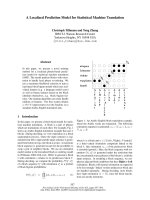
F
IG
. 1. To showcase the utility of Maya as an environment
for viewing the Visible Human Project data, a series of two-
dimensional planes, each mapped with a serial section from
the Visible Human Project data set, is shown. The planes have
been positioned in three-dimensional space with Maya. The
blue background represents the compound in which the
cadaver was suspended for sectioning. Note that to further
emphasize the versatility of this visualization technique, a
segment of the stacked slices has been removed. To further
orient the viewer, a parasagittal section of the nose is circled
in red, and to the right of this stack of Visible Human Project
sections, a schematic is shown. A number of individual planes
(left) are stacked close together to give the appearance of a
solid cube from the Visible Human Project. The green por-
tion in the planes and cube on the right represents the
segment removed from the stack of Visible Human Project
slices at left.
894
PLASTIC AND RECONSTRUCTIVE SURGERY
, September 1, 2005
can be rotated and viewed from any angle.
Individual structures can be viewed either in
isolation or in relation to one another. Any
structure may be highlighted or made com-
pletely or partially transparent to aid in the
illustration of a specific teaching point. The
model can be used for the illustration of any
surgical technique or problem involving the
depicted facial anatomy (Fig. 3).
By constructing the models with an eye to-
ward minimizing data density while maintain-
ing anatomical detail, we sought to produce
three-dimensional meshes that could easily be
manipulated or animated in Maya. The result-
ing models are thus Љlight,Љ in that they contain
relatively few data points for their high level of
anatomical detail.
D
ISCUSSION
Three-dimensional imaging has become
an integral part of the practice of craniofa-
cial surgery. Early work by Marsh et al. dis-
cusses three-dimensional computed tomog-
raphy and its use as a method of clarifying
the patient’s skeletal anatomy.
12,13
Cutting et
al. have described applications of three-
dimensional computed tomographic scan-
ning to craniofacial surgical planning as they
used virtual reality methods to intraopera-
tively track bone fragment movement to a
numerically optimized position.
14
Three-dimensional imaging is not limited to
skeletal anatomy. To name a few examples
from a wide selection, Nkenke et al. have used
three-dimensional surface imaging for exoph-
F
IG
. 2. This image illustrates the modeling process. (Above, left) One plane of those stacked in Figure
1 is viewed in isolation, with an EP curve (green) outlining the zygomaticus major muscle in horizontal
section. (Above, right) Texture maps are removed from this view, highlighting two EP curves (white,
representing the zygomaticus major as outlined on a more superior slice; and green, the outline of this
muscle as seen on the plane in Fig. 1). The vertical distance between the two curves is emphasized by
the blue arrow.(Below, left) A mesh (green) is created by ЉloftingЉ from superficial tracing to inferior
tracing. (Below, right) The texture map is again visible in this view; the mesh represents the beginnings
of the three-dimensional zygomaticus major model.
Vol. 116, No. 3 /
DESIGNING A VIRTUAL REALITY MODEL
895
thalmometry, Ferrario et al. have applied
three-dimensional surface scanning to the
analysis of facial morphology in ectodermal
dysplasia patients, and Ji et al. have used three-
dimensional surface scanning for the assess-
ment of facial tissue expansion.
15–17
In previous studies, we applied three-
dimensional imaging to soft-tissue structures
when we designed virtual reality animations to
teach cleft palate repair techniques and devel-
oped animations that illustrate the biomechan-
ics of eustachian tube dilation as it relates to
cleft palate repair.
18–20
The obvious difference
between these applications of three-dimen-
sional imaging and those of the skin and bone
discussed above is that many of the tissues key
to cleft surgery and eustachian tube biome-
chanics elude scanning by computed tomogra-
phy and surface digitization modalities alike.
As such, we developed a method to partially
hand-build three-dimensional models of rele-
vant anatomy as detailed in a previous study.
According to this protocol, tracings of histo-
logic sections were made in Adobe Photoshop
and essentially stacked using software devel-
oped by Dr. Cutting.
20
This technique repre-
sents the origin of the system described in the
Materials and Methods section of this article
for creation of the soft-tissue models in this
project.
The models of superficial facial anatomy de-
veloped in this project are intended to serve as
a three-dimensional atlas of the anatomy ger-
mane to aesthetic surgery of the face. Although
these models can be viewed from any angle
and made selectively transparent to illustrate
anatomical relationships difficult to appreciate
with other media, their greatest value lies in
their suitability for use in various emerging
teaching technologies. Examples of these tech-
nologies include three-dimensional anima-
tions, such as those mentioned above for illus-
tration of cleft repair technique, and three-
dimensional surgical simulators, such as that
currently being developed by Cutting et al.
18,21
The models are relatively “light” in terms of the
number of data points they contain, so it is
practical to manipulate them as the need arises
in animations, and their polygonal mesh con-
struction renders them compatible with modi-
fication for use in surgical simulators.
As mentioned earlier, there are currently no
technologies available to directly visualize an
individual patient’s facial soft-tissue anatomy in
three dimensions. Although the system de-
scribed here is clearly not useful for evaluating
a specific patient’s anatomy, future applica-
tions may allow for the warping of the idealized
anatomical models described here to best-fit
landmarks of individual patients. For example,
if a three-dimensional model of a patient’s skin
is derived from a laser scan, known relation-
ships between skin landmarks and underlying
soft-tissue structures could be used to warp the
model of facial soft tissue described here to
approximate that of the individual patient.
Such technologies could provide clinicians
with a reasonable—albeit indirect— depiction
of an individual patient’s soft-tissue structure.
Moreover, because these models are compati-
ble with evolving surgical simulators, and as
nascent simulator technology matures, these
models represent the basis for the capacity to
illustrate planned surgery and to simulate post-
operative changes.
C
ONCLUSIONS
This article presents a three-dimensional
computer model of anatomy for aesthetic sur-
gery. The protocol for designing such a model
is also discussed. The model illustrates the use-
fulness of virtual reality in the teaching and
practice of aesthetic plastic surgery and can
serve as a component of surgical educational
and (eventually) planning systems currently
being developed.
Sherrell J. Aston, M.D.
728 Park Avenue
New York, N.Y. 10021
F
IG
. 3. A final rendering of many of the model’s compo-
nents. Note the SMAS (S), which has been partially resected
for clarity. The portion of the SMAS that is visible is sus-
pended by two hooks. Branches of the facial nerve are visible
emerging superior to the cut edge of the SMAS.
896
PLASTIC AND RECONSTRUCTIVE SURGERY
, September 1, 2005
REFERENCES
1. Peitgen, H. The Complete Visible Human: The Complete
High-Resolution Male and Female Datasets from the Visible
Human Project. New York: Springer-Verlag, 1998.
2. Cutting, C., Bookstein, F., Haddad, B., and Kim, D.
Spline-based approach for averaging 3D curves and
surfaces. In Proceedings of the Society of Photo-Op-
tical Instrumentation Engineers, 1993.
3. Cutting, C., Dean, D., Bookstein, F. L., et al. A three-
dimensional smooth surface analysis of untreated
Crouzon’s syndrome in the adult. J. Craniofac. Surg. 6:
444, 1995.
4. Aiache, A. The suborbicularis oculi fat pad: An ana-
tomic and clinical study. Plast. Reconstr. Surg. 107:
1602, 2001.
5. Aston, S. J. Platysma-SMAS cervicofacial rhytidoplasty.
Clin. Plast. Surg. 10: 507, 1983.
6. Baker, D. C., and Conley, J. Avoiding facial nerve in-
juries in rhytidectomy: Anatomical variations and pit-
falls. Plast. Reconstr. Surg. 64: 781, 1979.
7. Barton, F. E., Jr., and Gyimesi, I. M. Anatomy of the
nasolabial fold. Plast. Reconstr. Surg. 100: 1276, 1997.
8. Hamra, S. T. Composite rhytidectomy. Plast. Reconstr.
Surg. 90: 1, 1992.
9. Mendelson, B. C. Surgery of the superficial muscu-
loaponeurotic system: Principles of release, vectors,
and fixation. Plast. Reconstr. Surg. 109: 824, 2002.
10. Mitz, V, and Peyronie, M. The superficial musculo-apo-
neurotic system (SMAS) in the parotid and cheek
area. Plast. Reconstr. Surg. 58: 80, 1976.
11. Owsley, J. Q. Lifting the malar fat pad for correction of
prominent nasolabial folds. Plast. Reconstr. Surg. 91:
463, 1993.
12. Marsh, J. L., and Vannier, M. W. Surface imaging from
computerized tomographic scans. Surgery 94: 159, 1983.
13. Marsh, J. L., Vannier, M. W., and Stevens, W. G. Surface
reconstructions from computerized tomographic
scans for evaluation of malignant skull destruction.
Am. J. Surg. 148: 530, 1984.
14. Cutting, C., Grayson, B., McCarthy, J. G., et al. A virtual
reality system for bone fragment positioning in mul-
tisegment craniofacial surgical procedures. Plast. Re-
constr. Surg. 102: 2436, 1998.
15. Nkenke, E., Benz, M., Maier, T., et al. Relative en- and
exophthalmometry in zygomatic fractures comparing
optical non-contact, non-ionizing 3D imaging to the
Hertel instrument and computed tomography.
J. Craniomaxillofac. Surg. 31: 362, 2003.
16. Ferrario, V. F., Dellavia, C., Serrao, G., and Sforza,
C. Soft-tissue facial areas and volumes in individuals
with ectodermal dysplasia: A three-dimensional non-
invasive assessment. Am. J. Med. Genet. 126A: 253, 2004.
17. Ji, Y., Zhang, F., Schwartz, J., Stile, F., and Lineaweaver,
W. C. Assessment of facial tissue expansion with
three-dimensional digitizer scanning. J. Craniofac.
Surg. 13: 687, 2002.
18. Cutting, C., Oliker, A., Haring, J., Dayan, J., and Smith,
D. Use of three-dimensional computer graphic an-
imation to illustrate cleft lip and palate surgery. Com-
put. Aided Surg. 7: 326, 2002.
19. Cutting, C., LaRossa, D., Sommerlad, B., et al. Virtual
Surgery CD Set: Volume I: Unilateral Cleft. Volume II: Bi-
lateral Cleft.Volume III: Cleft Palate.New York: The Smile
Train. 2001.
20. Dayan, J., Smith, D., Cutting, C., Oliker, A., and Haring,
J. A virtual reality model of eustachian tube dilation
and clinical implications for cleft palate repair. Plast.
Reconstr. Surg. 115: 236, 2005.
21. Cutting, C., Oliker, A., Khorammabadi, D., and Haddad,
B. A deformer-based surgical simulator program for
cleft lip and palate surgery. Submitted for publication.
Vol. 116, No. 3 /
DESIGNING A VIRTUAL REALITY MODEL
897