Báo cáo khoa học: BGN16.3, a novel acidic b-1,6-glucanase from mycoparasitic fungus Trichoderma harzianum CECT 2413 ppt
Bạn đang xem bản rút gọn của tài liệu. Xem và tải ngay bản đầy đủ của tài liệu tại đây (238.24 KB, 8 trang )
BGN16.3, a novel acidic b-1,6-glucanase from
mycoparasitic fungus Trichoderma harzianum CECT 2413
Manuel Montero
1
, Luis Sanz
1
, Manuel Rey
2
, Enrique Monte
1
and Antonio Llobell
3
1 Centro Hispano-Luso de Investigaciones Agrarias, Universidad de Salamanca, Spain
2 Newbiotechnic S.A., Sevilla, Spain
3 Instituto de Bioquı
´
mica Vegetal y Fotosı
´
ntesis, Universidad de Sevilla ⁄ CSIC, Spain
Trichoderma harzianum is a filamentous fungus that
has been proposed as a potential biocontrol agent
against phytopathogenic fungi [1] and more recently as
opportunistic, avirulent plant symbiont [2]. The antag-
onism by T. harzianum has been explained by different
mechanisms [3]. One of them, mycoparasitism, involves
the production of several hydrolytic enzymes for the
local degradation of the host fungal cell wall and fur-
ther penetration inside its hyphae as main steps [1].
Several mycoparasitic strains included in different
taxonomic groups in the Trichoderma genus [4,5]
secrete complex sets of enzymes [6]. Within these
enzymes we can find hydrolytic activities able to
degrade most components of fungal cell walls (chitin-
ases, glucanases, proteases, lipases, etc.). These are
usually present as isozyme groups composed by pro-
teins with the same activity but different catalytic and
molecular properties [7–12].
Chitinases and b-1,3-glucanases are considered the
main enzymes responsible for the degradation of the
host cell walls, as chitin and b-1,3-glucan are their two
major components. However, other enzymes hydrolyz-
ing less abundant, but structurally important compo-
nents (as b-1,6-glucan), can contribute to the efficient
disorganization and further degradation of the cell
wall by Trichoderma. b-1,6-glucan has been described
in budding yeasts as the link between cell wall proteins
and the main b-1,3-glucan ⁄ chitin polysaccharide [13]
supporting an important role for this polymer in the
structure of the fungal cell wall.
Although b-1,6-glucanases are widely distributed
among filamentous fungi, few of them have been
purified and characterized [10,14–17] and few gene
sequences have been published [18–22].
We have previously described two b-1,6-glucanases
in T. harzianum CECT 2413: BGN16.1 and BGN16.2
Keywords
b-1,6-glucanase; cell wall degrading enzyme;
mycoparasitism; Trichoderma
Correspondence
M. Montero, Sainsbury Laboratory, Colney
Lane, Norwich NR4 7UH, UK
Fax: +44 1603 450011
Tel: +44 1603 450404
E-mail: manuel.montero@
sainsbury-laboratory.ac.uk
(Received 3 March 2005, revised 8 May
2005, accepted 12 May 2005)
doi:10.1111/j.1742-4658.2005.04762.x
A new component of the b-1,6-glucanase (EC 3.2.1.75) multienzymatic
complex secreted by Trichoderma harzianum has been identified and fully
characterized. The protein, namely BGN16.3, is the third isozyme display-
ing endo-b-1,6-glucanase activity described up to now in T. harzianum
CECT 2413. BGN16.3 is an acidic b-1,6-glucanase that is specifically
induced by the presence of fungal cell walls in T. harzianum growth media.
The protein was purified to electrophoretical homogenity using its affinity
to b-1,6-glucan as first purification step, followed by chomatofocusing and
gel filtration. BGN16.3 has a molecular mass of 46 kDa in SDS ⁄ PAGE
and a pI of 4.5. The enzyme only showed activity against substrates with
b-1,6-glycosidic linkages, and it has an endohydrolytic mode of action as
shown by HPLC analysis of the products of pustulan hydrolysis. The
expression profile analysis of BGN16.3 showed a carbon source control of
the accumulation of the enzyme, which is fast and strongly induced by
fungal cell walls, a condition often regarded as mycoparasitic simulation.
The likely involvement b-1,6-glucanases in this process is discussed.
Abbreviations
CECT, Spanish type culture collection; CWDE, cell wall degrading enzyme.
FEBS Journal 272 (2005) 3441–3448 ª 2005 FEBS 3441
[10,16]. Both enzymes are secreted under conditions
where chitin is present as the only carbon source. In
this paper we report on the purification and characteri-
zation of a third isozyme: an acidic b-1,6-glucanase
[EC 3.2.1.75], namely BGN16.3, which is specifically
secreted in the presence of fungal cell walls, completing
the characterization of the b-1,6-glucanase isozyme sys-
tem of T. harzianum CECT 2413. The expression pro-
file of BGN16.3 is also analyzed.
Results
Enzyme production and purification
The purification and characterization of two b-1,6-glu-
canases from T. harzianum have been previously repor-
ted. Both proteins were produced in the presence of
chitin as carbon source [10,16]. The b-1,6-glucanase
described in this work (BGN16.3) was purified from
culture filtrates of T. harzianum CECT 2413 grown in
minimal medium supplemented with 0.5% cell walls of
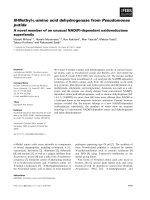
Botrytis cinerea as the only carbon source. Under these
conditions, two b-1,6-glucanases were detected by
chromatofocusing and activity staining (Fig. 1), one of
them corresponding to BGN16.2 (pI 5.8), which could
also be detected under chitin inductions, meanwhile
the other was a novel acidic isozyme which was named
BGN16.3 and showed a pI value around 4.5.
To purify BGN16.3 the filtrate of fungal cell walls-
supplemented cultures (1000 mL) was concentrated by
ammonium sulfate precipitation. The concentrate was
subjected to pustulan adsorption and further digestion.
Enzymes released after digestion of the polymer were
subjected to chromatofocusing and an acidic peak
(pH 4.1) with b-1,6-glucanase activity was obtained.
Fractions within this peak were pooled, concentrated
and subjected to FPLC gel filtration producing the
final purified protein with a yield of 31%. The purified
b-1,6-glucanase was analyzed by SDS ⁄ PAGE (Fig. 2A)
and a single protein band was observed using Coomas-
sie blue staining, suggesting a highly homogeneous
preparation. BGN16.3 was followed along all the puri-
fication steps using gel b-1,6-glucanase activity assay
after SDS ⁄ PAGE (Fig. 2B). Purification factors and
yields at each step are summarized in Table 1.
Physicochemical parameters
The molecular mass of the purified BGN16.3 was
approximately 46 kDa by SDS ⁄ PAGE, however, when
it was determined by S-200-HR gel filtration a value in
the range of 25–30 kDa was obtained.
The isoelectric point of the purified protein deter-
mined by isoelectrofocusing and acidic chromatofocus-
ing were 4.5 and 4.1, respectively.
No evidence was found of the presence of carbohy-
drates (glycosylation) in the purified protein as staining
with periodic acid ⁄ Schiff’s reagent [23] was negative
and no mobility shift was detected on SDS ⁄ PAGE
after treatment with endoglycosidase-F (Sousa, unpub-
lished results).
Kinetic parameters
The enzyme activity was measured at different pustu-
lan concentrations and Lineweaver–Burk representa-
tion was used to calculate Michaelis constants. A K
m
of 1.1 mg pustulanÆml
)1
and a V
max
of 390 lmol of
product per min
)1
Æ(mg protein)
)1
were estimated.
The optimal temperature for the BGN16.3 activity
was 50 °C and the inactivation temperature (50% of
the activity lost after 30 min incubation in the absence
of substrate) was calculated also 50 °C. This suggests
substrate protection against temperature inactivation
as previously described for other b-1,6-glucanases
[10,16]. Optimal pH was determined to be 5.0 and at
least 20% of maximum enzymatic activity was main-
tained between pH 4.0 and 7.0.
Substrate specificity and reaction products
The purified BGN16.3 was tested for activity towards
several glucan substrates (Table 2) by measuring the
release of reducing sugars. The highest activity was
Fig. 1. Isoelectrofocusing and further b-1,6-glucanase specific stain-
ing of extracellular proteins produced by T. harzianum CECT 2424
(1) and T. harzianum CECT 2413 (2) after 24 or 48 h growing on
chitin or B. cinerea cell walls as sole carbon source.
Acidic b-1,6-glucanase from Trichoderma harzianum M. Montero et al.
3442 FEBS Journal 272 (2005) 3441–3448 ª 2005 FEBS
detected for pustulan (linear b-1,6-glucan) and a lower
activity was measured towards yeast glucan (18% of
the maximum activity) and laminarin (8% of maxi-
mum) which are b-1,3-glucans with b-1,6-glycosidic
linkages at branches at the ratios of 4 : 1 and 7 : 1,
respectively [24]. No activity was found towards
colloidal chitin, pachyman, starch, cellulose, nigeran or
dextran, concluding that BGN16.3 is a specific b-1,6-
glucanase.
The most abundant oligomers detected by HPLC
after pustulan hydrolysis were di-, tri- and tetra-b-1,6-
glucosides as shown in Fig. 3. Low levels of glucose
could only be detected after longer incubations, sup-
porting an endolytic mode of action for BGN16.3.
This was confirmed later finding the lack of enzymatic
activity of BGN16.3 on gentiobiose (b-1,6-disacchar-
ide, not shown).
Protein sequences
The N-terminal and an internal peptide of the purified
protein were sequenced. Two 14 and 13 amino acid
sequences were obtained, respectively. These were:
N-terminal: Ala-Ala-Gly-Ala-Gln-Ala-Tyr-Ala-Ser-
Asn-Gln-Ala-Gly-Asn
Internal peptide: Gly-Leu-Asn-Ser-Asn-Leu-Gln-Ile-
Phe-Gly-Ser-Pro-Trp
Both sequences were compared to the existing
sequences in GenBank using blastp program. In the
case of the N-terminal no highly similar glucanase
sequences could be found, furthermore there was not
high similarity to the amino terminal ends of any of
Table 1. Purification of a b-1,6-glucanase (BGN16.3).
Step
Volume
(mL)
Total protein
(mg)
Total activity
(U)
Specific activity
(UÆmg
)1
)
Yield
(%)
Purification
(fold)
Crude enzyme 10 35.65 257 7.2 100 1
Pustulan digestion 1.8 2.02 215 106 75 15
Chromatofocusing eluate 0.425 0.550 100.4 182 39 25
Gel filtration eluate 0.400 0.425 80 188 31 26
Table 2. Substrate specificity of the purified BGN16.3. 100% activ-
ity corresponds to 185 U (mg protein)
)1
.
Substrate Linkage type
b-1,6-Glucanase
relative activity (%)
Pustulan b-1,6 (Glc) 100
Glucan (S. cerevisae) b-1,3: b-1,6 (Glc) 18
Laminarin b-1,3: b-1,6 (Glc) 8
Pachyman b-1,3 (Glc) 0
Carboxymethylcellulose b-1,4 (Glc) 0
Colloidal chitin b-1,4 (GlcNAc) 0
Nigeran a-1,3: a-1,4 (Glc) 0
Soluble starch a-1,4: a-1,6 (Glc) 0
AB
Fig. 2. Purification of BGN16.3. SDS ⁄ PAGE analysis (A) and activity staining by pustulan-agarose overlay (B) of the different purification steps
of BGN16.3. Proteins were stained with Coomassie blue. Lane 1, crude extract; lane 2, pustulan digestion; lane 3, chromatofocusing eluate
peak IP 4.1; lane 4, gel filtration eluate. The numbers of the left indicate the molecular masses of protein standards (lane M).
M. Montero et al. Acidic b-1,6-glucanase from Trichoderma harzianum
FEBS Journal 272 (2005) 3441–3448 ª 2005 FEBS 3443
the cloned b-1,6-glucanases confirming BGN16.3 as a
novel enzyme.
BGN16.3 internal peptide showed seven of 13 amino
acids identity with a fragment of a Neurospora crassa
b-1,6-glucanase named Neg1 [19]. No significant simi-
larity was found to BGN16.2 sequence previously
cloned from T. harzianum [18].
Regulation of the BGN16.3 production
To study the regulation of the expression of BGN16.3
under several different physiological conditions, we
used different induction media (replacement media)
after growth for 48 h in modified Czapek minimal
medium supplemented with glucose. Western blotting
with polyclonal antibodies raised against BGN16.3
was used in order to detect the presence of the enzyme
in seven different conditions after 48 h in the replace-
ment media. When glucose, glycerol, sorbitol or chitin
was used as a carbon source in the replacement media,
the presence of the protein could not be detected.
However it was clearly detected if 0.5% pustulan or
0.5% B. cinerea cell walls were used as the sole carbon
sources. A fainter band could be seen if no carbon
source was added to the minimal medium (Fig. 4A).
Similar results were obtained by b-1,6-glucanase activ-
ity staining after SDS ⁄ PAGE (not shown) on the same
samples. Further analyses were carried out on those
conditions where BGN16.3 could be detected studying
the expression of the enzyme at shorter time points: 12
and 24 h. Twelve hours after induction with fungal cell
walls BGN16.3 could already be clearly detected, it
was also detected in the absence of carbon source, but
not in the presence of pustulan. In this latter condition
24 h induction was required to detect the protein in
the supernatants (Fig. 4B).
Induction of BGN16.3 at a different pH or by nitro-
gen starvation was also tested, with negative results
(not shown).
Fig. 3. HPLC analysis of the mechanism of substrate degradation
by BGN16.3 on pustulan. The enzyme was incubated with pustulan
for 120 min, and aliquots of the reaction were taken at different
times. G
n
refers to glucose oligomers (n ¼ degree of polymeriza-
tion). Lower panels are substrate controls (C) where the enzyme
was not present. The incubation time is indicated in minutes in the
upper right corner of each graph.
Fig. 4. Expression profile of BGN16.3 under different induction conditions. (A) Western blot analysis on total extracellular protein from cul-
tures of T. harzianum CECT 2413 grown for 48 h on 2% glucose (1), 2% glycerol (2), 0.5% chitin (3), 0.5% pustulan (4), 0.5% B. cinerea cell
walls (5) or no carbon source (6). The purified BGN16.3 was used as positive control (7). (B) Accumulation of BGN16.3 was analyzed at shor-
ter times in the absence of carbon source (1), or in pustulan (2) or B. cinerea cell walls (3) inductions.
Acidic b-1,6-glucanase from Trichoderma harzianum M. Montero et al.
3444 FEBS Journal 272 (2005) 3441–3448 ª 2005 FEBS
Discussion
The implication of cell wall degrading enzymes
(CWDEs) in mycoparasitic processes carried out by
Trichoderma is widely accepted. Several dozen enzymes
putatively involved in the process have been identified,
many of them purified and their genes cloned [25].
Two extracellular b-1,6-glucanases had been previ-
ously purified from T. harzianum CECT 2413 [10,16].
In this paper we report the purification of a third
b-1,6-glucanase (BGN16.3), advancing the knowledge
on this diverse isozyme system. Interestingly the
BGN16.3 was identified using fungal cell walls in the
induction media, a condition often regarded as a simu-
lation of mycoparasitism, whereas it could not be
detected in chitin inductions, the condition most fre-
quently used to isolate enzymes from T. hazianum
[7,10,16].
The presence of different proteins displaying identi-
cal hydrolytic activity but with high sequence dissimi-
larities is a common fact in the CWDE complex
secreted by Trichoderma strains during mycoparatisic
interactions. In some strains, more than 10 different
chitinolytic enzymes and a similar number of b-1,3-glu-
canase isozymes have been described [9,25]. Differences
in their substrate specificity and ⁄ or regulatory proper-
ties [7,26,27] support the idea of a synergic and ⁄ or
complementary functional role for the different iso-
zymes during antagonistic processes to overcome the
problem of the complex nature of the fungal cell wall.
It is also interesting to consider the simultaneous pro-
duction of proteins with diverse structure but identical
substrate as a mechanism to avoid specific inhibitors
produced by the fungal host during the antagonistic
interaction. This phenomenon has been described in
plant–pathogen interactions [28]. Similar situations are
likely to occur in the fungus-to-fungus mycoparasitic
process.
The molecular mass of BGN16.3 is 46 kDa as deter-
mined by SDS ⁄ PAGE. Furthermore, the activity detec-
ted for BGN16.3 after SDS ⁄ PAGE and renaturation
suggests the monomeric nature of this protein. The
divergence with the molecular mass calculated from gel
filtration is probably due to an affinity of the protein
towards Sephacryl as previously described for other
extracellular proteins produced by T. harzianum [7].
Biochemical values obtained for this novel enzyme
are similar to the ones already described in the other
two endo-b-1,6-glucanases from T. harzianum [10,16],
although some differences can be found in isoelec-
tric point, K
m
value and substrate specificity, as
summarized in Table 3. BGN16.3 can degrade mixed
b-1,3- ⁄ b-1,6-glucans (i.e. laminarin, a b-1,3-glucan
polymer with b-1,6- branches), BGN16.1 can do this
as well, but not BGN16.2. However, unlike BGN16.1,
BGN16.3 cannot degrade isolated fungal cell walls of
S. cerevisiae. The fact that BGN16.3 cannot release
reducing sugars from the whole cell wall of S. cere-
visiae, but releases reducing sugars from b-glucan
obtained from this cell wall (by alkali lysis), suggests
that the enzyme is unable to reach its substrate in the
whole cell wall, probably due to the complex structure
of the fungal cell wall. This inability of BGN16.3 (and
probably other purified cell wall degrading enzymes) to
reach its substrate would not affect its participation in
the mycoparasitic process, as Trichoderma coordinately
produces a complex set of different enzymes with
synergistic action, able to complete the degradation of
the host cell wall [1,11].
BGN16.3 accumulation is mainly controlled by the
carbon source in the induction media, as could be expec-
ted for a glucanolytic extracellular enzyme. When
glucose is present in the induction media, no enzyme
is produced due to catabolite repression. Pustulan and
cell walls can induce the accumulation of BGN16.3 as
well as carbon source starvation. Western blots showed
a faster and higher accumulation of BGN16.3 when
T. harzianum was grown on fungal cell walls rather than
in pustulan or in the carbon source depletion condition.
This regulation pattern is different from that pre-
viously described for BGN16.1, which accumulates
abundantly under chitin induction, as do most of the
extracellular enzymes described from T. harzianum.
The fact that BGN16.3 accumulates strongly and spe-
cifically in fungal cell wall inductions suggests this
enzyme may play a role in mycoparasitism.
A thorough comparative study of the biochemical
properties of these three b-1,6-glucanases and the con-
ditions for the induction of each of them (including
the motifs present in their regulatory 5¢ region) could
give light to the detailed biological function of the dif-
ferent components of the b-1,6-glucanolytic system of
T. harzianum.
Table 3. Biochemical properties of the three b-1,6-glucanases puri-
fied from T. harzianum CECT 2413.
BGN16.1 BGN16.2 BGN16.3
Molecular mass (kDa) 51 43 46
pI 7.4–7.7 5.8 4.1–4.5
Optimum temperature (°C) 50 50 50
Glycosylation ND ND ND
K
m
(mg pustulanÆmL
)1
) 0.8 2.4 1.1
Degrades laminarin +++ – +
Degrades S. cerevisiae cell wall + – –
Degrades B. cinerea cell wall – – –
M. Montero et al. Acidic b-1,6-glucanase from Trichoderma harzianum
FEBS Journal 272 (2005) 3441–3448 ª 2005 FEBS 3445
Interestingly, there has recently been evidence for
the implication of a b-1,6-glucanase, Glu1, in the
mycoparasitic interaction of V. fungicola with Agaricus
bisporus [22]. In this process, the penetration into the
host occurs by a local degradation of its fungal cell
wall [29,30], as also occurs in Trichoderma mycopara-
sitic interactions. These results support an important
role for endo-b-1,6-glucanases in the degradation of
the fungal cell wall complex structure during mycopar-
asitic interactions. Further experiments will be carried
out to assess this possible role for BGN16.3.
The induction of the expression of BGN16.3 using
fungal cell walls has proven to be a valid approach to
identify novel enzymes produced by T. harzianum. The
use of fungal cell walls instead of chitin for inductions
would be closer (though maybe still not identical) to a
mycoparasitism situation, and has allowed us to iden-
tify of novel enzyme as shown here.
Experimental procedures
Strains and culture conditions
T. harzianum CECT 2413 [31] and T. harzianum CECT
2424 [4] were obtained from the Spanish Type Culture
Collection (Burjasot, Valencia, Spain). Botrytis cinerea
was isolated in our laboratory from infected strawberries.
Both strains were maintained in PDA [Potato ⁄ Dextrose ⁄
Agar (Difco, Detroit, MI, USA)] plates. For protein pro-
duction a two step growing method was used: Trichoderma
strains were grown (approximately 10
6
conidia per 400 mL
media) in modified Czapek minimal medium (0.5 gÆL
)1
MgSO
4
Æ7H
2
O, 0.01 gÆL
)1
FeSO
4
Æ7H
2
O, 0.425 gÆL
)1
KCl,
0.115 gÆL
)1
MgCl
2
Æ6H
2
O, 2.1 gÆL
)1
NH
4
Cl, 0.92 gÆL
)1
NaHPO
4
) supplemented with 2% glucose, in a rotatory
shaker at 180 r.p.m. After 48 h the mycelium was filtered,
thoroughly washed with 2% magnesium chloride and
water, and transferred to a new flask containing Czapek
minimal medium supplemented with different carbon
sources (replacement medium) and incubated for 48 h at
25 °C in a rotatory shaker at 180 r.p.m. In case of myco-
parasitic simulation, 0.5% B. cinerea cell walls, prepared
as previously described [10], were used as carbon source.
For carbon source starvation, modified Czapek minimal
medium without any supplement was used as replacement
medium.
Enzyme assays
b-1,6-Glucanase activity was determined by measuring the
amount of reducing sugars released from pustulan by the
Somogyi and Nelson procedure [32,33] using glucose as
standard. One unit of b-1,6-glucanase activity was defined
as the amount of enzyme that releases 1 lmol of reducing
sugar equivalents, expressed as glucose, per min under
standard assay conditions.
Thermal stability of the enzyme was determined incuba-
ting the purified protein at temperatures from 30 to 70 °C
in 50 mm sodium acetate buffer (pH 5.5) for 30 min and
then measuring the remaining enzymatic activity adding
pustulan as substrate and incubating as described. Inactiva-
tion temperature was defined as the temperature with a
reduction of 50% of the specific activity.
Optimum pH determination was performed using citrate–
acetic acid buffer for pH values between 3 and 5, phosphate
buffer for pH values between 6 and 8 and Tris ⁄ HCl buffer
was used for pH 9. In all cases the concentration was
50 mm.
Protein purification
(a) All purification steps, unless indicated, were performed
at 4 °C. T. harzianum CECT 2413 cultures grown at 28 °C
for 48 h on B. cinerea cell wall as the only carbon source
were filtered through filter paper and centrifuged for
10 min at 12 000 g. The supernatant was precipitated with
ammonium sulfate (90% saturation) and the precipitate
recovered by centrifugation at 25 000 g for 15 min, resus-
pended in a small volume of distilled water and dialyzed
against 50 mm sodium acetate buffer, pH 5.5.
(b) Dialyzed samples were adsorbed to alcohol precipita-
ted pustulan with magnetic stirring. Pustulan was then pre-
cipitated by centrifugation at 12 000 g for 10 min. The
adsorption was repeated twice with the nonadsorbed super-
natant. Pustulan pellets were washed three times with
50 mm sodium acetate buffer (pH 5.5), containing 1 m
NaCl and resuspended in the same buffer. These samples
were incubated overnight at 37 °C in the presence of 1 mm
phenylmethanesulfonyl fluoride and 1 mm sodium azide for
pustulan digestion. Clarified solutions were centrifuged at
12 000 g for 10 min and the supernatants recovered and di-
alyzed against 25 mm imidazole ⁄ HCl buffer (pH 6.5).
(c) A 0.5 mL sample of the dialyzed solution was applied
to a Polybuffer Exchanger PBE 94 column (Amersham Bio-
sciences, Barcelona, Spain) equilibrated with 25 mm imidaz-
ole ⁄ HCl buffer pH 6.5. Proteins were eluted at a flow rate
of 10 mLÆh
)1
with polybuffer 74 (1 : 10 pH 4.0) and collec-
ted fractions (1.6 mL each) were assayed for b-1,6-gluca-
nase activity as described above. Active fractions were
pooled and concentrated with a Centricon 10 (Amicon,
Beverley, MA, USA) device.
(d) The concentrated pool was subjected to FPLC gel fil-
tration with a Protein Pack 125 column (Waters, Milford,
MA, USA) using 50 mm sodium acetate buffer 0.1 m KCl
as eluent. The flow rate was 0.1 mLÆmin
)1
and fractions
were collected every minute. Fractions giving absorbance
at 280 nm were assayed for b-1,6-glucanase activity as
described above. Active fractions were pooled and concen-
trated using Centricon 10 devices.
Acidic b-1,6-glucanase from Trichoderma harzianum M. Montero et al.
3446 FEBS Journal 272 (2005) 3441–3448 ª 2005 FEBS
Gel electrophoresis and b-1,6-glucanase activity
staining
SDS ⁄ PAGE was performed by the method of Laemmli [34]
with 4% acrylamide in the stacking gel and 12% acryl-
amide in the separating gel. Detection of b-1,6-glucanase
specific activity in agar replicas of the SDS ⁄ PAGE gels was
carried out as described previously [35].
Isoelectrofocusing was performed using Pharmalyte gels
(Amersham Biosciences) following manufacturer’s direc-
tions. b-1,6 activity staining after electrofocusing was per-
formed as described earlier [35]. Standard marker proteins
with pI values within the range 3.5–9.3 (Amersham Bio-
sciences) were used to determine the apparent pI for
BGN16.3.
Substrate specificity
The purified BGN16.3 activity was tested against several
polymers with glycosidic linkages using 0.5 mgÆmL
)1
of
each substrate. Activity on these substrates was measured
by reducing sugar quantification using the Somogyi–Nelson
method, except for chitinase activity that was determined as
described previously [7].
Hydrolysis products determination
The resulting products from pustulan hydrolysis by the
purified BGN16.3 were applied to a HPLC Aminex HPX-42
A column (Bio-Rad, Barcelona, Spain) maintained at 45 °C.
Water was used as eluent at a flow rate of 0.4 mLÆmin
)1
;
diffraction index of the eluate was used for the detection of
the products. Glucose and cellulose oligosacharides (2–4
polymerization degree) were used as standards. Substrate
controls were carried out in each determination.
Preparation of antisera
Polyclonal antibodies were raised by subcutaneous injec-
tion of 250 lg of purified BGN16.3 into rabbits (New
Zealand) in complete Freund’s adjuvant. At 2-week inter-
vals, rabbits received additional injections with 125 lgof
protein in incomplete Freund’s adjuvant. Blood samples
were taken three times after the second injection with
2-week intervals. Samples were centrifuged 5 min at 3000 g
and the supernatant was stored at ) 20 °C and used for
western blotting.
Protein partial sequences
N-Terminal and internal peptide sequencing from the puri-
fied BGN16.3 was carried out by Eurosequence b. vs.
(Groningen, the Netherlands) following Edman degradation
method in an Applied Biosystem 494 Sequencer.
Acknowledgements
This work was supported in part by project FAIR
CT98-4140 from the European Union. M. Montero
was a recipient of a fellowship from program FPU
from Ministerio de Educacion y Ciencia, Spain, and
L. Sanz was a recipient of a fellowship from Junta de
Andalucia, Spain. We thank Andres Soler for his help-
ful advice on biochemical techniques and R. Sanchez
for help with HPLC experiments.
References
1 Papavizas GC (1985) Trichoderma and Gliocladium:
biology and potential for biological control. Annu Rev
Phytopathol 23, 23–54.
2 Harman GE, Howell CR, Viterbo A, Chet I & Lorito
M (2004) Trichoderma species – opportunistic, avirulent
plant symbionts. Nat Rev Microbiol 2, 43–56.
3 Howell CR (2003) Mechanisms employed by Trichoderma
species in the biological control of plant diseases: The
history and evolution of current concepts. Plant Dis 87,
4–10.
4 Hermosa MR, Grondona I, Iturriaga EA, Diaz-Minguez
JM, Castro C, Monte E & Garcia-Acha I (2000) Mole-
cular characterization and identification of biocontrol
isolates of Trichoderma spp. Appl Envir Microbiol 66,
1890–1898.
5 Hermosa MR, Keck EJ, Chamorro I, Rubio MB, Sanz
L, Vizcaı
´
no JA, Grondona I & Monte E (2004) Genetic
diversity shown in Trichoderma biocontrol isolates.
Mycol Res 108, 897–906.
6 Grondona I, Hermosa MR, Tejada M, Gomis MD,
Mateos PF, Bridge PD, Monte E & Garcia-Acha I
(1997) Physiological and biochemical characterization of
Trichoderma harzianum, a biological control agent of
soilborne fungal plant pathogens. Appl Envir Microbiol
63, 3189–3198.
7 De la Cruz J, Hidalgo-Gallego A, Lora JM, Benitez T,
Pintor-Toro JA & Llobell A (1992) Isolation and char-
acterization of three chitinases from Trichoderma harzia-
num. Eur J Biochem 206, 859–867.
8 Schickler H, Danin-Gehali BC, Haran S & Chet I
(1998) Electrophoretic characterization of chitinases as
a tool for the identification of Trichoderma harzianum
strains. Mycol Res 102, 373–377.
9Va
´
zquez-Garciduen
˜
as S, Leal-Morales CA & Herrera-
Estrella A (1998) Analysis of the b-1,3-glucanolytic sys-
tem of the biocontrol agent Trichoderma harzianum.
Appl Envir Microbiol 64, 1442–1446.
10 De la Cruz J, Pintor-Toro JA, Benitez T & Llobell A
(1995) Purification and characterization of an endo-beta-
1,6-glucanase from Trichoderma harzianum that is related
to its mycoparasitism. J Bacteriol 177, 1864–1871.
M. Montero et al. Acidic b-1,6-glucanase from Trichoderma harzianum
FEBS Journal 272 (2005) 3441–3448 ª 2005 FEBS 3447
11 Sanz L, Montero M, Grondona I, Vizcaı
´
no JA, Hermosa
R, Llobell A & Monte E (2004) Cell wall degrading
isoenzyme profiles of Trichoderma biocontrol strains
have correlation with rDNA taxonomical species. Curr
Genet 46, 277–286.
12 Sanz L, Montero M, Redondo J, Llobell A & Monte E
(2005) Expression of an a-1,3-glucanase during myco-
parasitic interaction of Trichoderma asperellum. FEBS J
272, 493–499.
13 Kapteyn JC, Montijn RC, Vink E, De la Cruz J, Llobell
A, Douwes JE, Shimoi H, Lipke PM & Klis FM (1996)
Retention of Saccharomyces cerevisiae cell wall proteins
through a phosphodiester-linked beta-1,3- ⁄ beta-1,6-glu-
can heteropolymer. Glycobiology 6, 337–345.
14 Schep GP, Shepherd MG & Sullivan PA (1984) Purifi-
cation and properties of a beta-1,6-glucanase from Peni-
cillium brefeldianum. Biochem J 223, 707–714.
15 Pitson SM, Seviour RJ, McDougall BM, Stone BA &
Sadek M (1996) Purification and characterization of an
extracellular (1-6)-beta-glucanase from the filamentous
fungus Acremonium persicinum. Biochem J 316, 841–846.
16 De la Cruz J & Llobell A (1999) Purification and prop-
erties of a basic endo-b-1,6-glucanase (BGN16.1) from
the antagonistic fungus Trichoderma harzianum . Eur J
Biochem 265, 145–151.
17 Fayad KP, Simao-Beaunoir AM, Gauthier A, Leclerc
C, Mamady H, Beaulieu C & Brzezinski R (2001) Puri-
fication and properties of a beta-1,6-glucanase from
Streptomyces sp. EF-14, an actinomycete antagonistic to
Phytophthora spp. Appl Microbiol Biotechnol 57, 117–
123.
18 Lora JM, De la Cruz J, Llobell A, Benitez T & Pintor-
Toro JA (1995) Molecular characterization and hetero-
logous expression of an endo-beta-1,6-glucanase gene
from the mycoparasitic fungus Trichoderma harzianum.
Mol Gen Genet 247, 639–645.
19 Oyama S, Yamagata Y, Abe K & Nakajima T (2002)
Cloning and expression of an endo-1,6-beta-d-glucanase
gene (neg1) from Neurospora crassa. Biosci Biotechnol
Biochem 66, 1378–1381.
20 Kim DJ, Baek JM, Uribe P, Kenerley CM & Cook DR
(2002) Cloning and characterization of multiple glycosyl
hydrolase genes from Trichoderma virens. Curr Genet
40, 374–384.
21 Moy M, Li HM, Sullivan E, White JF Jr & Belanger
FC (2002) Endophytic fungal b-1,6-glucanase expression
in the infected host grass. Plant Physiol 130, 1298–1308.
22 Amey RC, Mills PR, Bailey A & Foster GD (2003)
Investigating the role of a Verticillium fungicola beta-
1,6-glucanase during infection of Agaricus bisporus using
targeted gene disruption. Fungal Genet Biol 39, 264–275.
23 Dubray G & Bezard G (1982) A highly sensitive peri-
odic acid-silver stain for 1,2-diol groups of glycoproteins
and polysaccharides in polyacrylamide gels. Anal Bio-
chem 119, 325–329.
24 Hrmova M & Fincher GB (1993) Purification and prop-
erties of three (1-3)-beta-d-glucanase isoenzymes from
young leaves of barley (Hordeum vulgare). Biochem J
289, 453–461.
25 Lorito M (1998) Chitinolytic enzymes and their genes.
In Trichoderma and Gliocladium (Harman GE & Kubi-
cek CP, eds), Vol. 2, pp. 73–99. Taylor & Francis,
London.
26 Zeilinger S, Galhaup C, Payer K, Woo SL, Mach RL,
Fekete C, Lorito M & Kubicek CP (1999) Chitinase
gene expression during mycoparasitic interaction of
Trichoderma harzianum with its host. Fungal Genet Biol
26, 131–140.
27 Dana M, Limon MC, Mejias R, Mach RL, Benitez T,
Pintor-Toro JA & Kubicek CP (2001) Regulation of
chitinase 33 (chit33) gene expression in Trichoderma
harzianum. Curr Genet 38, 335–342.
28 Rose JKC, Ham KS, Darvill AG & Albersheim P
(2002) Molecular cloning and characterization of gluca-
nase inhibitor proteins: coevolution of a counterdefense
mechanism by plant pathogens. Plant Cell 14, 1329–
1345.
29 Calonje M, Garcia Mendoza C, Galan B & Novaes-
Ledieu M (1997) Enzymatic activity of the mycoparasite
Verticillium fungicola on Agaricus bisporus fruit body
cell walls. Microbiology 143, 2999–3006.
30 Calonje M, Garcia Mendoza C, Perez Cabo A,
Bernardo D & Novaes-Ledieu M (2000) Interaction
between the mycoparasite Verticillium fungicola and the
vegetative mycelial phase of Agaricus bisporus. Mycol
Res 104, 988–992.
31 Kullnig CM, Krupica T, Woo SL, Mach RL, Rey M,
Benı
´
tez T, Lorito M & Kubicek CP (2001) Confusion
abounds over identities of Trichoderma biocontrol iso-
lates. Mycol Res 105, 769–772.
32 Somogyi M (1952) Notes on sugar determination. J Biol
Chem 195, 19–23.
33 Nelson NJ (1955) Colorimetric analysis of sugars. Meth-
ods Enzymol 3, 85–86.
34 Laemmli UK (1970) Cleavage of structural proteins dur-
ing the assembly of the head of bacteriophage T4.
Nature 227, 680–685.
35 Soler A, De la Cruz J & Llobell A (1999) Detection of
beta-1,6-glucanase isozymes from Trichoderma strains in
sodium dodecyl sulphate-polyacrylamide gel electro-
phoresis and isoelectrofocusing gels. J Microbiol Methods
35, 245–251.
Acidic b-1,6-glucanase from Trichoderma harzianum M. Montero et al.
3448 FEBS Journal 272 (2005) 3441–3448 ª 2005 FEBS