Báo cáo khoa học: Reactions of gold(III) complexes with serum albumin docx
Bạn đang xem bản rút gọn của tài liệu. Xem và tải ngay bản đầy đủ của tài liệu tại đây (367.15 KB, 7 trang )
Reactions of gold(III) complexes with serum albumin
Giordana Marcon
1
, Luigi Messori
1
, Pierluigi Orioli
1
, Maria Agostina Cinellu
2
and Giovanni Minghetti
2
1
Department of Chemistry, University of Florence, Italy;
2
Department of Chemistry, University of Sassari, Italy
The reactions of a few representative gold(III) complexes –
[Au(ethylenediamine)
2
]Cl
3
, [Au(diethylentriamine)Cl]Cl
2
,
[Au(1,4,8,11-tetraazacyclotetradecane)](ClO
4
)
2
Cl, [Au(2,2¢,
2¢-terpyridine)Cl]Cl
2
,[Au(2,2¢-bipyridine)(OH)
2
][PF
6
]and
the organometallic compound [Au(6-(1,1-dimethylbenzyl)-
2,2¢-bipyridine-H)(OH)][PF
6
] – with BSA were investigated
by the joint use of various spectroscopic methods and
separation techniques. Weak metal–protein interactions
were revealed for the [Au(ethylenediamine)
2
]
3+
and
[Au(1,4,8,11-tetraazacyclotetradecane)]
3+
species, whereas
progressive reduction of the gold(III) centre was observed
in the cases of [Au(2,2¢-bipyridine)(OH)
2
]
+
and [Au(2,2¢,2¢-
terpyridine)Cl]
2+
. In contrast, tight metal–protein adducts
areformedwhenBSAisreactedwitheither[Au(diethylen-
triamine)Cl]
2+
and [Au(6-(1,1-dimethylbenzyl)-2,2¢-bipyri-
dine-H)(OH)]
+
. Notably, binding of the latter complex to
serum albumin results in the appearance of characteristic
CD bands in the visible spectrum. It is suggested that adduct
formation for both of these gold(III) complexes occurs
through coordination at the level of surface histidines. Sta-
bility of these gold(III) complexes/serum albumin adducts
was tested under physiologically relevant conditions and
found to be appreciable. Metal binding to the protein is tight;
complete detachment of the metal from the protein has been
achieved only after the addition of excess potassium cyanide.
The implications of the present results for the pharmacolo-
gical activity of these novel cytotoxic agents are discussed.
Keywords: gold(III) complexes; serum albumin; spectro-
scopic measurements.
Following the success of platinum(II) compounds in cancer
chemotherapy, several families of nonplatinum metal
complexes have been studied intensely as potential cytotoxic
and antitumour agents. In particular, in recent years,
various gold(III) complexes of sufficient stability in the
physiological environment have been prepared and evalu-
ated for in vitro anticancer properties. Some of them turned
out to exhibit relevant cytotoxic effects in vitro and were the
subject of further biochemical and pharmacological inves-
tigations [1]. Studies of the interactions of these gold(III)
complexes with DNA, the classical target of platinum(II)
complexes, pointed out that binding of these compounds to
nucleic acids is not as tight as in the case of platinum drugs,
suggesting the occurrence of a different mechanism for the
observed biological effects [2,3].
Surprisingly, at variance with the reactions with nucleic
acids, the reactions of antitumour metal complexes with
proteins have been poorly explored until now although they
may be of extreme relevance for the biodistribution, the
mechanism of action and the toxic effects of several
metallodrugs. For example, only a few studies exist on the
reactions of the well known anticancer platinum complexes
with proteins [4,5].
However, despite the results obtained so far often being
incomplete and fragmentary, we believe that the direct
damage inflicted on specific proteins by metal complexes,
following the formation of strong coordinate bonds, may
be of crucial relevance to explain the biological effects of
several metallodrugs, either established clinically or experi-
mentally.
In the present study, we have considered the reactions of
a series of representative gold(III) complexes, of different
structure and of known biological profile, developed in our
laboratory, with bovine serum albumin, selected both as the
most abundant plasma protein and as a general model for
globular proteins. Serum albumins have many physiological
functions. They contribute to colloid osmotic blood pres-
sure and are chiefly responsible for the maintenance of
blood pH [6]. There is evidence of a significant antioxidant
activity of serum albumins. These molecules may represent
the major plasma components that protect against oxidative
stress [7]. The most outstanding property of albumins is
their ability to reversibly bind a large variety of endogenous
and exogenous ligands. It is worthwhile remembering that
serum albumins have often been considered as general
ligands for fatty acids, which are otherwise insoluble in
blood, and exhibit a high affinity for hematin, bilirubin, and
small, negatively charged, hydrophobic molecules; more-
over albumins bind various metal ions [8].
The reactions of gold(III) complexes with serum albumin
were investigated primarily through the analysis of the
Correspondence to L. Messori, Department of Chemistry,
University of Florence, via della Lastruccia, 3, 50019 Sesto
Fiorentino (Florence), Italy.
Fax: + 39 055 4573385, Tel.: + 39 055 4573284,
E-mail: luigi.messori@unifi.it
Abbreviations: en, ethylenediamine (1,2-diaminoethane); dien, diethy-
lentriamine; cyclam, 1,4,8,11-tetraazacyclotetradecane; terpy, 2,2¢,
2¢-terpyridine; bipy, 2,2¢-bipyridine; bipy
c
, 6-(1,1-dimethylbenzyl)-
2,2¢-bipyridine; CDDP, cis-diammine dichloro platinum(II);
ESI-MS, electrospray ionization mass spectrometry; LMCT,
ligand to metal charge transfer.
(Received 11 July 2003, revised 29 September 2003,
accepted 2 October 2003)
Eur. J. Biochem. 270, 4655–4661 (2003) Ó FEBS 2003 doi:10.1046/j.1432-1033.2003.03862.x
characteristic bands of the gold(III) centre in the visible
spectrum. Our experiments show that markedly divergent
reactivity patterns with serum albumin have clearly emerged
for the various gold(III) complexes in relation to their
chemical structure and reactivity. The implications of such
differences in reactivity are discussed in relation to the
pharmacological properties of the individual compounds.
Materials and methods
Materials
[Au(ethylenediamine)
2
]Cl
3
([Au(en)
2
]Cl
3
)wasprepared
according to [9]. A gummy yellow precipitate was formed
by the addition of a solution of 1,2-ethylendiamine mono-
hydrate in ether to a solution of HAuCl
4
in ether; the yellow
precipitate was dissolved in water giving an orange solution.
A white precipitate of [Au(en)
2
]Cl
3
formed upon adding
ethyl alcohol to the latter solution.
[AuCl(diethylentriamine)]Cl
2
([AuCl(dien)]Cl
2
)waspre-
pared according to [10]. A solution of diethylenetriamine/
3HCl in water was added slowly and with stirring to a
solution of HAuCl
4
(20%, w/v) and a yellow precipitate
immediately formed. A solution of NaOH was added to the
mixture until pH 3 and stirred for 2 h at 0 °C. The yellow
precipitate was then filtered and washed with ethanol.
[Au(1,4,8,11-tetraazacyclotetradecane)](ClO
4
)
2
Cl ([Au
(cyclam)](ClO
4
)
2
Cl) was prepared by following the pro-
cedure reported by Kimura et al. [11]. Treatment of
NaAuCl
4
2H
2
O with equimolar amounts of cyclam in
CH
3
CN for 1 h yielded the [Au(cyclam)](ClO
4
)
2
Cl complex.
[Au(2,2¢,2¢-terpyridine)Cl]Cl
2
([Au(terpy)Cl]Cl
2
)waspre-
pared by addition of terpyridine to a HAuCl
4
solution under
a 1 : 1 stoichiometry according to [12]. [Au(2,2¢-bipyri-
dine](OH)
2
][PF
6
] ([Au(bipy])(OH)
2
][PF
6
]) was prepared
according to [13]. An aqueous suspension of Ag
2
Owas
added to a solution of [Au(bipy)Cl
2
][PF
6
] in acetone. The
mixture was stirred for 24 h at room temperature. AgCl was
removed by filtration and the solution evaporated to dryness
under reduced pressure. The residue was extracted with
acetonitrile and filtered over Celite (Sigma-Aldrich). The
pale-yellow filtrate was concentrated to a small volume and
diethyl ether was added to give a white precipitate of
[Au(bipy)(OH)
2
][PF
6
].
An aqueous solution of KOH (33 mg, 0.59 mmol) was
added to an aqueous suspension of [Au(bipy
c
-H)Cl][PF
6
]
(179 mg, 0.27 mmol) [14,15]. The mixture was refluxed for
1 h under stirring and filtered. The volume of the colourless
filtrate was reduced on a rotary evaporator until crystal-
lization was observed. The white product [Au(bipy
c
-
H)(OH)][PF
6
] was collected by filtration and dried under
vacuum.
All the products obtained were checked by elemental
analysis; in all cases, the purity of the compounds was
higher than 98%. Further evidence for the correct identi-
fication of the obtained compounds is provided by
electronic spectra and mass spectra (vide infra).
BSA was purchased from Fluka BioChemika (product
number 05470). The powder, lyophilized and crystallized,
was ‡ 98.0% pure (purified by HPCE) and of a molecular
mass 66 kDa. All the other reagents, purchased
from Sigma-Aldrich, were of analytical grade. Where
not stated otherwise, experiments were performed in
phosphate buffer containing 50 m
M
Na
2
HPO
4
, 100 m
M
NaCl, pH 7.4.
Spectroscopic measurements
The interaction of all complexes with BSA was analysed by
monitoring the electronic spectra of freshly prepared
solutions of each complex after mixing with BSA (in the
ratio 1 : 1) in the reference buffer. The concentration of
[Au(en)
2
]Cl
3
, [Au(dien)Cl]Cl
2
and [Au(cyclam)](ClO
4
)
2
Cl
was 1 · 10
)3
M
, while [Au(terpy)Cl]Cl
2
was 1 · 10
)4
M
,
[Au(bipy)(OH)
2
][PF
6
] and [Au(bipy
c
-H)(OH)][PF
6
]
2.25 · 10
)4
M
. Visible absorption spectra were carried out
with a PerkinElmer Lambda Bio 20 spectrophotometer.
The measurements were done at room temperature (25 °C).
Fluorescence spectra were registered with a Jasco FP-
750 spectrofluorimeter working at room temperature with
k
ex
¼ 295 nm; BSA 5 · 10
)5
M
was titrated with [Au(bi-
py
c
-H)(OH)][PF
6
] at the ratios [Au(bipy
c
-H)(OH)][PF
6
]/
BSA r ¼ 0.5–5.0 (where r is moles of drug per mole of
BSA).
Ultrafiltration experiments
The adducts between gold(III) compounds and BSA,
prepared as described above, were filtered after 24 h
incubation at room temperature, using Centricon YM-10
(Amicon Bioseparations, Millipore Corporation, USA) at
1370 g and the starting volume reduced by half; finally, the
absorption spectra of the upper and lower portions of the
solution were recorded.
Extensive ultrafiltration was applied to the same samples
and the absorption spectra were recorded after three cycles
of washing with the buffered solution.
Additional experiments were conducted by ultracentri-
fuging at half volume [Au(dien)Cl]Cl
2
/BSA solutions at
molar ratios of 1 : 1, 2 : 1, 4 : 1 and 8 : 1. Complex content
in the upper and lower solution was analysed spectro-
photometrically.
Circular dichroism spectra
CD spectra of BSA samples at increasing [Au(bipy
c
-
H)(OH)][PF
6
]/BSA molar ratios, in phosphate buffer, were
recorded on a Jasco J500C dichrograph and analysed
through the standard
JASCO
software. The time dependence
of the spectra was analysed over several hours; the final
spectra were recorded after 24 h incubation at 25 °C.
Reaction with cyanide
[Au(dien)Cl]Cl
2
/BSA and [Au(bipy
c
-H)(OH)][PF
6
]/BSA
adducts were treated with a 10 : 1 stoichiometric excess of
cyanide. The UV-Vis spectra were recorded before and
immediately after the addition of a concentrated solution
of sodium cyanide.
Reaction with imidazole
The interaction of [Au(bipy
c
-H)(OH)][PF
6
]2.5· 10
)4
M
and [Au(dien)Cl]Cl
2
1 · 10
)3
M
with imidazole (in the ratio
4656 G. Marcon et al.(Eur. J. Biochem. 270) Ó FEBS 2003
1 : 1) was analysed by monitoring the electronic spectra of a
freshly prepared solutions in the reference buffer at 25 °C,
5 h long.
Results
Structure and solution chemistry of the investigated
gold(III) complexes
In the present study we have considered the following six
gold(III) complexes: [Au(en)
2
]Cl
3
, [Au(dien)Cl]Cl
2
,[Au(cy-
clam)](ClO
4
)
2
Cl, [Au(terpy)Cl]Cl
2
, [Au(bipy)(OH)
2
][PF
6
]
and [Au(bipy
c
-H)(OH)][PF
6
], recently investigated in our
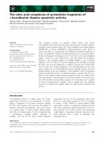
laboratory (Fig. 1). The choice of these gold(III) complexes
was dictated by their favourable chemical properties in
terms of solubility in water and stability within a physio-
logical-like environment; in addition, most of these com-
plexes are endowed with relevant cytotoxic properties
toward cultured human tumour cell lines, as previously
reported. The solution behaviour of these complexes, within
a reference physiological buffer, was further assayed by
monitoring the characteristic visible bands over several
hours. An appreciable stability was revealed for all men-
tioned gold(III) complexes in line with previous reports [1,3].
Spectrophotometric studies of the reaction with BSA
As all these gold(III) complexes, under physiological
conditions, exhibit intense and characteristic charge transfer
bands in the visible, their reactions with BSA were
monitored directly by visible absorption spectroscopy.
BSA was added in 1 : 1 stoichiometric amounts to buffered
solutions of each gold(III) complex and the visible spectra of
the resulting mixture recorded over several hours at room
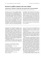
temperature. The obtained spectrophotometric patterns are
showninFig.2.
Different behaviours clearly emerge from direct inspec-
tion of the spectral profiles. It is apparent that the spectra of
either [Au(en)
2
]Cl
3
or [Au(cyclam)](ClO
4
)
2
Cl are not signi-
ficantly affected by addition of BSA. These observations
suggest that the gold(III) chromophore of these complexes
is not – or is only slightly – perturbed by protein addition.
Small changes are observed in the main charge transfer
band for both [Au(dien)Cl]Cl
2
and [Au(bipy
c
-H)(OH)
][PF
6
]. For [Au(dien)Cl]Cl
2
, the changes are complete within
about 2 h, while only a few minutes are needed in the case of
[Au(bipy
c
-H)(OH)][PF
6
].
In contrast, in the cases of [Au(terpy)Cl]Cl
2
and
[Au(bipy)(OH)
2
][PF
6
], a progressive decrease in intensity
of the visible bands is observed until complete disappear-
ance. Under the experimental conditions that we have
used, the process is complete within 2 h in the case of
[Au(terpy)Cl]Cl
2
and within about 6 h in the case of
[Au(bipy)(OH)
2
][PF
6
]. After ultrafiltration of the adducts
between BSA and [Au(terpy)Cl]Cl
2
or [Au(bipy)(OH)
2
]
[PF
6
], the lower solutions were spectrophotometrically
analysed and found to contain the free ligands terpyridine
and bipyridine. No gold was detected in these solutions. As
these gold(III) complexes are fairly stable, the best explan-
ation of the above observation is that gold(III) undergoes
reduction and the complexes break down with release of the
ligands. In turn, gold may be reduced to gold(I) or even to
colloidal gold associated with the protein.
Adduct formation as assessed by ultrafiltration
experiments
Further information on the reactions of gold(III) complexes
with BSA was gained by the application of classical
biochemical separation techniques. The main goal of these
experiments was to provide at least qualitative information
on the strength of the interactions between gold(III)
complexes and BSA. Buffered solutions of the individual
gold(III) complexes and BSA were prepared, at 1 : 1
stoichiometry, and incubated for 12–24 h at room tempera-
ture. Ultrafiltration with a Centricon device was carried out
to reduce sample volumes from 2 to 1 mL, and the upper
and lower solutions analysed spectrophotometrically. We
noticed that [Au(en)
2
]Cl
3
and [Au(cyclam)](ClO
4
)
2
Clare
readily removed from the protein by ultrafiltration, imply-
ing that the interaction is relatively weak and most likely
electrostatic in nature. Figure 3A shows the results
obtained with [Au(cyclam)](ClO
4
)
2
Cl). In the case of
[Au(bipy)(OH)
2
][PF
6
] (Fig. 3B), disruption of the gold(III)
complex is confirmed by the appearance of the characteristic
UV-Vis bands of the free ligand 2,2¢-bipyridine (at 230 and
280 nm) in the lower solution after ultrafiltration.
In contrast, both [Au(dien)Cl]Cl
2
and [Au(bipy
c
-
H)(OH)][PF
6
] are not easily displaced from the protein.
For [Au(dien)Cl]Cl
2
, the protein-bound complex after a
single ultrafiltration is about 80%, while for [Au(bipy
c
-
Fig. 1. Schematic drawings of some representative gold(III) complexes.
[Au(en)
2
]Cl
3
, [Au(dien)Cl]Cl
2
, [Au(cyclam)](ClO
4
)
2
Cl, [Au(terpy)Cl]
Cl
2
, [Au(bipy)(OH)
2
][PF
6
] and [Au(bipy
c
-H)(OH)][PF
6
].
Ó FEBS 2003 Interactions of cytotoxic gold(III) complexes with BSA (Eur. J. Biochem. 270) 4657
H)(OH)][PF
6
] it is more than 96%, suggesting that these
complexes are tightly bound to BSA through coordinate
bonds. However, [Au(dien)Cl]Cl
2
may be removed by
repeated cycles of ultrafiltration while [Au(bipy
c
-
H)(OH)][PF
6
] is not. Representative results of repeated
ultrafiltration experiments are shown in Fig. 4.
The [Au(dien)Cl]Cl
2
/BSA system
The appreciable stability of the [Au(dien)Cl]Cl
2
/BSA
adducts prompted us to analyse this system in more detail.
Specifically, we tested whether protein binding is reversible
and whether multiple binding sites are available for
[Au(dien)Cl]Cl
2
on the protein surface. To address these
issues, [Au(dien)Cl]Cl
2
/BSA solutions were prepared at
molarratiosof1:1,2:1,4:1and8:1;thegoldcontent
in the upper and lower solutions was analysed spectropho-
tometrically after extensive ultracentrifugation. From ana-
lysis of the experimental results, it is apparent that the
relative percentage of bound gold decreases as the
[Au(dien)Cl]Cl
2
/BSA ratio increases (Table 1). When BSA
is exposed to an 8 : 1 [Au(dien)Cl]Cl
2
molar excess, about
2.4 gold atoms are found associated with each protein
molecule after extensive washing. Overall, these findings
suggest that multiple binding sites for [Au(dien)Cl]Cl
2
are
present on BSA, of progressively lower affinity.
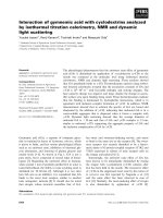
CD spectrum of the [Au(bipy
c
-H)(OH)][PF
6
]/BSA adduct
Further information on the spectral features of BSA-bound
gold(III) centres was obtained by CD spectroscopy, a
particularly well-suited technique to analyse the specific
environment of protein-bound metal centres [16].
Asampleof[Au(bipy
c
-H)(OH)][PF
6
]/BSA was prepared
at a 1 : 1 molar ratio, and analysed by CD, immediately
after mixing, at 25 °C (Fig. 5). Notably this adduct is
characterized by an intense CD negative band in the visible
spectrum, at k ¼ 410 nm, diagnostic of the fact that the
gold(III) species is bound to a chiral matrix such as the
protein.
With [Au(dien)Cl]Cl
2
, only minor modifications were
observed in the CD spectra of 10
)4
M
BSA when the
gold(III) complex was added in the ratios 1 : 1, 2 : 1, 4 : 1
and 8 : 1; however, no clear characteristic CD band
appeared in the visible spectrum (data not shown).
Gold removal from BSA by potassium cyanide
To further assess the stability of the adducts, either
[Au(dien)Cl]Cl
2
/BSA and [Au(bipy
c
-H)(OH)][PF
6
]/BSA
were treated with a 10 : 1 stoichiometric excess of cyanide.
It is well known that excess cyanide leads to the formation
of a very stable tetracyanoaurate complex and we therefore
wanted to check whether such a strong ligand is able to
remove gold(III) from the protein, both kinetically and
thermodynamically. Indeed, treatment with cyanide results
in quick disappearance of the peculiar visible bands of the
gold(III) centres in both adducts implying that the bound
gold is accessible and that the kinetics of release are fast. In
contrast, treatment of these derivatives with lower amounts
of cyanide did not result in complete detachment of gold
from the protein.
Fig. 2. Time-dependent spectral profiles of
gold(III) compounds/BSA adducts. Visible
absorption spectra of buffered solutions con-
taining gold(III) complexes and BSA in a 1 : 1
ratio. Spectra correspond to [Au(en)
2
]Cl
3
1 · 10
)3
M
(A), [Au(dien)Cl]Cl
2
1 · 10
)3
M
(B), [Au(cyclam)](ClO
4
)
2
Cl1· 10
)3
M
(C),
[Au(terpy)Cl]Cl
2
1 · 10
)4
M
(D), [Au(bipy)
(OH)
2
][PF
6
]2.25· 10
)4
M
(E) and
[Au(bipy
c
-H)(OH)][PF
6
]2.25· 10
)4
M
(F),
before (a) and after the addition of BSA. The
further evolution of the various systems over
time is reported until the spectral changes
reach completion. The buffer (pH 7.4) con-
tains 50 m
M
Na
2
HPO
4
and 100 m
M
NaCl.
4658 G. Marcon et al.(Eur. J. Biochem. 270) Ó FEBS 2003
Surface histidines as the probable binding site
for gold(III) complexes: the reaction with imidazole
Imidazoles of surface histidines are good candidates as
donors for the gold(III) centre. To elucidate this issue we
carried out the reaction of [Au(dien)Cl]Cl
2
and [Au(bipy
c
-
H)(OH)][PF
6
] with imidazole, within the same buffer, and
analysed the modifications of the visible spectra of the
gold(III) chromophore. Interestingly, spectral changes
similar to those observed upon reaction of the same
complex with albumin were detected. This observation,
although not conclusive, favours the view that histidines
are the probable binding sites for the gold(III) containing
fragments.
Fluorescence studies
Fluorescence measurements give information about the
molecular environment in the vicinity of the chromophore
molecules. The intensity of intrinsic fluorescence of two
tryptophan residues (Trp213 and Trp314) and a shift in
wavelength of their emission maxima were chosen as
indicators of protein conformational changes in serum
albumin.
Notably, the addition of [Au(bipy
c
-H)(OH)][PF
6
]to
BSA-buffered solutions results in a net decrease of fluores-
cence intensity; indeed, progressive fluorescence quenching
is observed as the [Au(bipy
c
-H)(OH)][PF
6
]/BSA molar ratio
increases from 0.5 to 5 (Fig. 6). At higher ratios, saturation
is reached and the final fluorescence spectrum is assigned
to the protein-bound form of [Au(bipy
c
-H)(OH)][PF
6
].
Whereas the residual fluorescence intensity is only 5%
of the original value, the position of the maximum moved
toward red wavelengths (a Dk ¼+13 nm has been deter-
mined for r ¼ 5).
The shift in the position of the emission maximum
corresponds to the changes of the polarity around the
chromophore molecule. The slight red-shift observed indi-
cates that tryptophan residues were placed in a more polar
environment and were more exposed to the solvent. It is
possible that [Au(bipy
c
-H)(OH)][PF
6
]stickstoBSAmole-
cules and consequently rearranges the tryptophan micro-
environment.
Fig. 4. The exhaustive ultrafiltration experiments of two representative
gold(III) compounds/BSA adducts. Visible absorption spectra of the
adduct before (a) and after exhaustive ultrafiltration: the spectra of
the lower (l) and the upper (u) solutions are shown. These data refer to
the [Au(dien)Cl]Cl
2
/BSA (A) and [Au(bipy
c
-H)(OH)][PF
6
]/BSA (B)
adducts (1 : 1).
Fig. 3. The ultrafiltration experiments at half volume of two represen-
tative gold(III) compounds/BSA adducts. Visible absorption spectra of
the lower (l) and upper (u) solution obtained after ultrafiltration
(reducing the volume to half). These data refer to the [Au(cy-
clam)](ClO
4
)
2
Cl/BSA (A) and [Au(bipy)(OH)
2
][PF
6
]/BSA (B) adducts
(1 : 1).
Table 1. Percentages of [Au(dien)Cl]Cl
2
in the upper and lower solutions
after ultracentrifugation. Percentage of complex found in the upper and
lower fractions after ultracentrifugation of solutions containing the
[Au(dien)Cl]Cl
2
/BSAsystemintheratios1:1,2:1,4:1and8:1.
Fraction 1 : 1 2 : 1 4 : 1 8 : 1
Upper solution 70.3 60.0 46.3 31.8
Lower solution 29.7 40.0 53.7 68.2
Ó FEBS 2003 Interactions of cytotoxic gold(III) complexes with BSA (Eur. J. Biochem. 270) 4659
Biological properties of the adduct
[Au(bipy
c
-H)(OH)][PF
6
]/BSA 1 : 1
It is still a matter of debate whether protein adducts of
cytotoxic metallodrugs retain, at least in part, the anti-
tumour properties of the free metal complex. In order to
address this point the biological activity of the adduct
[Au(bipy
c
-H)(OH)][PF
6
]/BSA 1 : 1 was tested toward some
representative human tumour cell lines. We observed that
the adduct retained to a good extent the cytotoxic activity of
the free metal complex; probably the protein behaves as a
ÔreservoirÕ of the free gold(III) compound (Table 2).
Discussion
The reactions of anticancer metal complexes with proteins
have been scarcely investigated until now. We believe that
this issue is of particular relevance in view of the established
reactivity of metal complexes with model proteins, and
deserves, in any case, greater attention. In fact, metal–protein
interactions may play key roles in the biodistribution, in the
mechanism of action and in the toxic effects of antitumour
metal complexes. Moreover, this subject is becoming more
important because the paradigm that DNA is a primary
target for antitumour metallodrugs is rapidly declining, and
seems to be no longer valid, at least for some families of
nonplatinum anticancer metal complexes. Obviously, this
observation has prompted new interest in the search of novel
proteins as possible targets for such metallodrugs.
Even in the case of cisplatin, the knowledge of the
interactions with proteins is limited to a few studies only,
from which, notwithstanding, it emerges that the largest
portion of administered platinum is associated with pro-
teins. Cole reported that cisplatin binds in vitro almost
irreversibly to BSA [17]; due to the apparent irreversibility
(both in vivo and in vitro) of the protein/195mPt–cisplatin
complex, it is unlikely that the protein-bound fraction of the
administered free drug will serve as a therapeutically useful
drug reservoir [18].
Other studies have been reported on the interactions of
some well known anticancer ruthenium(III) complexes and
of auranofin with plasma proteins [19–21].
Very scarce information exists on the reaction of gold(III)
complexes with proteins. In fact gold(III) complexes gen-
erally behave as strong oxidizing agents; hence it is
commonly believed that they are quickly reduced to gold(I)
compounds or to colloidal gold by low molecular mass
biomolecules and by protein side chains.
Thus, in the present paper, we have tried to detail the
reactions of a series of emerging antitumour gold(III)
complexes of appreciable redox stability with serum albu-
min, used as a general model for globular proteins. In the
compounds investigated the oxidizing properties of the
gold(III) centre are drastically decreased by the presence of
strong multidentate ligands in such a way that interaction
studies are feasible. However, the stronger oxidizing agents
in our series ([Au(terpy)Cl]Cl
2
and [Au(bipy)(OH)
2
][PF
6
])
are still able to slowly oxidize the protein side chains. At
variance with this, the complexes with less pronounced
oxidizing properties do not give rise to significant redox
chemistry but tend to form adducts with BSA that appear to
be of different strength. The tight adducts that formed with
either [Au(dien)Cl]Cl
2
or [Au(bipy
c
-H)(OH)][PF
6
]were
further investigated. Compared to the cisplatin–BSA
adduct, the adduct between the organometallic gold(III)
Fig. 5. Circular dichroism spectra of the [Au(bipy
c
-H)(OH)][PF
6
]/BSA
adduct. Circular dichroism spectra of BSA and of the [Au(bipy
c
-
H)(OH)][PF
6
]/BSA adduct in the 1 : 1 ratio. The spectrum of the
adduct was recorded immediately after mixing and after 3 h. BSA
concentration was 2 · 10
)4
M
.
Fig. 6. Titration of BSA with [Au(bipy
c
-H)(OH)][PF
6
] studied by
fluorescence. Fluorescence spectra of 5 · 10
)5
M
BSA upon addition of
increasing amounts of [Au(bipy
c
-H)(OH)][PF
6
], in the reference buffer
are shown. In the course of the experiment, r varies from 0.5 to 5.0.
Table 2. Cytotoxic activity of [Au(bipy
c
-H)(OH)][PF
6
] and of its adduct
with BSA. Inhibitory effects of [Au(bipy
c
-H)(OH)][PF
6
], the adduct
Au(bipy
C
-H)(OH)][PF
6
]/BSA and cisplatin on the growth of some
cisplatin-sensitive (A2780/S) and -resistant (A2780/R, SKOV3) human
tumour cell lines. ED
50
is defined as the concentration of drug required
to inhibit cell growth by 50% compared to control.
Cell line
ED
50
(l
M
)
[Au(bipy
C
-
H)(OH)][PF
6
]
[Au(bipy
C
-
H)(OH)][PF
6
]/BSA cisplatin
A2780/S 2.3 7.4 2.1
A2780/R 12.0 50 27.7
SKOV3 11.3 45 32.1
4660 G. Marcon et al.(Eur. J. Biochem. 270) Ó FEBS 2003
compound and BSA, once formed, is stable and retains its
cytotoxic activity; in other words it seems to be a good
candidate for further pharmacological evaluation. Notably,
the main features of the gold(III) centre are conserved after
association with BSA. The adducts are relatively stable and
may be destroyed only by the addition of strong ligands for
gold(III) such as cyanide.
This behaviour is interpreted in terms of either weak
electrostatic interactions or direct metal coordination to
surface residues of the protein. The ability of selected
complexes to tag either cysteine or histidine residues may
result in specific damaging of crucial proteins, which could
account for the pharmacological and toxic effects. Some
reports exist in the literature indicating that histidine
residues are preferred binding sites for ruthenium(III) on
the protein surface [22,23]. The antiarthritic gold(I) drug
Auranofin is known to bind specifically Cys34 of human
serum albumin [24]. In the light of the above examples it
might well be that selective modification of surface protein
residues by gold(III) complexes constitutes the molecular
basis for their biological effects.
Concluding remarks
In this study we have investigated the reactions of six
representative gold(III) complexes with bovine serum albu-
min used as a general model for plasma proteins. Different
patterns of reactivity emerge for the various compounds in
relation to the specific chemical properties of the gold(III)
complexes. In some cases tight adducts are formed in which
the bound gold(III) centres are probably coordinated to
surface histidines of the protein. It is hypothesized that the
ability of selected gold(III) complexes to tag either cysteine
or histidine residues may result in specific damaging of
crucial intracellular proteins thus accounting for the
relevant cytotoxic effects of these compounds.
Acknowledgements
The Cassa di Risparmio di Firenze and MIUR are gratefully
acknowledged for a generous grant. We thank Dr Costanza Landi
and Alessandro Vaccini for helping us in the experimental work.
References
1. Messori, L., Abbate, F., Marcon, G., Orioli, P., Fontani, M.,
Mini,E.,Mazzei,T.,Carotti,S.,O’Connell,T.&Zanello,P.
(2000) Gold (III) complexes as potential antitumor agents: solu-
tion chemistry and citotoxic properties of some selected gold (III)
compounds. J. Med. Chem. 43, 3541–3548.
2. Messori, L., Marcon, G., Tempi, C. & Orioli, P., (2001) Inter-
actions of selected gold (III) complexes with calf thymus DNA.
Biochem. Biophys. Res. Commun. 281, 352–360.
3. Marcon, G., Carotti, S., Coronnello, M., Messori, L., Mini, E.,
Orioli, P., Mazzei, T., Cinellu, M.A. & Minghetti, G. (2002)
Gold (III) complexes with bipyridyl ligands. solution chemistry,
cytotoxicity and DNA binding properties. J. Med. Chem. 45,
1672–1677.
4. Allardyce, C.S., Dyson, P.J., Coffey, J. & Johnson, N. (2002)
Determination of drug binding sites to proteins by electrospray
ionisation mass spectrometry: the interaction of cisplatin with
transferrin. Rapid Commun. Mass Spectrom. 16, 933–935.
5. LeRoy, A.F. & Thompson, W.C. (1989) Binding kinetics of tet-
rachloro-1,2-diaminocyclohexaneplatinum (IV) (tetraplatin) and
cis-diamminedichloroplatinum (II) at 37 degrees C with human
plasma proteins and with bovine serum albumin. Does aquation
precede protein binding? Natl. Cancer Inst 81, 427–436.
6. He, X.M. & Carter, D.C. (1992) Atomic structure and chemistry
of human serum albumin. Nature 358, 209–215.
7. Bourdon, E., Loreau, N. & Blache, D. (1999) Glucose and free
radicals impair the antioxidant properties of serum albumin.
FASEB J. 13, 233–244.
8. Bal, W., Christodoulou, J., Sadler, P.J. & Tucker, A. (1998) Multi-
metal binding site of serum albumin. J. Inorg. Biochem. 70, 33–39.
9. Block, B.P. & Bailar, J.C. (1951) The reaction of gold (III)
with some bidentate coordinating groups. J. Am. Chem. Soc. 73,
4722–4725.
10. Nardin,G.,Randaccio,L.,Annibale,G.,Natile,G.&Pitteri,B.
(1979) Comparison of structure and reactivity of bis (2-amino-
ethyl)amine- and bis(2-aminoethyl)amido-chlorogold (III)
complexes. J. Chem. Soc., Dalton Trans. 220–223.
11. Kimura, E., Kurogi, Y. & Takahashi, T. (1991) The first gold (III)
macrocyclic polyamine complexes and application to selective
gold (III) uptake. Inorg. Chem. 30, 4117–4121.
12. Hollis, L.S. & Lippard, S.J. (1983) Aqueous chemistry of (2,2¢,2¢-
terpyridine) gold (III). Preparation and structures of [Au (terpy)
Cl]Cl
2
3H
2
O and the mixed valence Au(I)-Au(III) salt [Au
(terpy)Cl]
2
[AuCl
2
]
3
[AuCl
4
]. J. Am. Chem. Soc. 105, 4293–4299.
13. Cinellu, M.A., Minghetti, G., Pinna, M.V., Stoccoro, S., Zucca, A.
& Manassero, M. (2000) Gold (III) derivatives with anionic
oxygen ligands: mononuclear hydroxo, alkoxo and acetato com-
plexes. Crystal structure of [Au(bipy)(OMe)
2
][PF
6
]. J. Chem. Soc.,
Dalton Trans. 1261–1265.
14. Cinellu, M.A., Zucca, A., Stoccoro, S., Minghetti, G., Manassero,
M. & Sansoni, M. (1996) Synthesis and characterization of gold
(III) adducts and cyclometallated derivatives with 6-benzyl- and
6-alkyl-2,2¢-bipyridines. J. Chem. Soc., Dalton Trans. 4217–4225.
15. Cinellu, M.A., Minghetti, G., Pinna, M.V., Stoccoro, S., Zucca, A.
& Manassero, M. (1999) Replacement of the chloride ligand
in [Au(C,N,N)Cl][PF
6
] cyclometallated complexes by C, N, O
and S donor anionic ligands. J. Chem. Soc., Dalton Trans.
2823–2831.
16. Rodger, A. & Norde
´
n, B., (1997) Circular Dichroism and Linear
Dichroism. Oxford University Press, Oxford, UK.
17. Cole, W.C. & Wolf, W. (1980) Preparation and metabolism of a
cisplatin/serum protein complex. Chem. Biol. Interact 30, 223–235.
18. Ivanov, A.I., Christodoulou, J., Parkinson, J.A., Barnham, K.J.,
Tucker, A., Woodrow, J. & Sadler, P.J. (1998) Cisplatin binding
sites on human albumin. J. Biol. Chem. 273, 14721–14730.
19. Messori,L.,Orioli,P.,Vullo,D.,Alessio,E.&Iengo,E.(2000)A
spectroscopic study of the reaction of NAMI, a novel ruthenium
(III) anti-neoplastic complex, with bovine serum albumin. Eur
J. Biochem. 267, 1206–1213.
20. Kratz, F., Hartmann, M., Keppler, B. & Messori, L., (1994) The
binding properties of two antitumor ruthenium (III) complexes to
apotransferrin. J. Biol. Chem. 269, 2581–2588.
21. Roberts, J.R., Xiao, J., Schliesman, B., Parsons, D.J. & Shaw,
C.F. III (1996) Kinetics and mechanism of the reaction between
serum albumin and auranofin (and its isopropyl analogue) in vitro.
Inorg. Chem. 35, 424–433.
22. Clarke, M.S. & Stubbs, H., (1987) Metal Ions in Biological Sys-
tems (Sigel, H., ed), Vol. 33, pp. 728–755. Marcel Dekker, New
York, USA.
23. Keppler, B.K. (1993) Metal Complexes in Cancer Chemotherapy.
VCH Publishers, Weinheim, Germany.
24. Shaw, C.F. III. (1999) Gold-based therapeutic agents. Chem. Rev.
99, 2589–2600.
Ó FEBS 2003 Interactions of cytotoxic gold(III) complexes with BSA (Eur. J. Biochem. 270) 4661