Head and Neck Cancer Edited by Mark Agulnik potx
Bạn đang xem bản rút gọn của tài liệu. Xem và tải ngay bản đầy đủ của tài liệu tại đây (16.01 MB, 440 trang )
HEAD AND NECK CANCER
Edited by Mark Agulnik
Head and Neck Cancer
Edited by Mark Agulnik
Published by InTech
Janeza Trdine 9, 51000 Rijeka, Croatia
Copyright © 2012 InTech
All chapters are Open Access distributed under the Creative Commons Attribution 3.0
license, which allows users to download, copy and build upon published articles even for
commercial purposes, as long as the author and publisher are properly credited, which
ensures maximum dissemination and a wider impact of our publications. After this work
has been published by InTech, authors have the right to republish it, in whole or part, in
any publication of which they are the author, and to make other personal use of the
work. Any republication, referencing or personal use of the work must explicitly identify
the original source.
As for readers, this license allows users to download, copy and build upon published
chapters even for commercial purposes, as long as the author and publisher are properly
credited, which ensures maximum dissemination and a wider impact of our publications.
Notice
Statements and opinions expressed in the chapters are these of the individual contributors
and not necessarily those of the editors or publisher. No responsibility is accepted for the
accuracy of information contained in the published chapters. The publisher assumes no
responsibility for any damage or injury to persons or property arising out of the use of any
materials, instructions, methods or ideas contained in the book.
Publishing Process Manager Jana Sertic
Technical Editor Teodora Smiljanic
Cover Designer InTech Design Team
First published March, 2012
Printed in Croatia
A free online edition of this book is available at www.intechopen.com
Additional hard copies can be obtained from
Head and Neck Cancer, Edited by Mark Agulnik
p. cm.
ISBN 978-953-51-0236-6
Contents
Preface IX
Part 1
Squamous Cell Carcinoma of the Head and Neck 1
Chapter 1
Laryngeal Cancers in Sub-Saharan Africa
Mala Bukar Sandabe, Hamman Garandawa
and Abdullahi Isa
Chapter 2
Hypopharyngeal Cancer
Valentina Krstevska
Part 2
Chapter 3
Chapter 4
3
13
Biology of Head and Neck Cancer
71
Molecular Genetics and Biology of
Head and Neck Squamous Cell Carcinoma:
Implications for Diagnosis, Prognosis and Treatment
Federica Ganci, Andrea Sacconi, Valentina Manciocco,
Renato Covello,Giuseppe Spriano, Giulia Fontemaggi
and Giovanni Blandino
Cell Signalings and the
Communications in Head and Neck Cancer
Yuh Baba, Masato Fujii, Yutaka Tokumaru
and Yasumasa Kato
73
123
Chapter 5
Role of ING Family Genes in Head
and Neck Cancer and Their Possible
Applications in Cancer Diagnosis and Treatment 141
Esra Gunduz, Mehmet Gunduz, Levent Beder,Ramazan Yigitoglu,
Bunyamin Isik and Noboru Yamanaka
Chapter 6
Arachidonic Acid Metabolism and
Its Implication on Head and Neck Cancer 167
Sittichai Koontongkaew and
Kantima Leelahavanichkul
VI
Contents
Part 3
Chapter 7
Therapeutic Options
185
Nasopharyngeal Carcinoma: The Role for
Chemotherapeutics and Targeted Agents
Jared Knol, Tiffany King and Mark Agulnik
187
Chapter 8
Novel Chemoradiotherapy Regimens
Incorporating Targeted Therapies in Locally
Advanced Head and Neck Cancers 201
Ritesh Rathore
Chapter 9
Advanced Radiation Therapy for
Head and Neck Cancer: A New
Standard of Practice 227
Putipun Puataweepong
Part 4
Post-Treatment Considerations
251
Chapter 10
Tumour Repopulation During Treatment
for Head and Neck Cancer: Clinical Evidence,
Mechanisms and Minimizing Strategies 253
Loredana G. Marcu and Eric Yeoh
Chapter 11
DNA Repair Capacity and
the Risk of Head and Neck Cancer 273
Marcin Szaumkessel, Wojciech Gawęcki
and Krzysztof Szyfter
Part 5
Prosthesis and Reconstruction 293
Chapter 12
Finesse in Aesthetic Facial Recontouring 295
Yueh-Bih Tang Chen, Shih-Heng Chen
and Hung-Chi Chen
Chapter 13
Prosthodontic Rehabilitation of
Acquired Maxillofacial Defects 315
Sneha Mantri and Zafrulla Khan
Chapter 14
Functional and Aesthetic Reconstruction of the
Defects Following the Hemiglossectomy in Patients
with Oropharyngeal Cancer 337
Mutsumi Okazaki
Part 6
Chapter 15
Health Outcomes
349
Pain Control in Head and Neck Cancer
Ping-Yi Kuo and John E Williams
351
Contents
Chapter 16
Health Related Quality of Life
Questionnaires: Are They Fit for Purpose? 371
Kate Reid, Derek Farrell and Carol Dealey
Chapter 17
A Health Promotion Perspective of
Living with Head and Neck Cancer 393
Margereth Björklund
VII
Preface
Head and neck cancer is a devastating illness affecting individuals around the globe.
The number of new cases of head and neck cancer in the US each year exceeds 40,000
individuals and accounts for about 3-5% of adult malignancies. In excess of 10,000
individuals will die of their disease each year. The worldwide incidence exceeds half a
million cases annually. In North America and Europe, the tumors usually arise in the
oral cavity, oropharynx, or larynx, whereas nasopharyngeal cancers the more common
in the Mediterranean countries and in the Far East. In Southeast China and Taiwan,
head and neck cancer, specifically nasopharyngeal cancer is the most common cause of
death in young man.
Head and neck cancer requires a multidisciplinary approach and a clear
understanding of human anatomy. Establishing a better understanding of the
pathogenesis behind the development of head and neck cancer will provide insight
into future therapies for this disease. While the treatment of head and neck cancer is
highly complicated, including chemotherapy, targeted therapy, radiation therapy, and
surgery, the complications and longer term effects of treatment can also be
devastating.
The purpose of this Head and Neck Cancer book is to highlight work currently being
done to give physicians, patients, scientists and researchers and better understanding
of this disease. Sections will look to educate about Squamous Cell Carcinoma
worldwide, elucidate new targets and biological aspects of the disease and then focus
on the existing and novel therapeutics available to these patients.
While most clinical trials and review articles stop at this point in the explanation and
evaluation of head and neck cancer, this book looks to move beyond treatment and
focus the second half on survivorship issues and aspects that can be utilized to
improve long term quality of life. Chapters will focus on post treatment side effects,
prostheses and reconstruction as well as health outcomes research for patients with
Head and Neck Cancers.
For those of us that dedicate our lives to the treatment of Head and Neck Cancers, it is
a passion, and a true desire to help patients overcome their devastating disease with
X
Preface
the least amount of long-term impact, on their lives. I trust that this book will be of
value to the reader and help to provide further understanding to this difficult disease.
Mark Agulnik, MD
Division of Hematology/Oncology
Robert H. Lurie Comprehensive Cancer Center
Northwestern University Feinberg School of Medicine
USA
Part 1
Squamous Cell Carcinoma
of the Head and Neck
1
Laryngeal Cancers in
Sub-Saharan Africa
Mala Bukar Sandabe,
Hamman Garandawa and Abdullahi Isa
Department of ENT University of Maiduguri
Teaching Hospital
Nigeria
1. Introduction
Laryngeal cancers are not common1. Squamous cell carcinomas of the larynx are the
commonest head and neck tumour in the western world. It represents approximately 1% of
all malignancy in males 1. It’s about five times common in men than in women. The cause is
unknown but tobacco smoking and alcohol acting synergistically increases the risk,
radiation, asbestos and a number of occupational factors are implicated. Patients usually
present with progressive hoarseness and difficulty in breathing, pain is an uncommon
symptom whereas dysphagia, neck swelling, cachexia and fetor indicate advance disease.
All patients in our series are black Africans and unfortunately they presents late. The cancer
is confirmed by biopsy of the tumour through direct laryngoscopy under general
anaesthesia. And tentative treatment depends on the stage of the tumour.
2. Research methodology
This would be 10 years retrospective studies of black African patients with laryngeal
carcinomas carried out in University of Maiduguri Teaching Hospital Maiduguri, Federal
Medical Centre Nguru, Federal Medical Centre, Yola. These hospitals are located in the
North Eastern region of Nigeria, Sub-Saharan Africa. These centers also receive patients
from neighboring countries of Niger, Chad and Cameroon. Clinical records of all patients
with histologically confirmed laryngeal carcinoma from January 2001 – December 2010 were
reviewed, data extracted from the records includes biodata, presenting complaints (the main
complaints for which the patient sought medical advice), and associated complaints
(complaints regarded as unimportant by the patient), duration of presenting complaints,
duration of symptoms on first presentation, Social habit, physical examination findings, Xray of soft tissue neck, CT-Scan/MRI of the Larynx findings, the site of the lesion in the
larynx, histopathological types, treatment offered and symptom free period after treatment
(last entry in the case note) . Data was analyzed using Statistical Package for Social Sciences
(SPSS) – version 15 software. Descriptive analysis done for all data; Chi square test, and
correlation studies were applied where appropriate. Results was presented in tables and
graphs. P – Value < 0.05 was considered significant.
4
Head and Neck Cancer
3. Literature review
Grossly the larynx extends from the superior border of the epiglottis to the inferior border of
the cricoid cartilage. Anteriorly, it is related to the lingual epiglottis, the thyrohyoid
membrane, the anterior commissure, thyroid cartilage, cricothyroid membrane and the
anterior arch of the cricoid cartilage. The posterior relations are the posterior commissure
the arytenoids, and the interarytenoid Space. 1. Squamous cell carcinoma of the larynx is the
commonest head and neck cancer in the Western world. In the UK it represents
approximately 1% of all malignancies in men. (Powell and Robin, 1983). It is about five times
commoner in males than in females. The incidence increases with age, but the peak age of
presentation is in the seventh decade. The cause of cancer of the larynx is not known, but
there is an indisputable relationship between tobacco smoking and alcohol consumption,
(US surgeon general, 1979; Hinds, Thomas and O’Reilly, 1979). Verrucous carcinoma is a
distinct variant of well differentiated Squamous cell carcinoma. (Ackerman’s tumour). Other
malignant tumour types include adenocarcinoma, adenoid cystic carcinoma, fibrosarcoma,
Chondrosarcoma and lymphomas. Spread and growth depends on the site of origin of the
primary tumour. Anatomical barriers are important factors in determining the direction and
extend of tumour growth.
1.
2.
3.
Supraglottis. This comprises the larynx superior to the apex of the ventricle. Exophytic
supraglottic cancers do not often extends to the glottic region and seldom involve the
thyroid cartilage, Ulcerative lesions may extend down below the anterior commissure,
Cranially supraglottic cancers extend to the vallecular and base of the tonque,
arytenoids cartilage and pyriform sinus is reach by deep invasion.1,2.
Glottis. This comprises the vocal cords and the anterior and posterior commissures. Most
of the tumours originates in the free margins of the vocal cords which are covered by
squamous epithelium. Tumour may extend along the cord to the anterior commissure and
to the muscles of the vocal cord. Fixation of the vocal cords indicate deep invasion
Subglottis. This extends from the inferior border of the glottis to the lower border of the
cricoids cartilage, tumours are rare, grow circumferentially, usually extensive before
symptoms appear which is mainly inspiratory sridor.1,2.
4. Clinical features
Hoarseness is the main symptoms; 1, 2, 3. Dyspnoea and stridor are late symptoms and
usually indicate an advanced tumour. Pain in the throat is an uncommon symptom.
Dysphagia indicates pharyngeal invasion Neck swelling indicate extra laryngeal extension
or lymph nodes involvement. Symptoms of anorexia, cachexia and fetor imply advanced
disease. Indirect laryngoscopy should reveal the site and size of the lesions however because
of difficulty in examining the subglottic and the laryngeal surface of the epiglottis. Flexible
Fibre optic laryngoscopy helps in visualizing all part of the larynx. The neck should be
palpated for the presence of enlarged lymph nodes. Laryngeal tumours usually metastasize
to the upper deep cervical lymph nodes, but supraglottic tumours may cause bilateral
nodes, and some subglottic tumours may spread to the upper mediastinal nodes.
Palpable lymph nodes are important in determining prognosis, about one-third of patients
with no palpable lymph nodes have histologically positive nodes, and a similar number of
palpable nodes are histologically negative.
5
Laryngeal Cancers in Sub-Saharan Africa
5. Investigations of patients with laryngeal cancer
The main stay of investigation in our center was radiography. Plain X-rays soft tissue neck
was done by the entire patient studied. Although plain X-rays soft tissue neck has no role in
the current management of patients with carcinoma of the larynx, prevertebral soft tissue
thickness, the epiglottis can be visualized; it is also affordable in the developing countries.
Cost about 8USD. Computerized tomography scan(CT-Scan) which include contrast
enhanced helical CT scanning has a high sensitivity 91% and high negative predictive values
of 95% in detecting cartilage invasion of CA larynx7. In our survey only 15(16.1%) of our
patients had CT scanning done. This is due to the high cost of CT scan per session. It cost
about 300USD and most of the patients live on less than a Dollar a day. Magnetic resonance
imaging(MRI) which has several advantages over CT-scan especially in pre- surgical
planning can only be done by 6(6.5%) of our patients due to the cost per session of 400USD.
The multiplanner capabilities of MRI are superior to the reformations available with the
traditional CT-scan. MRI has been found to have a sensitivity of 89-94%, specificity 74-88%
and a negative predictive value of 94-96% for the detection of neoplastic invasion7. Positron
emission tomography (PET) which is critical in detection of metastasis and for follow-up of
treated patients, but sadly such services is nonexistent in most developing nations.
6. Treatment options 1, 2, 3, 5, 6
The standard treatment of laryngeal carcinoma is surgery and radiotherapy in varying
combinations. Surgery involves partial or total removal of the larynx to achieve cure,
radiotherapy have been found to be effective in early laryngeal cancers (T1 and T2) with
local control ranging from 70-100%. In advance laryngeal cancers (T3 and T4) post operative
chemoradiation can achieve loco-regional control.4.
frequency
6
18
32
37
Partial laryngectomy and radiotherapy
Total laryngectomy and radiotherapy
Radiotherapy alone
Chemotherapy and radiotherapy
Percentage (%)
6.5
19.3
34.4
39.7
Table 1.
Male
Female
Total
1-2 years
30
6
36
3-4years
17
3
20
5-6 years
6
0
6
7-8 years
0
3
3
9-10 years
3
0
3
Total
78
15
93
Table 2. Symptom free period
supraglottic
glottic
subglottic
transglottic
total
<1year
9
0
3
13
25
1-2years
12
0
0
24
36
Table 3. Symptom free period
3-4years
5
6
6
3
20
5-6years
3
3
0
0
6
7-8years
3
0
0
0
3
9-10years
3
0
0
0
3
Total
35
9
9
40
93
6
Head and Neck Cancer
Most patients in our series where offered synchronous therapy of chemoradiation because
of late presentation 54(58%) and 27(29%) presented in stage III and stage IV respectively,
however the survival rate barely 1-2years and because of late presentation in our series most
glottis tumour have progress to transglottic on presentation with average symptom free
period of 3years after treatment. Overall 6(6.5%) had partial laryngectomy and postoperative radiotherapy, 18(19.3%) had total laryngectomy and post-operative radiotherapy,
32(34.4%) had radiotherapy alone and 37(39.7%) had chemotherapy and radiotherapy.
The common agents used in our series include cisplatin, 5-florouracil, docetaxel and
Adriamycin in varying combinations and administered either as neoadjuvant, adjuvant or
concomitant chemotherapy.
7. Discussion
Laryngeal cancer is the most common cancer of the aerodigestive track, it accounts for 20%
of all head and neck cancers. The incidence of these tumours is closely correlated with
smoking cigarettes, as head and neck tumours occur 6(six) times more often among
cigarettes smokers then among non smokers.
Cancer of the larynx has been found to be commoner in males, it occurs in increasing age
with the peak incidence being in the 5th decade.
In our study, 93 patients were surveyed with carcinoma of the larynx, 78 (83.9%) male and
females constituted 15 (16.1%) mean age of 56 years (+ 6- 8yrs), M: F=5.2:1.
Fig. 1. Age distribution
The estimated incidence of carcinoma of the larynx in the United States is about 12,000 per
annum while in Nigeria the incidence is estimated at 783 per annum. Squamous
cell carcinoma is the commonest histological type; in our series it constituted 90.3% other
were verrucous Carcinoma, 32% and Adenocarcinoma 6.5%. Studies conducted elsewhere
in the country by Amusa et al also showed the histological type to be predominantly
squamous cell. 8
7
Laryngeal Cancers in Sub-Saharan Africa
Fig. 2. Duration of symtoms
frequency
84
3
6
93
Squamous cell carcinoma
Verrucous carcinoma
Adenocarcinoma
Total
Percentage (%)
90.3
3.2
6.5
100.0
Table 4. Histological types
Transglottic carcinoma was found to be the commonest with 40 (43.0%), supraglottic, 35
(37.6%): table V. This is in contrast to other studies in which most laryngeal cancers arise
from the glottis, 9 this could be due to the late presentation in most of the patients with locoregional involvement, (images 1, 2 and 3)
Site
Transglottic
Supraglottic
Glottic
Subglottic
Total
N (%)
40(43.0)
35(37.6)
9(9.7)
9(9.7)
93(100.0)
Table 5.
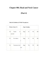
Most of the patient presented with stage – III tumours, this is in agreement with most head
and neck tumour presentation in developing countries.
Supraglottic
Glottic
Subglottic
Transglottic
Total
29
6
35
9
0
9
6
3
9
34
6
40
78
15
93
Male
Female
TOTAL
Table 6. Site of lesion
8
Head and Neck Cancer
Fig. 3. Clinical stage at presentation
There was a significant correlation between the clinical stage of the tumour at presentation
and the site of the lesion, most patient present with stages III &IV transglottic or supraglottic
tumour. P<0.05 (0.000).
Supraglottc
Glottic
Subglottic
Transglottic
Total
Stage II
3
9
0
0
12
Stage III
20
0
9
25
54
Stage IV
12
0
0
15
27
Total
35
9
9
40
93
Table 7. Correlation between clinical stage of patient and site of lesion
Correlation also exist between the site of the lesion and the social habit of the patients, with
those who smoke cigarettes and drink alcohol presenting more with glottis tumours
P< 0.05(0.00) This could be due to the synergistic effect of cigarette smoking and alcohol on
head and neck tumours.
smoke
alcohol
Smoke and alcohol
None
Total
Supraglottic
12
0
3
20
35
Glottic
0
0
6
3
9
Subglottic
0
0
3
6
9
Transglottic
15
3
3
19
40
P<0.05(0.000)
Table 8. Correlation between social habit of patients and site of lesion
Total
27
3
15
48
93
9
Laryngeal Cancers in Sub-Saharan Africa
Site of lesion
Stage II
Stage III
Stage IV
Total
Supraglottic
3
20
12
35
Glottic
9
0
0
9
Subglottic
0
9
0
9
Transglottic
0
25
15
40
Total
12
54
27
93
And the site of lesion P<0.05 (0.000)
Table 9. Correlation between the clinical stages of the tumour
8. Conclusion
In conclusion black African patients in our study typically present late which accounts for
the higher number of transglottic and supraglottic cancers. Among some of the reasons for
late presentations are lack of affordability and accessibility by most patients to tertiary
health facility in developing countries like Nigeria. The national health insurance scheme
covers less than 10% of the population of 150million Nigerians thus living the majority to
pay an exorbitant fee for health care services. Another reason is the absence of radiotherapy
centers in most tertiary health facility in developing countries such that patient have to
travel a long distance with their relatives to access such services further increasing the cost
of treatment and delay before presentation.
Finally there is a need to educate the general public and especially health care providers to
promptly refer patients with hoarseness of more than 2 weeks duration for direct
laryngoscopy and biopsy by an otolaryngologist.
Most countries in sub-Saharan Africa are now emerging democracies, and thus the
challenges of infrastructural development and health care reforms are central to effective
governance.
In Nigeria for instance in the last ten years about 20 tertiary health centers are established by
the governments and the existing teaching hospitals are completely overhauled to improve
service delivery particularly in the area of cancer management, new radiotherapy centers
are established to complement the old existing ones, which are also upgraded. Also most
states in Nigeria have upgraded some of their secondary health centers to specialist tertiary
health care centers while the existing secondary health centers are renovated and equipped
with modern facilities. Personnel are also trained to reduce the doctor to patient’s ratio and
also to manage the new and modern equipments, for example a decade ago there are about
30 trained ENT surgeons practicing in Nigeria but with better facilities and more training
centers there are now about 350 ENT surgeons in Nigeria. Patients are now seeking prompt
medical consultations to find solutions to their health problems, this is partly made possible
by continuous health education through both electronic and prints media. However there
are some problems militating against improved health care services particularly in cancer
management, these are, paucity of clinical pathologist, lack of regular maintenance of
medical hardware’s partly because of lack of spare parts and the technical knowhow in
sub-Saharan Africa.
10
Head and Neck Cancer
The future direction in head and neck cancer management in Africa is promising because
both governments and non-governmental organizations are establishing various cancer
treatment centers to complement the existing centers. Through the non-governmental
organizations doctors and other health care workers all over the world are visiting and
assisting African patients from all field of medical specialty.
Picture 1. Gluck Sorenson incision and flap Secured to the chin, with tracheostomy
Pre-operatively done to relieve airway obstruction.
Picture 2. A complete surgical specimen of the larynx with hyoid bone.
Laryngeal Cancers in Sub-Saharan Africa
11
Picture 3. A Longitudinal cut through laryngeal specimen showing the
Transglottic spread of the tumour
9. References
[1] P.E.Robin and Jan Olofsson; Scott-Brown’s otolaryngology and head and neck surgery,
vol 5, 6th ed. 1997.
[2] NJ Roland, RDR McRae, AW McCombe; Key Topics in otolaryngology and head and
neck surgery,2nd ed.2001.
[3] Iseh KR, Abdullahi M, Aliyu D; Laryngeal tumours: Clinical pattern in Sokoto,
Northwestern, Nigeria, Nig journal of medicine, vol. 20, No.1.2011.
[4] Babagana M. Ahmad; Laryngeal carcinoma-current treatment options, Nig journal of
medicine, vol. 8, No. 1.1999.
[5] Otoh EC, Johnson NW, Danfillo IS, Adeleke OA, Olasoji HA. Primary head and neck
cancers in Northeastern Nigeria. West Afr J. Med. 2004, oct-dec; 23(4): 305-13.
[6] Bhatia PL. Head and neck cancers in plateau state of Nigeria. West Afr J of Med.1990,
oct-dec; 9(4): 304-10.
[7] Becker M, Burkhardt K, Dulgnerov P, et al. imaging of the larynx and hypopharynx. Eur
J Radiol, Jun 2008, 66(3):460-79.
[8] YB Amusa, A Balmus, JK Olabanji, EO Oyebanjo. Laryngeal carcinoma: Experience in
Ile-ife, Nigeria, Nigerian Journal of clinical practice 2011, 14 (1):74- 78.
[9] Samuel W.B., Marshall M., Roy R.C. Laryngeal cancer, www.health.am/cr/laryngealcancer.
[10] Nasir Iqbal, James S, Simon L, Arthur J.F, Harold E.K, Michelle L.M, Ayeesha W,
Sameer R. K. Laryngeal carcinoma imaging,
www.emedicine/ medscape.com/article/383230. May 27, 2011
[11] Incidence (Annual) of larynx cancer,www.health24.com/medical/condition_centres.
12
Head and Neck Cancer
[12] Devleena M. A., Soumita P., Anondiya C. Comparison of vindrelbine with cisplatin in
concomitant chemoradiotherapy in head and neck cancer; Ind. J Med. Peadtr Onco
2010 31 (1): 4-7
2
Hypopharyngeal Cancer
Valentina Krstevska
Department of Head and Neck Cancer,
University Clinic of Radiotherapy and Oncology, Skopje
Republic of Macedonia
1. Introduction
Hypopharyngeal cancers arise from the mucosa of one of the three anatomical subsites of
the hypopharynx and are characterised by advanced disease at presentation mainly because
the hypopharynx, laying outside the glottis and being a silent area, allows tumours to grow
for a substantial period of time before symptoms occur (Elias et al., 1995; Sewnaik et al.,
2005). Hypopharyngeal cancers are relatively rare neoplasms and have one of the most
unfavourable prognosis among all cancers of the upper aerodigestive tract (Prades et al.,
2002; Samant et al., 1999). The reasons for the remarkably poor prognosis of hypopharyngeal
cancers is their aggressive behaviour represented by strong tendency for submucosal
spread, early occurrence of nodal metastatic involvement, propensity for direct invasion of
adjacent structures in the neck and high incidence of distant metastases (Elias et al., 1995;
Johansen et al., 2000).
Treatment options for early stage hypopharyngeal cancer include conservation or radical
surgery or radiotherapy, whereas total laryngectomy with partial or total pharyngectomy
followed by postoperative radiotherapy have been the standard form of treatment for
advanced stage disease. Over the past two decades, organ preservation strategies with either
altered fractionation radiotherapy or combination of chemotherapy and radiotherapy have
been used for the treatment of advanced hypopharyngeal cancers. Progressive tumourrelated dysphagia prior to diagnosis, associated tobacco and alcohol use, commonly older
age, medical comorbidities and social issues present in most of the patients, unequivocally
contribute to additional challenges for employment of aggressive treatment management,
and increase the risk of morbidity and mortality following therapy. The complex
management of these tumours creates an essential need for multidisciplinary team approach
involving a head and neck surgeon, radiation oncologist, medical oncologist, radiologist,
pathologist, nutritionist, speech and swallow therapist, and social worker. This chapter will
review the epidemiology and etiology, clinical presentation, diagnosis, prognosis, treatment
modalities for early and locally-regionally advanced resectable hypopharyngeal cancer,
management of unresectable disease, and treatment of recurrent and metastatic disease.
2. Epidemiology and etiology
Hypopharyngeal cancer is a rare disease representing about 0.5% of all human malignancies
with an incidence of less than 1 per 100,000 population and constituting only 3%–5% of all