Báo cáo khoa học: Functional analysis of disease-causing mutations in human galactokinase potx
Bạn đang xem bản rút gọn của tài liệu. Xem và tải ngay bản đầy đủ của tài liệu tại đây (276.87 KB, 8 trang )
Functional analysis of disease-causing mutations in human
galactokinase
David J. Timson and Richard J. Reece
School of Biological Sciences, University of Manchester, Manchester, United Kingdom
Galactokinase (EC 2.7.1.6) catalyzes the first committed
step in the catabolism of galactose. The sugar is phosphor-
ylated at position 1 at the expense of ATP. Lack of fully
functional galactokinase is one cause of the inherited disease
galactosemia, the main clinical manifestation of which is
early onset cataracts. Human galactokinase (GALK1) was
expressed in and purified from Escherichia coli.Therecom-
binant enzyme was both soluble and active. Product inhi-
bition studies showed that the most likely kinetic mechanism
of the enzyme was an ordered ternary complex one in which
ATP is the first substrate to bind. The lack of a solvent
kinetic isotope effect suggests that proton transfer is unlikely
to be involved in the rate determining step of catalysis. Ten
mutations that are known to cause galactosemia were con-
structed and expressed in E. coli.Ofthese,five(P28T,
V32M, G36R, T288M and A384P) were insoluble following
induction and could not be studied further. Four of the
remainder (H44Y, R68C, G346S and G349S) were all less
active than the wild-type enzyme. One mutant (A198V) had
kinetic properties that were essentially wild-type. These
results are discussed both in terms of galactokinase struc-
ture-function relationships and how these functional chan-
ges may relate to the causes of galactosemia.
Keywords: galactosemia; cataracts; GHMP family kinase;
GALK1.
Galactose is metabolized by the enzymes of the Leloir
pathway [1]. The sugar is first phosphorylated at position 1,
then converted to UDP-galactose and glucose-1-phosphate
(which can enter the glycolytic pathway) by reaction with
UDP-glucose. Defects in the enzymes of the Leloir pathway
can result in galactosemia in humans [2,3]. The main
symptom of this disease is early onset cataracts although
mental retardation is also seen in some patients. In the
absence of a functional Leloir pathway, galactose accumu-
lates in the lens of the eye where the enzyme aldose
reductase catalyzes its conversion to galactitol [4]. High
levels of this compound in lens fibre cells cause the uptake of
water by osmosis, swelling of the cells, cells lysis and
ultimately cataracts. The condition is treated by removal of
galactose and lactose from the diet.
Galactokinase belongs to a family of small molecule
kinases, the GHMP (galactokinase, homoserine kinase,
mevalonate kinase, phosphomevalonate kinase) family as
defined by sequence similarity [5]. Although there has been
no three-dimensional structure of a galactokinase reported
to date, structures of homoserine kinase [6,7], mevalonate
kinase [8,9] and phosphomevalonate kinase [10] have been
completed along with another family member mevalonate-
5-diphosphate decarboxylase [11]. Five highly conserved
motifs have been identified in galactokinases from different
species [12]. The structures of GHMP kinases show a high
degree of overall similarity. From this, functions can be
inferred for some of the conserved motifs in galactokinase.
Motif III is well conserved throughout the GHMP family
and interacts with the phosphates of ATP. Motif V, which
is also well conserved, is close to the substrate binding sites
and makes several interactions with residues that themselves
contact the substrates. Motif I is unique to galactokinases
but occurs in approximately the same place in the sequence
as the non-ATP ligand binding site in the other family
members. Therefore it is likely that this motif forms part of
the galactose-binding site.
A number of mutations in the first enzyme of the pathway,
galactokinase (GALK1), which are associated with reduced
blood galactokinase activity have been characterized
[13–17]. A variety of different mutations have been observed
including insertions, deletions, and single base changes.
Many of the latter group result in a change to a stop codon
and thus premature termination of the protein. However, 11
mutations that result in an altered amino acid sequence have
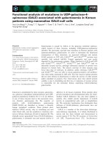
been reported. Of these, four (P28T, V32M, G36R and
H44Y) cluster in, or near, motif I (the galactokinase
signature motif). One (T288M) occurs in motif IV and two
(G346S and G349S) in motif V. Three others (R68C, A198V
and A384P) are located outside the conserved motifs. One
(M1I) abolishes the start codon of the gene (Fig. 1).
Disease causing mutations can be a valuable tool in
helping to assign functional roles to motifs and regions of
proteins. Furthermore, biochemical analysis of mutant pro-
teins can help in understanding the causes and symptoms of
Correspondence to R. J. Reece, School of Biological Sciences,
University of Manchester, 2.205 Stopford Building, Oxford Road,
Manchester, M13 9PT. United Kingdom.
Fax: + 44 161 275 5317, Tel.: + 44 161 275 5317,
E-mail:
Abbreviations: GHMP, galactokinase homoserine kinase mevalonate
kinase phosphomevalonate kinase; K
m,gal
, the Michaelis constant for
galactose; K
m,ATP
, the Michaelis constant for ATP; k
cat
, the turnover
number; k
cat
/K
m
, the specificity constant; K
IC
,thecompetitive
inhibition constant; K
IU
, the uncompetitive inhibition constant.
Enzymes: galactokinase (EC 2.7.1.6).
(Received 19 December 2002, revised 29 January 2003,
accepted 24 February 2003)
Eur. J. Biochem. 270, 1767–1774 (2003) Ó FEBS 2003 doi:10.1046/j.1432-1033.2003.03538.x
the inherited disease. We have established a bacterial
expression system for human galactokinase and have
purified active enzyme from this source. As there are a
number of different kinetic mechanisms reported for
galactokinases from different sources [18–23], we first deter-
mined the kinetic mechanism. The kinetic consequences of
the point mutations described above (with the exception of
M1I) were determined. Half were insoluble and four
exhibited altered kinetic constants with respect to the wild-
type enzyme. One was essentially unchanged in its enzymo-
logical properties compared to wild-type.
Experimental procedures
Cloning, expression and purification of GALK1
cDNA coding for the GALK1 gene was obtained from the
I.M.A.G.E. consortium (Clone ID: 3501788) [24]. The
sequence was amplified using PCR with primers designed to
introduce an NcoI restriction enzyme site and a His
6
-tag at
the 5¢ end and an EcoRI restriction enzyme site at the
3¢ end. This PCR fragment was then cloned into the NcoI
and EcoRI sites of pET21d (Novagen). The DNA sequence
of the entire GALK1 coding sequence was determined
(University of Manchester, Faculty of Medicine DNA
Sequencing Facility).
The recombinant plasmid was transformed into Escheri-
chia coli HMS174(DE3) cells (Novagen) for expression. One
to two litres of these cells were grown shaking in LB media
at 37 °C until the absorbance at 600 nm was approximately
0.6. The cultures were then induced with isopropyl thio-b-
D
-galactoside (2 m
M
, final concentration) and grown for a
further 2 h. Cells were harvested by centrifugation (10 min
at 5000 g), resuspended in approximately 20 mL 50 m
M
Hepes/OH pH 7.5, 150 m
M
NaCl, 10% (v/v) glycerol and
stored at )80 °C.
Cells were broken by sonication and cell debris removed
by centrifugation (20 min at 20 000 g). The supernatant
was passed over a column of 1–2 mL ProBond nickel-
agarose resin (Invitrogen) which had previously been
equilibrated in Buffer A (50 m
M
Hepes/OH, pH 7.5,
500 m
M
NaCl, 10% (v/v) glycerol). The column was
washed in this buffer until the absorbance at 280 nm was
negligible and then washed again in Buffer A supplemented
with 30 m
M
imidazole. Protein was eluted in Buffer A
supplemented with 250 m
M
imidazole. Fractions containing
GALK1 (as judged by SDS/PAGE) were dialysed overnight
at 4 °C against 50 m
M
Hepes/OH pH 7.5, 150 m
M
NaCl,
2m
M
EDTA, 1.4 m
M
2-mercaptoethanol, 10% (v/v)
glycerol. Protein concentrations were measured by the
method of Bradford [25]. The protein solution was frozen in
small aliquots in liquid nitrogen and stored at )80 °C.
Generation of point mutations
Mutations were introduced in to the GALK1-pET21d
construct using the Quik-Change method [26]. Briefly, the
PCR was used to amplify the entire plasmid from two,
complementary primers which both contained the desired
mutation. Template plasmid was then digested using the
restriction enzyme DpnI. Following transformation into
E. coli XL-1 Blue (Stratagene) and the isolation of single
colonies, plasmids were purified and the GALK1 coding
region sequenced in full to confirm the presence of the
mutation and that no other mutations had been introduced
during the PCR. All mutants were expressed and purified by
thesamemethodasthewild-type.
Galactokinase kinetics
Galactokinase activity was measured by coupling the
production of ADP to the reactions catalyzed by pyruvate
kinase and lactate dehydrogenase [12,23]. The decrease in
absorbance at 340 nm, which results from the oxidation of
NADH, was measured in a Multiskan Ascent microtitre
plate-reader. Reactions were carried out at 37 °C in a total
volume of 150 lL and each contained 20 m
M
Hepes/OH
pH 8.0, 150 m
M
NaCl, 5 m
M
MgCl
2
,1m
M
KCl, 10% (v/v)
glycerol, 1.0 m
M
NADH, 1 m
M
dithiothreitol, 400 l
M
phosphoenolpyruvate, 7.5 U pyruvate kinase (Sigma) and
10 U lactate dehydrogenase (Sigma). Reactions were initi-
ated by the addition of enzyme (concentrations ranged from
32 to 67 n
M
with the wild-type enzyme and from 67 to
700 n
M
with the mutants).
All data were analyzed by nonlinear curve fitting [27]
using the program GraphPad Prism (GraphPad Software
Inc.). Rates of reaction were obtained by fitting the
absorbance data to straight lines. These rates (v)were
fitted to the equation v ¼ V
max,app
[S]/(K
m,app
+ [S]) where
V
max,app
is the apparent maximum rate of reaction and
K
m,app
is the apparent Michaelis constant for the substrate,
S [28]. The turnover number (k
cat
) was calculated from the
equation k
cat
¼ V
max
/[E]
0
where [E]
0
is the total enzyme
concentration. From this the specificity constant, k
cat
/K
m
could be determined.
Product inhibition studies
The nature and magnitude of the inhibition by the product
galactose 1-phosphate was determined by observing the
effect of increasing concentrations of the compound on the
apparent turnover number and the apparent specificity
constants for both substrates. One substrate was held
constant at a saturating concentration (5 m
M
)andthe
kinetic constants determined over a range of inhibitor
concentrations. This was then repeated while holding the
other substrate at a constant concentration. Competitive
inhibition is characterized by an unchanging apparent
Fig. 1. Disease causing mutations in human galactokinase. The num-
bers I to V represent the conserved motifs in galactokinases [12].
Mutations that resulted in soluble protein on induction in E. coli are
shown above the bar representing the sequence of the protein, while
those that were insoluble are shown below.
1768 D. J. Timson and R. J. Reece (Eur. J. Biochem. 270) Ó FEBS 2003
turnover number and variation in the apparent specificity
constant according the equation (k
cat,app
/K
m,app
) ¼ (k
cat
/
K
m
) · K
IC
/([I] + K
IC
)whereK
IC
is the competitive inhibi-
tion constant and [I] is the concentration of the inhibitor. In
contrast, in uncompetitive inhibition the specificity constant
is invariant and the apparent turnover number varies
according to the equation k
cat,app
¼ k
cat
· K
IU
/([I] + K
IU
)
where K
IU
is the uncompetitive inhibition constant. In mixed
inhibition the apparent turnover number and specificity
constant vary and both K
IU
and K
IC
define the inhibition [28].
Solvent kinetic isotope effect
The solvent kinetic isotope effect was measured by deter-
mining the kinetic constants as described above in the
presence of increasing mole fractions of D
2
O(Aldrich).
Kinetic constants of the mutants
The equation for a two-substrate ternary complex
reaction is: v ¼ (k
cat
.[E]
0
.[gal].[ATP])/(K
I,ATP
.K
m,gal
+
K
m,gal
.[ATP] + K
mATP
.[gal] + [ATP].[gal]) where [gal] and
[ATP] are the concentrations of galactose and ATP,
respectively, K
I,ATP
is a constant relating to the dissociation
of the enzyme-ATP complex and K
m,gal
and K
m,ATP
are the Michaelis constants for galactose and ATP,
respectively. At any constant value of [gal] this simplifies
to v ¼ k
cat,app
.[E]
0
.[ATP]/(K
m,ATP,app
+[ATP]) where
k
cat,app
¼ k
cat
.[gal]/(K
m,gal
+ [gal]). A similar situation
holds if [ATP] is held constant [28]. Values for k
cat,app
were
obtained over a range of subsaturating constant concentra-
tions of ATP and galactose using a 5 · 5 concentration grid.
Nonlinear curve fitting was then used to derive values for
the kinetic constants.
Results
Active human galactokinase can be expressed
in
E. coli
Human galactokinase was expressed as an N-terminal His
6
fusion protein and purified on nickel-agarose resin (Fig. 2).
Typical yields were approximately 2 mg of GALK1 per litre
of bacterial culture. The protein is a monomer as judged by
analytical gel filtration (data not shown). The enzyme is
active (Fig. 3) with a turnover number (k
cat
)of8.7s
)1
,
K
m,gal
of 970 l
M
and K
m,ATP
of 34 l
M
. These values are of
the same order of magnitude as previously reported for the
yeast [23], rat [18,19] and human [29] enzymes. There is no
evidence for the glycosylation of human galactokinase
described during the purification or isolation of the enzyme
from human tissues, nor is there any anomalous migration
of bands on gels [29]. We therefore believe that post-
translational modifications do not play a significant role in
the functioning of the protein, and the activity that we
observe for the bacterially produced protein reflects that of
the native enzyme.
GALK1 has an ordered ternary complex mechanism
Galactokinases from different sources show a variety of
kinetic mechanisms. The enzyme from E. coli has been
Fig. 3. Kinetics of human galactokinase. (A) Determination of K
m,gal
.
Apparent turnover numbers were determined at different galactose
concentrations. The line shows the fit of these values to the equation
k
cat,app
¼ k
cat
.[gal]/(K
m,gal
+ [gal]) as described in Experimental pro-
cedures. (B) The determination of K
m,ATP
by the same method.
Fig. 2. Expression and purification of human galactokinase. The pro-
tein was expressed in E. coli HMS174(DE3) cells and purified on nickel
agarose.
Ó FEBS 2003 Disease-causing mutations in human galactokinase (Eur. J. Biochem. 270) 1769
reported to have a random ternary complex mechanism [20]
in which either ATP or galactose can be the first substrate to
bind. In contrast, galactokinases from rat [18,19] and yeast
[23] have an ordered, ternary complex mechanism in which
ATP binding precedes galactose binding. Plant galacto-
kinases also show an ordered mechanism, but one in which
galactose is the first substrate to bind [21,22]. Product
inhibition studies were undertaken with recombinant
human galactokinase in order to see which mechanistic
class it falls into (Fig. 4). a-
D
-Galactose 1-phosphate was
found to be an uncompetitive inhibitor with respect to
galactose (K
IU
¼ 28 ± 11 m
M
) and a mixed inhibitor with
respect to ATP (K
IU
¼ 39 ± 9 m
M
; K
IC
¼ 130 ±
90 m
M
). If galactose and galactose-1-phosphate bound to
the same form of the enzyme, competitive inhibition would
be observed [28]. As this is not the case, these two molecules
are unlikely to be the first substrate to bind and the last
product to be released from the enzyme. Therefore, the most
likely kinetic mechanism for GALK1 is an ordered ternary
complex one, in which ATP binds first. Using ADP as an
inhibitor was not possible using the enzyme-linked assay
system described here. The inhibition pattern we observed
was consistent only with an ordered ternary complex
mechanism with ATP binding first (out of all the common
mechanisms). If there were either a random mechanism or
an ordered one with galactose binding first, galactose-
1-phosphate would be a competitive inhibitor with respect
to galactose.
Proton transfer is unlikely to play a significant role
in the rate determining step of GALK1
Although the enzymes of the GHMP family share sequence
and structural similarity, there are differences in the
mechanism of catalysis. The structure of mevalonate kinase
shows an aspartate residue at an appropriate place in the
active site to act as catalytic base [9]. However, the active site
of homoserine kinase has no residues capable of acting as a
catalytic base [7] and catalysis is believed to be driven
through the stabilization of a transition state. A recent study
on the yeast enzyme, Gal1p, showed little variation of any
kinetic constant with pH and no significant deuterium
kinetic isotope effect [23]. This suggested that proton
transfer was unlikely to be important in the mechanism of
Gal1p and that this enzyme is likely to be similar
mechanistically to homoserine kinase.
Given the diversity of kinetic mechanisms among
galactokinases, we tested whether proton transfer is import-
ant in the reaction catalyzed by GALK1. Increasing the
mole fraction of D
2
O in the reaction mixture had essentially
no effect on the turnover number or the specificity constants
(Fig. 5). Other studies, in which there is a critical proton
transfer event in the rate determining step of the mechanism,
show a reduction in k
cat
of between 25 and 50% at a
deuterium mole fraction of 0.4 [30,31]. This level of
reduction would certainly have been observable in our
experimental system. Therefore in GALK1, like Gal1p and
Fig. 4. Human galactokinase has an ordered, ternary complex mechanism. Galactose 1-phosphate (G1P) is an uncompetitive inhibitor with respect
to galactose. (A) Galactose 1-phosphate causes a decrease in the apparent turnover number, k
cat,app
. The concentration of ATP was 5 m
M
.(B)
There is no change in the specificity constant, k
cat,app
/K
m,app
under the same conditions. Galactose 1-phosphate is a mixed inhibitor with respect to
ATP. (C) Galactose 1-phosphate causes a decrease in the apparent turnover number. The concentration of galactose was 5 m
M
. (D) Galactose
1-phosphate causes a decrease in the apparent specificity constant under the same conditions. This pattern of inhibition is consistent with an ordered
ternary complex mechanism in which ATP binds first.
1770 D. J. Timson and R. J. Reece (Eur. J. Biochem. 270) Ó FEBS 2003
homoserine kinase, proton transfer is unlikely to play a
major role in the rate-determining step of catalysis.
Several of the disease-causing mutations
are not soluble following induction in
E. coli
Although all the mutant galactokinases constructed could
be expressed in E. coli (as judged by the appearance of an
additional band of the expected molecular mass on SDS/
PAGE of cell extracts after induction), five (P28T, V32M,
G36R, T288M and A384P) were not present in the soluble
fraction after sonication and could not be purified (data not
shown).
The soluble mutants show altered kinetic constants
compared to the wild-type
The remaining mutants (H44Y, R68C, A198V, G346S and
G349S) were soluble on induction in E. coli and could be
purified in a similar manner to the wild-type enzyme. Yields
were comparable to that obtained with the wild-type, except
in the case of R68C where approximately fivefold less
soluble enzyme per litre of starting culture was obtained.
Each of these five mutants was an active galactokinase
and the kinetic constants for each could be determined
(Table 1). The kinetic consequences of a further mutation in
the highly conserved part of motif V, G347S were also
measured.
A variety of different kinetic phenotypes were observed.
G346S and G347S showed substantial reductions in turn-
over number. G347S also showed an increase in K
m,gal
as
did H44Y. Less dramatic effects were observed on K
m,ATP
with no mutant showing more than a fivefold change. The
most affected were H44Y and R68C. All three motif V
mutants (G346S, G347S and G349S) along with H44Y have
lower specificity constants for galactose and all the mutants
with the exception of A198V have lowered specificity
constants for ATP. Interestingly, A198V shows very similar
kinetic parameters to the wild-type enzyme.
Discussion
Human galactokinase, GALK1, has been expressed in and
purified from E. coli. The ability to produce good yields of
active protein in this way makes it possible to study the
biochemical consequences of mutations within the coding
sequence of the GALK1 gene.
The kinetic mechanism of GALK1 was shown to follow
an ordered ternary complex pathway in which ATP binds
first. GALK1 is therefore most similar to the rat and yeast
enzymes in its kinetic mechanism. The most likely cause of
this sort of mechanism is that ATP binding induces a
conformational change in the enzyme, which creates a
functional binding site for galactose. Identifying the nature
of this change and the residues involved in transmitting
information through the protein will be important chal-
lenges for the future. The absence of a deuterium kinetic
isotope effect suggests that GALK1 belongs to that group
of GHMP kinases in which proton transfer does not play a
major role in the rate determining step of catalysis.
That five of the 10 disease-causing mutations resulted in
insoluble protein in E. coli suggests that in these cases
protein folding and/or stability of the folded state may be
more important than enzymological defects. Generally these
mutations are associated with more severe clinical pheno-
types. Individuals who are homozygous for the P28T
mutation (which is common in Roma and Bosnian popu-
lations [32,33]) develop cataracts in the first few months or
years of life if galactose is not completely removed from the
diet. Blood galactokinase activities are low or zero [14].
Fig. 5. There is no solvent kinetic isotope effect in human galactokinase.
(A) The variation of k
cat
with mole fraction of deuterium oxide. These
values were obtained in an experiment in which the concentration of
ATP was varied and galactose was maintained at a saturating level
(5 m
M
). Similar results were obtained when ATP was the saturating
ligand and galactose concentration was varied (not shown). (B) The
variation in the specificity constant for galactose with mole fraction of
deuterium oxide. (C) The variation in the specificity constant for ATP
with mole fraction of deuterium oxide. Error bars show standard error.
Ó FEBS 2003 Disease-causing mutations in human galactokinase (Eur. J. Biochem. 270) 1771
A similar phenotype is seen in patients homozygous for
V32M [13]. When DNA encoding GALK1 with this
mutation was transfected into COS cells, no galactokinase
activity above background could be detected [13]. The
G36R mutation was detected in an individual who was
heterozygous for this mutation and a frameshift [15]. Blood
galactokinase activity was zero and transfection of this
mutant into COS cells also gave no activity [15]. T288M was
also observed in an individual who was heterozygous for
this and a frameshift mutation [16]. The patient had low
blood galactokinase activity and had been placed on a low
galactose diet and so no other symptoms had been observed.
A single individual was heterozygous for A384P and R68C
[16]. Like the T288M patient, the patient had been placed on
a low galactose diet before any symptoms could occur.
The M1I mutation [15] is assumed to cause loss of
galactokinase activity because the protein lacks its start
codon. If protein synthesis were to start at the next
methionine in the sequence, this would be M55 and would
result in deletion of the whole of motif I, the putative
galactose binding site. It is therefore not surprising that
transfection of this mutant sequence in to COS cells resulted
in no galactokinase activity [15].
H44Y and G349S were detected in a patient who was
heterozygous for these two mutations [15]. Although there
was zero blood galactokinase activity, transfection of either
mutant sequence in to COS cells gave low, but not zero,
levels of galactokinase activity. G346S (which was detected
in a patient who also had a seven base pair insertion in the
gene) gave similar results [15]. In general therefore the
soluble mutants tend to be those which occur in hetero-
zygotes along with more drastic mutations. Furthermore
where the activity of these mutants has been tested in vivo by
transfection into COS cells [15] they tend to give much
reduced, but not zero levels of activity in contrast to the
insoluble mutants. This gives us added confidence that our
conclusion that failure to produce soluble protein in E. coli
means that the protein is insoluble or unstable in humans is
correct.
Interestingly one mutant, A198V, has kinetic properties
that are very similar to the wild-type enzyme. This mutation
is also associated with the least severe clinical phenotype
[17]. Homozygotes show reduced blood galactokinase
activity (typically 10% of normal) and have a tendency to
develop cataracts later in life [17]. Studies on crude blood
extracts from homozygotes showed that K
m,gal
and K
m,ATP
were indistinguishable from the wild-type but that V
max
was
reduced by approximately 80%. The amount of protein that
could be detected immunologically was also reduced by
approximately the same amount [17]. This suggests that the
reduced blood galactokinase activity results not from
catalytic inefficiency of the enzyme but from reduced
amounts of the protein. This mutation may cause the
enzyme to be turned over more rapidly in human cells.
The five soluble mutations cause a variety of kinetic
consequences. The turnover number, k
cat
, reports on steps
in the reaction that occur after the formation of the enzyme-
ATP-galactose ternary complex including catalysis. All the
mutants have reduced turnover numbers, with the most
impaired being G346S and G347S. These residues are in
motif V which is believed (on the basis of comparison to the
structures of other GHMP family enzymes) to be adjacent
to the residues that form the active site. It is unlikely that
glycine can contribute much directly to stabilizing the
transition state. However the change of glycine to serine is
likely to make the peptide backbone much less flexible. This
in turn may make interactions between the active site
and the transition state less favourable, thereby reducing
catalytic efficiency.
Although K
m
values are often used as measures of
enzyme-substrate affinity, this is not strictly correct. More
accurately, it is an apparent dissociation constant referring
to all enzyme bound species of the substrate [34]. For
example, in the case of GALK1, K
m,ATP
does not just report
on the initial interaction between the enzyme and ATP, but
also on the dissociation of ATP from the ternary enzyme-
ATP-galactose complex and from any conformational
states that may occur prior to phosphate transfer. Two
mutants have large changes in K
m,gal
– H44Y and G347S.
H44 forms part of motif I, which is believed to interact with
galactose [12]. In the case of G347S, it seems that the
disruption of the peptide backbone that affects catalysis also
affects the binding of galactose at some point in the
reaction. Modest changes in K
m,ATP
are seen in H44Y and
R68C. That H44 influences the binding of both substrates
suggests that the binding sites are probably close in space.
R68 is not part of any conserved galactokinase motif, nor is
the residue well conserved between species. It is possible that
its kinetic changes result from structural alterations that are
propagated to the active site.
Specificity constants (k
cat
/K
m
) report on the interaction
between the enzyme and a particular substrate. Thus in the
case of GALK1, k
cat
/K
m,ATP
reports on the enzyme–ATP
interaction and k
cat
/K
m,gal
on the interaction between the
enzyme-ATP complex and galactose. The three mutations
in motif V (G346S, G347S and G349S) all have much
reduced specificity constants for galactose as does H44Y.
Failure to form a proper galactose-binding site is the most
Table 1. Kinetic constants of disease-causing mutations in GALK1.
Enzyme k
cat
(s
)1
) K
m,gal
(l
M
) K
m,ATP
(l
M
) k
cat
/K
m,gal
(LÆmol
)1
Æs
)1
) k
cat
/K
m,ATP
(10
5
· LÆmol
)1
Æs
)1
)
Wild-type 8.7 ± 0.5 970 ± 220 34 ± 4 8900 ± 2900 2.6 ± 0.4
H44Y 2.0 ± 0.1 7700 ± 4400 130 ± 9 270 ± 240 0.15 ± 0.02
R68C 3.9 ± 0.8 430 ± 150 110 ± 35 11000 ± 5600 0.35 ± 0.18
A198V 5.9 ± 0.1 660 ± 220 26 ± 1 8500 ± 4000 2.3 ± 0.2
G346S 0.4 ± 0.04 1100 ± 160 5 ± 2 400 ± 96 0.87 ± 0.37
G347S 1.1 ± 0.2 13000 ± 2000 89 ± 34 85 ± 21 0.12 ± 0.07
G349S 1.8 ± 0.1 1700 ± 480 39 ± 4 1100 ± 380 0.46 ± 0.07
1772 D. J. Timson and R. J. Reece (Eur. J. Biochem. 270) Ó FEBS 2003
likely cause of this in all these cases. All the mutants except
A198V have impaired specificity constants for ATP. Inter-
estingly, G346S has only a modest reduction in k
cat
/K
m,ATP
despite having a k
cat
that is approximately 20-fold reduced
compared to the wild-type. In this mutant K
m,ATP
is also
reduced (approximately sevenfold) and this compensates
partially. This must mean that although the free enzyme has
a slightly reduced affinity for ATP, a later stage in the
reaction pathway (perhaps the ternary complex) has an
enhanced affinity.
The enzymological consequences of disease-causing
mutations in human galactokinase have been investigated
in vitro. In general proteins produced from mutations which
give rise to the most severe clinical phenotypes are insoluble
when purified from E. coli, which may suggest that gross
structural changes have occurred in these proteins. The
results from the soluble mutants support the hypothesis that
motif I interacts with galactose and that motif V plays a
role in maintaining the structural integrity of the substrate
binding sites. The data represents the first step in the
analysis of the metabolic control of flux through the Leloir
pathway. Analysis of the galactokinase, its mutants, and the
other enzymes of the metabolic pathway using the principles
of a quantitative framework, such as metabolic control
analysis [35], may yield significant insights into the syn-
drome of galactosemia.
Acknowledgements
We are grateful for members of the Reece Laboratory for helpful
comments and suggestions. This work was funded by the Biotechno-
logy and Biological Sciences Research Council, UK and The
Leverhulme Trust, UK.
References
1. Frey, P.A. (1996) The Leloir pathway: a mechanistic imperative
for three enzymes to change the stereochemical configuration of a
single carbon in galactose. FASEB J. 10, 461–470.
2. Petry, K.G. & Reichardt, J.K. (1998) The fundamental
importance of human galactose metabolism: lessons from genetics
and biochemistry. Trends Genet. 14, 98–102.
3. Novelli, G. & Reichardt, J.K. (2000) Molecular basis of disorders
of human galactose metabolism: past, present, and future. Mol.
Genet. Metab. 71, 62–65.
4. Ai, Y., Zheng, Z., O’Brien-Jenkins, A., Bernard, D.J., Wynshaw-
Boris, T., Ning, C., Reynolds, R., Segal, S., Huang, K. & Stam-
bolian, D. (2000) A mouse model of galactose-induced cataracts.
Hum. Mol. Genet. 9, 1821–1827.
5. Bork, P., Sander, C. & Valencia, A. (1993) Convergent evolution
of similar enzymatic function on different protein folds: the
hexokinase, ribokinase, and galactokinase families of sugar
kinases. Protein Sci. 2, 31–40.
6. Zhou, T., Daugherty, M., Grishin, N.V., Osterman, A.L. &
Zhang, H. (2000) Structure and mechanism of homoserine kinase:
prototype for the GHMP kinase superfamily. Structure 8, 1247–
1257.
7. Krishna, S.S., Zhou, T., Daugherty, M., Osterman, A. & Zhang,
H. (2001) Structural basis for the catalysis and substrate specificity
of homoserine kinase. Biochemistry 40, 10810–10818.
8. Yang, D., Shipman, L.W., Roessner, C.A., Scott, A.I. & Sac-
chettini, J.C. (2002) Structure of the Methanococcus jannaschii
mevalonate kinase, a member of the GHMP kinase superfamily.
J. Biol. Chem. 277, 9462–9467.
9. Fu, Z., Wang, M., Potter, D., Miziorko, H.M. & Kim, J.J. (2002)
The structure of a binary complex between a mammalian meva-
lonate kinase and ATP. Insights into the reaction mechanism and
human inherited disease. J. Biol. Chem. 277, 18134–18142.
10. Romanowski, M.J., Bonanno, J.B. & Burley, S.K. (2002) Crystal
structure of the Streptococcus pneumoniae phosphomevalonate
kinase, a member of the GHMP kinase superfamily. Proteins 47,
568–571.
11. Bonanno, J.B., Edo, C., Eswar, N., Pieper, U., Romanowski,
M.J., Ilyin, V., Gerchman, S.E., Kycia, H., Studier, F.W., Sali, A.
& Burley, S.K. (2001) Structural genomics of enzymes involved in
sterol/isoprenoid biosynthesis. Proc. Natl Acad. Sci. USA 98,
12896–12901.
12. Platt, A., Ross, H.C., Hankin, S. & Reece, R.J. (2000) The
insertion of two amino acids into a transcriptional inducer con-
verts it into a galactokinase. Proc. Natl Acad. Sci. USA 97, 3154–
3159.
13. Stambolian, D., Ai, Y., Sidjanin, D., Nesburn, K., Sathe, G.,
Rosenberg, M. & Bergsma, D.J. (1995) Cloning of the galacto-
kinase cDNA and identification of mutations in two families with
cataracts. Nature Genet. 10, 307–312.
14. Kalaydjieva, L., Perez-Lezaun, A., Angelicheva, D., Onengut, S.,
Dye, D., Bosshard, N.U., Jordanova, A., Savov, A., Yanakiev, P.,
Kremensky, I., Radeva, B., Hallmayer, J., Markov, A., Nedkova,
V., Tournev, I., Aneva, L. & Gitzelmann, R. (1999) A founder
mutation in the GK1 gene is responsible for galactokinase defi-
ciency in Roma (Gypsies). Am.J.Hum.Genet.65, 1299–1307.
15. Kolosha, V., Anoia, E., de Cespedes, C., Gitzelmann, R., Shih, L.,
Casco, T., Saborio, M., Trejos, R., Buist, N., Tedesco, T., Skach,
W.,Mitelmann,O.,Ledee,D.,Huang,K.&Stambolian,D.
(2000) Novel mutations in 13 probands with galactokinase defi-
ciency. Hum. Mutat. 15, 447–453.
16. Hunter, M., Angelicheva, D., Levy, H.L., Pueschel, S.M. &
Kalaydjieva, L. (2001) Novel mutations in the GALK1 gene in
patients with galactokinase deficiency. Hum. Mutat. 17, 77–78.
17. Okano, Y., Asada, M., Fujimoto, A., Ohtake, A., Murayama, K.,
Hsiao, K.J., Choeh, K., Yang, Y., Cao, Q., Reichardt, J.K.,
Niihira, S., Imamura, T. & Yamano, T. (2001) A genetic factor for
age-related cataract: identification and characterization of a novel
galactokinase variant, ÔOsakaÕ,inAsians.Am.J.Hum.Genet.68,
1036–1042.
18. Ballard, F.J. (1966) Kinetic studies with liver galactokinase. Bio-
chem. J. 101, 70–75.
19. Walker, D.G. & Khan, H.H. (1968) Some properties of galacto-
kinase in developing rat liver. Biochem. J. 108, 169–175.
20. Gulbinsky, J.S. & Cleland, W.W. (1968) Kinetic studies of
Escherichia coli galactokinase. Biochemistry 7, 566–575.
21. Foglietti, M.J. & Percheron, F. (1976) Purification et me
´
canisme
d’action d’une galactokinase ve
´
ge
´
tale. Biochimie 58, 499–504.
22. Dey, P.M. (1983) Galactokinase of Vicia faba seeds. Eur. J.
Biochem. 136, 155–159.
23. Timson, D.J. & Reece, R.J. (2002) Kinetic analysis of yeast
galactokinase: Implications for transcriptional activation of the
GAL genes. Biochimie 84, 265–272.
24. Lennon, G., Auffray, C., Polymeropoulos, M. & Soares, M.B.
(1996) The I.M.A.G.E. Consortium: an integrated molecular
analysis of genomes and their expression. Genomics 33, 151–152.
25. Bradford, M.M. (1976) A rapid and sensitive method for the
quantitation of microgram quantities of protein utilizing the
principle of protein-dye binding. Anal. Biochem. 72, 248–254.
26. Wang, W. & Malcolm, B.A. (1999) Two-stage PCR protocol
allowing introduction of multiple mutations, deletions and inser-
tions using QuikChange site-directed mutagenesis. Biotechniques
26, 680–682.
27. Marquardt, D. (1963) An algorithm for least squares estimation of
nonlinear parameters. SIAM J. Appl. Math. 11, 431–441.
Ó FEBS 2003 Disease-causing mutations in human galactokinase (Eur. J. Biochem. 270) 1773
28. Cornish-Bowden, A. (1995) Fundamentals of Enzyme Kinetics.
Portland Press, London, UK.
29. Srivastava, S.K., Blume, K.G., van Loon, C. & Beutler, E. (1972)
Purification and kinetic properties of galactokinase from human
placenta. Arch. Biochem. Biophys. 150, 191–198.
30. Harris, M.N., Madura, J.D., Ming, L.J. & Harwood, V.J. (2001)
Kinetic and mechanistic studies of prolyl oligopeptidase from the
hyperthermophile Pyrococcus furiosus,. J. Biol. Chem. 276, 19310–
19317.
31. Zhou, J. & Adams, J.A. (1997) Is there a catalytic base in the active
site of cAMP-dependent protein kinase? Biochemistry 36, 2977–
2984.
32. Hunter, M., Heyer, E., Austerlitz, F., Angelicheva, D., Nedkova,
V.,Briones,P.,Gata,A.,dePablo,R.,Laszlo,A.,Bosshard,N.,
Gitzelmann,R.,Tordai,A.,Kalmar,L.,Szalai,C.,Balogh,I.,
Lupu, C., Corches, A., Popa, G., Perez-Lezaun, A. & Kalaydjieva,
L.V. (2002) The P28T mutation in the GALK1 gene accounts for
galactokinase deficiency in Roma (Gypsy) patients across Europe.
Pediatr. Res. 51, 602–606.
33. Reich, S., Hennermann, J., Vetter, B., Neumann, L.M., Shin, Y.S.,
Soling, A., Monch, E. & Kulozik, A.E. (2002) An unexpectedly
high frequency of hypergalactosemia in an immigrant Bosnian
population revealed by newborn screening. Pediatr. Res. 51,
598–601.
34. Fersht, A. (1999) Structure and Mechanism in Protein Science: a
Guide to Enzyme Catalysis and Protein Folding.W.H.Freeman,
New York.
35. Cascante, M., Boros, L.G., Comin-Anduix, B., de Atauri, P.,
Centelles, J.J. & Lee, P.W. (2002) Metabolic control analysis in
drug discovery and disease. Nature Biotechnol. 20, 243–249.
1774 D. J. Timson and R. J. Reece (Eur. J. Biochem. 270) Ó FEBS 2003