Báo cáo khoa học: Studies on structure–function relationships of indolepyruvate decarboxylase from Enterobacter cloacae, a key enzyme of the indole acetic acid pathway ppt
Bạn đang xem bản rút gọn của tài liệu. Xem và tải ngay bản đầy đủ của tài liệu tại đây (423.27 KB, 10 trang )
Studies on structure–function relationships of indolepyruvate
decarboxylase from
Enterobacter cloacae
, a key enzyme
of the indole acetic acid pathway
Anja Schu¨tz
1
, Ralph Golbik
1
, Kai Tittmann
1
, Dmitri I. Svergun
2,3
, Michel H. J. Koch
2
, Gerhard Hu¨ bner
1
and Stephan Ko¨ nig
1
1
Institut f
€
uur Biochemie, Fachbereich Biochemie/Biotechnologie, Martin-Luther-Universit
€
aat Halle-Wittenberg, Halle, Germany;
2
European Molecular Biology Laboratory, Hamburg Outstation, Hamburg, Germany;
3
Institute of Crystallography,
Russian Academy of Sciences, Moscow, Russia
Enterobacter cloacae, isolated from the rhizosphere of
cucumbers, produces large amounts of indole-3-acetic acid.
Indolepyruvate decarboxylase, the key enzyme in the
biosynthetic pathway of indole-3-acetic acid, catalyses the
formation of indole-3-acetaldehyde and carbon dioxide
from indole-3-pyruvic acid. The enzyme requires the cofac-
tors thiamine diphosphate and magnesium ions for catalytic
activity. Recombinant indolepyruvate decarboxylase was
purified from the host Escherichia coli strain JM109.
Specificity of the enzyme for the substrates indole-3-pyruvic
acid, pyruvic acid, benzoylformic acid, and seven benzoyl-
formic acid analogues was investigated using a continuous
optical assay. Stopped-flow kinetic data showed no indica-
tion for substrate activation in the decarboxylation reaction
of indole-3-pyruvic acid, pyruvic acid or benzoylformic acid.
Size exclusion chromatography and small angle X-ray
solution scattering experiments suggested the tetramer as
the catalytically active state and a pH-dependent subunit
association equilibrium. Analysis of the kinetic constants of
the benzoylformic acid analogues according to Hansch et al.
[Hansch, C., Leo, A., Unger, S.H., Kim, K.H., Nikaitani, D
& Lien, E.J. (1973) J. Med. Chem. 16, 1207–1216] and
comparison with indole-3-pyruvic acid conversion by pyru-
vate decarboxylases from Saccharomyces cerevisiae and
Zymomonas mobilis provided some insight into the catalytic
mechanism of indolepyruvate decarboxylase.
Keywords:
1
benzoylformate; small angle X-ray scattering;
steady-state kinetics; substrate specificity; thiamine
diphosphate.
The auxin indole-3-acetic acid, a phytohormone that
promotes cell growth and elongation and influences rooting,
is produced by plants [1,2] and plant-associated bacteria
[3,4]. Both tryptophan-dependent and -independent path-
ways of indole-3-acetic acid synthesis have been described
[5,6]. Plants use several mechanisms to control levels of the
active auxin indole-3-acetic acid. Thus, during different
developmental stages, indole-3-acetic acid may originate
from diverse sources for different auxin requirements, and
under different environmental conditions. Bacteria primar-
ily use tryptophan-dependent pathways. Phytopathogenic
strains follow the indoleacetamide pathway and plant
growth promoting strains the indolepyruvate pathway
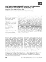
(Fig. 1). Indolepyruvate decarboxylase (IPDC), a key
enzyme in the second pathway, is a thiamine diphosphate
(ThDP)- and Mg
2+
-dependent homotetrameric enzyme
that catalyses the decarboxylation of indole-3-pyruvate to
indole-3-acetaldehyde [7–9]. Several microbial genes enco-
ding IPDC have been reported, including one from
Enterobacter cloacae isolated from the rhizosphere of
actively growing cucumbers [10]. DNA sequence analyses
revealed only one gene encoding EcIPDC. Its predicted
amino acid sequence comprises 552 residues and has
40% identity to PDC from Kluyveromyces lactis (DCPY
KLULA), 38% to PDC from Saccharomyces cerevisiae
(DCP1 YEAST), and % 32% to PDC from Zea mays
(DCP1 MAIZE), Oryza sativa (DCP1 ORYSA), Pisum
sativum (DCP1 PEA), and to PDC from Zymomonas
mobilis (DCPY ZYMO). In a previous study a molecular
mass of 240 kDa was determined for the native state of
EcIPDC, which corresponds to a tetramer with one type of
subunit [7]. A sharp pH optimum in the catalytic activity
of the enzyme assayed by quantitative HPLC was found at
pH 6.4–6.6. The native substrate indolepyruvate has a low
K
m
(15 l
M
) in contrast with that of pyruvate (2.5 m
M
)[7].
Correspondence to S. Ko
¨
nig, Institut fu
¨
r Biochemie, Fachbereich
Biochemie/Biotechnologie, Martin-Luther-Universita
¨
tHalle-
Wittenberg, Kurt-Mothes-Str. 3, 06099 Halle/Saale, Germany.
Fax: + 49 345 5527014, Tel.: + 49 345 5524829,
E-mail:
Abbreviations: IDPC, indolepyruvate decarboxylase; EcIPDC, IPDC
from Enterobacter cloacae;ScPDC,PDCfromSaccharomyces
cerevisiae; ZmPDC, PDC from Zymomonas mobilis;
ThDP, thiamine diphosphate.
Enzymes: indolepyruvate decarboxylase (indole-3-pyruvate carboxy
lyase; EC 4.1.1.74); pyruvate decarboxylase (2-oxoacid carboxy lyase;
EC 4.1.1.1).
Note:S
05
is the substrate concentration at half-maximum reaction
rate for enzymes displaying cooperativity characterized by sigmoid
reaction rate vs. substrate concentration plots.
(Received 5 February 2003, revised 17 March 2003,
accepted 2 April 2003)
Eur. J. Biochem. 270, 2322–2331 (2003) Ó FEBS 2003 doi:10.1046/j.1432-1033.2003.03602.x
The pyruvate derivatives a-keto glutarate and b-phenyl-
pyruvate inhibit EcIPDC activity. Indole and some similar
metabolites such as
L
-tryptophan, indole-3-lactate, indole-
3-acetaldehyde, tryptophol, and indole-3-acetate have
no effect on the enzymatic activity at a concentration of
0.5 m
M
[7].
Below, results on fast kinetics, substrate specificity, and
cofactor binding of EcIPDC are presented. For the kinetic
measurements a continuous optical assay was developed.
The pH- and cofactor-dependent subunit association beha-
viour was studied by small angle X-ray solution scattering.
The catalytic specificities of EcIPDC, ScPDC, and ZmPDC
for various substrates are discussed on the basis of their
crystal structures.
Materials and methods
Reagents
Horse liver alcohol dehydrogenase was from Roche
Molecular Biochemicals Inc., yeast alcohol dehydrogenase
and NADH were from Sigma-Aldrich Chemie GmbH.
Unless otherwise stated all reagents were purchased
from VWR International GmbH, Sigma-Aldrich Chemie
GmbH, Carl Roth GmbH, and AppliChem GmbH.
Bacterial strain and culture conditions
The plasmid (3.8 kb) pIP362 expressed in the Escherichia
coli strain JM109 (kindly provided by J. Koga, Meiji Seika
Kaisha Ltd, Satima, Japan) encodes the gene isolated from
E. cloacae [10]. A 6-L culture was grown for 24 h at 30 °C
in media containing 2% (w/v) tryptone, 1% (w/v) yeast
extract, 0.5% (w/v) sodium chloride, 0.1 m
M
thiamine,
0.1 m
M
magnesium sulphate, 0.01% (w/v) ampicillin, and
0.15
M
potassium phosphate pH 6.5. Expression of the
EcIPDC gene was induced by addition of 1 m
M
isopropyl
thio-b-
D
-galactoside. Cells were harvested by centrifugation,
quickly frozen in liquid nitrogen and stored at )80 °C.
Protein purification
About 25 g of cells were suspended in 40 mL 0.1
M
potassium phosphate pH 6.5, containing 10 m
M
ThDP,
Fig. 1. Scheme of the postulated biosynthesis pathway of indole-3-acetate from
L
-tryptophan in E. cloacae including the keto-enol tautomerism of
indolepyruvate, modified according to Koga et al.[7].1,
L
-tryptophan aminotransferase; 2, indolelactate dehydrogenase; 3, indolepyruvate
decarboxylase; 4, indoleacetaldehyde oxidase.
Ó FEBS 2003 Structure–function studies of E. cloacae IDPC (Eur. J. Biochem. 270) 2323
10 m
M
magnesium sulphate, 1 m
M
EDTA, 5 m
M
dithio-
threitol, and disrupted in a French press at 1200 bar
(Gaulin, APV Homogeniser GmbH, Lu
¨
beck, Germany).
The mixture was centrifuged at 70 000 g for 10 min and
the pellet was discarded. Nucleic acids were precipitated by
incubation with 0.1% (w/v) streptomycin sulphate for
45 min at 8 °C. A 15–30% (w/v) ammonium sulphate
fractionation was performed at a protein concentration of
20 mgÆmL
)1
. After centrifugation at 30 000 g for 5 min, the
precipitate was dissolved in 20 mL 50 m
M
Mes/NaOH
pH 6.5, containing 10 m
M
magnesium sulphate, 0.15
M
ammonium sulphate and 1 m
M
dithiothreitol. The solution
was applied to a Sephacryl S200HR column (5 · 95 cm,
Amersham Biosciences) and eluted with the same buffer at
1mLÆmin
)1
. The EcIPDC-containing fractions were pooled
and concentrated by precipitation with ammonium sulphate
(0.5 gÆmL
)1
). After centrifugation the precipitate was
dissolved in 20 m
M
Mes/NaOH pH 6.5, 1 m
M
dithiothre-
itol and this solution was desalted on a HiPrep desalting
column (2.6 · 10 cm, Amersham Biosciences) and applied
to a Source 15Q column (2.6 · 7 cm, Amersham Bio-
sciences). Elution was performed using a linear gradient of
120 mL 0–25% 20 m
M
Mes/NaOH pH 6.5, 1 m
M
dithio-
threitol, 0.25
M
ammonium sulphate. The fractions with the
highest catalytic activity and homogeneity were pooled,
quickly frozen in liquid nitrogen after addition of 0.2
M
ammonium sulphate, and stored at )80 °C.
SDS/PAGE
SDS/PAGE was carried out according to the method of
Laemmli [11]. Gels (10% (w/v) acrylamide) were stained
with Coomassie brillant blue G250.
Determination of enzyme concentration
The concentration of EcIPDC was determined spectro-
photometrically at 280 nm using a calculated molecular
absorption coefficient of
2
259 520
M
)1
Æcm
)1
[12]. ThDP-
containing samples were analysed using the method of
Bradford [13].
Syntheses of 4-substituted benzoylformates
Syntheses were performed according to Hallmann and
Ha
¨
gle [14] and Sultanov [15] by oxidation of the corres-
ponding acetophenones by SeO
2
.
Enzyme assays
EcIPDC was preincubated with 15 m
M
ThDP/Mg
2+
pH 6.5 at room temperature for 20 min to saturate the
enzyme with cofactors. Catalytic activities were measured
using a coupled optical test [16,17] in 10 m
M
Mes pH 6.5,
0.2 m
M
NADH and two different alcohol dehydrogenases
at 30 °C. Yeast alcohol dehydrogenase (15 UÆmL
)1
)was
used when the substrate was pyruvate, and horse liver
alcohol dehydrogenase (1 UÆmL
)1
) was used with the
substrates indolepyruvate, benzoylformate, and its 4-sub-
stituted analogues. The decarboxylation of indolepyruvate,
benzoylformate and its analogues was measured at 366 nm
to reduce interference with the substrates that considerably
absorb at 340 nm [17]. The conversion of pyruvate was
followed at 340 nm. Indolepyruvate was preincubated in
10 m
M
Mes pH 6.5 at 25 °C for 45 min to ensure the
generation of the ketone.
The ability of ScPDC and ZmPDC to decarboxylate
indolepyruvate was examined under the same conditions. In
the case of ZmPDC maximum enzyme concentration was
2.3 mgÆmL
)1
. Measurements with ScPDC were performed
at an enzyme concentration of 90 lgÆmL
)1
.
The plots of the reaction rate vs. substrate concentration
were fitted using the Michaelis–Menten equation in the case
of EcIPDC, or according to a substrate activation mech-
anism in the case of ScPDC [18]. For the substrate 4-NO
2
-
benzoylformate the kinetic constants were estimated from
the progress curves using the integrated Michaelis–Menten
equation.
Stopped-flow experiments were performed in 10 m
M
Mes
pH 6.5, 0.55 m
M
NADH, 450 UÆmL
)1
yeast alcohol dehy-
drogenase and 25 m
M
pyruvate at 10 °Cand30°C. With
0.5 m
M
indolepyruvate and 20 m
M
benzoylformate
160 UÆmL
)1
and 115 UÆmL
)1
horse liver alcohol dehydro-
genase were used, respectively
3
. For indolepyruvate the
EcIPDC concentration was 0.3 mgÆmL
)1
, for pyruvate it
was 85 lgÆmL
)1
, and for benzoylformate 3.5 lgÆmL
)1
.
The time-dependent inactivation of EcIPDC was exam-
ined under various conditions using the coupled optical test
with benzoylformate as substrate.
Cofactor binding experiments were performed in 10 m
M
Mes pH 6.5, 50 m
M
Mg
2+
,0.35m
M
NADH, 1 UÆmL
)1
horse liver alcohol dehydrogenase, and 25 m
M
benzoyl-
formate as substrate at 366 nm. To obtain the K
d
of the
primary binding of ThDP the measurements were started
with the apoenzyme–magnesium complex (10.7 lgÆmL
)1
)at
20 °C. The progress curves were fitted according to Wang
et al. [19] with an equation containing an exponential and
a linear term.
One unit of catalytic activity is defined as the amount of
enzyme converting 1 lmol substrateÆmin
)1
.
1
H NMR experiments on indolepyruvate
To study the keto-enol tautomerism of indolepyruvate,
1
H NMR spectra of a solution of 1 m
M
indolepyruvate
in 0.1
M
potassium phosphate pH 6.7 [10% (v/v) D
2
O]
were recorded 2–20 min after dissolving. Either presatu-
ration, or watergate pulse programs were used to
suppress the water signal. The chemical shifts refer to
3-(trimethylsilyl)-1-propane-sulphonate at 0 p.p.m. All
experiments were performed on a Bruker ARX 500
Avance NMR spectrometer (proton frequency
500.13 MHz) at 20 °C.
Determination of the molecular mass of EcIPDC
Size exclusion chromatography. A Fractogel EMD Bio-
SEC (S) column (2.6 · 70 cm, Merck KGaA) was equili-
brated with 100 m
M
Mes pH 6.0 and 100 m
M
ammonium
sulphate. EcIPDC was eluted with the same buffer at a flow
rate of 1 mLÆmin
)1
at 8 °C and detected by the protein
absorbance at 280 nm. Ferritin (450 kDa), catalase
(240 kDa), BSA (68 kDa), and ovalbumin (45 kDa)
(Combithek, calibration proteins for chromatography,
2324 A. Schu
¨
tz et al. (Eur. J. Biochem. 270) Ó FEBS 2003
Boehringer Mannheim GmbH) and ZmPDC (244 kDa)
were used as molecular mass standards.
Small angle X-ray solution scattering with synchrotron
radiation. Data were collected on the X33 camera of the
European Molecular Biology Laboratory outstation at
Hasylab at the storage ring DORIS of the Deutsches
Elektronen Synchrotron (DESY) in Hamburg [20–23].
Measurements were performed at a camera length of
1.9 m using multiwire proportional chambers with delay
line readout [22] at a temperature of 12 °CandEcIPDC
concentrations of about 5 mgÆmL
)1
in 60 m
M
buffer at
different pH values (citrate pH 5.6, Mes pH 6.1, BisTris
pH 6.4, Pipes pH 6.8, Mops pH 7.2, Hepes pH 7.5,
Tricine pH 8.1, Bicine pH 8.3, borate pH 9.2, Ches
pH 9.5, and Caps pH 10.2), 62.5 m
M
ammonium sul-
phate, 3 m
M
dithiothreitol in the presence or absence of
10 m
M
ThDP/Mg
2+
. The momentum transfer axis
(s ¼ 4psinh/k,where2h is the scattering angle and
k ¼ 0.15 nm, the X-ray wavelength) was calibrated using
collagen or tripalmitin as standards. The scattering
patterns were collected in 15 frames of 1 min to verify
the absence of radiation damage. The experimental data
was normalized to the intensity of the incident beam,
corrected for the detector response, and buffer scattering
was subtracted with propagation of statistical errors using
the program
SAPOKO
(D.I.SvergunandM.H.J.Koch,
unpublished data). To obtain the forward scattering
intensity I
0
and the radius of gyration (R
G
) the data
was processed with the program
GNOMOKO
[24]. The
molecular masses were calculated from the ratio of
the forward scattering intensity of the samples and of the
molecular mass standard BSA. The volume fractions of
monomers, dimers and tetramers were determined using
the program
OLIGOMER
(A. V. Sokolova, V. V. Volkov
4
and
D. I. Svergun, unpublished data). All protein concentra-
tions and pH values of the samples used for parameter
calculation were determined after the measurements.
Results
Purification of EcIPDC
The procedure, yielding the homogenous ThDP-free
enzyme, comprises four steps: streptomycin sulphate treat-
ment; ammonium sulphate precipitation; size exclusion
chromatography; and anion exchange chromatography.
After reconstitution of the holoenzyme the maximum
specific activity was % 1UÆmg
)1
using indolepyruvate as
substrate. EcIPDC is quite stable at 40 °C without any
further additions. A first-order rate constant of inactivation
of 10
)5
Æs
)1
was obtained in the elution buffer of the anion
exchange chromatography. Ammonium sulphate (0.2
M
)
stabilized the enzyme 14-fold. Further stabilization was
achieved by addition of ThDP/Mg
2+
. Addition of 10%
(v/v) glycerol had no effect. A molecular mass of 60 kDa per
subunit was determined by SDS/PAGE, corresponding to
the value calculated from the nucleotide sequence of the
structural gene. The N-terminal amino acid sequence of
the purified enzyme (Met-Arg-Thr-Pro-Tyr-Cys-Val-Ala) is
identical to that of the nucleotide sequence of the EcIPDC
gene (DCIP_ENTCL).
Molecular mass determination and pH dependence
of subunit association
A molecular mass of 245 kDa corresponding to a tetramer
was determined for EcIPDC at pH 6.0 by size exclusion
chromatography and confirmed by small angle X-ray
solution scattering with synchrotron radiation. Subunit
association depends on pH. At pH values between 5.6 and
6.0 the tetrameric form of EcIPDC predominates (R
G
, 3.95–
4.1 nm; R
G
is the so-called radius of gyration, one of the
structural parameters derived from a semi-logarithmic plot
of scattering data according to Guinier [25]) followed by
rapid dissociation into dimers at pH values between 6.7 and
7.4 (R
G
, 3.6–3.9 nm). At pH >8.0 R
G
values <3.1 nm
indicate a predominant monomeric state of the enzyme. In
the presence of cofactors the tetrameric holoenzyme is
stabilized in the range pH 5.6–7.5. Data analysis with the
program
OLIGOMER
demonstrated a pH-dependent equili-
brium between tetramers and dimers at lower pH and
dimers and monomers at higher pH. The presence of
cofactors strongly suppressed significant accumulation of
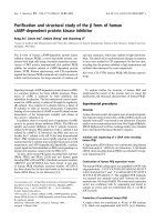
dimers (Fig. 2).
1
H NMR experiments on indolepyruvate
EcIPDC is unable to decarboxylate freshly prepared
solutions of indolepyruvate. Therefore, the chemical pro-
perties and purity of indolepyruvate were characterized by
1
H NMR spectroscopy. The
1
H NMR spectrum of freshly
dissolved indolepyruvate consists of the typical signals and
spin systems of the indole moiety (triplets of 5-H and 6-H at
7.12 and 7.18 p.p.m., doublets of 4-H and 7-H at 7.43 and
7.75 p.p.m and the singlet of 2-H at 7.81 p.p.m. with
identical integrals of all signals). The additional singlet of
the pyruvyl moiety at 6.65 p.p.m. with a relative integral of
1 with respect to the indole protons is consistent with the
occurrence of the enol form of indolepyruvate (Fig. 1). In
the course of the establishment of the equilibrium % 85% of
the enol form is converted into the ketone (half-time
% 8 min at 20 °C) as deduced from the appearance of
additional proton signals due to the indole part of
Fig. 2. pH dependence of the oligomeric state of EcIPDC. Volume
fractions were calculated from the scattering patterns with the program
OLIGOMER
in the absence of cofactors (A) and in the presence of 10 m
M
ThDP/Mg
2+
(B). (Circles and dotted lines, monomers; squares and
full lines, dimers; triangles and dashed lines, tetramers; lines are drawn
for better visualization only.)
Ó FEBS 2003 Structure–function studies of E. cloacae IDPC (Eur. J. Biochem. 270) 2325
indolepyruvate (triplets of 5-H and 6-H at 7.04 and
7.12 p.p.m., doublets of 4-H and 7-H at 7.39 and
7.42 p.p.m and the singlet of 2-H at 7.16 p.p.m. with
identical integrals of all signals) and to the b-CH
2
of the
pyruvyl part (singlet at 4.15 p.p.m., relative integral of 2),
respectively. As the ketone of indolepyruvate seems to be
the true substrate species of EcIPDC catalysis, indolepyru-
vate was always preincubated 45 min after dissolving to
ensure the equilibrium between the tautomers.
Steady state kinetics of EcIPDC
In all previous kinetic studies on EcIPDC, a discontinuous
assay based on HPLC was used [7]. To analyse the kinetic
behaviour of the enzyme in more detail, a coupled optical
assay was elaborated with alcohol dehydrogenase as
auxiliary enzyme, catalysing the aldehyde–alcohol conver-
sion similar to the assays established for pyruvate decarb-
oxylase (PDC) and benzoylformate decarboxylase [16,17].
A rather low substrate specificity of the auxiliary enzyme
horse liver alcohol dehydrogenase used in the latter assay
and the high k
cat
/K
m
value (330 s
)1
Æm
M
)1
) for the substrate
indole-3-acetaldehyde (data not shown) allowed application
of this assay. Under all conditions used, the reaction rate is
directly proportional to the EcIPDC concentration and
independent of the concentration of the auxiliary enzyme,
confirming that the coupled assay monitors the true rate of
EcIPDC catalysis. Figs 3 and 4 and Table 1 illustrate the
results of the steady-state kinetics for indolepyruvate,
pyruvate, benzoylformate, and 4-substituted benzoylfor-
mates (NO
2
-, Br-, Cl-, F-, C
2
H
5
-, CH
3
-, and CH
3
O-) as
substrates of EcIPDC. The enzyme has the highest catalytic
efficiency to the native substrate indolepyruvate, to 4-Cl-
benzoylformate and to 4-Br-benzoylformate (k
cat
/K
m
>100 s
)1
Æm
M
)1
). The K
m
of these substrates is <50 l
M
.
Benzoylformate has a rather low affinity to EcIPDC (K
m
1.65 m
M
), but its conversion resulted in the highest reaction
rate. Compared to benzoylformate all substitutions of this
substrate at the 4-position increase the affinity for the
enzyme and decrease the turnover rate considerably
(Table 1). The integrated Michaelis–Menten equation was
used for the determination of the kinetic constants of
4-NO
2
-benzoylformate, the substrate with the lowest K
m
(5 ± 0.5 l
M
) and a low k
cat
(0.4 ± 0.01 s
)1
). Pyruvate has
Fig. 3. Dependence of the catalytic activity of EcIPDC on the concen-
tration of substituted benzoylformates (Bf) measured in 10 m
M
Mes
pH 6.5 at 30 °C. The lines represent the fits to hyperbolic kinetics.
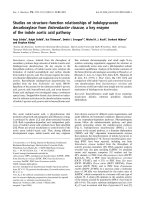
Fig. 4. Dependence of the catalytic activity of EcIPDC on the substrate concentration measured in 10 m
M
Mes pH 6.5 at 30 °C. The lines represent
the fits to hyperbolic kinetics. Insets, corresponding stopped-flow progress curves. Straight lines are linear fits. Measurements were monitored at
340 nm for pyruvate and at 366 nm for the other substrates with a coupled optical test. Ipyr, indolepyruvate; Bf, benzoylformate; Pyr, pyruvate.
2326 A. Schu
¨
tz et al. (Eur. J. Biochem. 270) Ó FEBS 2003
the lowest affinity of all substrates investigated (K
m
3.38 m
M
).
The straight lines in the plots according to Hanes [26]
(data not shown) demonstrate that there is no indication
for any substrate activation processes in EcIPDC catalysis.
The absence of lag phases in the progress curves obtained
from stopped-flow experiments using indolepyruvate,
pyruvate, and benzoylformate as substrates for EcIPDC
at 30 °C (Fig. 4 insets) and 10 °C (data not shown)
confirm these results. However, a weak substrate excess
inhibition (K
i
164 ± 16 m
M
) was observed for pyruvate
decarboxylation.
Examination of the decarboxylation of indolepyruvate
by ScPDC and ZmPDC
The ability of ScPDC and ZmPDC to decarboxylate
indolepyruvate was tested. In the case of ZmPDC no cata-
lytic activity was found with indolepyruvate as substrate,
even at very high enzyme concentrations (2.3 mgÆmL
)1
).
However, ScPDC is able to convert indolepyruvate and
displays, in contrast with EcIPDC, sigmoid kinetics as
illustrated in Fig. 5. A k
cat
of 3.81 ± 0.24 s
)1
andanS
0.5
-
value of 0.7 m
M
was calculated according to the rate
equation for substrate activation [18].
Cofactor binding experiments
Cofactor binding was studied by restoration of the catalytic
activity of the enzyme during reconstitution. Some progress
curves are presented in Fig. 6. The pseudo first-order rate
constants of reconstitution calculated from these time
courses show a hyperbolic dependence on the ThDP
concentration (at saturating Mg
2+
concentration), pointing
to a two-step mechanism of cofactor binding (Fig. 6 inset)
[27]. The calculated maximum rate constant of reconstitu-
tion is % 0.03 s
)1
and thus in the range of values determined
for other PDCs ([28]; J. Scha
¨
ffner
5,6
, unpublished data;
U. Mu
¨
cke
5,6
, unpublished data). A K
d
of 32.6 ± 4.6 l
M
determined for the binding of ThDP to EcIPDC is signifi-
cantly lower than that of other PDCs except ZmPDC [29].
Discussion
The purification procedure results in a homogenous ThDP-
free enzyme that is stabilized by the addition of 0.2
M
ammonium sulphate (inactivation rate constant 10
)6
s
)1
at
40 °C) or cofactors ThDP and Mg
2+
.Kogaet al.[7]also
described an effective stabilization of EcIPDC after addition
of the cofactors. The enzyme is destabilized at low ionic
strength. The stability of EcIPDC in aqueous solutions is
higher than that of other PDCs. The rate constant of
inactivation of PDC from Pisum sativum is about 10
)5
s
)1
at
37 °C, that of ScPDC is one order of magnitude higher [30].
Table 1. Catalytic constants for the decarboxylation of different substrates by EcIPDC. The K
m
for indolepyruvate was calculated under consid-
eration of the tautomer equilibrium (85% effective substrate concentration). k
cat
corresponds to the tetrameric enzyme, relative values to
indolepyruvate. The kinetic constants result from hyperbolic fits to the reaction rate vs. substrate concentration plots. In the case of 4-NO
2
-
benzoylformate values are obtained by fitting the progress curves using the integrated Michaelis–Menten equation (Ipyr, indolepyruvate, Pyr,
pyruvate, Bf, benzoylformate).
Substrates K
m
(l
M
)
K
m
(relative) k
cat
(s
)1
) k
cat (relative)
k
cat
/K
m
(s
)1
Æm
M
)1
)
Ipyr 20 ± 1.3 1.0 3.9 ± 0.07 1.0 199
Pyr 3381 ± 179 169.1 3.5 ± 0.08 0.9 1
Bf 1646 ± 32 82.3 46.4 ± 1.23 11.9 28
4-NO
2
-Bf 5 ± 0.5 0.25 0.4 ± 0.01 0.1 80
4-Cl-Bf 48 ± 2.0 2.4 5.3 ± 0.05 1.4 110
4-Br-Bf 19 ± 1.0 0.95 3.2 ± 0.03 0.8 168
4-F-Bf 617 ± 32.0 30.9 22.7 ± 0.57 5.8 37
4-C
2
H
5
-Bf 111 ± 3.5 5.6 5.7 ± 0.06 1.5 51
4-CH
3
-Bf 127 ± 7.0 6.4 4.5 ± 0.08 1.2 35
4-CH
3
O-Bf 1043 ± 33.0 52.2 3.5 ± 0.09 0.9 3
Fig. 5. Dependence of the catalytic activity of ScPDC on the indole-
pyruvate concentration. Measurements were carried out at 90 lgÆmL
)1
ScPDC in 0.1
M
Mes/NaOH pH 6.5 at 30 °Cand366nmwitha
coupled optical test. (Circles, experimental data; solid line, fit accord-
ing to the equation v([S]) ¼
V
max
Á½S
2
A þ BÁ½SþS
2
[18]).
Ó FEBS 2003 Structure–function studies of E. cloacae IDPC (Eur. J. Biochem. 270) 2327
Stowe [31] postulated that indolepyruvate crystallizes in
the enol form and is converted into the ketone at pH 8.0
and 25 °C within 20 min. Hydroxyphenylpyruvate and
phenylpyruvate behave in a similar manner [32]. Schwarz
and Bitancourt [33] demonstrated the tautomerism of
indolepyruvate by TLC. Our time-dependent
1
HNMR
measurements of aqueous solutions of indolepyruvate
confirm these results. After 20 min incubation at 20 °C,
85% of the substrate is present as ketone. The remaining
15% is probably responsible for the formation of highly
conjugated aromatic structures causing the well-known
reddish discoloration of aqueous solutions of the substance.
No EcIPDC activity is detectable with freshly prepared
solutions of indolepyruvate as substrate, but maximum
catalytic activity is obtained after incubation for about
45 min. Thus it can be concluded that only the ketone of
indolepyruvate is the substrate for the enzyme.
Application of a continuous optical assay for the steady-
state measurements modified according to Weiss et al.[17]
allowed detailed kinetic analysis of substrate specificity and
cofactor binding of EcIPDC. The enzymatic conversion of
all substrates studied in this work (pyruvate, the native
substrate of PDC, benzoylformate, the native substrate of
benzoylformate decarboxylase together with the 4-substi-
tuted derivatives, indolepyruvate, the native substrate of
IPDC) results in hyperbolic plots of catalytic activity vs.
substrate concentration (Figs 3 and 4). Corresponding
straight lines in Hanes plots (data not shown) and the
absence of lag phases in the stopped-flow time courses
(Fig. 4 insets) clearly demonstrates that there is no indica-
tion for substrate activation behaviour in the EcIPDC
catalysed reaction of the substrates indolepyruvate, pyru-
vate, and benzoylformate. The same holds true for the
ZmPDC catalysed reaction with pyruvate as substrate [34],
but contrasts with all other PDCs exhibiting sigmoid
dependencies in the plots of catalytic activity vs. substrate
concentration [30,35–37]. The kinetic constants of EcIPDC
summarized in Table 1 illustrate that indolepyruvate has the
highest catalytic efficiency (k
cat
/K
m
¼ 199 s
)1
Æm
M
)1
). Sur-
prisingly, benzoylformate is converted more rapidly than
thenativesubstrate(k
cat
46.4 s
)1
), but it shows a K
m
value
(1.65 m
M
) about 80 times higher. In contrast, the kinetic
constants of 4-Cl-benzoylformate and 4-Br-benzoylformate
are comparable to that of the native substrate indolepyru-
vate. Both halogenations seem to mimic the best substrate
surrogates of indolepyruvate. The highest K
M
value and
the lowest specificity are found for pyruvate and only this
substrate displays a weak substrate excess inhibition (K
i
164 m
M
). The K
m
values determined for indolepyruvate and
pyruvate correspond to those found by Koga et al.[7]using
a discontinuous quantitative HPLC assay (15 l
M
and
2.5 m
M
, respectively). Interestingly, the K
m
value of pyru-
vate in EcIPDC catalysis is similar to that found for all
other PDCs and the same holds true for the weak substrate
excess inhibition. However, the corresponding k
cat
value of
EcIPDC is only about 2% of that of other PDCs.
Hammett [38] developed a method to calculate the
electronic effect of a substituent from studies on the
dissociation of substituted benzoic acids in aqueous solu-
tion. The corresponding constants are only of restricted
value for other reactions. The modified substituent con-
stants r
p
recommended by Hansch et al.[39]werefoundto
be most suitable in the present case. The analysis of the
kinetic constants of the 4-substituted benzoylformates as
substrates for EcIPDC demonstrates that the dependence of
the logarithm of k
cat
/k
cat
0
vs. the substituent constant r
p
(Fig. 7) results in two linear plots with opposite slopes, one
for the electron-donating substituents with a value of about
4.4, and one for the electron-withdrawing substituents with
a value of about )2.5. This is indicative of an opposite effect
of the electron-withdrawing and electron-donating substi-
tuents on different rate-limiting steps in EcIPDC catalysis
(formation of mandelyl-ThDP, decarboxylation or alde-
hyde release), with a change in rate limiting step. To
summarize, in EcIPDC catalysed reactions all substituents
reduce the k
cat
value as compared with the unmodified
benzoylformate; this is also the case for catalysis by
benzoylformate decarboxylase from Pseudomonas putida
[17]. However, in ScPDC [40,41] all benzoylformates with
electron-withdrawing substituents exhibit a higher reaction
rate and all benzoylformates with electron-donating sub-
stituents have a lower one. EcIPDC binds all 4-substituted
benzoylformates with a higher affinity than the unsubsti-
tuted benzoylformate as is the case in ScPDC. With the
exception of 4-methoxybenzoylformate the substituted
benzoylformates have a lower affinity for benzoylformate
decarboxylase than does benzoylformate itself.
The hyperbolic dependence of the rate constants of
reconstitution, calculated from the corresponding progress
curves, on the concentration of ThDP (Fig. 6 inset) is
indicative of a two-step mechanism of cofactor binding as
Fig. 6. Progress curves of the reconstitution of EcIPDC with ThDP
measured by restoration of the catalytic activity of the formed holo-
enzyme for the substrate benzoylformate (25 m
M
)in10 m
M
Mes pH 6.5,
50 m
M
Mg
2+
,0.35m
M
NADH, and 1 UÆmL
-1
horse liver alcohol
dehydrogenase at 20 °C. The reaction was started with EcIPDC
(10.7 lgÆmL
)1
) at ThDP concentrations of 250, 120, 20, 12, 6, 3, 1.5, 1
and 0.5 l
M
(from left to right). Inset, dependence of the rate constant
of reconstitution on the ThDP concentration, calculated from the
progress curves.
2328 A. Schu
¨
tz et al. (Eur. J. Biochem. 270) Ó FEBS 2003
described previously by Schellenberger and Hu
¨
bner [27] and
Eppendorfer et al. [42] for ScPDC. The reconstitution starts
with binding of ThDP and Mg
2+
to the apoenzyme,
followed by a conformational change to the catalytically
active holoenzyme. Similar behaviour was found for
ZmPDC (J. Scha
¨
ffner
7
, unpublished data), ScPDC [28] and
PDC from Pisum sativum (U. Mu
¨
cke
8
, unpublished data). A
resulting K
d
value of % 33 l
M
for the primary binding of
ThDP to the enzyme saturated with magnesium ions
illustrates a significantly higher affinity of the cofactor
ThDPtoEcIPDCthantoScPDCandPDCfromPisum
sativum (150–300 l
M
). Even a higher affinity was found for
ZmPDC [29].
A molecular mass corresponding to a tetramer of
EcIPDC at pH 6.0 was determined by two independent
methods, size exclusion chromatography and small angle
X-ray solution scattering. These results suggest that the
tetramer is stable in aqueous solution even without cofac-
tors and that this oligomeric state is catalytically active in
the presence of cofactors. Evaluation of the scattering
experiments with ThDP-free EcIPDC demonstrates a pH-
dependent equilibrium between tetramers, dimers and even
monomers. A similar behaviour (without occurrence of a
monomer fraction) was described for PDCs from various
organisms, but not for ZmPDC, where the tetramer is stable
from pH 5 to pH 9 [43]. The cofactors ThDP and Mg
2+
stabilize the tetrameric state of EcIPDC up to pH 7.5
(Fig. 2). A similar stabilization up to pH 8.5 was found for
ScPDC [44]. The quality of the scattering patterns allowed
the calculation of volume fractions of different oligomeric
states of EcIPDC illustrating the pH-dependent subunit
association equilibrium and demonstrating a further disso-
ciation of EcIPDC into monomers at extreme alkaline pH
values also described by Koga et al.[7].
As the crystal structure analysis of EcIPDC revealed
some interesting similarities to other PDC species – a
ScPDC-like open topology of the substrate binding site
and a ZmPDC-like dimer assembly in the tetramer [45] –
the conversion of indolepyruvate by those related PDCs
was investigated. As expected from structural data
[46,47], ZmPDC is not able to cleave indolepyruvate
even at very high enzyme concentrations, whereas
ScPDC decarboxylates indolepyruvate with a k
cat
of
3.81 ± 0.24 s
)1
, a value similar to that of EcIPDC
(3.9 ± 0.07 s
)1
).InthecaseofZmPDCthesizeofthe
active site cavity is restricted by several amino acid
changes [45,46]. In contrast, this bulky substrate fits into
the active site of ScPDC and is decarboxylated. Differ-
ences between the catalytic cleavage of indolepyruvate by
ScPDC and EcIPDC can be found in the substrate affinity
and in the reaction rate vs. substrate concentration plot.
EcIPDC has a high affinity (K
M
20 l
M
) for indolepyruvate
and follows Michaelis–Menten kinetics, whereas ScPDC
exhibits sigmoid kinetics with a considerably lower affinity
for the substrate (S
0.5
¼ 0.7 m
M
) (Fig. 5). The S
0.5
values
for indolepyruvate and pyruvate (1.1 m
M
at pH 6.0;
J. Ermer
9
, unpublished data) are in the same range for
ScPDC.
As in ZmPDC and plant PDCs prominent amino acid
residues that restrict the size of the active site are conserved,
such as Trp392 and Trp551 (ZmPDC numbering), one can
assume that plant PDCs are also unable to accept indole-
pyruvate as substrate. Consequently, other pathways for the
biosynthesis of the phytohormone indoleacetic acid must
exist, not excluding the existence of a specific plant IPDC.
Yeast PDCs which do not possess such conserved space
filling amino acid residues, have a more open topology of
the substrate binding cavity and should thus presumably be
capable of using indolepyruvate as substrate, although with
a lower specificity than EcIPDC.
In the active site, several amino acid residues assumed to
play an important role in catalysis, such as Asp29, His115,
His116, and Glu468 (EcIPDC numbering), are conserved in
ZmPDC [48,49], ScPDC [50] and IPDCs suggesting a
similar catalytic mechanism. Differences in tetramer pack-
ing, several amino acid exchanges in the substrate binding
pocket and the diverse constitution of the C-terminal helix
covering the active site, are thought to be responsible for
different specificities of these enzymes [45].
Acknowledgements
We thank J. Koga (Meiji Seika Kaisha Ltd, Saitama, Japan) for
providing the plasmid pIP362 and K P. Ru
¨
cknagel (Max-Planck-
Society, Research Unit ÔEnzymology of protein foldingÕ,Halle)forthe
N-terminal sequencing. This work was supported by the travel expense
fund of Hasylab/Desy Hamburg, the Graduiertenkolleg of Sachsen-
Anhalt, the Deutsche Forschungsgemeinschaft, and the Fonds der
Chemischen Industrie.
References
1. Thimann, K.V. (1969) Physiology of Plant Growth and Develop-
ment (Wilkins, M.B., ed.), 3–45. McGraw-Hill, London.
2. Sheldrake, A.R. (1973) Production of hormones in higher plants.
Biol. Rev. Chamb. Philos. Soc. 48, 509–559.
Fig. 7. Plot of log(k
cat
/k
cat
0
) of EcIPDC catalysed decarboxylation of
the 4-substituted benzoylformates vs. the substituent constants according
to Hansch et a l.[39].The k
cat
value corresponds to unmodified ben-
zoylformate.
Ó FEBS 2003 Structure–function studies of E. cloacae IDPC (Eur. J. Biochem. 270) 2329
3. Patten, C.L. & Glick, B.R. (1996) Bacterial biosynthesis of indole-
3-acetic acid. Can. J. Microbiol. 42, 207–220.
4. Costacurta, A. & Vanderleyden, J. (1995) Synthesis of phyto-
hormones by plant-associated bacteria. Crit.Rev.Microbiol.21,
1–18.
5. Bartel, B. (1997) Auxin biosynthesis. Ann. Rev. Plant Mol. Biol.
48, 51–66.
6. Bartel, B., LeClere, S., Magidin, M. & Zolman, B.K. (2001) Inputs
to the active indole-3-acetic acid pool: De novo synthesis, con-
jugate hydrolysis, and indole-3-butyric acid b-oxidation. J. Plant
Growth Regul. 20, 198–216.
7. Koga,J.,Adachi,T.&Hidaka,H.(1992)Purificationandchar-
acterization of indolepyruvate decarboxylase. J. Biol. Chem. 267,
15823–15828.
8. Koga, J., Syono, K., Ichikawa, T. & Adachi, T. (1994) Involve-
ment of 1-tryptophan aminotransferase in indole-3-acetic acid
biosynthesis of Enterobacter cloacae. Biochim. Biophys. Acta 1209,
241–247.
9. Koga, J. (1995) Structure and function of indolepyruvate
decarboxylase, a key enzyme in indole 3-acetic acid biosynthesis.
Biochim. Biophys. Acta 1249, 1–13.
10. Koga, J., Adachi, T. & Hidaka, H. (1991) Molecular cloning of the
gene for indolepyruvate decarboxylase from Enterobacter cloacae.
Mol. Gen. Genet. 226, 10–16.
11. Laemmli, U.K. (1970) Cleavage of structural proteins during
the assembly of the head of bacteriophage T4. Nature 227,
680–685.
12. Gill, S.C. & Hippel, P.H. (1989) Calculation of protein extinction
coefficients from amino acid sequence data. Anal. Biochem. 182,
319–326.
13. Bradford, M.M. (1976) A rapid and sensitive method for the
quantitation of microgram quantities of protein utilizing the
principle of protein-dye binding. Anal. Biochem. 72, 248–254.
14. Hallmann, G. & Ha
¨
gle, H. (1963) Benzofuranderivate: Trypta-
min-, Serotonin- und Melatonin-Analoga. Liebigs Ann. Chem.
662, 147–159.
15. Sultanov, A.S. (1986) Organikum. (Becker, H., & Berger, W., eds.),
pp. 354–355. VEB Deutscher Verlag der Wissenschaften, Berlin.
16. Holzer, H., Schultz, G., Villar-Palasi, C. & Ju
¨
ntgen-Sell, J.
(1956) Isolierung der Hefecarboxylase und Untersuchung u
¨
ber
die Aktivita
¨
t des Enzyms in lebenden Zellen. Biochem. Z. 327,
331–344.
17. Weiss, P.M., Garcia, G.A., Kenyon, G.L. & Cleland, W.W. (1988)
Kinetics and mechanism of benzoylformate decarboxylase using
13
C and solvent deuterium isotope effects on benzoylformate and
benzoylformate analogues. Biochemistry 27, 2197–2205.
18. Krieger, F., Spinka, M., Golbik, R., Hu
¨
bner, G. & Ko
¨
nig, S.
(2002) Pyruvate decarboxylase from Kluyveromyces lactis. An
enzyme with an extraordinary substrate activation behaviour.
Eur. J. Biochem. 269, 3256–3263.
19. Wang, J., Golbik, R., Seliger, B., Spinka, M., Tittmann, K.,
Hu
¨
bner, G. & Jordan, F. (2001) Consequences of a modified
putative substrate-activation site on catalysis by yeast pyruvate
decarboxylase. Biochemistry 40, 1755–1763.
20. Koch, M.H.J. & Bordas, J. (1983) X-ray diffraction and scattering
on disordered systems using synchrotron radiation. Nucl. Instrum.
Methods 208, 461–469.
21. Boulin, C., Kempf, R., Koch, M.H.J. & McLaughlin, S.M. (1986)
Data appraisal, evaluation and display for synchrotron radiation
experiments: hardware and software. Nucl. Instrum. Methods
A249, 399–407.
22. Boulin, C.J., Kempf, R., Gabriel, A. & Koch, M.H.J. (1988) Data
acquisition systems for linear and area X-ray detectors using delay
line readout. Nucl. Instrum. Methods A269, 312–320.
23. Gabriel, A. & Dauvergne, F. (1982) The localisation method used
at EMBL. Nucl. Instrum. Methods 201, 223–224.
24. Svergun, D.I. (1992) Determination of the regularization param-
eter in indirect-transform methods using perceptual criteria.
J. Appl. Crystallogr. 25, 495–503.
25. Guinier, A. (1939) La diffraction de rayons X aux tre
`
spetits
angles: application a
`
l’e
´
tude de phe
´
nome
`
nes ultramicrosopiques.
Ann. Phys. (Paris) 12, 161–237.
26. Hanes, C.S. (1932) Studies on plant amylases. 1. The effect of
starch concentration upon the velocity of hydrolysis by the amy-
lase of germinated barley. Biochem. J. 26, 1406–1421.
27. Schellenberger, A. & Hu
¨
bner, G. (1967) Zur Theorie der
Thiaminpyrophosphat-Wirkung, IV. Mechanismus und Kinetik
der Rekombination und daraus abgeleitete Bindungsverha
¨
ltnisse
im aktiven Zentrum der Hefe-Pyruvatdecarboxylase. Hoppe-S. Z.
Physiol. Chem. 348, 491–500.
28. Killenberg-Jabs, M., Ko
¨
nig, S., Hohmann, S. & Hu
¨
bner, G.
(1996) Purification and characterisation of the pyruvate decar-
boxylase from a haploid strain of Saccharomyces cerevisiae. Biol.
Chem. Hoppe-S. 377, 313–317.
29. Diefenbach, R.J. & Duggleby, R.G. (1991) Pyruvate decarboxy-
lase from Zymomonas mobilis structure and reactivation of
apoenzyme by the cofactors thiamin diphosphate and magnesium
ion. Biochem. J. 276, 439–445.
30. Mu
¨
cke, U., Ko
¨
nig, S. & Hu
¨
bner, G. (1995) Purification and
characterisation of pyruvate decarboxylase from pea seeds (Pisum
sativum cv. Miko). Biol. Chem. Hope-Seyler 376, 111–117.
31. Stowe, B.B. (1955) The production of indoleacetic acid by bac-
teria. Biochem. J. 61, 9–10.
32. Knox, W.E. & Pitt, B.M. (1957) Enzymic catalysis of the keto-
enol tautomerization of phenylpyruvic acids. J. Biol. Chem. 225,
675–688.
33. Schwarz, K. & Bitancourt, A.A. (1960) Further evidence of tau-
tomerism in chromatograms of indolyl 3-pyruvic acid. Biochem. J.
75, 182–187.
34. Bringer-Meyer, S., Schimz, K.L. & Sahm, H. (1986) Pyruvate
decarboxylase from Zymomonas mobilis. Isolation and partial
characterization. Arch. Microbiol. 146, 105–110.
35. Hu
¨
bner, G., Weidhase, R. & Schellenberger, A. (1978) The
mechanism of substrate activation of pyruvate decarboxylase: a
first approach. Eur. J. Biochem. 92, 175–181.
36. Rivoal, J., Ricard, B. & Pradet, A. (1990) Purification and partial
characterization of pyruvate decarboxylase from Oryza sativa L.
Eur. J. Biochem. 194, 791–797.
37. Zehender, H., Tresher, D. & Ullrich, J. (1987) Improved puri-
fication of pyruvate decarboxylase from wheat germ. 1st partial
characterization and comparison with the yeast enzyme. Eur. J.
Biochem. 167, 149–154.
38. Hammett, L.P. (1937) The effect of structure upon the reactions of
organic compounds. Benzene derivatives. J. Am. Chem. Soc. 59,
96–103.
39. Hansch,C.,Leo,A.,Unger,S.H.,Kim,K.H.,Nikaitani,D.&
Lien, E.J. (1973) Aromatic substituent constants for structure-
activity correlations. J. Med. Chem. 16, 1207–1216.
40. Lehmann, H., Fischer, G., Hu
¨
bner, G., Kohnert, K.D. & Schel-
lenberger, A. (1973) The influence of steric and electronic para-
meters on the substrate behavior of oxo acids to yeast pyruvate
decarboxylase. Eur. J. Biochem. 32, 83–87.
41. Hu
¨
bner, G., Atanassova, M. & Schellenberger, A. (1986)
Investigations on the pyruvate decarboxylase catalysed oxidative
decarboxylation of 2-oxoacids by 2.6-dichlorophenolindophenol.
Biomed. Biochim. Acta 45, 823–832.
42. Eppendorfer, S., Ko
¨
nig,S.,Golbik,R.,Neef,H.,Lehle,K.,
Jaenicke, R., Schellenberger, A. & Hu
¨
bner, G. (1993) Effects
of metal ions, thiamine diphosphate analogues and subunit
interactions on the reconstitution behaviour of pyruvate decar-
boxylase from brewer’s yeast. Biol. Chem. Hoppe-Seyler 374,
1129–1134.
2330 A. Schu
¨
tz et al. (Eur. J. Biochem. 270) Ó FEBS 2003
43. Ko
¨
nig, S., Svergun, D.I., Volkov, V.V., Feigin, L.A. & Koch,
M.H.J. (1998) Small-angle X-ray solution scattering studies on
ligand induced subunit interactions of the thiamine diphosphate
dependent enzyme pyruvate decarboxylase from different organ-
isms. Biochemistry 37, 5329–5334.
44. Ko
¨
nig, S., Svergun, D., Koch, M.H.J., Hu
¨
bner, G. & Schellen-
berger, A. (1993) The influence of the effectors of yeast pyruvate
decarboxylase (PDC) on the conformation of the dimers and
tetramers and their pH-dependent equilibrium. Eur. Biophys. J.
22, 185–194.
45. Schu
¨
tz, A., Sandalova, T., Ricagno, S., Hu
¨
bner, G., Ko
¨
nig, S. &
Schneider, G. (2003) Crystal structure of indolepyruvate
decarboxylase from Enterobacter cloacae, an enzyme involved in
the biosynthesis of the plant hormone indole-3-acetic acid. Eur. J.
Biochem. 270, 2312–2321.
46. Dobritzsch, D., Ko
¨
nig, S., Schneider, G. & Lu, G. (1998) High
resolution crystal structure of pyruvate decarboxylase from
Zymomonas mobilis. J. Biol. Chem. 273, 20196–20204.
47. Arjunan, P., Umland, T., Dyda, F., Swaminathan, S., Furey, W.,
Sax,M.,Farrenkopf,B.,Gao,Y.,Zhang,D.&Jordan,F.(1996)
Crystal structure of the thiamine diphosphate-dependent enzyme
pyruvate decarboxylase from the yeast Saccharomyces cerevisiae
at 2.3 A
˚
resolution. J. Mol. Biol. 256, 590–600.
48. Chang, A.K., Nixon, P.F. & Duggleby, R.G. (1999) Aspartate-27
and glutamate-473 are involved in catalysis by Zymomonas
mobilis pyruvate decarboxylase. Biochem. J. 339 (2), 255–260.
49. Huang, C.Y., Chang, A.K., Nixon, P.F. & Duggleby, R.G. (2001)
Site-directed mutagenesis of the ionizable groups in the active site
of Zymomonas mobilis pyruvate decarboxylase: effect on activity
and pH dependence. Eur.J.Biochem.268, 3558–3565.
50.Liu,M.,Sergienko,E.A.,Guo,F.,Wang,J.,Tittmann,K.,
Hu
¨
bner, G., Furey, W. & Jordan, F. (2001) Catalytic acid-base
groups in yeast pyruvate decarboxylase. 1. Site-directed muta-
genesis and steady-state kinetic studies on the enzyme with the
D28A, H114F, H115F, and E477Q substitutions. Biochemistry
40, 7355–7368.
Ó FEBS 2003 Structure–function studies of E. cloacae IDPC (Eur. J. Biochem. 270) 2331