Báo cáo khoa học: Leishmania donovani methionine adenosyltransferase Role of cysteine residues in the recombinant enzyme pptx
Bạn đang xem bản rút gọn của tài liệu. Xem và tải ngay bản đầy đủ của tài liệu tại đây (354.43 KB, 8 trang )
Leishmania donovani
methionine adenosyltransferase
Role of cysteine residues in the recombinant enzyme
Yolanda Pe
´
rez-Pertejo
1
, Rosa M. Reguera
1
, Hector Villa
1
, Carlos Garcı
´
a-Estrada
1
, Rafael Balan
˜
a-Fouce
1
,
Maria A. Pajares
2
and David Ordo
´
n
˜
ez
1
1
Departamento de Farmacologı
´
a y Toxicologı
´
a (INTOXCAL), Universidad de Leo
´
n, Leo
´
n;
2
Instituto de Investigaciones
Biome
´
dicas ‘Alberto Sols’ (CSIC-UAM), Madrid, Spain
Methionine adenosyltransferase (MAT, EC 2.5.1.6)-medi-
ated synthesis of S-adenosylmethionine (AdoMet) is a two-
step process consisting of the formation of AdoMet and the
subsequent cleavage of the tripolyphosphate (PPP
i
)mole-
cule, a reaction induced, in turn, by AdoMet. The fact that
the two activities, AdoMet synthesis and tripolyphosphate
hydrolysis, can be measured separately is particularly useful
when the site-directed mutagenesis approach is used to
determine the functional role of the amino acid residues
involved in each. The present report describes the cloning
and subsequent functional refolding, using a bacterial
expression system, of the MAT gene (GenBank accession
number AF179714) from Leishmania donovani, the etiolog-
ical agent of visceral leishmaniasis. The absolute need to
include a sulfhydryl-protection reagent in the refolding
buffer for this protein, in conjunction with the rapid inacti-
vation of the functionally refolded protein by N-ethylma-
leimide, suggests the presence of crucial cysteine residues in
the primary structure of the MAT protein. The seven
cysteines in L. donovani MAT were mutated to their iso-
sterical amino acid, serine. The C22S, C44S, C92S and C305S
mutants showed a drastic loss of AdoMet synthesis activity
compared to the wild type, and the C33S and C47S mutants
retained a mere 12% of wild-type MAT activity. C106S
mutant activity and kinetics remained unchanged with
respect to the wild-type. Cysteine substitutions also modified
PPP
i
cleavage and AdoMet induction. The C22S, C44S and
C305S mutants lacked in tripolyphosphatase activity alto-
gether, whereas C33S, C47S and C92S retained low but
detectable activity. The behavior of the C92S mutant was
notable: its inability to synthesize AdoMet combined with its
retention of tripolyphosphatase activity appear to be indi-
cative of the specific involvement of the respective residue in
the first step of the MAT reaction.
Keywords: methionine adenosyltransferase; S-adenosyl-
methionine; polyamines; site-directed mutagenesis; cysteine;
Leishmania donovani; trypanosomatids.
S-Adenosylmethionine (AdoMet) is a key compound in
transmethylation reactions [1], a donor of propylamine
groups in the metabolism of spermidine and spermine [2]
and an intermediary in the trans-sulfuration pathway to
cysteine, one of the three amino acids involved in glutathi-
one and trypanothione synthesis [3]. Methionine adenosyl-
transferase (MAT; EC 2.5.1.6) catalyses the enzymatic
condensation of ATP and
L
-methionine in all living
organisms, yielding AdoMet as the major product, as well
as pyro- and orthophosphate [4–6].
Two enzymatic activities have been found for all MATs
described to date. In the first step, AdoMet and tripolyphos-
phate (PPP
i
) are synthesized, while in the second reaction,
PPP
i
is hydrolyzed to pyro- and orthophosphate, a reaction
highly induced by the final product AdoMet [3–5]. Two
MAT isozymes have been described in mammals: MAT I/III
[7], expressed in the liver only, and MAT II, which is
expressed in all tissues including the liver [8]. MAT I/III
includes a catalytic subunit (native a
1
), a precursor of
oligomeric (dimeric or tetrameric) active enzymes [7]. Extra-
hepatic MAT II, in turn, is a heteroligomeric enzyme with
two catalytic subunits (a
1
/a
2
)andab regulatory subunit [9].
The enzymatic activity of all MATs studied depends on
the redox state of the sulfhydryl moieties on the cysteine
residues in the amino acid sequence of the protein [10].
Chemical modification of bacterial MAT with the sulfhyd-
ryl-reagent N-ethylmaleimide leads to time-dependent inac-
tivation of the purified enzyme. Furthermore, site-directed
mutagenesis of cysteines 90 and 240 from Escherichia coli
MAT reveals a significant loss of AdoMet condensation
activity and lower oligomeric status [11]. Mingorance et al.
[12], found that the substitution of any of the cysteine
residues in positions 35–105 in recombinant rat liver MAT
causes changes in the MAT oligomeric status.
Several authors have sustained that AdoMet is relevant
in trypanosomatids on the grounds of results obtained
with the AdoMet-resembling antibiotic sinefungin [13].
Correspondence to D. Ordo
´
n
˜
ez, Departamento de Farmacologı
´
a
y Toxicologı
´
a (INTOXCAL), Universidad de Leo
´
n,
Campus de Vegazana s/n; 24071 Leo
´
n, Spain.
Fax: + 34 987291252, Tel.: + 34 987291257,
E-mail:
Abbreviations: AdoMet, S-adenosylmethionine; DFMO, 1-difluoro-
methylornithine; IPTG, isopropyl thio-b-
D
-galactoside; MAT,
methionine adenosyltransferase; PPP
i
, tripolyphosphate.
Enzymes: methionine adenosyltransferase (EC 2.5.1.6).
(Received 26 July 2002, revised 26 September 2002,
accepted 7 November 2002)
Eur. J. Biochem. 270, 28–35 (2003) Ó FEBS 2003 doi:10.1046/j.1432-1033.2003.03355.x
Long-term exposure of African trypanosomes to the irre-
versible ornithine decarboxylase inhibitor a-difluoromethyl-
ornithine (DFMO) leads to massive intracellular build-up of
AdoMet and a potential state of hypermethylation, causing
cellular death in the parasite [14]. The information on MAT
in trypanosomatids is scant, however. Yarlett et al.[15]
described two isozymes with different kinetic constants
isolated from Trypanosoma brucei extracts, which, unlike
the host enzyme, are only poorly inhibited by AdoMet.
A recent report describes the molecular cloning and
characterization of a recombinant MAT-II enzyme from
Leishmania infantum (similar to mammalian MAT II). The
protein contains seven cysteine residues which function in
AdoMet synthesis and tripolyphosphatase activities has yet
to be ascertained [16].
The present paper describes the molecular cloning,
expression and functional folding of MAT from the
etiological agent of visceral leishmaniasis, Leishmania
donovani, as well as the relevance of cysteine residues to
both the AdoMet synthesis and the PPP
i
hydrolysis
activities in the recombinant enzyme.
Materials and methods
Reagents, cells and libraries
DNA modification and restriction enzymes were from
Boehringer Mannheim. Thermus aquaticus (Taq) poly-
merase was from Promega and Pyrococcus furiosus (Pfu)
polymerase was from Stratagene (La Jolla, CA, USA).
Leishmania donovani promastigotes (insect flagellated form)
and L82 (Ethiopian) genomic library EMBL-3 were a
kindly gift by J. C. Meade (University of Mississippi, USA).
Heterologous expression in bacteria was performed in
E. coli XL-1Blue strain. All other chemical and reagents
were of the highest quality available.
Amplification of MAT2-encoding fragment
To generate a DNA probe, the polymerase chain reaction
(PCR) was employed using degenerated oligonucleotides in
both, forward and reverse orientations, corresponding to
the phylogenetically conserved regions EGHPDK and
PGGIVF, respectively. The reaction mixture contained
50 m
M
KCl, 10 m
M
Tris/HCl, pH 9.0, 1% (v/v) Triton
Ò
X-100, 2.5 m
M
MgCl, 200 l
M
of each deoxynucleotide
triphosphate, 50 pmol of each oligonucleotide primer and
2.5 units of Taq polymerase in a final volume of 50 lL. The
amplified product (639 bp) was originated using the sense
primer 5¢-GAG GGC CAC/T CCC/G GAC/T AAG-3
and the antisense primer 5¢-C/GGG GCC GCC G/
AAT G/CAC GAA-3¢ corresponding to the above expec-
ted amino acid sequences, was subcloned into pGEM-T
(Promega) and sequenced.
Cloning of
L. donovani
MAT-II (MAT2 gene)
To isolate the full-length clone, a leishmanial L82 (Ethio-
pian) genomic library was screened as follows. 50 000
bacteriophages were blotted onto nylon membranes and
further treated at 42 °Cfor4hin5· Denhardt’s reagent,
5 · NaCl/Cit (75 m
M
sodium citrate, 750 m
M
NaCl,
pH 7.0), 50 m
M
sodium phosphate pH 6.5, 100 lgÆmL
)1
single-stranded calf thymus DNA and 50% formamide [17].
Membranes were hybridized at 42 °C in the same solution
containing 10
6
)10
7
cpm of the random-priming
32
P-labeled
639 bp PCR fragment. By stringent washings and auto-
radiography exposure, one positive recombinant colony was
obtained. After three rounds of screening of a L. donovani
L82 (Ethiopian) EMBL-3 genomic library, only one
bacteriophage was isolated with the use of colony plaque
hybridization [17]. The isolated bacteriophage was digested
with restriction endonucleases, electrophoresed on 0.8%
(w/v) agarose gels and transferred onto nylon membranes
by the method of Southern [18]. The Southern blot was
probed to the labeled 639 bp PCR fragment under the
conditions described above. A 6.0 kb SmaI fragment that
hybridized to the probe was ligated to pGEM-3Zf(+) and
transformed into XL1-Blue E. coli. Large-scale plasmid
preparations of pGEM-3Z containing the 6.0 kbp SmaI
fragment were prepared using Qiagen columns following
the manufacturer’s instructions. A restriction map of the
6.0 kb SmaI fragment was generated using a variety of
restriction endonucleases, and the labeled 639 bp PCR
product as probe. A new fragment of 2.0 kb obtained by
digestion with AvaI was isolated, subcloned into pGEM-
3Zf(+) and sequenced on both strands, using synthetic
oligonucleotide primers by the Sanger method [19]. Analy-
ses of nucleotide and amino acid sequences were performed
on BLAST algorithm from the National Centre of
Biotechnology Information database.
Site-directed mutagenesis
The full-length MAT2 gene was amplified by PCR using
L. donovani genomic DNA as template. The sense and
antisense primers were: 5¢-CGG GAT CCA TGT
CTG TCC ACA GCA TTC TCT TC-3¢ and 5¢-GGG
GTA CCC CTT ACT CGA CCA TCT TCT TTG GCA
C-3¢ containing the nucleotides 1–24 and 1179–1156 of
L. donovani coding sequence, respectively. The 1.2 kb
fragment containing the MAT2 gene was subcloned into
BamHI/KpnI sites of pBluescript M13(+)SK (Stratagene,
La Jolla, CA, USA). This plasmid, called pSK-MAT2 was
the template for mutagenesis experiments using the Quick-
Change site-directed mutagenesis kit following the manu-
facturer’s instructions.
The oligonucleotides employed for site-directed mutagen-
esis were: 5¢-CATCCAGACAAGCTGAGCGATCAGGT
ATCCGAC-3¢ (C22S); 5¢-GCTGTGCTT GACGCGAGC
CTCGCCGGCGACCCGCCG-3¢ (C33S), 5¢-GTTCTCG
AAGGTGTG TGCGAGCGAGTCGTCCGCGAAG-3¢
(C44S), 5¢-GTTGCGTGCGAGTCGAGCGCGAAGAC
GGGCATG-3¢ (C47S), 5¢-CTG GACTACGAGTCGAG
CAATGTGCTGGTTGCG-3¢ (C92S), 5¢-CAGTCGCC
GGACATCAGCCAGGGTCTGGGCAAC-3¢ (C106S),
5¢-GGCCTGGCGCGCCGCAGCCTTGTGCAGCTCG
CG-3¢ (C305S), respectively. The substituted bases are in
italic, sense to the coding strand. The PCR reaction
contained 20 ng of plasmid pSK-MAT2 as template,
250 ng each of the mutagenic oligonucleotide, 100 l
M
dNTPs, 5 lLof10· Pfu-buffer and 2.5 units of
Ó FEBS 2003 Cysteine residues in leishmanial MAT (Eur. J. Biochem. 270)29
Pfu-polymerase in a total volume of 50 lL. Reactions
were carried out in a Mastercycler gradient thermocycler
(Eppendorf) and consisted of a 5-min cycle at 94 °C,
followed by 12 cycles at 94 °C for 30 s, 55 °Cfor1minand
68 °C for 12 min and ending with 10 min at 68 °C. PCR-
products were incubated in presence of DpnIinorderto
digest the parental DNA template. The purified fragments
were used to transform XL1-Blue E. coli, and four to six
clones were sequenced to assure that site-directed mutation
had been introduced accurately. Typically, the efficiency of
mutagenesis was around 80%. Selected mutants were
sequenced to confirm the lack of undesirable additional
mutations, and then subcloned into the BamHI/KpnIsites
of the bacterial expression vector pQE30 (QIA-express
System, Qiagen).
MAT-II overexpression and refolding
MAT was overexpressed as described previously [20].
Briefly, the 1.2 kb MAT2 gene was subcloned into pQE30
as described above, and transformed into XL1-Blue E. coli
competent cells. Overnight cultures prepared from single
colonies were used to inoculate 100 mL of LB medium plus
ampicillin (100 lgÆmL
)1
). Cells were grown to D
600
0.5
and isopropyl thio-b-
D
-galactoside (IPTG) was added to a
final concentration of 0.1 m
M
. After induction, growth was
continued for 3 h. Cells were harvested by centrifugation,
washed with saline solution, and stored at )70 °C until use.
Cell pellets were disrupted by sonication at 4 °Cwitha
U50-control (Kika Labortechnick) sonifier [10 pulses of
30 s at 30-s intervals, at 50 W in 10 mL of 50 m
M
Tris/HCl,
pH 8.0, containing 0.5
M
NaCl, 0.1% (v/v) 2-mercaptoeth-
anol, and protease inhibitors (2 lgÆmL
)1
aprotinin,
1 lgÆmL
)1
pepstatin A, 0.5 lgÆmL
)1
leupeptin, 2.5 lgÆmL
)1
antipain, 0.1 m
M
benzamidine, 0.1 m
M
phenylmethylsulfo-
nyl fluoride)].
Soluble and insoluble fractions (including inclusion
bodies), were separated by centrifugation at 10 000 g for
10 min. Pellets from the insoluble fraction, were washed
four times with 0.1
M
Tris/HCl, pH 7.0, 10 m
M
MgSO
4
,
5% (v/v) Triton X-100 and 4
M
urea. A final wash with the
same buffer without Triton X-100 and urea was performed.
The inclusion bodies were solubilized with 50 m
M
Tris/HCl
pH 8.0 (refolding buffer) containing 8
M
urea and 10 m
M
MgSO
4
, during 24 h at 10 °C. Protein refolding was
performed by a fourfold dilution refolding buffer up to a
concentration of 2
M
urea. The resulting suspension was
then dialysed three times at 4 °C against refolding buffer
containing the additives to be assayed, in order to remove
the urea.
Refolding process from denatured MAT-enriched inclu-
sion bodies was followed by measuring MAT-activity in
withdrawn aliquots at different times, and by monitoring
MAT intrinsic fluorescence quenching [21]. Fluorescence
measurements were performed at 30 °C using excitation
and emission wavelengths of 290 and 350 nm, respectively.
Samples were maintained in the cuvettes for 12 min for the
fluorescence signal to reach a constant value. All fluores-
cence data were corrected when necessary for dilution and
for fluorescence background of the refolding buffer used.
MAT assay
MAT activity was assayed as described previously [22]. The
assay contained in 250 lL total volume, 5 m
ML
-methion-
ine, 1 m
M
ATP (containing [2,8-
3
H]adenosine 5¢-triphos-
phate, 46 CiÆmmol
)1
, Amersham), in 100 m
M
Tris/HCl,
pH 8.0, 240 m
M
KCl, 12 m
M
MgCl
2
,and10m
M
dithio-
threitol. The reaction was stopped with 4 mL ice-cold water.
Reaction mixtures were loaded onto AG 50 W-X4 cationic
exchanger columns, washed twice with 10 mL water and
eluted with 4 mL of 3
M
NH
4
OH. Samples, previously
neutralized with 1 mL of glacial acetic acid, were measured
in a scintillation counter using 10 mL of Optiphase-Hisafe 3
(Wallac) cocktail for aqueous mixtures. One unit of MAT
activity is defined as the amount of enzyme that catalyses
the formation of 1 lmol AdoMet per hour and per
milligram of protein. Protein was determined using the
Bradford method [23]. Each data point was measured by
triplicate and presented as the mean. Kinetic parameters for
L
-methionine an ATP were assessed under steady-state
conditions. Kinetic constants for
L
-amino acid were deter-
mined in presence of 0.05–5 m
M
ATP and 0.025–2.5 m
M
L
-methionine, whereas nucleoside constants were ascer-
tained in presence of 0.05–1 m
ML
-methionine and
0.05–5 m
M
ATP.
Tripolyphosphatase activity
The final reaction volume was 400 lL containing 100 lLof
the enzymatic solution and 50 m
M
Tris/HCl, pH 7.8,
100 m
M
KCl, 7 m
M
MgCl
2
,1m
M
dithiothreitol and 20–
500 l
M
range of PPP
i
. The reaction was carried out for
30 min at 30 °C and stopped by the addition of 1 mL of the
stop solution [0.5% ammonium molibdate (w/v), 2%
H
2
SO
4
(v/v), 0.5% SDS (w/v) and 10 lL 10% (w/v)
ascorbate]. After 5 min, absorption at 750 nm was meas-
ured.
Results and discussion
A probe for screening the L. donovani EMBL-3 genomic
library was developed on the grounds of a partial amino
acid sequences from the organisms appearing in Fig. 1 and
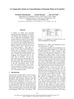
Fig. 1. Scheme of the alignment of cysteine residues of MATs from different origins based on the previously reported [16]. GenBank
TM
accession
numbers are as follows: L. donovani (AF179714); P. falciparum (AF097923); Saccharomyces cerevisiae (M23368); E. coli (K02129) and rat liver
(S06114).
30 Y. Pe
´
rez-Pertejo et al. (Eur. J. Biochem. 270) Ó FEBS 2003
PCR amplification (see ‘Materials and methods’). Transla-
tion of the 639 bp PCR fragment in all six possible reading
frames revealed that several stretches of the peptide
sequence predicted from one of the six, conserved high
homology with the amino acid sequences of others MAT
proteins submitted to GenBank
TM
database. The PCR
product was used to screen the DNA library for this parasite
to isolate the full-length clone. The isolated bacteriophage
was used for subsequent analysis; a 6.0 kb SmaI-digested
fragment was hybridized to the PCR fragment and
redigested with AvaI to obtain a 2.0 kb fragment, the
product ultimately sequenced. The nucleotide sequence of
the L. donovani MAT2 clone (GenBank
TM
accession num-
ber AF179714) was identical to the sequence found for
L. infantum [16], with a single 1179 bp long ORF encoding
394 amino acid residues and a calculated molecular mass
of 42 000 Da (data not shown). The MAT2-encoding
sequence conserved a high G/C codon bias. L. donovani
MAT2 contains all the motifs that bind to ATP, metals and
the active site: the nonapeptide GGGAFSGKD, at position
269–277, corresponds to a P-loop that forms a part of the
ATP binding site [24]. The c-phosphate moiety is hydrolized
from tripolyphosphate at conserved Arg255. The Asp19
and Asp282 residues have been described to bind Mg
2+
[25]
and Glu45 is involved in K
+
binding [26]. L. donovani also
conserved the hexapeptide GAGDQG at position 118–123,
associated with the active site of the enzyme. The genomic
organization of the MAT2 gene in the L. donovani genome
was ascertained by digestion of the entire genomic DNA
from L. donovani L82 (Ethiopian) cells with different
endonucleases, after which the fragments were blotted and
probed with the labeled 639 bp PCR fragment. The number
of bands obtained after cleavage with the restriction
enzymes is indicative of the presence of two copies of the
MAT2 gene in the L. donovani genome, a result that concurs
with findings described previously for L. infantum [16].
L. donovani MAT contains seven cysteine residues
(Cys22, 33, 44, 47, 92, 106 and 305) per enzyme subunit.
The location of the cysteine residues, based on the alignment
recently reported for L. infantum MAT [16], is shown in
Fig.1.Whencomparedtothemammalianenzyme,the
cysteine residues in L. donovani MAT-II at positions 22
(which corresponds to Cys35 in rat liver MAT), 44 (Cys57
in rat liver), and 92 (Cys105 in rat liver) are found to remain
invariable in most of the sequences aligned. The cysteines at
positions 33 and 47 are found in the Plasmodium falciparum
MAT sequence [27]. Cys305 aligns with the cysteine at
position 295 in the E. coli enzyme. In addition to these Cys
residues, L. donovani contains a specific cysteine at position
106.
E. coli strain XL-1Blue cells transformed with pQE30-
MAT, were induced with 0.1 m
M
IPTG. Aliquots were
harvested at different times (30 min, 1 h, 2 h, 3 h), lysed
and spun at 13 000 r.p.m. in a microfuge for 15 min.
Proteins from the supernatants and pellets were analyzed by
SDS/PAGE under reducing conditions. In the absence of
IPTG, MAT expression was nil. With IPTG induction,
however, a protein with an estimated molecular mass of
48 kDa was found to accumulate. The recombinant protein
formed primarily in inclusion bodies. Successive washes of
inclusion bodies with 4
M
urea and 5% (v/v) Triton X-100
removed most of the contaminating proteins, producing a
homogeneous MAT-II preparation, as shown by SDS/
PAGE gels (Fig. 2, lane 2). His-tag affinity chromatography
(Fig. 2, lane 3) showed that no further purification was
obtained with this step. A single band with an estimated
molecular weight of 48 kDa (Fig. 2, lane 4) was observed
when Western analysis was conducted using a polyclonal
MAT antibody [16] and whole L. donovani extract.
The protocol for functional folding of the MAT-protein
enriched insoluble aggregates was based on the procedure
described by Lo
´
pez-Vara et al. [20]. Briefly, two successive
washes with 4
M
urea containing 5% (v/v) Triton X-100
yielded the protein overexpressed in the inclusion bodies in a
very pure (over 99%) state. Removal of the excess urea
added, protein dilution and equilibrium dialysis was
requisite to proper MAT folding, which was monitored by
both enzyme activity and fluorescence quenching.
The presence of seven cysteines in L. donovani MAT-II
suggests that sulfhydryl-protection reagents may be required
for optimum refolding. Conformational transitions were
observed (Fig. 3A) via fluorescence quenching during the
refolding of L. donovani MAT-II in the presence of 10 m
M
dithiothreitol. Such transitions, which provoked fast and
large fluorescence quenching effects (indicating strong
stimulation of MAT activity), took place during the first
2 h. MAT activity (Fig. 3B) shows a sharp rise after 2 h of
dialysis with 10 m
M
dithiothreitol, to plateau thereafter.
Notably, when the MAT molecule refolded in the absence
of dithiothreitol, it only quenched about half of the
Fig. 2. SDS/PAGE analysis of the expression and purification of
recombinant L. donovani MAT, from XL-1Blue E. coli extracts. (A)
Purification of MAT recombinant protein. Coomassie Blue-stained
SDS/PAGE gel. Lane 1, molecular weight markers; Lane 2, washed
inclusion bodies obtained from lysates of E. coli XL-1Blue trans-
formedwithpQE30-MATplasmidandinducedwith0.1m
M
IPTG.
Lane 3, result of the purification with His-tag affinity chromatography.
(B) MAT expression in L. donovani promastigotes. Lane 1, molecular
weight markers; lane 4, Western blot performed with cellular extracts
from logarithmic-phase cultures and MAT-polyclonal antibodies [16].
Ó FEBS 2003 Cysteine residues in leishmanial MAT (Eur. J. Biochem. 270)31
fluorescence quenched in the presence of the thiol, and no
activity at all was recovered. All MAT enzymes require a
divalent cation and most have binding sites for both the
Mg
2+
-ATP substrate and for free Mg
2+
[25]. Another
experiment, similar to the one described, was conducted in
which MAT activity was monitored during the refolding
process in the presence and absence of MgSO
4
(Fig. 3C,D).
The Mg
2+
cation does not affect the refolding of wild-type
MAT, and the MAT activity was observed to be similar in
both cases.
The synthetic reaction catalyzed by MAT occurs in two
consecutive steps: AdoMet and PPP
i
are first synthesized
from methionine and ATP and then PPP
i
is subsequently
hydrolyzed to PP
i
and P
i
to allow the products to be
released from the active site of the enzyme. Recombinant
MAT activity was linear in terms of both time (up to
90 min) and protein concentration (data not shown). The
steady-state activity at saturation of both substrates, i.e.
5m
M
ATP and 5 m
ML
-methionine, was 12 lmolÆ
mg
)1
Æh
)1
(k
cat
¼ 0.32 s
)1
) (Table 1). The enzyme showed
slight sigmoid behavior with both
L
-methionine and ATP.
Hill plots and the software package Enzfitter, were used
for kinetic parameters calculations. Co-operativity, esti-
matedtoben ¼ 2.3 (ATP ¼ 0.5 m
M
) declined with rising
ATP levels to nearly 1 (ATP ¼ 5m
M
). S
0.5
-values for
L
-methionine, not significantly affected by ATP, were
estimated to be 250 ± 25 l
M
. Conversely, when assessed
as a function of ATP at different
L
-methionine levels,
MAT activity was sigmoid (n ¼ 1.8). The S
0.5
-values for
ATPwerecalculatedtobe27±5l
M
and the curve
retained its sigmoid shape as concentrations of the
L
-amino acid were increased. The tripolyphosphatase
activity of L. donovani recombinant MAT-II was meas-
ured under the standard assay conditions described in
‘Material and methods’. Tripolyphosphatase activity was
linear over time and for protein concentration, and no P
i
was observed to be released in the absence of recombinant
MAT. Sigmoid behavior was found under steady state
conditions, with a k
cat
0.04 s
)1
and an S
0.5
-value of 40 l
M
(Table 1).
The feedback inhibition of MAT by AdoMet, was
analyzed using the Dixon approach at
L
-methionine
concentrations of 0.5–5.0 m
M
, resulting in a noncompetitive
pattern with a K
i
value of 4 m
M
.Bycontrast,inasimilar
Fig. 3. Functional refolding of L. donovani
MAT from E. coli inclusion bodies. Time
course of the fluorescence intensity during the
folding of wild-type MAT-II (A). Fluores-
cence emission intensity signal at 350 nm was
monitored (excitation, 290 nm) in the presence
(s) and absence (j)of10m
M
dithiothreitol.
Time-course of the reactivation process (B) in
thepresence(s) and absence (j)of10m
M
dithiothreitol. MAT-II activity was deter-
mined at the indicated time points from
aliquots of the corresponding incubation
mixtures. (C) and (D) show the effect of
10 m
M
MgSO
4
(j) or absence (s)onMAT
refolding process. Points are means ± SD of
three different experiments. *P < 0.001 using
Student’s t-test.
Table 1. Kinetic characterization of refolded L. donovani MAT. Ado-
Met synthesis and tripolyphosphatase activity were measured under
steady-state conditions established in Material and methods. Hill plots
were used to determine the kinetic constants of both activities. Results
are the mean of four independent determinations ± SD.
Parameter AdoMet synthesis
Tripolyphosphatase
activity
Specific activity 12 lmolÆmg
)1
Æh
)1
3.5 lmolÆmg
)1
Æh
)1
k
cat
(s
)1
) 0.32 s
)1
0.04 s
)1
S
0.5
(
L
-methionine) 250 ± 25 l
M
S
0.5
(ATP) 27 ± 5 l
M
S
0.5
(PPP
i
)40±3l
M
32 Y. Pe
´
rez-Pertejo et al. (Eur. J. Biochem. 270) Ó FEBS 2003
analysis with ATP concentrations of 0.5–5.0 m
M
,acom-
petitive inhibitory effect was found with a K
i
value for the
nucleobase of 0.8 m
M
. Regardless to AdoMet synthesis,
AdoMet was a nonessential activator of tripolyphosphatase
activity in the range of 5–100 l
M
.
The k
cat
values determined at saturating concentrations
of tripolyphosphate, ATP and
L
-methionine showed that
the rate of AdoMet synthesis is higher than the rate of
tripolyphosphate cleavage. As the overall process should be
dependent on the slowest reaction, tripolyphosphatase
activity may be thought to be responsible for the rate of
AdoMet synthesis in leishmania. However, tripolyphos-
phate hydrolysis is activated several-fold when AdoMet
occupies the active site, thus suggesting that the reaction
forming AdoMet and PPP
i
is the step that determines the
rate of the overall process, in which PPP
i
cleavage would be
requisite to enzyme turnover [28].
The dependence of MAT activity on cysteine residues was
assessed for both the AdoMet synthesis and trypolyphos-
phatase activities in the presence of the sulfhydryl reagent
N-ethylmaleimide. The panels shown in Fig. 4 represent the
time-course of AdoMet synthesis (Fig. 4A) and tripoly-
phosphatase (Fig. 4B) inactivation when 1 m
M
N-ethylma-
leimide was added to the incubation media. In both cases
time-dependent inactivation was observed, which is indi-
cative of irreversible binding to one/several of the sulfhydryl
moieties involved in the enzymatic process. The semi-
inactivation times estimated for AdoMet synthesis and PPP
i
cleavage processes were estimated to be 3.9 and 11.8 min,
respectively. However, the presence of 50 l
M
AdoMet in the
incubation media caused bi-exponential decay in the
presence of the sulfhydryl reagent. The semi-inactivation
time of the rapid process was calculated to be 1.6 min,
whereas the slope of the slow semi-inactivation process was
rather similar to the slope of the curve found when the
medium did not contain AdoMet, with an estimated half-
life of 10.5 min.
The role of the seven cysteine residues in L. donovani
MAT was studied using the site-directed mutagenesis
approach, in which seven single mutants were produced,
each lacking one of the sulfhydryl moieties. The amino
acid chosen to replace Cys was serine, as it is regarded to
be isosterical to cysteine and does not impact hydropho-
bicity [12]. All the mutants, named C22S, C33S, C44S,
C47S, C92S, C106S and C305S, were expressed in E. coli
and their products were refolded as described in ‘Material
and methods’. MAT was measured for both AdoMet
synthesis and tripolyphosphatase activities in all the
cysteine mutants for comparison to the wild-type protein
(Fig. 5A–C).
Figure 5A shows the ability of the various L. donovani
MAT cysteine mutants to synthesize AdoMet under the
standard assay conditions. Site-directed mutations on the
phylogenetically conserved cysteines Cys22, Cys44 and
Cys92 yielded mutants completely lacking in any synthetic
activity. The C305S mutant retained a scant 1% of the
activity displayed by the wild type. The C33S and C47S
mutants retained only 15% and 10%, respectively, of the
V
max
under saturating conditions for both substrates, and
no changes in affinity were found. Unlike the other
mutants, C106S was not kinetically different from the
wild-type protein. Structural analyses of mammalian
MAT show that the cysteines positioned between Cys35
and Cys105 are located in the central domain of each
subunit, in the interface between the two dimers compri-
sing the tetrameric structure [29]. This domain contains
five cysteine residues, two of them (Cys35 and Cys61)
forming a disulfide bond which may be necessary for the
tetrameric state of the enzyme. In addition, Cys69 is
involved in the correct folding of the monomer, support-
ing the establishment of the disulfide bond [29,30].
Chromatographic and modeling studies show that Leish-
mania [16] and Plasmodium [27] MATs are dimers whose
identical cysteine compositions in the central domain lack
the homologous mammalian enzyme amino acids at
positions 61 and 69 which are, in turn, involved in
establishing the disulfide bond and proper folding to the
tetrameric structure. Nevertheless, the substitution of two
specific cysteines, Cys33 and Cys47, present in both
species, originated a significant loss of enzymatic activity
but no change in affinity.
The behavior observed for the cysteine mutants differed
in terms of tripolyphosphatase activity. Figure 5B shows the
residual activity of single cysteine mutants assayed under
standard saturation conditions, in the absence of AdoMet.
The single cysteine mutants of L. donovani MAT, C22S,
C44S and C305S, lacked tripolyphosphatase activity. C33S
maintained a mere one-tenth of the activity displayed by the
wild type. However, the ability of C92S, C106S and C47S to
cleave PPP
i
remained high, and in the case of C47S, even
Fig. 4. Time-course of L. donovani
recombinant MAT inactivation with 1 m
M
N-ethylmaleimide. (A)AdoMetsynthesis.
(B) Tripolyphosphatase activity; s,with
50 l
M
AdoMet; d, without 50 l
M
AdoMet.
No loss of activity was found without
N-ethylmaleimide during the incubation time,
either for MAT activity (s) or PPP
i
cleavage.
Points are the mean of three separate
experiments.
Ó FEBS 2003 Cysteine residues in leishmanial MAT (Eur. J. Biochem. 270)33
higher than the wild-type protein. The addition of 50 l
M
AdoMet (Fig. 5C) activated PPP
i
hydrolysis more than
12-foldinthewildtypeandtoasimilarextentintheC106S
mutant. Significant activation was also observed in the
C47S and C33S mutants, although with different kinetic
constants and sigmoid behavior. There was a notable lack
of any AdoMet stimulation in the C92S mutant (Table 2).
Whilst it showed significant tripolyphosphatase activity in
the absence of AdoMet, its activity was not enhanced in the
presence of AdoMet. The Cys92 residue is thus involved in
the stimulatory effect of PPP
i
cleavage induced by AdoMet
and may be the amino acid residue to be rapidly inactivated
by N-ethylmaleimide (Fig. 4B). The residues homologous
with leishmanial Cys92 in mammalian and E. coli MATs
are amino acid residues Cys105 and Cys90, respectively
[11,12]. Both are involved in the dimer/tetramer equilibrium
of the enzyme, and E. coli Cys90isalsoinvolvedinthe
binding of ATP to the active site. Because C92S is unable to
synthesize AdoMet but can cleave PPP
i
, the respective
cysteine may plausibly be thought to be involved in the first
step of the reaction (synthesis of AdoMet and PPP
i
), with
no role in PPP
i
cleavage.
The results of these studies show that recombinant
L. donovani MAT only folds properly in reducing environ-
ments. With the exception of Cys106, all the cysteine
residues in the enzyme are needed for AdoMet condensa-
tion, PPP
i
hydrolysis or AdoMet activation. The structural
involvement of the cysteines at positions 22, 44 and 305
appears to be crucial to the overall process. By contrast, the
mutants lacking cysteine residues at positions 33 and 47
Fig. 5. Leishmania donovani recombinant
MAT activity and effect of cysteine substitu-
tions on AdoMet synthesis (A) and tripoly-
phosphatase activity in absence of AdoMet
(B) and presence of 50 l
M
AdoMet (C).
Freshly refolded MAT (16 lg) and cysteine
mutants, were assayed in presence of 1 m
M
ATP for AdoMet synthesis activity and under
standard saturation conditions for tripoly-
phosphatase activity. Each bar represents the
average ± SD of triplicates.
Table 2. Kinetic parameters of tripolyphosphatase activity of wild-type MAT and cysteine-mutants from L. donovani. Kinetics were performed under
standard assay conditions using 16 lg of freshly folded recombinant protein. Activation kinetics were performed in the presence of 50 l
M
AdoMet.
V
max
,S
0.5
-andn-values are the average of three different experiments; n.d., not determined.
Mutant
Without AdoMet With AdoMet
V
max
(lmolÆh
)1
Æmg
)1
)S
0.5
(l
M
) nV
max
(lmolÆh
)1
Æmg
)1
)S
0.5
(l
M
) n
Wild type 3.7 40 2.4 50 287 1.2
C22S No activity n.d. n.d. No activity n.d. n.d.
C33S 0.3 n.d. n.d. 3.2 38 2,6
C44S No activity n.d. n.d. No activity n.d. n.d.
C47S 5.3 63 2.6 20 252 2.1
C92S 1.9 52 2.7 1.9 52 2.7
C106S 3.5 38 2.3 49 275 1.1
C305S No activity n.d. n.d. No activity n.d. n.d.
34 Y. Pe
´
rez-Pertejo et al. (Eur. J. Biochem. 270) Ó FEBS 2003
retained part of their AdoMet synthesis and tripolyphos-
phatase activities. As the C92S mutant, which completely
lacked the ability to synthesize AdoMet, retained tripoly-
phosphatase activity but was not stimulated by exogenous
AdoMet, it may be concluded that the cysteine residue at
position 92 participated in the first of the two reactions that
comprise the process.
Acknowledgements
We thank Jose
´
Marı
´
a Requena and his group (Centro de Biologı
´
a
Molecular Severo Ochoa, Universidad Auto
´
noma de Madrid, Spain)
for their help in molecular techniques. We also want to thank John
Chris Meade (University of Mississippi Medical Center, Jackson, MS)
for the Leishmania donovani genomic library. This research was
supported by Comisio
´
n Interministerial de Ciencia y Tecnologı
´
a
(grants PM98/0036 and PB96/0159), Junta de Castilla y Leo
´
n(grants
LE05/01 and LE06/02) and Fondo de Investigacio
´
n Sanitaria del
Ministerio de Sanidad y Consumo (grant FIS 01/1077).
References
1. Mato, J.M., Corrales, F.J., Lu, S.C. & Avila, M.A. (2002)
S-Adenosylmethionine: a control switch that regulates liver func-
tion. FASEB J. 16, 15–26.
2. Tabor, C.W. & Tabor, H. (1985) Polyamines in microorganisms.
Microbiol. Rev. 49, 81–99.
3. Mato, J.M., Corrales, E.C., Martı
´
n-Duce, A., Ortiz, P., Pajares,
M.A. & Cabrero, C. (1990) Mechanisms and consequences of the
impaired trans-sulphuration pathway in liver disease: part I. Bio-
chemical implications. Drugs 40, 58–64.
4. Chiang, P.K. & Cantoni, G. (1977) Activation of methionine by
transmethylation. Purification of the S-adenosylmethionine syn-
thetase from Baker’s yeast and its separation into two forms.
J. Biol. Chem. 252, 4506–4513.
5. Kotb, M. & Geller, A.L. (1993) Methionine adenosyltransferase:
structure and function. Pharmacol. Ther. 59, 125–142.
6. Tabor, C.W. & Tabor, H. (1984) Methionine adenosyltrans-
ferase (S-adenosylmethionine synthetase) and S-adenosyl-
methionine decarboxylase. Adv. Enzymol. Relat. Area Mol. Biol.
56, 251–282.
7. Alvarez, L., Corrales, F., Martı
´
n-Duce,A.&Mato,J.M.(1993)
Characterization of a full-length cDNA encoding human liver
S-adenosylmethionine synthetase: tissue-specific gene expression
and mRNA levels in hepatopathies. Biochem. J. 293, 481–486.
8. De La Rosa, J., Ostrowski, J., Hryniewicz, M.M., Kredich, N.M.,
Kotb,M.,LeGros,H.L.Jr,Valentine,H.&Geller,A.M.(1995)
Chromosomal localization and catalytic properties of the
recombinant alpha subunit of human lymphocytes methionine
adenosyltransferase. J. Biol. Chem. 270, 21860–21868.
9. LeGros, H.L. Jr, Halim, A.B., Geller, A.M. & Kotb, M. (2000)
Cloning, expression and functional characterization of the beta
regulatory subunit of human methionine adenosyltransferase
(MAT II). J. Biol. Chem. 275, 2359–2366.
10. Markham, G.D. & Satishchandran, C. (1988) Identification of
the reactive sulfhydryl groups of S-adenosylmethionine synthe-
tase. J. Biol. Chem. 263, 8666–8670.
11. Reckowski, R.S. & Markham, G.D. (1995) Structural and func-
tional roles of cysteine 90 and cysteine 240 in S-adenosylmethio-
nine synthetase. J. Biol. Chem. 270, 18484–18490.
12. Mingorance, J., Alvarez, L., Sa
´
nchez-Go
´
ngora, E., Mato, J.M. &
Pajares, M.A. (1996) Site-directed mutagenesis of rat liver S-ade-
nosylmethionine synthetase. Biochem. J. 315, 761–766.
13. Neal, R.A., Iwobi, M.V. & Robert-Gero, M. (1989) Antil-
eishmanial effect of free and encapsulated sinefungin against
Leishmania donovani infections in BALB/c mice. C. R. Acad. Sci.
III (308), 485–488.
14. Bacchi, C.J. & Yarlett, N. (1993) Effects of antagonists of
polyamine metabolism on African trypanosomes. Acta Trop. 54,
225–236.
15. Yarlett, N., Garofalo, J., Goldberg, B., Ciminelli, M.A., Ruggiero,
U., Sufrin, J.R. & Bacchi, C.J. (1993) S-Adenosylmethionine
synthetase in bloodsteam Trypanosoma brucei. Biochim. Biophys.
Acta 1181, 68–76.
16. Reguera, R.M., Balan
˜
a-Fouce, R., Pe
´
rez-Pertejo, Y., Ferna
´
ndez,
F.J., Garcı
´
a Estrada, C., Cubrı
´
a, J.C., Ordo
´
n
˜
ez, C. & Ordo
´
n
˜
ez, D.
(2002) Cloning, expression and characterization of methionine
adenosyltransferase in Leishmania infantum promastigotes. J. Biol.
Chem. 277, 3158–3167.
17. Sambrook,J.,Fritsch,E.,E.F.&Maniatis,T.(1989)Molecular
Cloning: a Laboratory Manual, 2nd edn. Cold Spring Harbor
Laboratory, Cold Spring Harbor, New York.
18. Southern, E.M. (1975) Detection of specific sequences among
DNA fragments separated by gel electrophoresis. J. Mol. Biol. 98,
503–517.
19. Sanger, F., Nicklen, S. & Coulson, A.R. (1977) DNA sequencing
with chain-terminating inhibitors. Proc. Natl Acad. Sci. USA 74,
5463–5467.
20. Lo
´
pez-Vara, M.C., Gasset, M. & Pajares, M.A. (2000) Refolding
and characterization of rat liver methionine adenosyltransferase
from Escherichia coli inclusion bodies. Protein Expr. Purif. 19,
219–226.
21. Go
´
mez-Gallego, F., Garrido-Pertierra, A., Mason, P.J. &
Bautista, J.M. (1996) Unproductive folding of the human G6PD-
deficient variant A(–). FASEB J. 10, 153–158.
22. Alvarez, L., Mingorance, J., Pajares, M.A. & Mato, J.M. (1994)
Expression of rat liver S-adenosylmethionine synthetase in
Escherichia coli results in two active oligomeric forms. Biochem.
J. 301, 557–561.
23. Bradford, M.M. (1976) A rapid and sensitive method for the
quantitation of microgram quantities of protein utilizing the
principle of protein-dye binding. Anal. Biochem. 72, 248–254.
24. Deigner, H.P., Mato, J.M. & Pajares, M.A. (1995) Study of the rat
liver S-adenosylmethionine synthetase active site with 8-azido
ATP. Biochem. J. 308, 565–571.
25. Taylor, J.C. & Markham, G.D. (1999) The bifunctional active
site of S-adenosylmethionine synthetase. Roles of the active site
aspartates. J. Biol. Chem. 274, 32909–32914.
26. McQueney, M.S. & Markham, G.D. (1995) Investigation of
monovalent cation activation of S-adenosylmethionine synthe-
tase, using mutagenesis and uranyl inhibition. J. Biol. Chem. 270,
18277–18284.
27. Chiang, P.K., Chamberlin, M.E., Nicholson, D., Soubes, S., Su,
X Z., Subramanian, G., Lanar, D.E., Prigge, S.T., Scovill, J.P.,
Miller, L.H. & Chou, J.Y. (1999) Molecular characterization of
Plasmodium falciparum S-adenosylmethionine synthetase. Bio-
chem. J. 344, 571–576.
28. McQueney, M.S., Anderson, K.S. & Marlham, G.D. (2000)
Energetics of S-adenosylmethionine synthetase catalysis. Bio-
chemistry 39, 4443–4454.
29. Gonza
´
lez, B., Pajares, M.A., Hermoso, J.A., A
´
lvarez, L., Garrido,
B., Sufrin, J. & Sanz-Aparicio, J. (2000) The crystal structure of
tetrameric methionine adenosyltransferase from rat liver reveals
the methionine-binding site. J. Mol. Biol. 300, 363–375.
30. Martı
´
nez-Chantar, M.L. & Pajares, M.A. (2000) Assignment of a
single disulfide bridge in rat methionine adenosyltransferase. Eur.
J. Biochem. 267, 132–137.
Ó FEBS 2003 Cysteine residues in leishmanial MAT (Eur. J. Biochem. 270)35