Báo cáo khoa học: Plasmodium falciparum merozoite surface protein 1 Glycosylation and localization to low-density, detergent-resistant membranes in the parasitized erythrocyte pdf
Bạn đang xem bản rút gọn của tài liệu. Xem và tải ngay bản đầy đủ của tài liệu tại đây (531.78 KB, 10 trang )
Plasmodium falciparum
merozoite surface protein 1
Glycosylation and localization to low-density, detergent-resistant membranes
in the parasitized erythrocyte
Daniel C. Hoessli
1
, Monique Poincelet
1
, Ramneek Gupta
2
, Subburaj Ilangumaran
3
and Nasir-ud-Din
4
1
Department of Pathology, Centre me
´
dical universitaire, Geneva, Switzerland;
2
Center for Biological Sequence Analysis,
Technical University of Denmark, Lyngby, Denmark;
3
Department of Experimental Therapeutics, Ontario Cancer Institute,
Toronto, Canada;
4
Institute of Biomedical Sciences, Pakistan and HEJ Institute of Chemistry,
University of Karachi, Lahore, Pakistan
In addition to the major carbohydrate moieties of the
glycosylphosphatidylinositol (GPI) anchor, we report that
Plasmodium falciparum merozoite surface protein 1 (MSP-1)
bears O-GlcNAc modifications predominantly in b-ano-
meric configuration, in both the C- and N-terminal portions
of the protein. Subcellular fractionation of parasitized
erythrocytes in the late trophozoite/schizont stage reveals
that GPI-anchored C-terminal fragments of MSP-1 are
recovered in Triton X-100 resistant, low-density membrane
fractions. Our results suggest that O-GlcNAc-modified
MSP-1 N-terminal fragments tend to localize within the
parasitophorous vacuolar membrane while GPI-anchored
MSP-1 C-terminal fragments associate with low-density,
Triton X-100 resistant membrane domains (rafts), redis-
tribute in the parasitized erythrocyte and are eventually shed
as membrane vesicles that also contain the endogenous,
GPI-linked CD59.
Keywords: detergent-resistant membranes; malaria; mer-
ozoite surface protein; O-GlcNAc modification; vesicles.
In the blood-stage forms of the malarial parasite Plasmodium
falciparum, the merozoite surface protein 1 (MSP-1) is a
major surface component [1] that undergoes selective
proteolytic processing and reassembly in preparation for
erythrocyte invasion [1–4]. MSP-1 is linked to the parasite
plasma membrane via a glycosylphosphatidylinositol (GPI)
anchor [5], but the functional consequences of this mode of
anchoring for the merozoite to interact with the erythrocyte
have not been fully evaluated [6]. In addition to the GPI-
anchor modification, MSP-1 also contains mono- or oligo-
saccharides in O-linkage to serines or threonines
[7–10]. N-linked carbohydrates have also been described in
association with asparagines on MSP-1 [9], despite the
reported lack of N-glycosylating machinery in P. falciparum
parasites [11]. As P. falciparum merozoite maturation takes
place within an intraerythrocytic network of modified
(parasitophorous vacuolar membrane) and newly made
(tubo-vesicular network) membranes [12,13], it is possible
that parasite surface proteins also constitute substrates for
carbohydrate-modifying enzymes of the erythrocyte. In
normal erythrocytes, O-GlcNAc modifications of serines/
threonines in intracellular proteins occur in a manner
reciprocal to phosphorylation [14] and O-GlcNAc addition
is considered a widespread and general mechanism for
protein modification [15]. In this study, we have analysed
MSP-1 for the presence of O-GlcNAc-modified serines and
threonines, using specific antibodies to map the biosynthe-
tically labelled modifications to the N and C terminus of the
MSP-1 protein [16]. The presence of O-GlcNAc on both the
C- and N-terminal ends of MSP-1 was confirmed by
exogalactosylation and two-thirds of the [
3
H]GlcN label
incorporated into MSP-1 was sensitive to Jack Bean
b-N-acetylglucosaminidase, suggesting the presence of
O-GlcNAc moieties in b-anomeric linkage [15]. Predictions
for a-andb-anomeric O-GlcNAc sites in five known MSP-1
sequences were made using methods based on artificial
neural networks which are competent in recognizing fuzzy
sequence motifs, and two distinct sets of a-and
b-O-GlcNAc sites have been predicted. The GPI-anchored
19-kDa C-terminal fragment was found associated with
detergent-resistant, low-density membranes of the parasi-
tized erythrocyte, suggesting that GPI-linked MSP-1 prod-
ucts redistribute within the membrane network of the
parasitized host cell aboard detergent-resistant membrane
domains. Infected erythrocytes were also found to release
membrane vesicles containing parasitic 19-kDa MSP-1
fragments and endogenous CD59, both GPI-linked proteins.
Materials and methods
Materials
Anti-MSP-1 mAbs reactive with the C (3B10) and N
terminus (7B2) [16], were obtained from J.A. Lyon (Walter
Reed Army Institute of Research, Washington, USA). A
human immune serum against blood stage antigens was
used to detect all MSP-1 epitopes [10]. Anti-CD59 mAb
Correspondence to D. C. Hoessli, Department of Pathology,
Centre me
´
dical universitaire, 1, rue Michel-Servet,
1211 Geneva 4, Switzerland.
Fax: +41 22 7025746, Tel.: +41 22 7025893,
E-mail:
Abbreviations: GPI, glycosylphosphatidylinositol; MSP-1,
merozoite surface protein 1.
(Received 9 September 2002, revised 13 November 2002,
accepted 26 November 2002)
Eur. J. Biochem. 270, 366–375 (2003) Ó FEBS 2003 doi:10.1046/j.1432-1033.2003.03397.x
MEM-43 was supplied by V. Horejsi (Academy of Sciences
of the Czech Republic, Prague). Recombinant, bovine
b-1,4-galactosyltransferase was from Calbiochem. b-galac-
tosidase (bovine brain) and b-N-acetylglucosaminidase
(Jack Bean) were from Sigma.
Metabolic radiolabelling of M25 Zaire
P. falciparum
parasites
Asexual blood stage parasites were cultured in the asyn-
chronous mode in 10-mL cultures at 37 °C in a candle jar.
The culture medium contained 5% v/v human group A+
erythrocytes, in RPMI medium supplemented with 10%
ARh+ human serum (HD Supplies, Aylesbury, Bucks,
UK) and 0.1% glucose. Labelling was carried out for 16 h
with 50 lCiÆmL
)1
D
-[6-
3
H]glucosamine hydrochloride
([
3
H]GlcN; 40 CiÆmmol
)1
, American Radiolabeled Chemi-
cals, supplied by Anawa, Wangen, Switzerland), as previ-
ously carried out to show carbohydrate modification of
plasmodial proteins [9,17].
At the end of the labelling time, culture supernatants were
collected and centrifuged at low speed (2000 r.p.m., 5 min)
to remove uninfected and parasitized erythrocytes. The
supernatant was centrifuged sequentially at 15000 g for
10 min at 4 °C and the 15000 g supernatant again at
100 000 g for 30 min at 4 °C (Beckman SW41 rotor) to
collect the released membrane vesicles. The 100 000 g pellet
was analysed as a source of parasite-free membrane
nanovesicles (Fig. 1A).
To study the distribution of MSP-1 and its fragments, the
[
3
H]GlcN-labelled, parasitized erythrocytes at the late
trophozoite/schizont stage were collected by sedimentation
over a 70% Percoll (Pharmacia) gradient [18] and
hemolysedinH
2
O in the presence of protease inhibitors
(0.19 m
M
leupeptin, 0.17 m
M
chymostatin, 2 m
M
N-p-tosyl-
L
-lysine chloromethyl ketone (TLCK), 2 m
M
N-p-tosyl-
L
-phenylalanine chloromethyl ketone (TPCK), 1 m
M
phenylmethanesulfonyl fluoride and 1 m
M
ortho-phenanthro-
line; Fig. 1B). A small aliquot (5%) of the [
3
H]GlcN-labelled
parasites and haemoglobin-free erythrocyte membranes was
directly solubilized in SDS/PAGE sample buffer (see Fig. 2,
[
3
H]GlcN; tot), resolved on SDS/PAGE and the labelled
proteins revealed by fluorography. The gel was soaked in
Enlightning (NEN), dried and exposed to Hyperfilms
(Amersham).
The bulk of this material was resuspended in TKM buffer
(50 m
M
Tris/HCl pH 7.4, 25 m
M
KCl, 5 m
M
MgCl
2
,1m
M
EGTA) containing 1% Triton X-100, 36% sucrose and
protease inhibitors, and centrifuged at 250 000 g in a SW 50
rotor for 16 h at 4 °C. This procedure allows the parasites
to be pelleted with remnants of the parasitophorous
vacuolar membrane [19] (P, Fig. 1B), and to be separated
from Triton X-100 soluble and Triton X-100 resistant
components of the parasitized erythrocyte membranes that
are recovered in the supernatant (S, Fig. 1B).
SDS/PAGE, immunoprecipitation and Western blotting
In 10% of the supernatant material, the proteins were
precipitated with chloroform/methanol for Western blotting
analysis [20] with anti-(C-ter) or anti-(N-ter) mAbs. The
precipitated proteins were separated on a 10% minigel
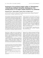
Fig. 1. Subcellular fractionation protocols utilized to generate extracts
containing parasite proteins from [
3
H]GlcN-labelled and unlabelled,
P. falciparum-infected erythrocytes. (A) Isolation of microvesicles and
nanovesicles from [
3
H]GlcN-labelled, infected erythrocytes. (B) Isola-
tion of parasite extracts (P) and Triton X-100 extracted proteins (S)
from [
3
H]GlcN-labelled, infected erythrocytes. (C) Isolation of Triton
X-100 insoluble membranes and Triton X-100 soluble membrane and
cytosolic proteins from infected erythrocytes.
Ó FEBS 2003 Glycosylation and membrane localization of MSP-1 (Eur. J. Biochem. 270) 367
and transferred to nitrocellulose (Hybond-C, Amersham
Pharmacia Biotech) with a semidry blotting apparatus
(Bio-Rad). After 2 h of blocking at room temperature in
NaCl/Tris/Tween (10 m
M
Tris/HCl pH 7.4, 100 m
M
NaCl,
0.05% Tween 20) containing 5% low-fat, dry milk powder
(NaCl/Tris/Tween/5% MP), the filters were incubated with
antibodies in NaCl/Tris/Tween/5% MP overnight at 4 °C.
Thoroughly washed filters were incubated with horseradish-
peroxidase-conjugated secondary antibodies for 1 h at
room temperature. Chemiluminescence development was
carried out with the Immun-Star Pack reagents (Bio-Rad)
and the filters exposed to X-Omat Kodak films.
The bulk (90%) of the supernatant (S, Fig. 1B) was
dialysed to remove sucrose and sequentially immunopre-
cipitated with Sepharose 4B-coupled anti-(N-ter), followed
by anti-(C-ter) mAb. The antibodies were covalently
coupled to CNBr-activated Sepharose 4B beads according
to the manufacturer’s instructions. Incubation with each
mAb was carried out for 6–10 h at 4 °C on a rotating wheel,
the antibody beads were washed in TKM/Triton X-100
containing protease inhibitors, and the bound antigens
extracted with SDS/PAGE sample buffer. The [
3
H]GlcN-
labelled, immunoprecipitated proteins were revealed by
fluorography, as described above.
The parasite pellet (P, Fig. 1B) was extracted with 10%
SDS (1 h at room temperature), 10% of the extract sampled
for Western blotting and the remainder diluted with Triton
X-100 and BSA to obtain a final concentration of 0.05%
SDS, 0.5% Triton X-100, 10 lgÆmL
)1
BSA, suitable for
immunoprecipitation. Sequential immunoprecipitation with
Sepharose-coupled anti-N followed by anti-(C-ter) mAb
was carried out as with the supernatant (S) material and the
[
3
H]GlcN-labelled, immunoprecipitated proteins visualized
by fluorography as described above.
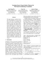
Fig. 3. Vesicular release of C-terminal MSP-1 fragments by parasitized
erythrocytes. The [
3
H]GlcN-labelled, parasitized erythrocytes in the
culture were purified over Percoll and processed for immunoprecipi-
tation as outlined in Materials and methods. Immunoprecipitation was
carried out with Sepharose-coupled 3B10 mAb and human malaria-
immune serum coupled to protein A/G beads, and the immunopre-
cipitated, labelled bands revealed by fluorography (Whole extract: IP
anti C-ter; IP immune serum). The supernatant of [
3
H]GlcN-labelled
cultures at the late trophozoite/schizont stage were allowed to settle
and the supernatant collected. Remaining erythrocytes were sedi-
mented at 2000 r.p.m. for 10 min. The resulting supernatant was
centrifuged once at 16 000 g to remove pelletable parasites, parasitized
erythrocytes and uninfected erythrocytes. The last supernatant was
ultracentrifuged at 100 000 g and the membrane pellet (nanovesicles)
wasresuspendedinTKM.One-tenthwasextractedwith10%SDS
sample buffer (vesicles: unselected) and the [
3
H]GlcN-
labelled bands revealed by fluorography. The remaining 90% was
divided into two aliquots: one aliquot was extracted with 10% SDS
and diluted to 0.5% Triton X-100, 0.05% SDS, 10 lgÆmL
)1
BSA for
immunoprecipitation with Sepharose-coupled anti-MSP-1 C terminus
(vesicles: IP anti-C-ter) and the labelled bands revealed by fluorogra-
phy. The other aliquot was kept in TKM and incubated for 6–10 h
with Sepharose-coupled anti-(C-ter). The immunoselected vesicles
were washed in TKM and extracted with SDS/PAGE buffer, trans-
ferred to nitrocellulose and probed with MEM-43 anti-CD59 mAb
(vesicles: WB anti CD59) and revealed by chemiluminescence.
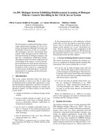
Fig. 2. Both C-terminal and N-terminal fragments of MSP-1 biosyn-
thetically incorporate [
3
H]GlcN. The parasitized erythrocytes were
isolated by Percoll gradient centrifugation and lysed in hypotonic
buffer. The resulting parasites and membrane ghosts were extracted in
TKM-1% Triton X-100/35% sucrose and ultracentrifuged to yield a
pellet (P) of parasites and a supernatant (S) containing Triton X-100-
resistant complexes and Triton X-100-soluble membrane proteins.
[
3
H]GlcN: fluorogram of [
3
H]GlcN-labelled P. falciparum proteins in a
total (tot), SDS extract of Percoll-purified parasitized erythrocytes
following hemolysis. C-ter and N-ter: probing with the 3B10 (C-ter) or
7B2 (N-ter) mAb following transfer to nitrocellulose and detection by
chemiluminescence (WB), or immunoprecipitation with Sepharose-
bound mAb and fluorography (IP). Results shown are representative
of three separate experiments.
368 D. C. Hoessli et al. (Eur. J. Biochem. 270) Ó FEBS 2003
One-tenth of the nanovesicle pellet (Fig. 1A) was directly
solubilized in SDS/PAGE sample buffer, resolved on a 10%
minigel and processed for fluorography (unselected, Fig. 3).
The remainder of the nanovesicle pellet was divided into two
aliquots. One aliquot was solubilized in 10% SDS and
subsequently diluted with TKM/Triton X-100 and BSA to a
final concentration of 0.5% Triton X-100, 0.05% SDS,
10 lgÆmL
)1
BSA. The MSP-1 19 kDa C-terminal fragment
was immunoprecipitated with Sepharose-coupled anti-
(C-ter) mAb as described above. The bound MSP-1
fragments were eluted in SDS/PAGE sample buffer,
resolved on a 10% minigel and processed for fluorography.
The other aliquot was incubated in TKM with Sepharose
4B-coupled anti-(C-ter) mAb for 6–10 h at 4 °C(rotating
wheel). The antibody-bound membranes were extracted in
SDS/PAGE sample buffer, resolved on a 10% minigel,
transferred to Hybond, probed with MEM-43 anti-CD59
mAb and visualized by chemiluminescence.
Exogalactosylation and deglycosylation of MSP-1
Auto-galactosylated, recombinant b-1,4-galactosyltrans-
ferase (20 mU, Calbiochem) was used to probe nitrocel-
lulose-immobilized parasite proteins for nonreducing
terminal GlcNAc residues [21], using UDP-[6-
3
H]galac-
tose (40 CiÆmmol
)1
, American Radiolabeled Chemicals)
as galactose donor. The MSP-1 proteins were specifically
immunoprecipitated from a 10% SDS extract of Percoll-
purified parasitized erythrocytes. This SDS extract was
diluted with Triton X-100 as described above for the
parasite pellets, and incubated with Sepharose-coupled
antibodies. Affinity-purified C- or N-terminal MSP-1
proteins were eluted from the solid-phase antibodies with
SDS/PAGE sample buffer, electrophoretically separated
and transferred to nitrocellulose. The presence of the
C- and N-terminal fragments was confirmed by probing
a parallel lane containing identically immunoprecipitated
MSP-1 proteins with anti-C and anti-(N-ter) mAbs and
the adjacent nitrocellulose lane containing the appropri-
ate protein was cut and subjected to exogalactosylation.
Cut nitrocellulose pieces corresponding to the 195 (whole
MSP-1), 56 (C-terminal) and 86 (N-terminal) kDa MSP-
1 proteins (marked by asterisks in Fig. 2) were incubated
with 20 mU autogalactosylated, recombinant galactosyl-
transferase overnight at 37 °Cin0.1
M
cacodylate buffer
pH 7.2, with 100 l
M
MnCl
2
,with1lCi UDP-[6-
3
H]
galactose as galactose donor. After washing in 0.1
M
citrate-phosphate buffer pH 4.3, degalactosylation was
carried out on the labelled proteins with 10 mU
b-galactosidase in the same buffer. Radioactivity in the
exogalactosylated and the degalactosylated bands was
counted in a liquid scintillation counter. Control exoga-
lactosylation reactions included anti-C or anti-(N-ter),
sham selected material from lysates of uninfected eryth-
rocytes, and nitrocellulose-transferred BSA, that does not
bear the O-GlcNAc modification and thus cannot be
exogalactosylated.
b-N-acetylglucosaminidase from Jack Bean (Sigma) was
utilized to remove biosynthetically incorporated [
3
H]GlcN
on MSP-1 retained on Sepharose-coupled anti-(N-ter) or
anti-(C-ter) mAbs. b-N-acetylglucosaminidase treatment of
MSP-1 bound to antibodies was carried out as described
[22]. The radioactivity remaining on the beads after
b-N-acetylglucosaminidase or control b-galactosidase treat-
ments was counted.
Prediction of O-GlcNAc addition sites on MSP-1 protein
Sequences for the Ghana-RO33, Png-MAD20, Uganda,
Thai-K1 and Wellcome (Swiss-Prot accession no. P19598,
P08569, P50495, P04932, P04933) isolates were aligned as
recommended in [23] using the sequence editor Jalview
(M. Clamp, unpublished data). Alignment for the C-ter-
minal third of the Ghana-RO33 isolate was missing in [23],
and this was performed manually. O-GlcNAc modified sites
in the a-anomeric configuration were predicted using the
DICTYOGLYC
1.1 prediction server />services/
DICTYOGLYC
/[24]andb-anomeric O-GlcNAc sites
were predicted using the YinOYang 1.2 prediction server
(R. Gupta, S. Brunak & J. Hansen, unpublished data)
available at />Both prediction methods are based on neural networks
and incorporate a surface-accessibility derived threshold
which makes it more probable for a predicted site to be on a
surface exposed Ser/Thr in the protein. The design of these
methods is similar to NetOGlyc, a successful predictor for
O-GalNAc mucin type glycosylation sites [25]. The methods
have been rigorously cross-validated and have at least one
experimental verification for prediction of each type of
linkage. DictyOGlyc, the O-a-GlcNAc predictor, was
trainedonanin vivo set of secreted and membrane proteins
of Dictyostelium discoideum,andtheO-b-GlcNAc predictor
was trained on a set of intracellular eukaryotic (mostly
mammalian) proteins. Predictions from the servers were
then mapped onto the alignment.
Equilibrium sucrose density gradient centrifugation
of
P. falciparum
-parasitized erythrocytes
Lysates of Percoll-purified, late trophozoites/schizonts in
TKM/1% Triton X-100 were adjusted to 40% sucrose,
placed at the bottom of a Beckman SW41 tube, overlaid
with 6 mL 36% and 3.5 mL 5% sucrose in TKM buffer
(Fig. 1C). Following centrifugation at 250 000 g for 16 h at
4 °C, 1-mL fractions were collected from the top. Equal
volumes (50 lL) of the floating, detergent-resistant mem-
branes containing GPI-linked proteins (fractions 3 and 4)
and the Triton X-100 soluble proteins (fractions 5–10) were
concentrated and analysed by Western blotting [20] as
described above. The parasite pellet containing remnants of
the parasitophorous membrane (fraction 11) was solubilized
in SDS/PAGE sample buffer and a matching amount
subjected to Western blotting. MSP-1 was detected with the
anti-(C-ter) mAb and the erythrocyte surface molecule
CD59, a GPI-linked complement defence protein, was
detected with the MEM-43 mAb.
Results
MSP-1 is O-GlcNAc-modified in the N and C termini
Fig. 2 compares the MSP-1 protein and fragments detected
in extracts of [
3
H]GlcN-labelled, parasitized erythrocytes by
immunoprecipitation or Western blotting. The parasites
Ó FEBS 2003 Glycosylation and membrane localization of MSP-1 (Eur. J. Biochem. 270) 369
recovered in the pelletable material of the Triton X-100/
36% sucrose extract were contained within residual para-
sitophorous vacuolar membranes [19]. The supernatant (S)
contained both the Triton X-100 solubilized proteins and
the Triton X-100 resistant complexes emanating from the
membranes of the parasitized erythrocyte. The fluorograph-
ic pattern of [
3
H]GlcN-labelled proteins from the total SDS
extract of the parasitized, hemolysed erythrocytes is shown
for reference ([
3
H]GlcN; tot).
The 195-kDa MSP-1 protein was labelled in the total
[
3
H]GlcN extract and immunoprecipitated by both anti-
(C-ter) and anti-(N-ter) mAbs in the parasite pellet as well as
in the supernatant. Western blotting with the anti-(C-ter)
mAb showed a higher ratio of intact MSP-1 to the 19-kDa
fragment in the parasite pellet than in the supernatant
suggesting that MSP-1 C-terminal 19-kDa peptides were
preferentially found in the membrane network of the
parasitized erythrocyte. One 100-kDa peptide bearing both
N- and C-terminal epitopes was detected by both antibodies
on Western blots of the pellet and supernatant. The 86-kDa,
N-terminal specific peptide was detected by Western
blotting and immunoprecipitated as a [
3
H]GlcN-labelled
fragment only in the parasite pellet. Likewise a further
N-specific and [
3
H]GlcN-labelled peptide of 40 kDa was
also immunoprecipitated from the parasite pellet.
The C-terminal specific peptides consisted of one group
of three bands between 48 and 58 kDa, detectable by
Western blotting and immunoprecipitated as [
3
H]GlcN-
labelled peptides. The other C-terminal peptide of 19 kDa
formed a heterogeneous group of peptides between 10 and
19 kDa (on Western blot) and predominated in the
supernatant. The electrophoretic heterogeneity of these
C-terminal fragments is compatible with their being modi-
fied by GPI anchors [26]. The immunoprecipitated 19 kDa
protein was detectable only as a single 19-kDa band. The
only strong [
3
H]GlcN-labelledbandinthetotalextract
matching the Western blotted material was a 17-kDa band.
The majority of the [
3
H]GlcN-labelled material ( 70% of
the total label) ran between 5 and 10 kDa and did not
comigrate with either Western blotted or immunoprecipi-
tated material. It is likely that this fast-moving [
3
H]GlcN-
labelled material corresponds to the GPI-anchored peptides
no longer associated with MSP-1 C-terminal epitopes. The
incorporated
3
H-label in this 5–10 kDa material ran as
glucosamine by paper chromatography (data not shown),
indicating that [
3
H]GlcN had not been chemically trans-
formed. With both antibodies, immunoprecipitation of
intact MSP-1195 kDa was more efficient than that of the
fragments. On the contrary, Western blotting detected the
fragments more efficiently. This probably reflects conform-
ational differences between MSP-1 intact protein and its
fragments in solution and adsorbed onto nitrocellulose. The
detectability of [
3
H]GlcN-labelled, immunoprecipitated
fragments is therefore likely to be suboptimal.
Low
M
r
C-terminal fragments are released
in membrane vesicles by parasitized erythrocytes
The 5- to 17-kDa [
3
H]GlcN-labelled material pelleted with
in vitro released membrane vesicles (Fig. 3, unselected)
corresponding to the nanovesicles released from normal
erythrocytes following Ca
++
exposure [27]. This high-speed
pellet of released vesicles contained labelled 5- to 10- and
17-kDa fragments, as well as a labelled 19-kDa fragment
immunoprecipitable with the anti-(C-ter) mAb (Fig. 2; IP
anti C-ter). The [
3
H]GlcN-labelled MSP-1 fragments
detectable in the released vesicles were predominantly of
low M
r
. No intact MSP-1 protein and no other C- or N-
terminal fragments were detected by immunoprecipitation
in the released vesicles. Importantly, endogenous CD59 was
detected by Western blotting in the released membrane
vesicles immunoselected with solid-phase anti-MSP-1 C-ter
mAb (Fig. 3, WB anti-CD59). The parasite extract from the
same culture (whole extract) contained the full spectrum of
MSP-1 protein and fragments, including [
3
H]GlcN-labelled
86 and 40 kDa N-terminal fragments detected by a
polyclonal antibody (immune serum) directed against
MSP-1. The absence of intact MSP-1 in the vesicles strongly
suggested that they were free of parasites (merozoites) and
consisted only of membranes emanating from the parasi-
tized erythrocyte. Moreover, the coexistence of MSP-1
19 kDa and CD59 in nanovesicle membranes selected with
anti-(C-ter) mAb further suggests that MSP-1 and CD59
proteins are released in the same membrane vesicles from
P. falciparum-infected erythrocytes.
Analysis of the non-GPI-anchored carbohydrate
moieties of MSP-1
It is therefore possible that part of the remaining protein-
bound, non GPI-anchored [
3
H]GlcN label could be incor-
porated on the surface of the molecule. This contention was
further supported by the observation that the 86- and
40-kDa N-terminal fragments which cannot carry the GPI
anchor were strongly labelled fragments compared to the
C-terminal ones. These results were confirmed by exo-
galactosylation of the affinity-purified MSP-1 proteins
transferred to nitrocellulose membranes. The specifically
immunoprecipitated 195-kDa MSP-1, and 56-kDa C-ter-
minal and 86-kDa N-terminal fragments (marked with
asterisks in Fig. 2) were exogalactosylated with
3
H-UDP-
Gal at levels significantly above control labelling of the non
O-GlcNAc-modified BSA (Table 1). Sham immunopreci-
pitations with uninfected erythrocyte lysates did not yield
exogalactosylated material above the BSA control at 195, 86
and 56 kDa (data not shown). Specificity of the b-1,4-
galactosyl transferase-mediated labelling was confirmed by
removal of the incorporated label following treatment with
b-galactosidase. Further, the Jack Bean b-N-acetylglucosa-
minidase, an enzyme that specifically cleaves O-GlcNAc
residues in b-anomeric linkage [22], released 65% of the
[
3
H]GlcN label from biosynthetically labelled proteins im-
munoprecipitated with either 3B10 or 7B2 mAbs (compare
with C-ter and N-ter immunoprecipitates of pellet, Fig. 2).
Prediction of potential O-glycosylation sites on MSP-1
The potential for O-GlcNAc modification of five known
and verified MSP-1 sequences (Ghana [28]; Uganda-Palo
Alto; Papua New Guinea MAD 20; Thai K1 and Wellcome
isolates [23]) was evaluated using the DictyOGlyc 1.1
predictor [24]. Fig. 4 (left panel) shows that Thr at position
1278 and Ser at 1280 (single cross), and Ser at positions 1498
and 1506 (asterisk) in the MSP-1 sequences of Ghana,
370 D. C. Hoessli et al. (Eur. J. Biochem. 270) Ó FEBS 2003
PnGMAD20 and Uganda isolates had the potential to be
modified by a-GlcNAc. The allelomorphic sequences of the
Wellcome and Thai-K1 strains bear deletions at these
positions [23] and thus could not be evaluated. One Ser
however (1353, single cross) had a potential close to the
threshold line in the Thai and Wellcome sequences. All of
these sites are located within the C terminus and correspond
to the allelomorphic block 5–16 of the MSP-1 sequence [23]
but do not encompass the GPI-anchored 19-kDa fragment.
In contrast to the C-terminal region, the N-terminal region
does not contain any predictable a-GlcNAc sites.
Interestingly, screening for potential b-O-GlcNAc modi-
fication sites revealed a wider and different set of sites (Fig. 4,
right panel). b-O-GlcNAc sites occured both in the
N-terminal, nonpolymorphic (nonallelomorphic) part of
MSP-1 (blocks 1–4), as well as in the polymorphic (allelo-
morphic) partof MSP-1 (blocks 5–16). In block 1–4,multiple
threonines between the aligned positions 80–135 were
detectable in the Uganda sequence (filled triangles). In the
other four sequences, one cluster of threonines between 75
and 80 and another cluster of serines (135–145) were also
predicted. Inblocks 5–16, serines at aligned positions 931 and
957 were positive in the Ghana, Png and Uganda sequences
(filled triangles). The next positively predicted O-GlcNAc
sites occured at the aligned positions 1271 (Ser), 1278 (Thr)
and 1283 (Ser) (filled circles). Position 1271 was positive in
the Thai and Wellcome sequences while position 1283 was
positive for the Ghana, Png and Uganda sequences. Position
1278 was the only position of the alignment where both
a-andb-O-GlcNAchavebeenpredictedintheGhana,Png
and Uganda sequences. The Thr1503 was predicted positive
for b (Ghana, Png and Uganda: filled circles, right panel),
while Ser1498 and 1506 were positive for a (double cross, left
panel). Thr1693 in block 17 was found positive for b only in
the Png sequence and suggests that the 19 kDa C-terminal
fragment does not usually contain an O-GlcNAc site in
MSP-1.
The M25 Zaı
¨
re MSP-1 shows substantial labelling in its
N terminus and thus fits the b-O-GlcNAc predictions made
on the Ghana, Papua and Uganda sequences.
Distribution of MSP-1 fragments in Triton X-100
resistant and soluble membranes of the
parasitized erythrocyte
GPI-anchored membrane proteins favour the environment
of ordered lipids [29] and accumulate in the low density,
detergent-resistant membranes recovered after equilibrium
density centrifugation of Triton X-100 lysates of mamma-
lian cells [20]. Parasitized erythrocytes were isolated by
sedimentation in Percoll, washed and hypotonically lysed to
remove haemoglobin (Fig. 1C). The resulting ghosts were
extracted in Triton X-100 before equilibrium density
centrifugation in sucrose. In this gradient system (Fig 1C
and Fig. 5), the Triton X-100 resistant membranes floated
to the 5–36% sucrose interface (fractions 3–4) while the
membrane proteins that lack strong interactions with
membrane lipids were solubilized (fractions 5–10) and the
parasites were pelleted with remnants of the parasitopho-
rous vesicular membrane (fraction 11). The floating mem-
branes from such a Triton X-100 extract of parasitized
erythrocytes were enriched in the 19-kDa C-terminal
fragments of MSP-1 and in the erythrocytic, GPI-linked
CD59. The Triton X-100 soluble fractions of the gradient
contained the bulk of the proteins displayed in the gradient,
but only small quantities of MSP-1 19 kDa and CD59,
suggesting that the two GPI-linked proteins seek similar
lipid-rich membrane environments.
Discussion
MSP-1 not only ensures adhesion of newly released
merozoites to fresh erythrocytes, but its C-terminal GPI-
linked fragments also appear to redistribute in the parasi-
tized erythrocyte in detergent-resistant membrane domains.
During the initial phase of this process, vesicle-borne MSP-1
fragments [30] could come in contact with glycosyltrans-
ferases present in the erythrocyte cytosol [31,32], or in
intracellular membranes [33,34]. The intracellular localiza-
tion of the glycosyltransferases that catalyse O-GlcNAc
addition remains undefined and the O-GlcNAc transferase
activity has been found in membrane-free reticulocyte
lysates [32], as well as membrane-associated. Most
O-GlcNAc-modified proteins are indeed cytoplasmic or
nuclear [15], but are also found at the cell surface [35]. This
implies that GPI-linked MSP-1 fragments exposed to the
lumenal side of intracellular membranes such as the tubo-
vesicular network [13,36] could become O-GlcNAc-modi-
fied similarly to the O-GlcNAc-modified proteins found at
thecellsurface[35].
Our evidence for carbohydrate modifications of MSP-1
other than the GPI anchor is based on the following: (a)
[
3
H]GlcN biosynthetic labelling occurs in both the C- and
N-terminal fragments; (b) exogalactosylation of terminal
Table 1. Exogalactosylation of MSP-1 protein and fragments. From an
SDS extract of parasitized erythrocytes (four 10-mL culture plates)
obtained following hypotonic lysis of Percoll-isolated erythrocytes
containing late trophozoites and schizonts (P in Fig. 1), C- and
N-terminal MSP-1 fragments were immunoprecipitated with 3B10
(anti-C-ter) or 7B2 (anti-(N-ter)) mAbs coupled to Sepharose. The 195-
kDa whole protein, and the 56-kDa C-terminal and the 86 kDa
N-terminal fragments were identified by immunoblotting of an aliquot
of the immunoprecipitate run in parallel. The corresponding 195-, 86-
and 56-kDa proteins were subjected to exogalactosylation in duplicate.
The data presented are representative of three different experiments.
Substrate
Galactosyl
transferase
b-galacto-
sidase c.p.m.
195 kDa 0 0 283
MSP-1 20 mU 0 1469
20 mU 0 1365
20 mU 10 mU 198
86 kDa 0 0 264
N-ter 20 mU 0 1354
20 mU 0 1607
20 mU 10 mU 264
56 kDa 0 0 210
C-ter 20 mU 0 1263
20 mU 0 1277
20 mU 10 mU 200
BSA, 2 lg0 0 80
20 mU 0 158
Ó FEBS 2003 Glycosylation and membrane localization of MSP-1 (Eur. J. Biochem. 270) 371
O-GlcNAc also occurs in both termini of the MSP-1
molecule; and (c) 65% of the incorporated [
3
H]GlcN
associated with MSP-1 in an SDS parasite extract is
removed by Jack Bean glucosaminidase, an enzyme that
releases O-GlcNAc moieties in b-anomeric configuration
[22]. The remaining 35% of incorporated [
3
H]GlcN that is
resistant to the Jack Bean hexosaminidase could be either
GPI-linked, N-linked to asparagines or linked to the
surface of the protein in a-O-GlcNAc configuration. Two
a-O-GlcNAc sites are indeed predicted in the Ghana,
PngMAD20 and Uganda strain MSP-1 proteins and it is
remarkable that the three b-O-GlcNAc sites predicted in the
allelomorphic portion of MSP-1 (blocks 5–16) are distinct
from the predicted a-O-GlcNAc sites (with the exception of
Thr1278), and distinguish the two dimorphic forms (Ghana,
Png and Uganda vs. Thai-K1 and Wellcome). Such
b-O-GlcNAc sites are not localized in the regions of
homology (387–413 and 1100–1187) within the sequences
of blocks 5–16 of the two dimorphic forms. The comparison
could not be extended to the a-O-GlcNAc sites because the
MSP-1 proteins of Thai-K1 and Wellcome strains contain
deletions [23] in the regions of MSP-1 where a-O-GlcNAc
sites have been predicted.
Our previous findings indicated that [
3
H]GlcN is linked to
serines and [17,37,38] threonines [7,10] and we now show
that O-GlcNAc addition takes place in both the C- and
N-terminal ends of the protein, while the GPI-anchor
remains the major carbohydrate modification of MSP-1, as
established by others [17,37,38]. However, following SDS/
PAGE separation of MSP-1 fragments, the labelled GPI
anchors appear dissociated from the 19-kDa fragment
carrying the C-terminal epitope (compare Figs 2 and 3).
The lower M
r
[
3
H]GlcN-labelled material amounts to
80% of the biosynthetically incorporated GlcNAc
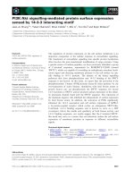
Fig. 4. a-GlcNAc and b-GlcNAc predictions on the aligned sequences of MSP-1 from Ghana (RO-33), Papua New Guinea (MAD20), Uganda (Palo
Alto), Thailand (K1) and Wellcome strains. Sequences extracted from SwissProt (accession no. P19598, P08569, P50495, P04932, P04933) were
aligned according to [23] and designated as G (Ghana), P (Png MAD20), U (Uganda), T (Thai) and W (Wellcome). Alpha- and b-GlcNAc site
predictions, made using methods based on neural networks, were marked on the alignment. The x-axis shows the position of the alignment, and the
y-axis marks the predicted potentials. The horizontal wavy line is a surface-accessibility derived threshold. A vertical impulse crossing the threshold
is said to represent a (predicted) glycosylated site. While the GlcNAc linkages in the N-terminal half of the protein are probably entirely the b form,
the C-terminal half has a mix of a and b forms. (Left) ÔXÕ marks represent potential a-GlcNAc positions. For Ghana, PnG and Uganda strains, out
of the four potential positions 1278, 1280, 1498, 1506, 1278 may be a b-O-GlcNAc. (Right) The triangles and circles represent N- and C-terminal
predicted b-O-GlcNAc positions. Empty circles and triangles are other ÔpossiblesÕ (negative predictions but very close to the threshold). The
positions in the figure are alignment positions. Exact sequence positions may vary slightly from strain to strain. The prediction methods are
available at />372 D. C. Hoessli et al. (Eur. J. Biochem. 270) Ó FEBS 2003
(5–10, 17 and 19 kDa), but only the [
3
H]GlcN-labelled
19-kDa band is immunoprecipitated by the anti-(C-ter)
mAb. The same mAb however, detects the 17-kDa fragment
andpartofthelowerM
r
fragments by Western blotting, but
still does not recognize the bulk of the [
3
H]GlcN-labelled
peptides between 5 and 10 kDa. Those [
3
H]GlcN-labelled,
lipophilic peptides are contained in sedimentable vesicular
membranes (nanovesicles, see [27]) released by parasitized
erythrocytes in culture. This lipophilic behaviour strongly
suggest that MSP-1 C-terminal peptides carry [
3
H]GlcN-
labelled GPI anchors. Vesicular membranes enriched in
GPI-anchored peptides could be a vehicle for the bioactive
inositol glycan moieties released by P. falciparum parasites
[39]. As the tyrosine kinase activity of macrophages was
shown to respond to the hydrophilic, carbohydrate moieties
of GPI molecules, released vesicles should exert their
biological effect by simple contact, whereas protein kinase
C enzymes were modulated only by acylated inositol glycans
and would thus require fusion of the vesicles with the target
cell membrane [39].
The low M
r
C-terminal MSP-1 fragments, like the
endogenous GPI-linked CD59, are selectively enriched in
low density, detergent-resistant membranes of the para-
sitized erythrocytes, suggesting that the parasite GPI-
anchored proteins seek a similar environment as the
endogenous ones in the membranes of the parasitized
erythrocyte. The presence of CD59 in nanovesicles immu-
noselected with anti-(C-ter) mAb strongly suggests that
both GPI-linked proteins are inserted in membrane subdo-
mains of similar properties that vesiculate as a unit in form
of nanovesicles.
The Triton X-100 resistant membranes described in this
study are most probably derived from erythrocytes and not
from the parasite membranes, as intact parasites were
removed by centrifugation prior to sucrose gradient float-
ation. Sphingomyelin is synthesized de novo in the parasi-
tized erythrocyte under the control of P. falciparum [40] and
may also contribute to the formation of detergent-resistant
membrane domains in the newly made membranes. The
Triton X-100 resistant membranes originally described in
erythrocyte ghosts were characteristically rich in sphingo-
lipids and cytoskeletal proteins spectrin, actin and band 4.1
[41]. Using standard equilibrium sucrose density gradients,
Civenni et al. have shown that GPI-linked surface proteins
such as acetylcholinesterase, CD55 and CD59 are also
included in the detergent-resistant membranes of normal
erythrocytes [42] and released as vesicles by the stressed or
aging erythrocytes [43]. The considerable remodelling of the
cytoskeleton–membrane interface taking place in the para-
sitized erythrocyte [44] makes it difficult to define precisely
the relationship of the GPI-rich, detergent-resistant mem-
branes we describe with the detergent-resistant membranes
of normal erythrocytes. However, a recent study proposes
that vacuolar uptake of erythrocyte components (CD59,
Duffy antigen) could be carried out by membranes with
detergent-resistant properties in the parasitized erythrocyte
[19]. We further show in this study that membranes
containing GPI-linked MSP-1 C-terminal 19-kDa fragment
and endogenous CD59 are released in vesicular form by the
cultured parasitized erythrocyte. The in vivo implication of
this finding is that MSP-1 C-terminal antigens may disperse
in the bloodstream and possibly integrate other cellular
membranes [45].
Acknowledgements
This work was supported by the UNDP/World Bank/WHO Special
Programme for Research and Training in Tropical Diseases Grant ID
970604 and Swiss National Science Foundation Grant 31–57696.99.
R. G. thanks J. Hansen for useful discussions, and the Danish National
Research Foundation for funding. We are grateful to Drs J. A. Lyon
and V. Horejsi for their kind gifts of antibodies.
References
1. Holder, A.A. (1988) The precursor to major surface antigens:
structure and role in immunity. Progr. Allergy. 41, 72–97.
2. Blackman, M.J., Whittle, H. & Holder, A.A. (1991) Processing of
the Plasmodium falciparum merozoite surface protein-1: identifi-
cation of a secondary processing product which is shed prior to
erythrocyte invasion. Mol. Biochem. Parasitol. 49, 35–44.
3. Blackman, M.J. & Holder, A.A. (1992) Secondary processing
of the Plasmodium falciparum merozoite surface protein
(MSP-1) by a calcium-dependent membrane-bound serine pro-
tease: shedding of MSP-1 (33) as a noncovalently associated
complex with other fragments of the MSP-1. Mol. Biochem.
Parasitol. 50, 307–316.
4. Blackman, M.J., Chappel, J.A., Shai, S. & Holder, A.A. (1993) A
conserved parasite serine protease processes the Plasmodium fal-
ciparum merozoite surface protein-1. Mol. Biochem. Parasitol. 62,
103–114.
Fig. 5. The MSP-1 C-terminal 19-kDa fragment and the endogenous
CD59 distribute to low-density, detergent-resistant membranes. Percoll-
purified parasitized erythrocytes were washed once in TKM and lysed
in TKM containing 1% Triton X-100 and protease inhibitors. The
lysate was adjusted to 40% sucrose and subjected to equilibrium
gradient centrifugation as described in Methods. At equilibrium, 11
1-mL fractions were collected from the top. Each fraction was con-
centrated and run on SDS/PAGE. After transfer to nitrocellulose the
blots were probed with MEM-43 mAb against erythrocyte CD59 (top
panel) or 3B10 mAb against the MSP-1 C terminus (bottom panel).
Fractions 3 and 4 correspond to the 5–36% sucrose interface where
detergent-resistant membranes accumulate. Fractions 5–10 contain
solubilized proteins. Fraction 11 is the gradient pellet containing whole
parasites and remnants of the parasitophorous membranes.
Ó FEBS 2003 Glycosylation and membrane localization of MSP-1 (Eur. J. Biochem. 270) 373
5. Gerold, P., Schofield, L., Blackman, M.J., Holder, A.A. &
Schwarz, R.T. (1996) Structural analysis of the glycosyl-phos-
phatidylinositol membrane anchor of the merozoite surface pro-
teins-1 and – 2 of Plasmodium falciparum. Mol. Biochem. Parasitol.
75, 131–143.
6. Gratzer, W.B. & Dluzewski, A.R. (1993) The red blood cell and
malaria parasite invasion. Semin. Hematol. 30, 232–247.
7. Dayal-Drager, R., Hoessli, D.C., Decrind, C., Del Guidice, G.,
Lambert, P H. & Nasir-ud-Din (1991) Presence of O-glycosylated
glycoproteins in the Plasmodium falciparum parasite, Carbohydr.
Res. 209, c5–c8.
8. Dieckmann-Schuppert, A., Bause, E. & Schwarz, R.T. (1994)
Glycosylation reactions in Plasmodium falciparum, Toxoplasma
gondii,andTrypanosome brucei brucei probed by the use of syn-
thetic peptides. Biochim. Biophys. Acta. 1199, 37–44.
9. Kimura,E.A.,Couto,A.S.,Peres,V.J.,Casal,O.L.&Katzin,
A.M. (1996) N-linked glycoproteins are related to schizogony of
the intraerythrocytic stage in Plasmodium falciparum. J. Biol.
Chem. 271, 14452–14461.
10. Nasir-ud-Din, Drager-Dayal, R., Decrind, C., Hu, B.H., Del
Giudice, G. & Hoessli, D.C. (1992) Plasmodium falciparum
synthesizes O-glycosylated glycoproteins containing O-linked
N-acetylglucosamine. Biochem. Int. 27, 55–64.
11. Dieckmann-Schuppert, A., Bender, S., Odenthal-Schnittler, M.,
Bause,E.&Schwarz,R.T.(1992)ApparentlackofN-glycosyla-
tion in the asexual intraerythrocytic stage of Plasmodium falci-
parum. Eur. J. Biochem. 205, 815–825.
12. Lingelbach, K. & Joiner, K.A. (1998) The parasitophorous
vacuole membrane surrounding Plasmodium and Toxoplasma:an
unusual compartment in infected cells. J. Cell Sci. 111, 1467–1475.
13. Haldar, K. (1996) Sphingolipid synthesis and membrane forma-
tion by Plasmodium. Trends Cell Biol. 6, 398–405.
14. Holt, G.D., Haltiwanger, R.S., Torres, C R. & Hart, G.W. (1987)
Erythrocytes contain cytoplasmic glycoproteins. J. Biol. Chem.
262, 14847–14850.
15. Hart, G.W. (1997) Dynamic O-linked glycosylation of nuclear and
cytoskeletal proteins. Ann.Rev.Biochem.66, 315–335.
16. Lyon, J.A., Haynes, J.D., Diggs, C.L., Chulay, J.D., Haidaris,
C.G. & Pratt-Rossiter, J. (1987) Monoclonal antibody char-
acterization of the 195-kilodalton major surface glycoprotein of
Plasmodium falciparum malaria schizonts and merozoites: identi-
fication of additional processed products and a serotype-restricted
repetitive epitope. J. Immunol. 138, 895–901.
17. Gowda, D.C., Gupta, P. & Davidson, E.A. (1997) Glycosylphos-
phatidylinositol anchors represent the major carbohydrate
modification in proteins of intraerythrocytic stage Plasmodium
falciparum. J. Biol. Chem. 272, 6428–6439.
18. Dluzewski, A.R., Ling, I.T., Rangachari, K., Bates, P.A. &
Wilson, R.J. (1984) A simple method for isolating viable mature
parasites of Plasmodium falciparum from cultures. Trans. R. Soc.
Trop. Med. Hyg. 78, 622–624.
19. Lauer,S.,VanWye,J.,Harrison,T.,McManus,H.,Samuel,B.U.,
Hiller, N.L., Mohandas, N. & Haldar, K. (2000) Vacuolar uptake
of host components, and a role for cholesterol and sphingomyelin
in malarial infection. EMBO J. 19, 3556–3564.
20. Ilangumaran, S., Briol, A. & Hoessli, D. (1997) Distinct interac-
tions among GPI-anchored, transmembrane and membrane
associated intracellular proteins, and sphingolipids in lymphocyte
and endothelial cell plasma membranes. Biochim. Biophys. Acta.
1328, 227–236.
21. Parchment,R.E.,Ewing,C.M.&Shaper,J.H.(1986)Theuseof
galactosyltransferase to probe nitrocellulose-immobilized glyco-
proteins for non-reducing terminal N-acetylglucosamine residues.
Anal. Biochem. 154, 460–469.
22. Previato, J.O., Sola-Penna, M., Agrellos, O.A., Jones, C.,
Oeltmann, T., Travassos, L.Z. & Mendonca-Previato, L. (1998)
Biosynthesis of O-N-acetylglucosamine-linked glycans in Trypa-
nosoma cruzi. J. Biol. Chem. 273, 14982–14988.
23. Miller, L.H., Roberts, T., Shahabuddin, M. & McCutchan, T.F.
(1993) Analysis of sequence diversity in the Plasmodium falciparum
merozoite surface protein-1 (MSP-1). Mol. Biochem. Parasitol. 59,
1–14.
24. Gupta, R., Jung, E., Gooley, A.A., Williams, K.L., Brunak, S. &
Hansen, J. (1999) Scanning the Dictyostelium discoideum pro-
teome for O-linked GlcNAc glycosylation sites using neural net-
works. Glycobiology 9, 1009–1022.
25. Hansen, J., Lund, O., Tolstrup, N., Gooley, A.A., Williams, K.L.
& Brunak, S. (1998) NetOGly: prediction of mucin type O-gly-
cosylation sites based on sequence context and surface accessi-
bility. Glycoconjugate J. 15, 115–130.
26. Hooper, N.M. & Turner, A.J. (1988) Ectoenzymes of the kidney
microvillar membrane: differential solubilization by detergents can
predict a glycosylphosphatidylinositol membrane anchor.
Biochem. J. 250, 865–869.
27. Salzer, U., Hinterdorfer, P., Hunger, U., Borken, C. & Prohaska,
R. (2002) Ca
++
-dependent vesicle release from erythrocytes
involves stomatin-specific lipid rafts, synexin (annexin VII), and
sorcin. Blood 99, 2569–2577.
28. Certa, U., Rotmann, D., Matile, H. & Reber-Liske, R. (1987) A
naturally-occurring gene encoding the major surface antigen
precursor P190 of Plasmodium falciparum lacks tripeptide repeats.
EMBO J. 6, 4137–4142.
29. Brown, D.A. & London, E. (1998) Structure and origin of ordered
lipid domains in biological membranes. J. Membrane Biol. 164,
103–114.
30. Hibbs, A.R. & Saul, A.J. (1994) Plasmodium falciparum:highly
mobile small vesicles in the malaria-infected red blood cell cyto-
plasm. Exp. Parasitol. 79, 260–269.
31. Chakraborty, A., Saha, D., Bose, A., Chatterjee, M. & Gupta,
N.K. (1994) regulation of eIF-2 alpha-subunit phosphorylation in
reticulocyte lysate. Biochemistry 33, 6700–6706.
32. Starr, C. & Hanover, J.A. (1990) Glycosylation of nuclear pore
protein p62. J. Biol. Chem. 265, 6868–6873.
33. Haltiwanger, R.S., Holt, G.D. & Hart, G.W. (1990) Enzymatic
addition of O-GlcNAc to nuclear and cytoplasmic proteins.
Identification of a uridine diphospho-N-acetyglucosamine: peptide
beta-N-acetylglucosaminyltransferase. J. Biol. Chem. 265, 2563–
2568.
34. Capasso, J.M., Abeijon, C. & Hirschberg, C.B. (1988) An intrinsic
membrane glycoprotein of the Golgi apparatus with O-linked
N-acetylglucosamine facing the cytosol. J. Biol. Chem. 263, 19778–
19782.
35. Torres, C R. & Hart, G.W. (1984) Topography and polypeptide
distribution of terminal N-acetylglucosamine residues on the
surfaces of intact lymphocytes. J. Biol. Chem. 259, 3308–3317.
36. Lauer, S.A., Rathod, P.K., Ghori, N. & Haldar, K. (1997) A
membrane network for nutrient import in red cells infected with
the malaria parasite. Science. 276, 1122–1125.
37. Gowda, D.C. & Davidson, E.A. (1999) Protein glycosylation in
the malaria parasite. Parasitol. Today. 15, 147–152.
38. Kimura, E.A., Katzin, A.M. & Couto, A.S. (2000) More on
protein glycosylation in the malaria parasite. Parasitol. Today 16,
38–39.
39. Tachado, S.D., Gerold, P., Schwarz, R., Novakovic, S., McCon-
ville, M. & Schofield, L. (1997) Signal transduction in macro-
phages by glycosylphosphatidylinositols of Plasmodium,
Trypanosoma,andLeishmania: Activation of protein tyrosine
kinases and protein kinase C by inositolglycan and diacylglycerol
moieties. Proc.NatlAcad.Sci.USA94, 4022–4027.
40. Lauer, S.A., Ghori, N. & Haldar, K. (1995) Sphingolipid synthesis
as a target for chemotherapy against malaria parasites. Proc. Natl
Acad. Sci. USA 92, 9181–9185.
374 D. C. Hoessli et al. (Eur. J. Biochem. 270) Ó FEBS 2003
41. Yu, J., Fischman, D.A. & Steck, T.L. (1973) Selective solubli-
zation of proteins and phospholipids from red cell membranes by
nonionic detergents. J. Supramol. Struc. 1, 233–247.
42. Civenni, G., Test, S.T., Brodbeck, U. & Butikofer, P. (1998) In
vitro incorporation of GPI-anchored proteins into human
erythrocytes and their fate in the membrane. Blood 91,
1784–1792.
43. Butikofer,P.,Kuypers,F.A.,Xu,C.M.,Chiu,D.T.Y.&Lubin,B.
(1989) Enrichment of two glycosyl-phosphatidylinositol-anchored
proteins, acetylcholinesterase and decay accelerating factor, in
vesicles released from human red blood cells. Blood. 74, 1481–
1485.
44. Deitsch, K.W. & Wellems, T.E. (1996) Membrane modifications
in erythrocytes parasitized by Plasmodium falciparum. Mol. Bio-
chem. Parasitol. 76, 1–10.
45. Ilangumaran, S., Robinson, P.J. & Hoessli, D.C. (1996) Transfer
of exogenous glycosylphosphatidylinositol (GPI)-linked molecules
to plasma membranes. Trends Cell Biol. 6, 163–167.
Ó FEBS 2003 Glycosylation and membrane localization of MSP-1 (Eur. J. Biochem. 270) 375