Tài liệu Báo cáo khoa học: Chromatin under mechanical stress: from single 30 nm fibers to single nucleosomes pdf
Bạn đang xem bản rút gọn của tài liệu. Xem và tải ngay bản đầy đủ của tài liệu tại đây (182.95 KB, 13 trang )
MINIREVIEW
Chromatin under mechanical stress: from single 30 nm
fibers to single nucleosomes
Jan Bednar
1,2,3
and Stefan Dimitrov
4
1 CNRS, Laboratoire de Spectrometrie Physique, St Martin d’Heres, France
2 Charles University in Prague, First Faculty of Medicine, Institute of Cellular Biology and Pathology, Prague, Czech Republic
3 Department of Cell Biology, Institute of Physiology, Academy of Science, Prague, Czech Republic
4 Institut Albert Bonniot, Grenoble, France
Introduction
Since the pioneering use of micromechanical and single
molecule manipulation approaches to probe biological
systems back in the late 1980s and 1990s (e.g. [1–5]),
their use has continuously expanded. In this review we
will focus mainly on the approaches using optical and
magnetic tweezers for studying the structure and con-
formational transitions of chromatin.
The basic repeating unit of chromatin, the nucleo-
some, represents the first level of the chromatin organi-
zation [6]. The major part of the nucleosome (termed
the chromatosome [7]) is composed of an octamer of
core histones (two each of H2A, H2B, H3 and H4), a
linker histone and 166 bp ( 56 nm) of DNA [6].
The histone octamer alone associates with 146 bp of
DNA ( 50 nm) wrapped round in 1.65 left-handed
superhelical turns (Fig. 1) to form the nucleosome core
particle (NCP), the structure of which has been solved
to 1.9 A
˚
resolution by X-ray crystallography [8]. The
neighboring chromatosomes are connected by linker
DNA.
The linear array of nucleosomes folds into 30 nm
fiber, the second level of chromatin organization. The
linker histones and the core histone NH
2
tails and
their post-translational modifications are essential for
both the folding process and the maintenance of the
chromatin fiber [9–11] as well as for the maintenance
Keywords
chromatin, micro-manipulation, nucleosome,
optical tweezers
Correspondence
J. Bednar, CNRS, Laboratoire de
Spectrometrie Physique, UMR 5588, BP87,
140 Av. de la Physique, 38402 St Martin
d’Heres Cedex, France
Fax: +33 476 51 45 44
Tel: +33 476 51 47 61
E-mail:
(Received 22 November 2010, revised 7
April 2011, accepted 28 April 2011)
doi:10.1111/j.1742-4658.2011.08153.x
About a decade ago, the elastic properties of a single chromatin fiber and,
subsequently, those of a single nucleosome started to be explored using
optical and magnetic tweezers. These techniques have allowed direct mea-
surements of several essential physical parameters of individual nucleo-
somes and nucleosomal arrays, including the forces responsible for the
maintenance of the structure of both the chromatin fiber and the individual
nucleosomes, as well as the mechanism of their unwinding under mechani-
cal stress. Experiments on the assembly of individual chromatin fibers have
illustrated the complexity of the process and the key role of certain specific
components. Nevertheless a substantial disparity exists in the data reported
from various experiments. Chromatin, unlike naked DNA, is a system
which is extremely sensitive to environmental conditions, and studies car-
ried out under even slightly different conditions are difficult to compare
directly. In this review we summarize the available data and their impact
on our knowledge of both nucleosomal structure and the dynamics of
nucleosome and chromatin fiber assembly and organization.
Abbreviations
ACF, ATP-dependent chromatin assembly and remodeling factor; HMG, high-mobility group; NAP-1, nucleosome assembly protein 1;
NCP, nucleosome core particle.
FEBS Journal 278 (2011) 2231–2243 Journal compilation ª 2011 FEBS. No claim to original French government works 2231
of mitotic chromosomes [12,13]. The globular domain
of the linker histone is internally located in the 30 -nm
chromatin fiber [14], although how it interacts with
both the NCP and the linker DNA remains a subject
of debate [15,16].
The conformation of the 30 nm chromatin fiber is
sensitive to ionic conditions [9]. The fiber adopts a
relaxed zigzag structure at low ionic strength and
undergoes compaction with increasing salt concentra-
tion, reaching a very compact form under physiologi-
cal conditions. The linker DNA arrangement in the
most compact form of the chromatin fiber continues to
be a controversial issue [15–19].
Micromechanical approaches were used to study
three different aspects of chromatin organization: the
mechanical properties of (a) mitotic chromosomes and
(b) an individual nucleosome or a single 30 nm chro-
matin fiber, and (c) the rheology of chromatin in vivo
(e.g. [20–22]). Mitotic chromosomes have been the sub-
ject of several ‘mechanical’ studies [23–29] and some of
the stretching experiments were performed long before
the invention of optical tweezers [30–33]. These studies
have recently been thoroughly reviewed [34] and this
review will thus concentrate on reviewing single mole-
cule studies of individual nucleosomes, nucleosomal
arrays and 30 nm chromatin fibers.
Chromatin samples ‘eligible’ for single
molecule experiments
All micromechanical experiments applied to a nucleo-
some or chromatin fiber require an adaptation of the
substrate in order to make it suitable for attachment
to a ‘micro-handle’. In the case of experiments with
optical tweezers, micro-beads of dielectric material (sil-
ica, polystyrene) are the most frequently used type of
‘handle’. A typical configuration of the optical twee-
zers stretching experiment is depicted in Fig. 2. The
chromatin substrate is tethered between the two beads
by means of a very tight interaction, typically using
biotin ⁄ streptavidin or digoxigenin ⁄ anti-digoxigenin
coupling between the fiber ends and beads.
Four distinct types of chromatin substrates have
been used for stretching experiments: (a) native chro-
matin, isolated from nuclei after microccocal nuclease
digestion, (b) chromatin reconstituted in vitro by salt
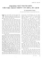
Fig. 1. Chromatin organization and scheme of chromatin array stretching under different force regimes. A nucleosomal core particle, formed
by 147 bp of DNA and a histone octamer, is complemented with linker histone (H1) and an additional 20 DNA bp to form the chromato-
some. Linker DNA completes and links consecutive nucleosomes which fold into the 30-nm chromatin fiber. During stretching the nucleoso-
mal array is first stretched to its contour length. Additional stretching leads to the rupture of inter-nucleosomal interactions and the array is
stretched to the beads-on-a-string configuration. Further force increase results in progressive eviction of histone octamers. The force values
are approximate (see text) (adapted from [69,80]).
Fig. 2. Optical tweezers experimental setup. The laser beam (LB)
is conducted via a dichroic mirror (DM) to the back aperture of the
objective lens (OL) which focuses the beam and creates the optical
trap (OT) at the focal point. The filament (F) (DNA, chromatin fiber
etc.) is tethered between a trapped bead (TB) and a bead (FB) held
by suction onto a micropipette (MP). The micropipette is coupled to
a high precision micro-positioning system (typically a piezoelectric
XY plate). The image of the bead is projected onto a position sen-
sor (PS). As the fiber is stretched beyond its curvilinear length, the
bead in the trap will start to displace from the center of the trap
and the force which is trying to bring the bead back is linearly pro-
portional to this displacement. Thus, the change of the fiber length
as a function of the force can be measured, resulting in a so-called
force–extension curve.
Chromatin under mechanical stress J. Bednar and S. Dimitrov
2232 FEBS Journal 278 (2011) 2231–2243 Journal compilation ª 2011 FEBS. No claim to original French government works
dialysis, (c) nucleosome assembly protein 1 (NAP-1)
and ATP-dependent chromatin assembly and remodel-
ing factor (ACF) assembled chromatin and (d) chro-
matin assembled in nuclear extracts. These distinct
substrates have different properties and advanta-
ges ⁄ disadvantages for the experiments. Native chro-
matin fibers, isolated after light micrococcal nuclease
digestion from nuclei, exhibit heterogeneous lengths
(different numbers of nucleosomes per individual
fiber) and both their protein composition and state of
histone modifications are poorly defined. The nucleos-
omal arrays reconstituted by salt dialysis on tandem
repeats of positioning DNA sequences (601 [35] or 5S
[36]) have defined length, and the nature and modifi-
cation state of histones can be controlled, but the
proper association of linker histones in vitro is diffi-
cult. Therefore, this material is mostly studied in their
absence. The use of NAP-1 ⁄ ACF systems has allowed
the reconstitution of chromatin using any DNA
sequence, but it does not solve the issue of linker his-
tone assembly. The preparation of chromatin frag-
ments in nuclear extracts does not require a DNA
substrate bearing positioning sequences and the num-
ber of nucleosomes will depend only on the length of
DNA used. The linker histone will be present,
although its type will vary depending on the type of
extract used. Unfortunately, in addition to the chro-
matin assembly proteins, the extracts contain a large
number of other proteins which can eventually form
distinct DNA–protein complexes. These could affect
the physical properties of the assembled chromatin
fiber and consequently the interpretation of the mea-
sured elastic parameters.
Mechanical properties of native 30 nm
fiber
The first ever single molecule micromanipulation
experiment on chromatin focused mostly on the elastic
behavior of the 30-nm chromatin fiber as a function of
its environmental conditions [37]. In the low ionic
strength (5 mm NaCl) and low force regime
(< 10 pN), the measured stretching curve exhibited a
rather extended plateau, which was interpreted as fiber
accordion-like extension and disruption of inter-nucle-
osomal interactions. The energy of these interactions
was estimated to be around 3.4 k
B
T per nucleosome.
The authors observed the onset of a hysteresis in
repeated stretch ⁄ relaxation cycle curves at a force of
about 20 pN. Its origin was attributed to eviction of
some histone octamers from the fiber by mechanical
stress. These experiments allowed the determination of
several physical parameters. The persistence length of
the fiber and its stretch modulus at low salt conditions
were determined to be 30 nm and 5 pN, respectively.
The chromatin fiber showed similar elastic behavior
when the experiments were performed in 40 mm NaCl.
However, the forces necessary to achieve the same
extension of the fiber were significantly higher. This
was attributed to the more compact initial conforma-
tion of the fiber. Although the compaction level of
native chromatin fibers (containing the linker histone)
is significantly higher in 150 mm NaCl than in 40 mm
[9], quite surprisingly the experiments in 150 mm did
not show significant differences in the fiber elastic
characteristics compared with those at 40 mm.
As mentioned earlier, about 166 bp of DNA is asso-
ciated with the chromatosome and 145 bp with the
NCP. The nucleosome structure can thus be consid-
ered as a ‘DNA length’ buffer. When the stretching
forces applied on the chain of nucleosomes exceeds the
mechanical resistance of the DNA ⁄ histone contacts, a
mechanical unwrapping of DNA from the histone oct-
amer will occur. If this release is discontinuous (i.e. a
certain characteristic length of DNA is released in an
all-or-none event) this will lead to a drop of instant
stretching force and a sawtooth profile of the
force ⁄ extension curve will appear. This is referred to as
a ‘disruption’ event. As the nucleosome will not reas-
semble, the length of the fiber will remain increased
and in the next stretch ⁄ relaxation cycle a different
extension curve will be observed. In this study [37], the
sawtooth pattern could not be directly observed as the
stretching was effected in discrete steps of about
50 nm, a value similar to the total length of nucleoso-
mal DNA.
Mechanical properties of chromatin
reconstituted in egg extract
Another work used chromatin fibers reconstituted in
Xenopus laevis egg extract [38]. Stretching these fibers
at a continuous speed of 1 lmÆs
)1
revealed a sawtooth
profile, which started to appear at forces above 20 pN
and continued until about 40 pN. The analysis
revealed three distinct characteristic DNA release
lengths: 65, 130 and 195 nm. The 65 nm was attributed
to single nucleosome disruption and the two others
were attributed to the simultaneous dissociation of two
and three nucleosomes, respectively.
The direct attribution of the observed released
lengths to a single nucleosomal DNA unwrapping,
however, appeared not to be straightforward. Upon
eviction of the histone octamer and histone H1 (i.e.
the disruption of the chromatosome) 166 bp of DNA
is expected to be released, i.e. 56.4 nm and not 65 nm.
J. Bednar and S. Dimitrov Chromatin under mechanical stress
FEBS Journal 278 (2011) 2231–2243 Journal compilation ª 2011 FEBS. No claim to original French government works 2233
To explain this, it was suggested [38] that other non-
histone proteins, namely high-mobility group (HMG)
family members, abundant in the X. laevis egg extract,
were associated with the nucleosome. This would result
in reinforcing the nucleosome mechanical resistance
and in locking of additional DNA into the complex.
These suggestions were not experimentally addressed,
however.
These experiments allowed also calculation of the
assembly rate of the nucleosomes, which was found to
be about three nucleosomes per second. For the length
of k DNA (48 kbp) and the nucleosomal repeat length
(200 bp), the assembly of chromatin would thus be
complete in about 80 s under these experimental condi-
tions. This is far shorter than the chromatin assembly
time (typically a few hours) in bulk in vitro reconstitu-
tion in egg extracts [39]. When a force countering the
DNA shortening (due to nucleosome formation) was
applied, the rate of DNA shortening gradually
decreased and was finally halted at forces above 10
pN. A similar fast rate of nucleosome assembly was
also observed in experiments where DNA was
stretched by hydrodynamic shear forces and incubated
in nuclear extracts [40]. This apparent contradiction
between the rates of assembly of single molecules and
bulk chromatin could be explained by the very high
histone : DNA ratio in the single molecule experiment
compared with those in the bulk experiments. When
competitive DNA (in amounts needed to reach the
DNA : histone ratio typical for bulk experiments) is
added to such a system, the nucleosome assembly rate
dramatically decreases (Claudet, Bednar and Dimitrov,
unpublished results).
Unwrapping individual nucleosomes in
reconstituted nucleosomal arrays
A detailed study of the mechanical behavior of in vitro
(by salt dialysis) reconstituted nucleosomal arrays (on
DNA templates containing 17 tandem repeats of the
5S positioning sequence from sea urchin) was accom-
plished by Brower-Toland and colleagues [41]. In their
experiments, the arrays did not contain linker histones
and the stretching was performed in 100 mm NaCl,
1.5 mm MgCl
2
. The stretching profiles were recorded
with either constant stretching speeds or at constant
force. The force–extension curves showed characteristic
sawtooth patterns at forces starting at about 20 pN
with 17 nominal peaks and a regular length of sub-
strate elongation steps of about 27 nm. Very similar
values were observed in the constant force regime.
Interestingly, the authors also observed a continuous
non-DNA stretching profile under a low force regime
(< 15 pN). This part of the curve was interpreted as a
continuous unwinding of nucleosomal DNA from the
histone octamer, mainly from contacts with histones
H2A ⁄ H2B where the DNA–histone interactions are
supposed to be weak [42]. The total amount of DNA
released was calculated to be 158 bp per nucleosome, a
value slightly higher than the 147 bp expected. It was
concluded that 76 bp of DNA per nucleosome is
unwound continuously in the low force regime, and
82 bp dissociates under stresses higher than 20 pN in
an all-or-none fashion. The calculated energy (using
dynamic force spectroscopy theory [43]) necessary to
dissociate the DNA from the histone octamer was 21–
22 kcalÆmol
)1
.
In the multiple stretching cycle experiments, the
reappearance of peaks was observed when the time
gap between successive cycles was sufficiently long (at
least 10 s) and the stretching force in the preceding
cycle did not exceed 50 pN. It was suggested that
forces below this value did not cause a complete evic-
tion of all histone octamers. Some of the octamers
may have remained attached to the DNA (probably at
the dyad region, where the DNA–histone interactions
are the strongest), and upon DNA relaxation nucleo-
some reassembly could occur. This phenomenon was
not observed in the case of stretching experiments
using chromatin reconstituted in X. laevis egg extracts
[38].
The part of the stretching profile interpreted as ‘con-
tinuous release of the outer DNA turn’ [41] is very
similar to the initial plateau in the experiments with
native chromatin under similar ionic conditions [37],
interpreted as chromatin fiber accordion-like extension
and reflecting the disruption of the nucleosome–nucle-
osome interactions. Unfortunately, in [41] there is no
comparison with stretching profiles in low salt condi-
tions, which would help to clarify the contribution of
inter-nucleosomal interactions or elastic contributions
of chromatin fiber compaction. Note that the direct
comparison of results reported in these two studies
[37,41] is rather difficult as the fibers used in [37] were
about 15-fold longer and contained linker histones. In
addition, the experiments were carried out under dif-
ferent ionic conditions.
The experiments were further refined with arrays
reconstituted with tail-less histones or histones with
modified NH
2
-termini [44]. The removal of N-termini
of all core histones had a strong impact on both the
length of the outer DNA turn and the peak force,
which dropped by nearly 40 bp (from 65 bp in intact
to 28 bp in tail-less octamer nucleosomes) and to 3
pN, respectively. Also the released DNA lengths in the
case of the nucleosome with intact tails were revised
Chromatin under mechanical stress J. Bednar and S. Dimitrov
2234 FEBS Journal 278 (2011) 2231–2243 Journal compilation ª 2011 FEBS. No claim to original French government works
and found to be 65 bp (instead of 76 bp in [41]) for
continuous release of the outer DNA turn and 72 bp
(instead of 82 bp) for disruption of the inner turn. In
all studied cases the removal or modification of histone
tails influenced the stretching profile and the effect
concerned mainly the outer DNA turn while the inner
turn was only minimally affected. A similar phenome-
non was also observed for nucleosomal arrays reconsti-
tuted with the H2A.Bbd histone variant octamer. In
this case a 2 pN drop of threshold disruption forces
(from 19 pN for conventional nucleosomes to 17 pN
for H2A.Bbd nucleosomes) was measured [45]. The
last result is in agreement with the data obtained from
other methods showing a weaker association of the
variant H2A.Bbd octamer with DNA [45,46].
Obviously, similar experiments performed on nucle-
osomal arrays prepared by salt dialysis and by assem-
bly in egg extracts (see above) gave quite divergent
results. While the threshold force values were very sim-
ilar (about 20 pN), the lengths of DNA released upon
mechanical disruption of nucleosomes were quite dif-
ferent. The values of 65 nm or 130 nm measured in
the case of egg extract assembled fibers [38] were never
observed for chromatin reconstituted by salt dialysis.
Gemmen et al. [47] performed analogous experi-
ments on nucleosomal arrays prepared in vitro by
using the histone chaperone NAP-1 and ACF which
forms nucleosomal arrays on random DNA sequences
with nucleosomal repeat of about 168 bp [48,49].
Although the features of the measured stretching pro-
files were generally comparable with the results of [41]
(including the DNA re-wrapping in repeated stretching
cycles), some important differences were observed. The
disruption length varied from 55 bp to 95 bp and the
threshold forces ranged from 5 to 65 pN. In addition,
the authors found a clear dependence of the average
threshold force on ionic conditions, ranging from 24
pN in 100 mm NaCl to 31 pN in 5 mm NaCl. The
wide range of measured threshold forces was attributed
to the variation of histone octamer affinities to the
given underlying DNA sequence.
We have studied the elastic properties of both native
chromatin samples (isolated from chicken erythrocytes
and containing linker histones) and nucleosomal arrays
reconstituted by salt dialysis [50]. We found the same
values of basic characteristics of the majority of dis-
ruption events, i.e. the peak force and the released
DNA length, as reported in [42] (20 pN and 25 nm).
However, a minor population of events exhibited a dis-
ruption length centered at 50 nm, thus corresponding
very closely to the 147 bp of DNA released upon dis-
ruption. How can we explain this finding? It was previ-
ously reported that the integrity of the nucleosomal
structure depends on two factors: (a) the ionic condi-
tions and (b) the concentration of the chromatin itself
[51]. At very low concentrations of chromatin, the
structure is destabilized and a progressive dissociation
of the linker histone and H2A–H2B dimers from the
nucleosome is observed [50,51]. When the stretching
experiments with native chromatin fibers were repeated
under conditions favoring histone octamer stability
(presence of exogenous chromatin or low ionic
strength) a significant increase in the number of 50-nm
events was observed. This was interpreted as an effect
of histone octamer stabilization and the release of all
the DNA associated with a histone octamer in an all-
or-none event [50]. A similar effect was observed with
arrays containing 12 nucleosomes reconstituted on 5s
positioning sequences. Further analysis showed that
indeed under conditions typical for single molecule
experiments (where the chromatin concentration is
usually extremely low) H2A–H2B dimers as well as lin-
ker histones readily dissociated from the nucleosomes
even at moderate ionic concentrations [50]. The
remaining (H3–H4)
2
tetramers associate with only one
superhelical turn of DNA and consequently, upon
stretching, the release of only 25 nm in a single disrup-
tion event will be observed.
Why then were the peaks with 25-nm release length
not observed in experiments with egg extract reconsti-
tuted chromatin? One of the possible explanations is
the association of non-histone proteins (e.g. HMG
proteins) with chromatin, leading to an additional sta-
bilization, mainly of the outer turn. The study of Pope
and colleagues [52] showed that the situation might be
even more complex. In their work they focused mainly
on the elastic response of chromatin fibers assembled
in X. laevis egg extract to different loading rates (i.e.
the force increase per time unit). They detected three
typical disruption lengths: 30 nm, 59 nm and 117 nm.
These data differed from the results of Bennink et al.
[38], where the 30-nm disruption length was not
detected, and revealed two distinct energy barriers hav-
ing values of 25 and 28 k
B
T (14.5 kcalÆmol
)1
and
16 kcalÆmol
)1
). With high loading rates the value of
the first barrier dropped to 20 k
B
T (12 kcalÆmol
)1
).
The individual lengths were attributed to a disruption
of the entire nucleosome in one event (60 nm), simulta-
neous release of the DNA from two nucleosomes
(117 nm) or the partial unraveling of one DNA turn
(30 nm). In addition to the explanation of Brower-To-
land and Wang [53], Pope et al. [52] also considered
the possibility of disruption of an incomplete nucleo-
some – missing either one or both H2A–H2B
dimers. The individual energy barriers were attributed
to nucleosomes with and without linker histone B4
J. Bednar and S. Dimitrov Chromatin under mechanical stress
FEBS Journal 278 (2011) 2231–2243 Journal compilation ª 2011 FEBS. No claim to original French government works 2235
(the embryonic linker histone variant present in the
egg extract). Based on the analysis, the linker histone
contribution to nucleosomal ‘stability’ was estimated
to be rather low, about 3 k
B
T, which would reflect the
fact that no significant difference in threshold force
was observed between nucleosomal arrays with and
without linker histones [50]. In repeated stretching
experiments the number of events with high energy
barriers (28 k
B
T) rapidly decreased suggesting the per-
manent removal of B4 from the nucleosomes during
the initial stretching. No correlation between the dis-
ruption length and the energy barrier was found. The
value of the barrier was significantly lower than that
reported in [41] (16 kcalÆmol
)1
versus 20–22 kcalÆ-
mol
)1
) but again the experiments were carried out in
different ionic conditions (10 mm Tris ⁄ HCl, pH 7.5,
1mm EDTA, 150 mm NaCl, 0.05% BSA and 0.01%
NaN
3
) and the chromatin samples were assembled by
different techniques.
The mechanical properties of nucleosomal arrays
reconstituted on African green monkey alpha-satellite
DNA were studied by Bussiek et al. [54]. They found
disruption of alpha-satellite nucleosomes to occur at a
higher force on average – 26.4 pN versus 21.7 pN for
random DNA nucleosomes. The authors hypothesized
that the increased bending flexibility of alpha-satellite
DNA (due to the presence of clustered CA⁄ TG steps)
would result in the formation of more stable nucleo-
somes as less energy is needed for DNA bending.
Zooming in on the stretching of a
single nucleosome
Analysis of the experiments with nucleosomal arrays is
always complicated by the elastic contribution of the
inter-nucleosomal interactions at different ionic con-
centrations. This could be overcome by analyzing the
properties of a single mononucleosomal template.
Mihardja et al. [55] prepared a mononucleosomal tem-
plate on a 2582-bp long DNA construct containing a
single 601 positioning sequence [35]. Stretching profiles
of these particles showed several features not previ-
ously observed. Pulling the template at very low load-
ing rates, the first discontinuity in the stretching curve
was observed at forces centered at 3 pN and the
length of the released DNA was determined to be
21 nm. A second peak occurred at forces around
8–9 pN with a similar length, 22 nm. These events
were interpreted as a successive release of the outer
and the inner wrap of the nucleosomal DNA. The first
unwrapping was reversible, provided the stretching
curve did not reach the second discontinuity. Experi-
ments conducted under a constant force regime
ranging between 2 and 3 pN revealed a bistable char-
acter of the first event with a dwell time in the
unwrapped state depending on the force value, increas-
ing with increased force. From these measurements the
free energy of the outer turn unwrapping was calcu-
lated to be 6 kcalÆmol
)1
. The unwrapping of the sec-
ond, inner turn represented by the second peak at
about 8 pN was not reversible. Its analysis with load-
ing rates in the range 2.4–11 pNÆs
)1
revealed that the
dependence of the probability of unwrapping on the
force was not linear. Therefore, the unwrapping of the
inner turn cannot be considered as a simple two-state
process but will involve some intermediate states as
well. The same experiments were also performed at
high salt concentrations (200 mm potassium acetate).
Under these conditions, the first low force transition
was transformed into a nearly continuous plateau
rather than a sharp peak and the high force transition
was shifted to lower forces.
These experiments identified at least two novel fea-
tures of the nucleosome elasticity behavior. First, the
value of the disruption force was lowered to about
half of that originally reported (9 pN versus 20 pN)
and, second, the experiments clearly showed that
unwrapping of the outer turn was not continuous as
reported previously [41]. The differences in these
experimental data from those obtained with nucleoso-
mal arrays could reflect both the differences in the
experimental conditions and the nature of the starting
material. The single nucleosome experiments avoid all
contributions coming from the fiber-like behavior of
the nucleosomal arrays, which is strongly dependent
on the ionic conditions. The forces needed to stretch
the fibers containing native linker histone without dis-
rupting the nucleosomes (5 pN [37]) are roughly equal
to or greater than the threshold forces for unwrapping
of the outer turn (3 pN [55]). It is thus likely that at
forces up to 5 pN two events are happening simulta-
neously – an unwrapping of the outer DNA turn and
stretching of the folded nucleosomal array. The result-
ing elastic profile would reflect a superposition of
these two events. This would in turn result in a
smeared, plateau-like characteristic of the stretching
curve at low forces rather than resolved peaks. The
different composition of buffers used in the experi-
ments make the comparison even more difficult. As
the nucleosome stability strongly depends on ionic
conditions, in order to directly compare the data the
experiments have to be carried out under exactly the
same ionic conditions. Rather high concentrations of
Mg
2+
(10 mm magnesium acetate) together with
50 mm potassium acetate, however, were used in
the single nucleosome experiments [55], while 100 mm
Chromatin under mechanical stress J. Bednar and S. Dimitrov
2236 FEBS Journal 278 (2011) 2231–2243 Journal compilation ª 2011 FEBS. No claim to original French government works
NaCl and 1.5 mm MgCl
2
were used for nucleosomal
array stretching [41].
An interesting approach to investigate the stability
of a single nucleosome was used by Shundrovsky et al.
[56]. Instead of pulling tethered nucleosomal templates,
they ‘unzipped’ the DNA of a reconstituted template
containing a single 601-positioned nucleosome. The
nucleosome was flanked by free DNA arms and, upon
stretching, the first 220 bp of naked DNA were
unzipped before the histone octamer was reached. The
unzipping of DNA associated with the histone octamer
was affected by histone–DNA contacts within the
nucleosome and reflected the strength of the histone–
DNA interactions. The unzipping profile of the nucleo-
some showed three distinct high force regions
(contrary to the first two, the third region was not reg-
ularly observed). Within these regions, forces up to 45
pN had to be applied in order to overcome the barrier.
The first peak was observed at about 50 bp from the
dyad upon applying an average force of 31 pN, while
the second one was observed in the vicinity of the
nucleosome dyad and at 37 pN average force. These
peaks were attributed to the disruptions of the strong
interactions between H2A–H2B dimers and H3–H4
tetramers, respectively. The attribution of the first peak
to the disruption of the H2A⁄ H2B–DNA interaction
was confirmed by stretching a particle reconstituted
with the (H3–H4)
2
tetramer only. The unzipping pro-
file of this tetrameric particle exhibited only the sec-
ond, high force peak. According to the authors, the
third peak was associated with the instability of the
nucleosome when most of the nucleosomal DNA was
unzipped.
These experiments were further refined [57], allowing
analysis of the DNA–histone interactions with near
base-pair resolution. The unzipping was carried out
under a constant force regime using a 28-pN trapping
force. The strength of the interaction was found to be
proportional to the time needed for its disruption. This
allowed mapping of the interaction strength of the dif-
ferent regions with a resolution of about 1.5 bp. The
recorded data again revealed three regions of strong
interactions (longer dwell times): one was located close
to the dyad, while the other two were symmetrically
located at positions ±40 bp from the dyad. All three
exhibited a 5-bp periodicity. The data demonstrated
that the unzipping of the first 20 bp of nucleosomal
DNA had the same characteristics as those of naked
DNA, indicating a loose interaction of the histones
with DNA at the entry ⁄ exit points of the NCP. Very
similar results were obtained in continuous stretching
regime measurements with loading rates of 8 pNÆs
)1
,
as well as when random DNA sequences instead of
positioning sequences were used for nucleosome recon-
stitution.
Magnetic tweezer experiments
Several experiments with magnetic tweezers have also
been reported (for the principles of magnetic tweezers
see for example [58,59]). Magnetic tweezers can mea-
sure forces about 1–2 orders smaller than optical twee-
zers and, unlike optical tweezers, they can also control
the torsion of the fiber.
Leuba et al. [60] studied NAP-1 mediated assembly
of chromatin fibers on k DNA using magnetic twee-
zers. They observed an inhibition of the fiber assembly
at forces of 10 pN, but they also registered disas-
sembly events (in an otherwise progressive assembly
process) at forces of about 5 to 7.5 pN. This suggested
that the equilibrium forces were in this range.
Experiments using a similar strategy, but in X. laevis
egg extracts, were realized by Yan et al. [61]. The
experiments were carried out either in ATP-depleted
extract or in extract containing a defined concentration
of ATP or non-hydrolyzable ATP. They found that in
ATP-depleted extract forces of only 4 pN resulted in
inhibition of nucleosome assembly. At forces below
3.5 pN, the extract was able to accomplish the assem-
bly although the number of assembled nucleosomes
was significantly lower relative to the nucleosomal
array reconstituted under optimal conditions (the mea-
sured nucleosomal repeat was only 280 bp in contrast
to the 180–160 bp repeat reported for fully extract-
assembled chromatin [39]). The 3.5 pN value was
determined as an equilibrium force of ATP-indepen-
dent nucleosome assembly giving straightforwardly the
free energy of DNA–histone octamer association as
27 kcalÆmol
)1
. Once the assembly was completed, the
fibers were stretched with different loading rates. Dur-
ing this process, a step-wise fiber lengthening was
observed with a predominating step value of 50 nm,
attributed to an unwrapping of one complete nucleo-
some. The presence of 30- and 100-nm steps was also
detected. Interesting changes were induced by addition
of ATP to the extract. In this case, the disassembly
threshold force decreased to 1 pN. Non-hydrolyz-
able ATP did not affect the nucleosome assembly ⁄
disassembly equilibrium force determined in ATP-
depleted extract.
Why did these two very similar experiments give rise
to such different results? First, the egg extract contains
a poorly defined composition of proteins compared
with the purified NAP-1 assembly system. It is quite
possible that some ATP-independent protein com-
plexes present in the egg extract can associate with the
J. Bednar and S. Dimitrov Chromatin under mechanical stress
FEBS Journal 278 (2011) 2231–2243 Journal compilation ª 2011 FEBS. No claim to original French government works 2237
nucleosomes and modify their mechanical stability.
The steep drop to 1 pN in the stall force in the pres-
ence of ATP, however, is quite surprising. The events
observed in the stretching profile under these condi-
tions did not correspond to an assembly of individual
nucleosomes, but rather to formation and release of
rather long ‘loops’ (200–400 nm). The fact that the
energy provided by the added ATP in the system was
not even partly used for assisted nucleosome assembly
is also surprising. However, the authors have observed
nucleosome-like disassembly steps of 50 and 100 nm
when the force was increased to over 5 pN. Impor-
tantly, no reverse (i.e. assembly) events were detected
even at low forces.
Kruithof et al. [62] carried out experiments on
strongly subsaturated oligonucleosomal arrays (one to
four nucleosomes present on 17 tandem repeats of 5s
DNA) using magnetic tweezers with sub picoNewton
resolution. This experiment is directly comparable with
the work of Mihardja et al. [55]. Although both groups
used very similar conditions, Kruithof et al. did not
observe any DNA unwrapping from the nucleosome
below forces of 6 pN, even though they used a posi-
tioning sequence with lower affinity for the histone
octamer (5s versus 601).
The data obtained by force spectroscopy of chroma-
tin are not always easy to interpret unambiguously
and to explain in terms of changes in nucleosome and
fiber structure and dynamics. While in lower salt con-
centrations (50 mm) and in the absence of bivalent
ions the inter-nucleosomal interactions can be
neglected, the situation becomes more complex when
the fiber is studied in its compact form, where the pres-
ence ⁄ absence of linker histones, the higher concentra-
tion of monovalent ions, and the presence of bivalent
or polyvalent ions contribute significantly to the fiber
properties. Kruithof et al. [63] used improved tech-
niques of linker histone association [64,65] to prepare
defined chromatin arrays of 25 nucleosomes with two
different nucleosomal repeat lengths (197 and 167 bp)
and used magnetic tweezers to study their elastic
behavior. The stretching curves of these samples exhib-
ited four major regions. The first was attributed to the
extension of the DNA segments flanking the array of
25 nucleosomes which serve as a handle for tethering.
The second region (at forces up to 4 pN) represented
the extension of the chromatin fiber. The third region
(a plateau observed at 4–4.5 pN) was attributed to the
disruption of inter-nucleosomal interactions. The last
region was interpreted to reflect extension of the
beads-on-a-string fiber. The incorporation of the linker
histone had only a minor effect on the overall form of
the stretching profile. Upon H5 association, the third
region (plateau) was shifted to a higher force value –
7 pN – suggesting that linker histone stabilizes
nucleosomal stacking. However, its absence did not
compromise chromatin folding when Mg
2+
was pres-
ent (1.5 mm MgCl
2
). When Mg
2+
ions were depleted
from the solution, the behavior of the fibers without
linker histones changed. A disruption of the inter-nu-
cleosomal interactions at forces of about 3.5 pN and
an increasing irreversibility upon repeated stretching
cycles (in the presence of 100 mm NaCl) were
observed. Reintroduction of Mg
2+
resulted in a com-
plete recovery of the original folding pattern, suggest-
ing that, at least under these conditions, the linker
histone might not be required for proper chromatin
folding. The analysis of fiber stretching profiles, their
Hookian behavior, their length and transition to
extended beads-on-a-string structures in the third and
fourth regions of the stretching curve led the authors
to conclude that in its compact form the fiber is orga-
nized in a one-start solenoidal topology. The data
obtained on fibers with 167 bp nucleosomal repeat
were significantly different [63]. Surprisingly, their con-
tour length at 0.5 pN stretching force was longer than
for fibers with 197 bp nucleosomal repeat and their
measured stiffness was found to be 2.7-fold higher
(0.052 versus 0.019 pNÆnm
)1
). This was interpreted as
a consequence of their different topological organiza-
tion and a two-start helix topology was suggested as
best fitting the observed data.
However, the story of chromatin fiber folding is
apparently more complex. Other studies have demon-
strated that for longer nucleosomal repeats both linker
histone and Mg
2+
ions are required in order to reach
maximal packing levels of the chromatin [66]. This is
not valid for short nucleosomal repeats where, even in
the absence of linker histone, the fiber can maximally
pack in a regular manner [66]. It would therefore be
interesting to see whether the same elastic behavior
(i.e. linker-histone-independent compaction in the pres-
ence of bivalent ions) would be observed for longer
nucleosomal repeats. The presence of Mg
2+
also
resulted in a substantial increase of the inter-nucleoso-
mal stacking energy to about 17 k
B
T, compared with
the value of 3.4 k
B
T observed in [37] for native chro-
matin fibers in the absence of bivalent ions. This
clearly demonstrates an important role for bivalent
ions in chromatin fiber stabilization. It should also be
mentioned that the presence of bivalent ions not only
influences chromatin stability [10,67,68] but may also
direct the topology of its folding. Indeed, analysis of
the data obtained on native chromatin fibers with
rather long nucleosomal repeat and in the absence
of bivalent ions [37], using metropolis–Monte Carlo
Chromatin under mechanical stress J. Bednar and S. Dimitrov
2238 FEBS Journal 278 (2011) 2231–2243 Journal compilation ª 2011 FEBS. No claim to original French government works
Table 1. Comparison of experimental conditions and results of selected experiments.
References Type of chromatin substrate Threshold force (pN) Disruption length
Calculated energy of
DNA–histone
octamer dissociation Ionic conditions
Cui & Bustamante [37] Native, chicken erythrocytes > 20 pN 10 m
M Tris, 2 mM EDTA pH 7.5, 5,
40 and 150 m
M NaCl, 2 mgÆmL
)1
BSA, exogenous chromatin
Bennink et al. [38] Chromatin reconstituted in
Xenopus egg extract on k DNA
> 20 pN 65, 130, 195 nm 10 m
M Tris ⁄ HCl pH 7.5, 1 mM
EDTA, 150 mM NaCl and 0.01%
(w ⁄ v) NaN
3
Brower-Toland et al. [41] Chromatin reconstituted on 17
tandem repeats of 5S DNA, no
linker histone
< 15 pN for outer turn
20 pN for inner turn
76 bp outer turn,
continuously
unwrapped, 82 bp
for inner turn
22 kcalÆmol
)1
10 mM Tris ⁄ HCl pH 8.0, 1 mM
Na
2
EDTA, 100 mM NaCl, 1.5 mM
MgCl
2
, 0.02% (v ⁄ v) Tween-20,
0.01% (w ⁄ v) milk protein
Gemmen et al. [47] Chromatin reconstituted on random
DNA using NAP-1 ⁄ ACF system,
no linker histone
24 pN in 100 m
M M
+
31 pn in 5 mM M
+
55–95 bp 20 mM Tris pH 7.8, 1 mM EDTA
and 5–100 m
M NaCl
Claudet et al. [50] Chromatin reconstituted on 12
tandem repeats of 5S DNA, no
linker histone, Native chromatin
from chicken erythrocytes
20 pN 25 and 50 nm 10 m
M Tris ⁄ HCl pH 7.5, 1 mM
EDTA, 50–100 mM NaCl,
exogenous chromatin
Pope et al. [52] Chromatin reconstituted in
Xenopus egg extract
20 pN 30, 59 and 117 nm 14.5 and
16 kcalÆmol
)1
10 mM Tris ⁄ HCl pH 7.5, 1 mM
EDTA, 150 mM NaCl, 0.05% BSA
and 0.01% NaN
3
Mihardja et al. [55] Single nucleosome reconstituted
on 601 sequence
3 pN outer turn
8–9 pN inner turn
21 nm outer turn
22 nm inner turn
6 kcalÆmol
)1
for
outer turn
10 mM Tris-acetate, 50 mM
potassium acetate, 10 mM
magnesium acetate, 1 mM
dithiothreitol, 0.1 mgÆml
)1
BSA
Kruithof et al. [62] Strongly subsaturated nucleosomal
arrays reconstituted on 17 tandem
repeats of 5S DNA, no linker
histone
> 6 pN (no DNA
unwrapping observed
below)
10 m
M Hepes, pH 7.6, 100 mM
KAc, 2 mM MgAc, 10 mM NaN
3
,
0.1% (v ⁄ v) Tween-20, 0.2%
(w ⁄ v) BSA
Yan et al. [61] Chromatin reconstituted in
Xenopus egg extract either ATP
depleted or ATP enriched
3.5 pN ATP)
< 2 pN ATP+
50 nm (ATP)) 27 kcalÆmol
)1
(ATP))
Egg extract ATP depleted or
enriched
Buissek et al. [54] Chromatin reconstituted on tandem
repeat alpha-satellite DNA and
random DNA using NAP-1 ⁄ ACF
system, no linker histone
22 pN random DNA
26 pN alpha-satellite
DNA
23 nm 10 m
M Tris ⁄ HCl pH 7.5, 0.05%
BSA, 100 m
M NaCl
J. Bednar and S. Dimitrov Chromatin under mechanical stress
FEBS Journal 278 (2011) 2231–2243 Journal compilation ª 2011 FEBS. No claim to original French government works 2239
simulation [69], proposed the zigzag organization of
the fiber as the best fitting to measured elastic profiles.
Therefore, the organization of the chromatin fiber in
its compact state remains an open issue and it is very
likely that variable topologies can be adopted depend-
ing on the given conditions [18].
Chromatin arrays under twist
The group of Viovy used magnetic tweezers to study
the behavior of a 36 nucleosome long array reconsti-
tuted on the tandem repeat of 5s DNA under torsional
stress [70]. The acquired data allowed the determina-
tion of several elastic parameters of the fiber, namely
the persistence length (28 nm) and the stretch modulus
(8 pN), which are quite close to the values obtained
for native chromatin fibers (30 nm persistence length
and 8 pN stretch modulus) determined in [37]. How-
ever, the determined torsional persistence length
(5 nm) differed markedly from the value of 35 nm
obtained by WLC (worm-like chain) modeling of simi-
lar arrays, using canonical nucleosomes [71]. A new
model of the fiber was therefore proposed, where the
nucleosomes could exist in three different configura-
tions according to the crossing of the entry ⁄ exit DNA
segments: negatively crossed, open and positively
crossed. Transitions between the different configura-
tions are possible and energies of 0.4 kcalÆmol
)1
and
1.2 kcalÆmol
)1
from negative to open and positive to
open nucleosome states, respectively, fitted the experi-
mental data very well. As linker histone was not pres-
ent in the system, transitions between individual
configurations (crossings) of nucleosomes could be
facilitated.
Further experiments have revealed that the behavior
of the fiber differed significantly during stress relaxa-
tion [70]. While in the case of negative twist the pro-
cess was essentially reversible, in the case of positive
twist a very significant hysteresis was observed, as if
the stress (and the resulting shortening) was released in
time by an internal structural rearrangement of the
fiber. When the H2A–H2B histone dimers were selec-
tively removed from the nucleosomes, the hysteresis
disappeared. Based on previous detailed studies on
nucleosomal polymorphism [72–76], the authors pro-
posed a specific mechanism for this rearrangement,
which required a flip of the nucleosomal chirality from
left-handed to right-handed.
Concluding remarks
In this review, we have summarized available data on
the mechanical properties of nuclesomes and chroma-
tin. We did not include single molecule experiments in
which functional aspects of nucleosomal interactions
with other complexes were examined (e.g. [77,78]) or
experiments where single molecule techniques other
than micromechanical manipulation were used (e.g.
[79]). Still, the situation appears to be rather complex,
as documented in Table 1 where data obtained in
selected studies are compared. As can be seen, in some
cases the data from very similar experiments are quite
divergent. This reflects the high sensitivity of the stud-
ied chromatin samples to a number of parameters.
Obviously, the traction parameters, i.e. the loading
rate, turns out to be particularly important. It is there-
fore not surprising that data from early experiments,
using in general quite high loads, are quite similar (e.g.
a disruption force around 20 pN), but very different
from the latest data (3–11 pN). The ionic conditions
and the buffer composition are also very important
factors, as they can influence the octamer stability or
the DNA–octamer association strength. It is also clear
that the choice of DNA substrate has an impact on
the results [57]. The question of the effect of the linker
histone association still remains an open issue as most
of the array stretching experiments were carried out in
the absence of linker histone. Although substantial
progress has been made in the micromanipulation of
chromatin substrates, many additional experiments will
certainly be needed in order to evaluate the effects of
individual factors that potentially influence the
mechanical properties of chromatin substrates.
Acknowledgement
This work was supported by grants from INSERM
and CNRS. S.D. acknowledges ANR-09-BLAN-
NT09-485720 ‘CHROREMBER’. J.B. acknowledges
the support of the Ministry of Education, Youth and
Sports (MSM0021620806 and LC535) and the Acad-
emy of Sciences of the Czech Republic (Grant
#AV0Z50110509).
References
1 Ashkin A & Dziedzic JM (1987) Optical trapping and
manipulation of viruses and bacteria. Science 235,
1517–1520.
2 Ashkin A, Dziedzic JM & Yamane T (1987) Optical
trapping and manipulation of single cells using infrared
laser beams. Nature 330, 769–771.
3 Svoboda K, Schmidt CF, Schnapp BJ & Block SM
(1993) Direct observation of kinesin stepping by
optical trapping interferometry. Nature 365,
721–727.
Chromatin under mechanical stress J. Bednar and S. Dimitrov
2240 FEBS Journal 278 (2011) 2231–2243 Journal compilation ª 2011 FEBS. No claim to original French government works
4 Smith SB, Cui Y & Bustamante C (1996) Overstretching
B-DNA: the elastic response of individual double-
stranded and single-stranded DNA molecules. Science
271, 795–799.
5 Wang MD, Yin H, Landick R, Gelles J & Block SM
(1997) Stretching DNA with optical tweezers. Biophys J
72, 1335–1346.
6 van Holde KE (1988) Chromatin. In Springer Series
in Molecular Biology (Rich A ed). Springer-Verlag,
New York.
7 Simpson RT (1978) Structure of the chromatosome, a
chromatin particle containing 160 base pairs of DNA
and all the histones. Biochemistry 17, 5524–5531.
8 Davey CA, Sargent DF, Luger K, Maeder AW &
Richmond TJ (2002) Solvent mediated interactions in
the structure of the nucleosome core particle at 1.9 a
resolution. J Mol Biol 319, 1097–1113.
9 Thoma F, Koller T & Klug A (1979) Involvement of
histone H1 in the organization of the nucleosome and
of the salt-dependent superstructures of chromatin.
J Cell Biol 83, 403–427.
10 Dorigo B, Schalch T, Bystricky K & Richmond TJ
(2003) Chromatin fiber folding: requirement for the
histone H4 N-terminal tail. J Mol Biol 327, 85–96.
11 Shogren-Knaak M, Ishii H, Sun JM, Pazin MJ, Davie
JR & Peterson CL (2006) Histone H4-K16 acetylation
controls chromatin structure and protein interactions.
Science 311, 844–847.
12 de la Barre AE, Angelov D, Molla A & Dimitrov S
(2001) The N-terminus of histone H2B, but not that of
histone H3 or its phosphorylation, is essential for
chromosome condensation. EMBO J 20, 6383–6393.
13 Scrittori L, Hans F, Angelov D, Charra M, Prigent C
& Dimitrov S (2001) pEg2 aurora-A kinase, histone H3
phosphorylation, and chromosome assembly in
Xenopus egg extract. J Biol Chem 276, 30002–30010.
14 Dimitrov SI, Russanova VR & Pashev IG (1987) The
globular domain of histone H5 is internally located in
the 30 nm chromatin fiber: an immunochemical study.
EMBO J 6, 2387–2392.
15 Zlatanova J, Seebart C & Tomschik M (2008) The
linker-protein network: control of nucleosomal DNA
accessibility. Trends Biochem Sci 33, 247–253.
16 Graziano V, Gerchman SE, Schneider DK &
Ramakrishnan V (1994) Histone H1 is located in the
interior of the chromatin 30-nm filament. Nature 368,
351–354.
17 Tremethick DJ (2007) Higher-order structures of
chromatin: the elusive 30 nm fiber. Cell 128, 651–654.
18 Wu C, Bassett A & Travers A (2007) A variable topol-
ogy for the 30-nm chromatin fibre. EMBO Rep 8,
1129–1134.
19 Syed SH, Goutte-Gattat D, Becker N, Meyer S, Shukla
MS, Hayes JJ, Everaers R, Angelov D, Bednar J &
Dimitrov S (2010) Single-base resolution mapping of
H1-nucleosome interactions and 3D organization of the
nucleosome. Proc Natl Acad Sci USA 107 , 9620–9625.
20 de Vries AH, Krenn BE, van Driel R, Subramaniam V
& Kanger JS (2007) Direct observation of nanomecha-
nical properties of chromatin in living cells. Nano Lett
7, 1424–1427.
21 Kanger JS, Subramaniam V & van Driel R (2008)
Intracellular manipulation of chromatin using magnetic
nanoparticles. Chromosome Res 16, 511–522.
22 Verstraeten VL & Lammerding J (2008) Experimental
techniques for study of chromatin mechanics in intact
nuclei and living cells. Chromosome Res 16, 499–510.
23 Houchmandzadeh B, Marko JF, Chatenay D &
Libchaber A (1997) Elasticity and structure of
eukaryote chromosomes studied by micromanipulation
and micropipette aspiration. J Cell Biol
139, 1–12.
24 Paliulis LV & Nicklas RB (2004) Micromanipulation of
chromosomes reveals that cohesion release during cell
division is gradual and does not require tension. Curr
Biol 14, 2124–2129.
25 Poirier MG, Monhait T & Marko JF (2002) Reversible
hypercondensation and decondensation of mitotic
chromosomes studied using combined chemical-
micromechanical techniques. J Cell Biochem 85,
422–434.
26 Poirier MG, Nemani A, Gupta P, Eroglu S & Marko
JF (2001) Probing chromosome structure with dynamic
force relaxation. Phys Rev Lett 86, 360–363.
27 Zhang D & Nicklas RB (1999) Micromanipulation of
chromosomes and spindles in insect spermatocytes.
Methods Cell Biol 61, 209–218.
28 Almagro S & Dimitrov S (2005) Assembly and
micromanipulation of Xenopus in vitro-assembled
mitotic chromosomes. Curr Protoc Cell Biol, Chapter
22, Unit 22.9, 1–23.
29 Almagro S, Riveline D, Hirano T, Houchmandzadeh B
& Dimitrov S (2004) The mitotic chromosome is an
assembly of rigid elastic axes organized by structural
maintenance of chromosomes (SMC) proteins and
surrounded by a soft chromatin envelope. J Biol Chem
279, 5118–5126.
30 Nicklas RB (1967) Chromosome micromanipulation. II.
Induced reorientation and the experimental control of
segregation in meiosis. Chromosoma 21, 17–50.
31 Nicklas RB & Koch CA (1969) Chromosome
micromanipulation. 3. Spindle fiber tension and the
reorientation of mal-oriented chromosomes. J Cell Biol
43, 40–50.
32 Nicklas RB & Koch CA (1972) Chromosome
micromanipulation. IV. Polarized motions within the
spindle and models for mitosis. Chromosoma 39,
1–26.
33 Nicklas RB & Staehly CA (1967) Chromosome
micromanipulation. I. The mechanics of chromosome
attachment to the spindle. Chromosoma 21, 1–16.
J. Bednar and S. Dimitrov Chromatin under mechanical stress
FEBS Journal 278 (2011) 2231–2243 Journal compilation ª 2011 FEBS. No claim to original French government works 2241
34 Marko JF & Poirier MG (2003) Micromechanics of
chromatin and chromosomes. Biochem Cell Biol 81,
209–220.
35 Lowary PT & Widom J (1998) New DNA sequence
rules for high affinity binding to histone octamer and
sequence-directed nucleosome positioning. J Mol Biol
276, 19–42.
36 Simpson RT, Thoma F & Brubaker JM (1985) Chro-
matin reconstituted from tandemly repeated cloned
DNA fragments and core histones: a model system for
study of higher order structure. Cell 42, 799–808.
37 Cui Y & Bustamante C (2000) Pulling a single chroma-
tin fiber reveals the forces that maintain its higher-order
structure. Proc Natl Acad Sci USA 97 , 127–132.
38 Bennink ML, Leuba SH, Leno GH, Zlatanova J,
de Grooth BG & Greve J (2001) Unfolding individual
nucleosomes by stretching single chromatin fibers with
optical tweezers. Nat Struct Biol 8, 606–610.
39 Rodriguez-Campos A, Shimamura A & Worcel A
(1989) Assembly and properties of chromatin containing
histone H1. J Mol Biol 209, 135–150.
40 Ladoux B, Quivy JP, Doyle P, du Roure O, Almouzni
G & Viovy JL (2000) Fast kinetics of chromatin assem-
bly revealed by single-molecule videomicroscopy and
scanning force microscopy. Proc Natl Acad Sci USA 97,
14251–14256.
41 Brower-Toland BD, Smith CL, Yeh RC, Lis JT, Peter-
son CL & Wang MD (2002) Mechanical disruption of
individual nucleosomes reveals a reversible multistage
release of DNA. Proc Natl Acad Sci USA 99, 1960–
1965.
42 Luger K, Mader AW, Richmond RK, Sargent DF &
Richmond TJ (1997) Crystal structure of the nucleo-
some core particle at 2.8 A
˚
resolution. Nature 389,
251–260.
43 Evans E (2001) Probing the relation between force life-
time and chemistry in single molecular bonds. Annu Rev
Biophys Biomol Struct 30, 105–128.
44 Brower-Toland B, Wacker DA, Fulbright RM, Lis JT,
Kraus WL & Wang MD (2005) Specific contributions
of histone tails and their acetylation to the mechanical
stability of nucleosomes. J Mol Biol 346, 135–146.
45 Doyen CM, Montel F, Gautier T, Menoni H, Claudet
C, Delacour-Larose M, Angelov D, Hamiche A, Bednar
J, Faivre-Moskalenko C et al. (2006) Dissection of the
unusual structural and functional properties of the
variant H2A.Bbd nucleosome. EMBO J 25,
4234–4244.
46 Angelov D, Verdel A, An W, Bondarenko V, Hans F,
Doyen CM, Studitsky VM, Hamiche A, Roeder RG,
Bouvet P et al. (2004) SWI ⁄ SNF remodeling and p300-
dependent transcription of histone variant H2ABbd
nucleosomal arrays. EMBO J 23 , 3815–3824.
47 Gemmen GJ, Sim R, Haushalter KA, Ke PC,
Kadonaga JT & Smith DE (2005) Forced unraveling of
nucleosomes assembled on heterogeneous DNA using
core histones, NAP-1, and ACF. J Mol Biol 351,
89–99.
48 Fyodorov DV & Kadonaga JT (2003) Chromatin
assembly in vitro with purified recombinant ACF and
NAP-1. Methods Enzymol 371
, 499–515.
49 Ito T, Bulger M, Pazin MJ, Kobayashi R & Kadonaga
JT (1997) ACF, an ISWI-containing and ATP-utilizing
chromatin assembly and remodeling factor. Cell 90,
145–155.
50 Claudet C, Angelov D, Bouvet P, Dimitrov S &
Bednar J (2005) Histone octamer instability under single
molecule experiment conditions. J Biol Chem 280,
19958–19965.
51 Yager TD, McMurray CT & van Holde KE (1989)
Salt-induced release of DNA from nucleosome core
particles. Biochemistry 28, 2271–2281.
52 Pope LH, Bennink ML, van Leijenhorst-Groener KA,
Nikova D, Greve J & Marko JF (2005) Single
chromatin fiber stretching reveals physically distinct
populations of disassembly events. Biophys J 88,
3572–3583.
53 Brower-Toland B & Wang MD (2004) Use of optical
trapping techniques to study single-nucleosome
dynamics. Methods Enzymol 376, 62–72.
54 Bussiek M, Hoischen C, Diekmann S & Bennink ML
(2009) Sequence-specific physical properties of African
green monkey alpha-satellite DNA contribute to
centromeric heterochromatin formation. J Struct Biol
167, 36–46.
55 Mihardja S, Spakowitz AJ, Zhang Y & Bustamante C
(2006) Effect of force on mononucleosomal dynamics.
Proc Natl Acad Sci USA 103, 15871–15876.
56 Shundrovsky A, Smith CL, Lis JT, Peterson CL &
Wang MD (2006) Probing SWI ⁄ SNF remodeling of the
nucleosome by unzipping single DNA molecules. Nat
Struct Mol Biol 13, 549–554.
57 Hall MA, Shundrovsky A, Bai L, Fulbright RM,
Lis JT & Wang MD (2009) High-resolution dynamic
mapping of histone-DNA interactions in a nucleosome.
Nat Struct Mol Biol 16, 124–129.
58 Neuman KC & Nagy A (2008) Single-molecule force
spectroscopy: optical tweezers, magnetic tweezers and
atomic force microscopy. Nat Methods 5, 491–505.
59 Zlatanova J & Leuba SH (2003) Magnetic tweezers:
a sensitive tool to study DNA and chromatin at the
single-molecule level. Biochem Cell Biol 81, 151–159.
60 Leuba SH, Karymov MA, Tomschik M, Ramjit R,
Smith P & Zlatanova J (2003) Assembly of single
chromatin fibers depends on the tension in the DNA
molecule: magnetic tweezers study. Proc Natl Acad Sci
USA 100, 495–500.
61 Yan J, Maresca TJ, Skoko D, Adams CD, Xiao B,
Christensen MO, Heald R & Marko JF (2007)
Micromanipulation studies of chromatin fibers in
Chromatin under mechanical stress J. Bednar and S. Dimitrov
2242 FEBS Journal 278 (2011) 2231–2243 Journal compilation ª 2011 FEBS. No claim to original French government works
Xenopus egg extracts reveal ATP-dependent chromatin
assembly dynamics. Mol Biol Cell 18, 464–474.
62 Kruithof M, Chien F, de Jager M & van Noort J
(2008) Subpiconewton dynamic force spectroscopy using
magnetic tweezers. Biophys J 94, 2343–2348.
63 Kruithof M, Chien FT, Routh A, Logie C, Rhodes D
& van Noort J (2009) Single-molecule force spectro-
scopy reveals a highly compliant helical folding for
the 30-nm chromatin fiber. Nat Struct Mol Biol 16,
534–540.
64 Huynh VA, Robinson PJ & Rhodes D (2005) A method
for the in vitro reconstitution of a defined ‘‘30 nm’’
chromatin fibre containing stoichiometric amounts of
the linker histone. J Mol Biol 345, 957–968.
65 Robinson PJ & Rhodes D (2006) Structure of the
‘30 nm’ chromatin fibre: a key role for the linker
histone. Curr Opin Struct Biol 16, 336–343.
66 Routh A, Sandin S & Rhodes D (2008) Nucleosome
repeat length and linker histone stoichiometry determine
chromatin fiber structure. Proc Natl Acad Sci USA 105,
8872–8877.
67 d’Erme M, Yang G, Sheagly E, Palitti F & Bustamante
C (2001) Effect of poly(ADP-ribosyl)ation and Mg2+
ions on chromatin structure revealed by scanning force
microscopy. Biochemistry 40, 10947–10955.
68 Strick R, Strissel PL, Gavrilov K & Levi-Setti R (2001)
Cation-chromatin binding as shown by ion microscopy
is essential for the structural integrity of chromosomes.
J Cell Biol 155, 899–910.
69 Katritch V, Bustamante C & Olson WK (2000) Pulling
chromatin fibers: computer simulations of direct physi-
cal micromanipulations. J Mol Biol 295, 29–40.
70 Bancaud A, Wagner G, Conde ESN, Lavelle C, Wong
H, Mozziconacci J, Barbi M, Sivolob A, Le Cam E,
Mouawad L et al. (2007) Nucleosome chiral transition
under positive torsional stress in single chromatin fibers.
Mol Cell 27, 135–147.
71 Schalch T, Duda S, Sargent DF & Richmond TJ (2005)
X-ray structure of a tetranucleosome and its implica-
tions for the chromatin fibre. Nature 436, 138–141.
72 Hamiche A, Carot V, Alilat M, De Lucia F,
O’Donohue MF, Revet B & Prunell A (1996)
Interaction of the histone (H3-H4)2 tetramer of the
nucleosome with positively supercoiled DNA
minicircles: Potential flipping of the protein from a left-
to a right-handed superhelical form. Proc Natl Acad Sci
USA 93, 7588–7593.
73 Sivolob A, De Lucia F, Alilat M & Prunell A (2000)
Nucleosome dynamics. VI. Histone tail regulation of
tetrasome chiral transition. A relaxation study of
tetrasomes on DNA minicircles. J Mol Biol 295, 55–69.
74 Alilat M, Sivolob A, Revet B & Prunell A (1999)
Nucleosome dynamics. Protein and DNA contributions
in the chiral transition of the tetrasome, the histone
(H3-H4)2 tetramer-DNA particle. J Mol Biol 291,
815–841.
75 De Lucia F, Alilat M, Sivolob A & Prunell A (1999)
Nucleosome dynamics. III. Histone tail-dependent
fluctuation of nucleosomes between open and closed
DNA conformations. Implications for chromatin
dynamics and the linking number paradox. A relaxation
study of mononucleosomes on DNA minicircles. J Mol
Biol 285, 1101–1119.
76 Sivolob A, De Lucia F, Revet B & Prunell A (1999)
Nucleosome dynamics. II. High flexibility of nucleo-
some entering and exiting DNAs to positive crossing.
An ethidium bromide fluorescence study of mononu-
cleosomes on DNA minicircles. J Mol Biol 285,
1081–1099.
77 Hodges C, Bintu L, Lubkowska L, Kashlev M & Busta-
mante C (2009) Nucleosomal fluctuations govern the
transcription dynamics of RNA polymerase II. Science
325, 626–628.
78 Zhang Y, Smith CL, Saha A, Grill SW, Mihardja S,
Smith SB, Cairns BR, Peterson CL & Bustamante C
(2006) DNA translocation and loop formation mechan-
ism of chromatin remodeling by SWI ⁄ SNF and RSC.
Mol Cell 24, 559–568.
79 Tomschik M, Zheng H, van Holde K, Zlatanova J &
Leuba SH (2005) Fast, long-range, reversible conforma-
tional fluctuations in nucleosomes revealed by single-
pair fluorescence resonance energy transfer. Proc Natl
Acad Sci USA 102, 3278–3283.
80 Annunziato AT (2008) DNA packaging: nucleosomes
and chromatin. Nat Educ 1. />scitable/topicpage/dna-packaging-nucleosomes-and-
chromatin-310
J. Bednar and S. Dimitrov Chromatin under mechanical stress
FEBS Journal 278 (2011) 2231–2243 Journal compilation ª 2011 FEBS. No claim to original French government works 2243