Báo cáo khoa học: Cytotoxic activity of nucleoside diphosphate kinase secreted from Mycobacterium tuberculosis pptx
Bạn đang xem bản rút gọn của tài liệu. Xem và tải ngay bản đầy đủ của tài liệu tại đây (303.03 KB, 10 trang )
Cytotoxic activity of nucleoside diphosphate kinase secreted
from
Mycobacterium tuberculosis
Puneet Chopra
1,2
, Anubha Singh
1,3
, Anil Koul
1
, S. Ramachandran
1
, Karl Drlica
4
, Anil K. Tyagi
2
and Yogendra Singh
1,3
1
Institute of Genomics and Integrative Biology, Mall Road, Delhi, India,
2
Department of Biochemistry, South Campus,
University of Delhi, N. Delhi, India,
3
Ambedkar Centre for Biomedical Research, University of Delhi, India and
4
International Center for Public Health, NJ, USA
Pathogenicity of Mycobacterium tuberculosis is closely rela-
ted to its ability to survive and replicate in the hostile envi-
ronment of macrophages. For some pathogenic bacteria,
secretion of ATP-utilizing enzymes into the extracellular
environment aids in pathogen survival via P2Z receptor-
mediated, ATP-induced death of infected macrophages. A
component of these enzymes is nucleoside diphosphate
kinase (Ndk). The ndk gene was cloned from M. tuberculosis
H
37
Rv and expressed in Escherichia coli. Ndk was secreted
into the culture medium by M. tuberculosis, as determined by
enzymatic activity and Western blotting. Purified Ndk
enhanced ATP-induced macrophage cell death, as assayed
by the release of [
14
C]adenine. A catalytic mutant of Ndk
failed to enhance ATP-induced macrophage cell death, and
periodate-oxidized ATP (oATP), an irreversible inhibitor of
P2Z receptor, blocked ATP/Ndk-induced cell death. Purified
Ndk was also found to be autophosphorylated with broad
specificity for all nucleotides. Conversion of His117fiGln,
which is part of the nucleotide-binding site, abolished
autophosphorylation. Purified Ndk also showed GTPase
activity. Collectively, these results indicate that secreted Ndk
of M. tuberculosis acts as a cytotoxic factor for macrophages,
which may help in dissemination of the bacilli and evasion of
the immune system.
Keywords: cytotoxic; Mycobacterium; nucleoside diphos-
phate kinase; tuberculosis; GTPase.
Mycobacterium tuberculosis, the causative agent of tuber-
culosis, normally replicates in host macrophages. The
pathogen has evolved several mechanisms to circumvent
the hostile environment of macrophages. These include, (a)
inhibition of phagosome–lysosome fusion [1], (b) inhibition
of phagosome acidification [2], (c) recruitment and retention
of tryptophan/aspartate-containing coat protein on phago-
somes to prevent their delivery to lysosomes [3], and (d)
expression of members of the host-induced PE-PGRS
family of proteins [4]. Another process that occurs with
many bacterial pathogens concerns surface-associated P2Z
receptors of macrophages. These receptors are involved in
the killing of infected macrophages via external ATP that is
effluxed from macrophages after activation by the invading
pathogen [5]. A component of this system is the bacterial
ATP-utilizing enzymes, that are secreted by bacterial
pathogens such as, Pseudomonas aeruginosa [6,7], Vibrio
cholerae [8], Burkholderia cepacia [9], and from Trichinella
spiralis, an intracellular, parasitic nematode [10]. Culture
supernatant from P. aeruginosa, V. cholerae,andB. cepacia,
harboring Ndk and other ATP-utilizing enzymes, is cyto-
toxic for macrophages and mast cells when ATP is present
at millimolar concentrations [7–9]. Ndk is also secreted by
the nonpathogenic bacterium M. bovis BCG [11], but
addition of culture supernatant of M. bovis BCG prevents
ATP-mediated cell death [11]. The culture supernatant of
M. bovis BCG also contains an ATPase that can modulate
ATP concentrations. As studies on Ndk have been
performed using culture supernatant, the role of Ndk alone
in the cytotoxicity process is not well understood.
In the present study, Ndk from M. tuberculosis was
expressed in E. coli and purified. Antiserum elicited by the
purified protein was used to show that Ndk is secreted from
M. tuberculosis. Purified Ndk enhanced the cytotoxic effect
of ATP on mouse macrophages. Further characterization of
Ndk revealed the presence of GTPase and GTP-binding
activities. Ndk, that probably functions as part of nucleo-
tide metabolism, may contribute to pathogenicity by facili-
tating the destruction of host cells when secreted by
M. tuberculosis.
Experimental procedures
Materials
Biochemicals, reagents and chromatography materials were
purchased from Sigma Chemicals. Bacterial culture media
and albumin–dextrose complex (ADC) were purchased
from Difco Laboratories (BBL-Difco, Becton Dickinson,
Correspondence to Y. Singh, Institute of Genomics and Integrative
Biology, Mall Road, Near Jubilee Hall, Delhi 110 007, India,
Fax: + 91 11 27667471, Tel.: + 91 11 27666156,
E-mail:
Abbreviations: ADC, albumin–dextrose complex; Ndk, nucleoside
diphosphate kinase; Ni-NTA, nickel nitrilotriacetic acid;
oATP, periodate-oxidized ATP; LPS, lipopolysaccharide.
(Received 11 September 2002, revised 9 November 2002,
accepted 27 November 2002)
Eur. J. Biochem. 270, 625–634 (2003) Ó FEBS 2003 doi:10.1046/j.1432-1033.2003.03402.x
New Delhi, India). Affinity resin (nickel nitrilotriacetic
acid; Ni-NTA) was purchased from Qiagen. DNA modi-
fying enzymes were obtained from New England
Biolabs. Enhanced chemiluminescence (ECL) reagent and
[
14
C]adenine (uniformly labeled) were obtained from
Amersham Pharmacia Biotech (Buckinghamshire, UK).
[c-
32
P]ATP, [c-
32
P]GTP and [a-
32
P]GTP were purchased
from BRIT (Hyderabad, India).
Cell culture and preparation of culture supernatant
The J774A.1 macrophage cell line was maintained in
Dulbecco’s Modified Eagle’s Medium (DMEM) supple-
mented with 10% fetal bovine serum and 50 lgÆmL
)1
gentamycin sulfate (Life Technologies Gaithersburg, MD,
USA).
M. tuberculosis H
37
R
v
(obtained from Dr J. S. Tyagi,
AIIMS, N. Delhi, India) was grown in Middlebrook 7H9
medium supplemented with 10% ADC and 0.2% tween 80
at 37 °C with shaking at 220 r.p.m. for 3–4 weeks. The
mid log-phase culture supernatant was filtered through a
0.22-lm filter and concentrated 50-fold using Centricon-10
concentrators (Millipore).
Plasmid construction and mutagenesis
M. tuberculosis genomic DNA was used as a template for
PCR-based amplification of the Rv2445c gene, which
encodes Ndk. The nucleotide sequence of two primers
were: 5¢-CTA GTG TTG GGA TCC GTG ACC GAA-3¢
carrying a BamHI site at the 5¢ end (forward primer) and
5¢-TCG GCG CAC AAG CTT CTA GGC GCC-3¢,that
carried a HindIII site (reverse primer). The amplified
product was digested with BamHI and HindIII, and the
resulting fragment was inserted into pQE-30 plasmid
(Qiagen), which was previously digested with the same
restriction enzymes. The recombinant plasmid was desig-
nated as pNdk.
Site-directed mutagenesis of His49, -53 and -117fiGln
was performed by overlapping PCR. The oligonucleotides
used included a forward primer 5¢-CAC CAT CAC GGA
TCC GTG ACC GAA-3¢, carrying BamHI at its 5¢-end and
a reverse primer 5¢-TCC GGA TGA GCA TTC ATC
AGG-3¢. The internal primers were 5¢-GCC AGC CAG
CAA TAC GCC GAA-3¢ and 5¢-TTC GGC GTA TTG
CTG GCT GGC-3¢ for mutation at position 49; 5¢-TAC
GCC GAA
CAG GAA GGC AAA-3¢ and 5¢-TTT GCC
TTC CTG TTC GGC GTA-3¢ for mutation at position 53
internal primers were 5¢-C AAC CTG GTG
CAG GGG
TCT G-3¢ and 5¢-C AGA CCC CTG CAC CAG GTT G-3¢
for mutation at position 117 (underlined bases indicate His
to Gln codon changes).
Purification of Ndk Protein
Ndk protein was purified as described previously [12]. In
brief, E. coli SG13009 (pREP4) was transformed with
recombinant plasmid pNdk. E. coli carrying recombinant
plasmid was grown in Luria broth containing 100 lgof
ampicillin and 25 lg of kanamycin per mL at 37 °Cwith
shaking at 250 r.p.m. When D
600
reached 0.6, isopropyl-1-
thio-b-
D
-galactopyranoside was added to a final concentra-
tion of 1 m
M
. After 5 h of induction, the cells were
harvested at 5000 g. For purification of protein, 1 L of
culture pellet was resuspended in 20 mL of sonication buffer
(50 m
M
NaP
i
at pH 7.8 and 300 m
M
NaCl). Lysozyme
(1 mgÆmL
)1
) was added to the slurry followed by incubation
on ice for 30 min. Phenylmethylsulfonyl fluoride was added
to a final concentration of 1 m
M
. Cells were sonicated at
4 °C (1 min burst, 1 min of cooling, 200–300 W) for five
cycles. The resulting cell lysate was centrifuged at 15 000 g
for 30 min. The supernatant fluid was mixed with 4 mL of
Ni-NTA resin equilibrated previously with sonication
buffer. The slurry was packed into a column and allowed
to settle. The matrix was washed first with sonication buffer
followed by wash buffer (50 m
M
NaP
i
at pH 6.0, 500 m
M
NaCl and 10% glycerol). Protein was eluted with a linear
gradient of 15 mL each of 0 and 500 m
M
imidazole chloride
in elution buffer (50 m
M
NaP
i
at pH 7.0, 100 m
M
NaCl and
10% glycerol). Fractions of 1 mL were collected and
analyzed by 15% SDS/PAGE. The fractions containing
purified Ndk were pooled.
Autophosphorylation assay
Autophosphorylation activity of the purified Ndk and
mutant proteins were measured as described previously [13].
In brief, 1 lg of the purified Ndk or mutant proteins were
incubated with 10 lCi of [c-
32
P]ATP or [c-
32
P]GTP
(3000 CiÆmmol
)1
) in a final reaction volume of 20 lL
prepared with TMD buffer (50 m
M
Tris/HCl, 10 m
M
MgCl
2
and 1 m
M
of dithiothreitol, pH 7.4). The reaction
was allowed to continue for 10 min and was terminated by
the addition of 2 lL of 10% SDS. The samples were boiled
for 10 min and separated by 15% SDS/PAGE. Analysis
was by autoradiography.
Enzymatic activity of Ndk
Enzymatic activity of purified Ndk or its activity in culture
supernatant of M. tuberculosis was assayed as described
previously [14]. In brief, 1 lg of purified protein was
incubated with 1 m
M
(final concentration) of each of NDP
(where N is G, C or U) and 10 lCi of [c-
32
P]ATP
(3000 lCiÆmmol
)1
) along with 0.1 m
M
ATP or with NDP
(where N is A, C or U) and 10 lCi of [c-
32
P]GTP (3000 lCiÆ
mmol
)1
) along with 0.1 m
M
GTP, in a final volume of
20 lL of TMD buffer. The reaction was initiated by the
addition of ATP or GTP and continued for 10 min at room
temperature. Then, 2 lLof10· SDS sample buffer was
added. One lL of the reaction mixture was spotted onto a
polyethyleneimine-thin layer chromatography (PEI-TLC)
plate using 0.75
M
KH
2
PO
4
as the moving phase and
visualized by autoradiography [14].
Production of polyclonal anti-Ndk Ig
Purified Ndk protein (50 lg) was solubilized in 500 lLof
Freund’s incomplete adjuvant and injected into Swiss albino
mice. Subsequently, three injections of 25 lgeachofNdkin
250 lL of Freund’s incomplete adjuvant were given after an
interval of 14 days. Ten days after the final injection, animals
were bled, and the titer of Ndk antiserum was determined by
enzyme-linked immunosorbent assay (ELISA).
626 P. Chopra et al. (Eur. J. Biochem. 270) Ó FEBS 2003
GTPase assay
Three methods were used to determine the GTPase activity
associated with purified Ndk and in the culture supernatant
of M. tuberculosis. In one [15], GTP hydrolysis was
measured after purified Ndk (1 lg) was incubated with
1.0 lCi of [c-
32
P]GTP in 20 lL of reaction volume in TMD
buffer for different times at 25 °C. The reaction was
terminated by addition of 2 lL of 4% SDS solution, and
the reactants were resolved by polyethyleneimine thin layer
chromatography (PEI-TLC) using 0.75
M
KH
2
PO
4
(pH 3.75). The decrease in the amount of [c-
32
P]GTP was
determined by the increase in the amount of the
32
P
i
.The
same procedure was used for the ATPase assay.
In the second method [16], GTPase activity was deter-
mined after purified Ndk (1 lg) was mixed with 10 lCi of
[c-
32
P]GTP in 20 lL of buffer (20 m
M
Tris/HCl (pH 7.6),
5m
M
EDTA, 1 m
M
didithiothreitol) and 3 lLofmixwas
diluted 10 times using dilution buffer (20 m
M
Tris, pH 7.6,
0.1 m
M
didithiothreitol, 1 m
M
GTP and BSA 1 mgÆmL
)1
).
Diluted mix (5 lL) was removed (0 min) and further
incubated for different times at room temperature. Then,
5 lL of samples were removed and spotted on nitrocellulose
filters (Millipore), washed extensively with cold assay buffer
and air-dried. Filter-associated radioactivity was determined
by liquid scintillation counter.
In the third method [17], GTPase activity was measured
after purified Ndk (1 lg) was incubated with 3 lCi of
[a-
32
P]GTP in a buffer consisting of 50 m
M
Tris/HCl
(pH 7.4), 1 m
M
MgCl
2
,1 m
M
dithiothreitol and 1 mgÆmL
)1
bovine serum albumin at 25 °C for 10 min. The reaction
was stopped by addition of 4 lLof4· SDS sample buffer.
Reaction mixture (1 lL) was loaded onto the PEI-TLC
plate to resolve GTP and GDP. Analysis was by auto-
radiography.
GTP binding assay
GTP binding assay was measured by the nitrocellulose filter
binding method as described previously [15]. Binding was
carried out in TMD buffer. One microgram of the purified
protein was spotted on the nitrocellulose filter paper
(2 · 2 cm), air dried for 10 min and placed in a Petriplate
with 10 mL of TMD buffer containing 1 lCi of [c-
32
P]GTP
(3000 CiÆmmol
)1
). The binding reaction was carried out for
various times at 25 °C. After completion of the binding
reaction, each filter was washed several times with an excess
of TMD buffer, air dried and autoradiographed.
Cytotoxicity assay
Cytotoxic activity of purified Ndk and concentrated culture
supernatant of M. tuberculosis were measured as described
earlier [8,18]. Macrophages (J774A.1) were cultured in a
12-well tissue culture plate in 1 mL of DMEM media
supplemented with 10% fetal bovine serum and incubated
overnight at 37 °CinaCO
2
incubator (5% CO
2
). Cells were
labeled with [
14
C]adenine by adding media containing
1 lCiÆmL
)1
for 6 h. The labeled cells were washed three
times with the same medium to remove unincorporated
[
14
C]adenine. Cells were incubated with medium containing
50 ng of lipopolysaccharide (LPS) per mL for 12 h. LPS-
primed cells were washed three times and incubated
with 3 m
M
of ATP with or without purified Ndk or
M. tuberculosis culture supernatant for different times. At
the end of each incubation, 150 lL of supernatant was
aspirated from each well and radioactivity was determined
by liquid scintillation counting. In experiments with P2Z
receptor antagonist, macrophages were preincubated with
1m
M
of periodate oxidized ATP (oATP) for 2 h prior to
addition of ATP.
Results
Expression and purification of Ndk
M. tuberculosis gene Rv2445c (Ndk) was amplified by PCR
from genomic DNA of M. tuberculosis H
37
Rv and cloned
into the pQE30 expression plasmid. The resulting plasmid,
designated as pNdk, was transferred to E. coli SG13009
(pREP4) by bacterial transformation, and the Ndk protein
was purified using Ni-NTA affinity matrix chromatogra-
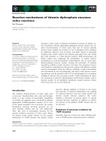
phy. The protein migrated with an apparent molecular mass
of 14.4 kDa during 15% SDS/PAGE (Fig. 1). This result
was consistent with the calculated molecular mass of Ndk.
Ndk is defined by its ability to catalyze the transfer of
terminal phosphate from any NTP to any NDP. Enzyme
activity was assayed by incubating purified protein with
[c-
32
P]ATP and 1 m
M
of unlabelled G-, U- or CDP or
[c-
32
P]GTP and 1 m
M
of A-, C- or UDP. After 10 min
incubation at room temperature, the mixture was separated
by PEI-TLC. As shown in Fig. 2A and 2B, Ndk transferred
Fig. 1. Electrophoretic analysis of recombinant pNdk and mutants.
Affinity purified Ndk and mutant proteins (2 lg) were separated by
15% SDS/PAGE and stained with coomassie blue. Lane: 1, molecular
mass marker; lane 2, Ndk; lane 3, Ndk H49Q; lane 4, Ndk H53Q and
lane 5, Ndk H117Q.
Ó FEBS 2003 Nucleoside diphosphate kinase of Mycobacterium (Eur. J. Biochem. 270) 627
a terminal phosphate from [c-
32
P]ATP or [c-
32
P] GTP to all
NDP, converting them to the corresponding triphosphates.
Heat inactivated (100 °C for 10 min) purified Ndk failed to
show phosphotransferase activity (Fig. 2A and 2 B).
In M. tuberculosis, Ndk contains His at amino acid
positions 49, 53 and 117. Each His was replaced individually
with Gln by overlapping PCR. The resulting mutant
plasmids were designated as pNdk H49Q, pNdk H53Q
and pNdk H117Q. Mutant proteins were purified by
Ni-NTA affinity matrix chromatography and assayed for
enzymatic activity. All the mutants showed similar phos-
photransferase activity as that of native Ndk (Fig. 2C).
ATPase activity of purified Ndk
Purified Ndk, and mutant proteins (H49Q, H53Q and
H117Q) were also analyzed for their ability to bind and
hydrolyze ATP. Purified Ndk showed ATPase activity as
evidenced by the decrease in amount of [c-
32
P]ATP and the
simultaneous increase in
32
P
i
(Fig. 3). The activities of two
mutants (H49Q and H53Q) were similar to those of wild-
type Ndk. However, mutation at position 117 (H117Q)
resulted in loss of both ATP binding and hydrolysis activity
(Fig. 3). Thus, H117 is crucial for ATPase activity.
Secretion of nucleoside diphosphate kinase
by
M. tuberculosis
M. tuberculosis H
37
Rv culture supernatant exhibited Ndk
activity when assayed by transfer of terminal c)
32
Pfrom
[c-
32
P]ATP or [c-
32
P]GTP to any of the four NDP (Fig. 4A,
4B).
To confirm that Ndk was secreted from M. tuberculosis
H
37
Rv, proteins of concentrated, mid log-phase culture
Fig. 2. Nucleoside diphosphate kinase activity of Ndk. Purified Ndk and mutant proteins (1 lg) were incubated with 10 lCi of [c-
32
P]ATP and 1 m
M
NDP (G-, C- or UDP) or [c-
32
P]GTP and 1 m
M
NDP (A-, C- or UDP) for 10 min at room temperature. Reaction was stopped by the addition of
2 lLof10· SDS/PAGE buffer and resolved by PEI-TLC. (A) Experiment with Ndk and [c-
32
P]ATP: (Lane 1, [c-
32
P]ATP control; lane 2,
[c-
32
P]ATPplusGDP;lane3,[c-
32
P]ATP plus GDP and Ndk; lane 4, [c-
32
P]ATP plus CDP and Ndk; lane 5, [c-
32
P]ATP plus UDP and Ndk; lane 6,
[c-
32
P]ATP plus UDP and heat inactivated Ndk; lane 7, [c-
32
P]GTP as a control). (B) Experiment with Ndk and [c-
32
P]GTP: (Lane 1, [c-
32
P]GTP
control; lane 2, [c-
32
P]GTP plus ADP; lane 3, [c-
32
P]GTP plus ADP and Ndk; lane 4 [c-
32
P]GTP plus CDP and Ndk; lane 5, [c-
32
P]GTP plus UDP
and Ndk; lane 6, [c-
32
P]GTP plus UDP and heat inactivated Ndk; lane 7, [c-
32
P]ATP as a control). (C) Experiment with His mutants (pNdk H49Q,
H53Q and H117Q) of Ndk with [c-
32
P]ATP: (Lane 1, [c-
32
P]ATPcontrol;lane2,[c-
32
P]ATP plus GDP and H49Q; lane 3, [c-
32
P]ATP plus CDP
and H49Q; lane 4, [c-
32
P]ATP plus UDP and H49Q; lane 5, [c-
32
P]ATP plus GDP and H53Q; lane 6, [c-
32
P]ATP plus CDP and H53Q; lane 7,
[c-
32
P]ATP plus UDP and H53Q; lane 8, [c-
32
P]ATP plus GDP and H117Q; lane 9, [c-
32
P]ATP plus CDP and H117Q; lane 10, [c-
32
P]ATP plus
UDP and H117Q).
628 P. Chopra et al. (Eur. J. Biochem. 270) Ó FEBS 2003
supernatant were separated by SDS/PAGE, transferred to
nitrocellulose, and probed with immune serum prepared
from mice injected with purified, recombinant Ndk. The
presence of Ndk was observed in the culture supernatant
(Fig. 5A). In contrast, adenylate kinase (a cytoplasmic
protein) was not detected by Western blot using polyclonal
antibody against purified adenylate kinase (Fig. 5B).
Autophosphorylation activity
The autophosphorylating activity of Ndk was determined
by incubating purified protein with [c-
32
P]ATP at room
temperature for 5 min. Proteins were separated by 15%
SDS/PAGE and analyzed by autoradiography. A sharp
band at 14.4 kDa was observed, indicating that Ndk is an
autophosphorylating enzyme (Fig. 6A). Both the H49Q
and H53Q mutant proteins were autophosphorylated, while
the H117Q Ndk protein was not (Fig. 6A). These data
indicate that in Ndk of M. tuberculosis H117 is required for
autophosphorylation. The presence of native and mutant
Ndk protein in each reaction was shown by Western blot
using anti-Ndk antibodies (Fig. 6B).
GTPase activity
We next examined the ability of Ndk to bind and hydrolyze
GTP by three methods. In the first, Ndk was incubated with
[c-
32
P]GTP for various times at 25 °C. A time-dependent
increase in
32
P
i
formation and decrease in [c-
32
P]GTP was
observed that was proportional to Ndk concentration
(Fig. 7A). Second, Ndk-associated GTPase activity was
demonstrated in a filter-binding assay by incubating Ndk
with [c-
32
P]GTP which resulted in hydrolysis of 60% bound
GTP in 30 min (Fig. 7B). Third, GTPase activity was
Fig. 3. ATPase activity in Ndk of M. tuberculosis. Purified Ndk and
mutant H117Q were incubated with 10 lCi of [c-
32
P]ATP at 25 °Cfor
various time periods and release of
32
P
i
was monitored as an indicator
of ATPase activity. Lane 1, [c-
32
P]ATP; lane 2, [c-
32
P]ATP plus
H117Q at 30 min; lane 3, [c-
32
P]ATP plus Ndk at 15 min; lane 4,
[c-
32
P]ATP plus Ndk at 30 min.
Fig. 4. Ndk activity in the supernatant of M. tuberculos is culture.
M. tuberculosis was grown in 7H9 media and mid log-phased cells were
harvested. Culture supernatant was filtered through 0.22 lmfilterand
concentrated 50-fold by Centricon and filtrate was used for the enzyme
assay as described in the experimental procedure. Culture supernatant
(10 lL) was incubated with 10 lCi of [c-
32
P]ATP and 1 m
M
NDP (G, C
or UDP) or [c-
32
P]GTP and 1 m
M
NDP (A, C or UDP) for 10 min at
room temperature. Reaction was stopped bythe addition of 2 lLof10·
SDS/PAGE buffer and resolved by PEI-TLC. (A) Experiment with
[c-
32
P]ATP: (Lane 1, [c-
32
P]ATP control; lane 2, [c-
32
P]ATP plus GDP;
lane 3, [c-
32
P]ATP plus CDP; lane 4, [c-
32
P]ATP plus UDP; and lane 5,
[c-
32
P]GTP as a control). (B) Experiment with [c-
32
P]GTP: (Lane 1,
[c-
32
P]GTP control; lane 2, [c-
32
P]GTPplusADP;lane3,[c-
32
P]GTP
plus CDP; lane 4 [c-
32
P]GTP plus UDP; and lane 5, [c-
32
P]ATP as a
control).
Ó FEBS 2003 Nucleoside diphosphate kinase of Mycobacterium (Eur. J. Biochem. 270) 629
measured by incubating purified Ndk with [a-
32
P]GTP for
10 min followed by separation of the products by PEI-TLC
to observe the formation of [a-
32
P]GDP (Fig. 7C). Ndk was
bound to [c-
32
P]GTP in a time-dependent fashion, suggest-
ing that binding of GTP to Ndk is important for its GTPase
activity (data not shown).
The H49Q, H53Q and H117Q mutant Ndk proteins were
also analyzed for their ability to bind and hydrolyze GTP.
The activities of two mutants (H49Q and H53Q) were
similar to those of wild-type Ndk. However, mutation at
position 117 (H117Q) resulted in loss of both GTP binding
and GTP hydrolysis activity (Fig. 7C). Thus, H117 is crucial
for both activities.
Enhancement of cytotoxic action by Ndk
Macrophages expel ATP upon activation by either bacterial
LPS or intact bacteria [5]. The ATP then activates P2Z
receptors on the surface of macrophages, which in turn
trigger macrophage cell death by formation of large,
nonselective membrane pores that are permeable to mole-
cules up to a mass of 900 Da [19]. In the present study, ATP
alone was cytotoxic to macrophages and resulted in the
leakage of [
14
C]adenine up to 29% in 8 h. Ndk, in
combination with ATP, increased cytotoxicity in a time-
dependent manner (Fig. 8A). Addition of purified Ndk to
the macrophage cells, in combination with 3 m
M
ATP,
resulted in 79% leakage of [
14
C]adenine in 8 h. Ndk alone
had no significant effect on release of [
14
C]adenine. Mutant
H117Q Ndk failed to stimulate ATP-dependent cytotoxicity
(Fig. 8A). This result was expected, as the mutant also
lacked ATP binding and ATP hydrolysis activity.
To further investigate the role of Ndk in ATP-mediated
cytotoxicity, culture supernatant of M. tuberculosis H37Rv
was examined for ATP-dependent cytotoxicity. Culture
supernatant, in combination with 3 m
M
ATP, resulted in
48% leakage of [
14
C]adenine in 5 h. Addition of anti-Ndk
polyclonal antibody to the culture supernatant halted the
ATP-mediated leakage of adenine (Fig. 8B). Cytotoxicity of
purified Ndk was also measured in the presence of a mixture
of 3 m
M
ADP and 1 m
M
each of G-, C- and UTP. It was
observed that Ndk was cytotoxic to the macrophages in the
presence of the mixture, while alone the mixture was not
toxic (Data not shown).
As a test for involvement of surface P2Z receptors, we
examined the effect of oATP, a well-known P2Z receptor
antagonist [20]. When macrophages were pretreated with
1m
M
oATP prior to the addition of ATP and Ndk, oATP
prevented the ATP- and Ndk-induced leakage of [
14
C]
adenine (Fig. 8A). Thus, the cytotoxicity associated with
purified Ndk appears to be mediated by the macrophage cell
surface P2Z receptors.
Fig. 5. Western blot analysis of culture supernatant of M. tuberculosis.
Concentrated culture supernatant, purified Ndk and adenylate kinase
were separated on 15% SDS/PAGE, proteins were transferred to a
nitrocellulose membrane incubated with anti-Ndk (A) or anti-adeny-
late kinase antibodies (B) and developed with ECL reagent. Lane 1,
purified Ndk or adenylate kinase and lane 2, culture supernatant.
Fig. 6. Autophosphorylation of recombinant Ndk and mutant proteins.
(A) Ndk and mutant proteins (1 lg) were incubated in the presence of
10 lCi of [c-
32
P]ATP in 20 lL of reaction volume. The reaction was
stopped by the addition of 2 lL of 10% SDS/PAGE loading buffer.
Fractions were resolved by 15% SDS/PAGE and autoradiographed.
Lane 1, Ndk; lane 2, Ndk H49Q; lane 3, Ndk H53Q; lane 4, Ndk
H117Q. (B) Detection of Ndk and three His mutants of Ndk by anti-
Ndk antibody. Ndk and mutant proteins (1 lg) were separated on
15% SDS/PAGE, proteins were transferred to nitrocellulose mem-
brane. Probed with anti-Ndk antibody raised in mice and developed
using ECL reagent. Lane 1, Ndk; lane 2, H49Q; lane 3, H53Q and lane
4, H117Q.
630 P. Chopra et al. (Eur. J. Biochem. 270) Ó FEBS 2003
Discussion
The results presented above indicate that Ndk is secreted by
M. tuberculosis as a cytotoxic factor that facilitates ATP-
dependent P2Z receptor-mediated macrophage death. The
Ndk gene was cloned and expressed in E. coli,andNdkwas
purified as a His-tagged protein. Antibody was raised
against purified Ndk in mice and used to study secretion of
Ndk from M. tuberculosis. Western blot analysis of concen-
trated supernatant of M. tuberculosis suggested that Ndk is
secreted in the culture media. In order to determine whether
the detection of Ndk in the culture supernatant of M.
tuberculosis H37Rv is caused by the secretion rather than by
the autolysis of the cells, culture supernatant was also
analysed for the presence of a cytoplasmic protein, adenylate
kinase. Western blot analysis showed that adenylate kinase
of M. tuberculosis was absent from the culture supernatant
suggesting that the presence of Ndk in culture supernatant is
due to secretion and not autolysis (Fig. 5B). Secretion of
Ndk, a crucial enzyme of metabolism seems unusual, but its
secretion has been reported from several organisms such as
P. aeruginosa, V. cholerae, B. cepacia, T. spiralis, M. bovis
and M. smegmatis [6–10,14]. Purified Ndk stimulated ATP-
induced cytotoxicity in cultured murine macrophage cells
(Fig. 8A). Thus, secreted Ndk from M. tuberculosis, like
culture supernatant of V. cholerae and B. cepacia that
harbors Ndk and other ATP-utilizing enzymes, acts as a
cytotoxic virulence factor [8,9].
Ndk was also cytotoxic to macrophages in the presence of
a mixture of ADP, G-, C- and UTP, while alone this mixture
was less cytotoxic (data not shown). This observation
suggests that ADP was converted to ATP by Ndk through
the transfer of a terminal phosphate from a pool of other
triphosphates (C-, G- and UTP) present in the medium. It
has been observed that different ionic forms of ATP and
adenine nucleotides differ in their agonist activities towards
P2Z receptor activation [19,21]. The enhancement in ATP-
mediated cytotoxicity of Ndk as compared to ATP alone
might be due to Ndk-mediated conversion of ATP into
various adenine nucleotides that may act as better agonists
than ATP itself. Such speculations have also been made in
the cases of P. aeruginosa, V. cholerae and B. cepacia [7–9].
Pretreatment of macrophages with an antagonist of the
P2Z receptor, oATP, protected the cells from Ndk-mediated
cytotoxicity, suggesting that Ndk of M. tuberculosis acts via
the P2Z receptors. The mechanism of Ndk-mediated
cytotoxicity is ATP-mediated, as mutant H117Q, which is
deficient in ATP binding and hydrolysis activities failed to
stimulate ATP-mediated cytotoxicity (Fig. 8A).
Culture supernatant of M. tuberculosis was found to be
cytotoxictomacrophagesinthepresenceof3m
M
ATP.
Addition of anti-Ndk polyclonal antibody resulted in a time-
dependent decrease in ATP-mediated cytotoxicity of culture
supernatant of M. tuberculosis H37Rv (Fig. 8B), suggesting
that this cytotoxicity was induced by Ndk present in the
culture supernatant. Several other intracellular pathogens,
such as Salmonella typhimurium, Legionella pneumophila and
Listeria monocytogenes, induce apoptosis in immune cells
[22–24]. It has been suggested that the induction of
programmed cell death before macrophages can synthesize
pro-inflammatory cytokines may play an important role in
bacterial evasion of the host immune system [22]. The ability
Fig. 7. GTPase activity of purified Ndk. (A)
[c-
32
P]GTP hydrolysis. Purified Ndk (1 lg)
was incubated with 10 lCi of [c-
32
P]GTP at
25 °C for various time periods (0–30 min),
and release of
32
P
i
was noted as an indicator of
GTPase activity. Lane 1, [c-
32
P]GTP alone;
lane 2, [c-
32
P]GTP plus Ndk at 5 min; lane 3,
[c-
32
P]GTP plus Ndk at 15 min; lane 4,
[c-
32
P]GTP plus Ndk at 30 min. (B) Filter
binding assay. purified protein (1 lg) was
incubated with 10 lCi of [c-
32
P]GTP for
various time intervals (0–30 min). GTPase
activity was analyzed by filter binding assay as
described in the experimental procedure.
Shown is the remaining GTP at each time
points as percent of bound [c-
32
P]GTP before
incubation at 37 °C. (C) Hydrolysis of
[a-
32
P]GTP. Purified Ndk (1 lg) was incuba-
tedwith3lCi of [a-
32
P]GTP, for 10 min and
mixture was resolved by PEI-TLC and auto-
radiographed. Lane 1, [a-
32
P]GTP; lane 2,
[a-
32
P]GTP incubated with H117Q, lane 3
[a-
32
P]GTP plus Ndk.
Ó FEBS 2003 Nucleoside diphosphate kinase of Mycobacterium (Eur. J. Biochem. 270) 631
of M. tuberculosis to promote apoptosis may also be
important for dissemination of infection.Aknockoutmutant
of Ndk in M. tuberculosis would give important insight into
the in vivo role of Ndk. Experiments are in progress to
construct an ndk knockout mutant of M. tuberculosis.
TheroleofM. tuberculosis Ndk is to produce nucleoside
triphosphates (NTP) as precursors for RNA, DNA and
polysaccharide synthesis. Ndk catalyzes the reversible
transfer of the 5¢-terminal P
i
from NTP to NDP [25]. The
central importance of such a function is consistent with the
failure of attempts to isolate knockout mutants of ndk in
Myxococcus xanthus [26]. However, in a few organisms,
such as E. coli and P. aeruginosa, Ndk activity is comple-
mented by adenylate kinase and pyruvate kinase [6,27]. Ndk
also plays a vital role in the physiology of the eukaryotes.
For example, in Drosophila, a null mutation in ndk causes
abnormalities in larval development that lead to tissue
necrosis and death at the prepupal stage [28]. Thus, Ndk
might have multiple functions. In humans, reduction of ndk
transcript level is associated with lowered metastatic poten-
tial in tumor cells [29]. In the present study it was observed
that purified Ndk from M. tuberculosis was able to transfer
terminal P
i
both from [c-
32
P]ATP and [c-
32
P]GTP to all
nucleoside diphosphates and to convert them to their
corresponding triphosphates (Fig. 2A and B). Ndk from
M. tuberculosis is thermostable upto 75 °C and becomes
inactivated completely at 82 °C [30]. In this study, heat
inactivated Ndk (100 °C, 10 min) was also checked for
enzymatic activity and found to lack phosphotransferase
activity (Fig. 2A and B).
All three His mutants of Ndk (pNdk-H49Q, H53Q and
H117Q) showed similar phosphotransferase activity
(Fig. 2C). The presence of phosphotransferase activity in
mutant pNdk-H117Q was surprising, as this mutant lost
both ATP-binding and hydrolysis activity (Fig. 3). Similar
activity has been reported for the His mutant of Ndk from
Dictyostelium discoideum. It has beenshown that nucleophilic
His can be rescued by other exogenous small nucleophiles
including water [31,32].
Ndk is autophosphorylated, and His117 is the only His
residue that is conserved in all known Ndk characterized to
date [33]. In Myxococcus xanthus it has been reported that
replacement of His117 with Gln in Ndk abolishes the
autophosphorylation and nucleotide binding activity [33].
Ndk of M. tuberculosis has three His residues at positions
49, 53 and 117 that were replaced individually with Gln.
Fig. 8. ATP-induced macrophage cytotoxicity
from purified Ndk and culture supernatant of
M. tuberculosis. J774A.1 cells were labeled
with [
14
C]adenine (1 lCiÆmL
)1
)for6hand
stimulated with LPS (50 ngÆmL
)1
) for 12 h.
For the experiment with oATP, cells were
pretreated with 1 m
M
oATP for 2 h before the
cytotoxicity assay was carried out. Release of
[
14
C]adenine into media was counted using
liquid scintillation counter. Each value is the
average ± SEM and representative of four
experiments with duplicate wells for each
treatment. (A) Experiment with purified Ndk
(25 lgÆmL
)1
) in presence or absence of
exogenous ATP (3 m
M
).
(B) Experiment with concentrated culture
supernatant of M. tuberculosis in presence or
absence of exogenous ATP (3 m
M
).
632 P. Chopra et al. (Eur. J. Biochem. 270) Ó FEBS 2003
Replacement of H117Q but not H49Q or H53Q resulted in
the loss of both autophosphorylation and nucleotide
binding activity (Figs 3, 6 and 7). Thus only His117 is
critical for autophosphorylation and nucleotide binding.
In this report, we show that Ndk has intrinsic GTPase
and GTP binding activity (Fig. 7A–C). M. tuberculosis Ndk
lacks the GXXGK and DXXG motifs that are character-
istic features of GTP binding proteins [34,35]. The sequence,
NKKD, which is known to be involved in guanine base
recognition [36] is also absent from M. tuberculosis Ndk.
In summary, our results suggest that Ndk secreted by
M. tuberculosis is a cytotoxic factor that induces ATP-
dependent P2Z receptor-mediated macrophage death. In
addition, we showed that Ndk has GTPase activity. The
ability of M. tuberculosis to promote apoptosis may be
important for the initiation of infection, bacterial survival,
and escape of the host immune response.
Acknowledgements
We thank Prof. S. K. Brahmachari for making this work possible.
P. C. and A. S. were supported by University Grant Commission
(UGC), N. Delhi. We are also thankful to L. S Meena, P. K. Gupta,
H. Chandra, H. Khanna Parampal, R. Gaur for valuable discussions
and Vineet and Neeraj for helping with bioinformatics work. Financial
support for the project was provided by NMITLI, Council of Scientific
and Industrial Research (CSIR).
References
1. Armstrong, J.A. & Hart, P.D. (1975) Phagosome–lysosome
interaction in cultured macrophages infected with virulent tubercle
bacilli. Reversal of the usual non-fusion pattern and observation
on bactericidal survival. J. Exp. Med. 142, 1–16.
2. Sturgill-Koszycki, S., Schlesinger, P.H., Chakraborty, P., Haddix,
P.L., Collins, H.L., Fok, A.K., Allen, R.D., Gluck, S.L., Heuser,
J. & Russell, D.G. (1994) Lack of acidification in Mycobacterium
phagosome produced by exclusion of the vesicular proton-
ATPase. Science. 263, 678–681.
3. Ferrari, G., Langen, H., Naito, M. & Piters, J. (1999) A coat
protein onphagosomes involved in the intracellular survival of
mycobacteria. Cell. 97, 435–447.
4. Ramakrishnan, L., Federspiel, N.A. & Falkow, S. (2000) Granu-
loma-specific expression of Mycobacterium virulence proteins
from the glycine rich PE-PGRSfamily. Science 288, 1436–1439.
5. Ferrari, D., Chiozzi, P., Falzoni, S., Dalsusino, M., Melchiorri, L.,
Baricordi, O.R. & Di Virgilio, F. (1997) Extracellular ATP triggers
IL-1 b release by activating the purinergic P2Z receptor of human
macrophages. J. Immunol. 159, 1451–1458.
6. Zaborina, O., Misra, N., Kostal, J., Kamath, S., Kapatral, V.,
El-Idrissi, M.E., Prabhakar, B.S. & Chakrabarty, A.M. (1999) A)
P2Z-independent and P2Z receptor-mediated macrophage killing
by Pseudomonas aeruginosa isolated from cystic fibrosis patients.
Infect. Immun. 67, 5231–5242.
7. Zaborina, O., Dhiman, N., Chen, M.L., Kostal, J., Holder, I.A. &
Chakrabarty, A.M. (2000) Secreted products of a nonmucoid
Pseudomonas aeruginosa strain induce two modes of macrophage
killing: external-ATP-dependent, P2Z-receptor-mediated necrosis
and ATP-independent, caspase-mediated apoptosis. Microbiology
146, 2521–2530.
8. Punj, V., Zaborina, O., Dhiman, N., Falzari, K., Bagdasarian, M.
& Chakrabarty, A.M. (2000) Phagocytic cell killing mediated by
secreted cytotoxic factors of Vibrio cholerae. Infect. Immun. 68,
4930–4937.
9. Melnikov, A., Zaborina, O., Dhiman, N., Prabhakar, B.S.,
Chakrabarty, A.M. & Hendrickson, W. (2000) Clinical and
environmental isolates of Burkholderia cepacia exhibits differential
cytotoxicity towards macrophage and mast cells. Mol. Microbiol.
36, 1481–1493.
10. Gounaris, K., Thomas, S., Najarro, P. & Selkirk, M.E. (2001)
Secreted variant of nucleoside diphosphate kinase from the
intracellular parasitic nematode Trichinella spiralis. Infect. Immun.
69, 3658–3662.
11. Zaborina,O,Li,X.,Cheng,G.,Kapatral,V.&Chakrabarty,
A.M. (1999) B). Secretion of ATP utilizing enzymes, nucleoside
diphosphate kinase and ATPase, by Mycobacterium bovis BCG:
sequestration of ATP from macrophage P2Z receptors? Mol.
Microbiol. 31, 1333–1343.
12. Gupta, P., Batra, S., Chopra, A.P., Singh, Y. & Bhatnagar, R.
(1998) Expression and purification of recombinant lethal factor of
Bacillus anthracis. Infect. Immun. 66, 862–865.
13. Kapatral, V., Bina, X. & Chakrabarty, A.M. (2000) Succinyl
coenzyme a synthetase of Pseudomonas aeruginosa with a broad
specificity for nucleoside triphosphate (NTP) synthesis modulate
specificity for NTP synthesis by the 12-kilodalton form of
nucleoside diphosphate kinase. J. Bacteriol. 182, 1333–1339.
14. Shanker, S., Hershberger, C.D. & Chakrabarty, A.M.
(1997) The nucleoside diphosphate kinase of Mycobacterium
smegmatis: identification of protein that modulate specificity of
nucleoside triphosphate synthesis by the enzyme. Mol. Microbiol.
24, 477–487.
15. Chopade, B.A., Shankar, S., Sundin, G.W., Mukhopadhyay, S. &
Chakrabarty, A.M. (1997) Characterization of membrane–asso-
ciated Pseudomonas aeruginosa Ras-Like proteins Pra, a GTP
binding protein that forms complexes with truncated nucleoside
diphosphate kinase and pyruvate kinase to modulate GTP syn-
thesis. J. Bacteriol. 179, 2181–2188.
16. Black, D.S. & Bliska, J.B. (2000) The Rho GAP activity of the
Yersinia pseudotuberculosis cytotoxin YopE is required for anti-
phagocytic function and virulence. Mol. Microbiol. 37, 515–527.
17. Zhu, J., Tseng, Y., Kantor, J.D., Rhodes, C.J., Zetter, B.R.,
Moyers, J.S. & Kahn, R.C. (1999) Interaction of the Ras-related
protein associated with diabetes Rad and the putative tumor
metastasis suppressor NM23 provides a novel mechanism of
GTPase regulation. Proc.NatlAcad.Sci.96, 14911–14918.
18. Shirhatti, V. & Krishna, G. (1985) A simple and sensitive method
for monitoring drug-induced cell injury in cultured cells. Anals
Biochem. 147, 410–418.
19. Di Virgilio, F. (1995) The P2Z purinoreceptor-an intriguing role
in immunity, inflammation and cell death. Immunol. Today 16,
524–528.
20. Lammas, D.A., Stober, C., Harvey, C.J., Kendrick, N., Panch-
alingam, S. & Kumararatne, D.S. (1997) ATP-induced killing of
Mycopbacteria by human macrophages is mediated by purinergic
P2Z (P2X
7
) receptors. Immunity 7, 433–444.
21. Harden, T.K., Boyer. J.L. & Nicholas R.A. (1995) P2-Purinergic-
receptors: subtype-associated signaling and structure. Annu. Rev.
Pharmocol. Toxicol. 35, 541–579.
22. Monack, D.M., Raupach, B., Hromockyj, A.E. & Falkow, S.
(1996) Salmonella typhimurimum invasion induces apoptosis in
infected macrophages. Proc.NatlAcad.Sci.USA93, 9833–9838.
23. Purcell, M. & Shuman, H.A. (1998) The Legionella pneumophila
icmGCDJBFgenes are required for killing of human macro-
phages. Infect. Immun. 66, 2245–2255.
24. Chen, Y. & Zychlinsky, A. (1994) Apoptosis induced by bacterial
pathogens. Microb. Pathog. 17, 203–212.
25. Chakrabarty, A.M. (1998) Nucleoside diphosphate kinase: role in
bacterial growth, virulence, cell signaling and polysaccharide
synthesis. Mol. Microbiol. 28, 875–882.
Ó FEBS 2003 Nucleoside diphosphate kinase of Mycobacterium (Eur. J. Biochem. 270) 633
26. Munoz-Dorado, J., Inouye, M. & Inouye, S. (1990) Nucleoside
diphosphate kinase from Myxococcus xanthus; Cloning and
sequencing of the gene. J. Biol. Chem. 265, 2702–2706.
27. Lu, Qing & Inouye, Masayori. (1996) Adenylate kinase comple-
ments nucleoside diphosphate kinase deficiency in nucleotide
metabolism. Proc. Natl Acad. Sci. USA 95, 57720–55725.
28. Biggs,J.,Hersperger,E.,Steeg,P.S.,Liotta,L.A.&Shearn,A.
(1990) A Drosophila gene that is homologous to a mammalian
gene associated with tumor metastasis codes for a nucleoside
diphosphate kinase. Cell 63, 933–940.
29. Steeg, P.S., De la Rosa, A., Flatow, U., MacDonald, N.J.,
Benedict, M. & Leone, A. (1993) Nm23 and breast cancer
metastasis. Breast Cancer Res. Treat. 25, 175–187.
30. Chen, Y., Morera, S., Mocan, J., Lascu, I. & Janin, J. (2002) X-ray
structure of Mycobacterium tuberculosis nucleoside diphosphate
kinase. Proteins 17, 556–557.
31. Admiraal, S.J., Meyer, P., Schneider, B., Deville-Bonne, D., Janin,
J. & Herschlag, D. (2001) Chemical rescue of phosphoryl transfer
in a cavity mutant: a cautionary tale for site-directed mutagenesis.
Biochemistry 40, 403–413.
32. Admiraal, S.J., Schneider, B., Meyer, P., Janin, J., Veron, M.,
Deville-Bonne, D. & Herschlag, D. (1999) Nucleophilic activation
by positioning in phosphoryl transfer catalyzed by nucleoside
diphosphate kinase. Biochemistry. 38, 4701–4711.
33. Munoz-Dorado, J., Alamula, N, Inouye, S. & Inouye, M. (1993)
Autophosphorylation of nucleoside diphosphate kinase from.
Myxococcux xanthus. J. Bacteriol. 175, 1176–1181.
34. March, P.E. (1992) Membrane associated GTPases in bacteria.
Mol. Microbiol. 6, 1253–1257.
35. McCormick, F., Clark, B.F.C., la Cour, T.F.M., Kjedgaard, M.,
Norskov-Lauritsen, L. & Nyborg, J. (1985) A model for the ter-
tiary structure of p21, the product of Ras oncogene. Science 230,
78–82.
36. Dever,T.E.,Glynias,M.J.&Merrick,W.C.(1987)GTPbinding
domain: three consensus sequence elements with distinct spacing.
Proc.NatlAcad.Sci.USA84, 1814–1818.
634 P. Chopra et al. (Eur. J. Biochem. 270) Ó FEBS 2003