Báo cáo khoa học: a-Fetoprotein positively regulates cytochrome c-mediated caspase activation and apoptosome complex formation docx
Bạn đang xem bản rút gọn của tài liệu. Xem và tải ngay bản đầy đủ của tài liệu tại đây (319.54 KB, 12 trang )
a-Fetoprotein positively regulates cytochrome
c
-mediated caspase
activation and apoptosome complex formation
Lidia Semenkova
1,
*, Elena Dudich
1,
*, Igor Dudich
1
, Natalie Tokhtamisheva
1
, Edward Tatulov
2
,
Yury Okruzhnov
3
, Jesus Garcia-Foncillas
3
, Juan-Antonio Palop-Cubillo
4
and Timo Korpela
5
1
Institute of Immunological Engineering, Moscow, Russia;
2
Anticancer Drug Research Center, Moscow, Russia; Departments of
3
Oncology and
4
Organic Chemistry and Pharmacology, University of Navarra, Pamplona, Spain;
5
Joint Finnish-Russian
Biotechnology Laboratory, Turku University, Finland
Previous results have shown that the oncoembryonic marker
a-fetoprotein (AFP) is able to induce apoptosis in tumor
cells through activation of caspase 3, bypassing Fas-
dependent and tumor necrosis factor receptor-dependent
signaling. In this study we further investigate the molecular
interactions involved in the AFP-mediated signaling of
apoptosis. We show that AFP treatment of tumor cells is
accompanied by cytosolic translocation of mitochondrial
cytochrome c. In a cell-free system, AFP mediates process-
ing and activation of caspases 3 and 9 by synergistic
enhancement of the low-dose cytochrome c-mediated sig-
nals. AFP was unable to regulate activity of caspase 3 in cell
extracts depleted of cytochrome c or caspase 9. Using
high-resolution chromatography, we show that AFP posit-
ively regulates cytochrome c/dATP-mediated apoptosome
complex formation, enhances recruitment of caspases and
Apaf-1 into the complex, and stimulates release of the active
caspases 3 and 9 from the apoptosome. By using a direct
protein–protein interaction assay, we show that pure human
AFP almost completely disrupts the association between
processed caspases 3 and 9 and the cellular inhibitor of
apoptosis protein (cIAP-2), demonstrating its release from
the complex. Our data suggest that AFP may regulate cell
death by displacing cIAP-2 from the apoptosome, resulting
in promotion of caspase 3 activation and its release from the
complex.
Keywords: apoptosis; apoptosome; cytochrome c;IAP-2;
a-fetoprotein.
Apoptotic cell death is characterized by biochemical and
morphological changes, which are largely caused by caspase
activity. A class of cysteine proteases, known as caspases,
which are constitutively expressed in cells as inactive
proenzymes, require proteolytic cleavage to be activated.
In general, either receptor-induced or mitochondrion-
induced death signals stimulate activation of specific
adapterproteinsFADD/MORT1orApaf-1byformation
of the high-molecular-mass death-inducing complex or
apoptosome. The adapter proteins recruit initiator caspases
8 and 9 to activate them by autoprocessing. Once activated,
initiator caspases are ready to induce processing of down-
stream effector caspases 3 and 7 [1]. The mitochondrial
apoptosis pathway is mediated by cytochrome c (cyt-c)
release with the subsequent formation of the Apaf-1/cyt-c/
dATP/procaspase 9 apoptosome complex, leading to acti-
vation of caspase 9 and downstream effector caspases [2].
Chromatographic analysis of the apoptosome assembly
indicated that, in native cell lysates, Apaf-1 oligomerizes
into multimeric complexes of molecular mass 1.4 MDa
and 700 kDa, which in addition to processed caspase 9,
contain fully processed caspase 3 and 7 [3]. Caspases are
inhibited by a number of cellular inhibitor of apoptosis
proteins (cIAPs), which bind directly to procaspases 9 and 3
to prevent their cyt-c-mediated processing and activation
[4,5]. During apoptosis, a mitochondrial protein named
Smac/DIABLO [6] that directly binds to IAPs to remove
them from the apoptosome complex [4,7], cancels the
IAP-mediated caspase inhibition. Recently, another IAP-
inhibitory protein Omi/HtrA2 was characterized, which
operates by abrogation of the IAP–caspase interaction [8].
AFP is the major serum protein of embryonic plasma
that is involved in regulation of gene expression, differen-
tiation, proliferation and apoptosis in developing cells
[9–12]. Although, the biological role of this protein is not
yet fully understood, it has been well characterized as a
physiological carrier/transport protein for various ligands,
including fatty acids, drugs, hormones, heavy metals,
delivering them to developing and malignant cells [9,12].
The specific expression and internalization of AFP is
restricted to developing cells, such as embryonic cells,
activated immune cells and tumor cells, which suggests its
important regulatory role in cell growth and differentiation
[9,10,12]. Various researchers have documented the exist-
ence of specific receptor-dependent mechanisms responsible
for the active endocytosis of AFP by malignant cells [13,14].
Microscopic data have demonstrated that fluoresceinated
Correspondence to E. Dudich, Institute of Immunological Engineering,
142380, Lyubuchany, Moscow Region, Chekhov District, Russia.
Tel./Fax: + 7 095 996 15 55, E-mail:
Abbreviations: AFP, a-fetoprotein; cyt-c, cytochrome c; cIAP, cellular
inhibitor of apoptosis protein; Ac-DEVD-AMC, Ac-Asp-Glu-Val-
Asp-7-amino-4-methylcoumarin; LEHD-AFC, Leu-Glu-His-Asp-
aminotrifluoromethylcoumarin; IETD-AMC, Ile-Glu-Thr-Asp-7-
amino-4-methylcoumarin; CHO, aldehyde.
*Note: These authors contributed equally to this work.
(Received 11 February 2003, revised 28 August 2003,
accepted 16 September 2003)
Eur. J. Biochem. 270, 4388–4399 (2003) Ó FEBS 2003 doi:10.1046/j.1432-1033.2003.03836.x
AFP is specifically bound to the cell surface at 4 °Cand
internalized into the cytoplasm at 37 °C [15,16]. It has been
shown that AFP is internalized via coated pits and vesicles
before being delivered to endosomes [15,16]. Much evidence
of cell growth regulatory activity, including tumor suppres-
sion, has been reported for various species of the full-length
AFP molecule [17–22], its proteolytic fragments [23],
recombinant domains [24] and synthetic peptides [25–27].
It has been demonstrated that AFP realizes its tumor-
suppressive activity by triggering apoptosis, characterized
by typical morphological changes, growth arrest, cytotoxi-
city, and DNA fragmentation [20–22]. It was shown that
AFP induces apoptosis in malignant cells through activa-
tion of caspase 3, bypassing Fas/FasL and tumor necrosis
factor (TNF)/TNFR-dependent pathways and does not
require upstream activation of receptor-dependent initiatory
caspase 8 and caspase 1 [21]. Although these studies have
shown that a caspase cascade is initiated during AFP-
induced apoptosis, the mechanisms by which AFP triggers
caspase activation are unknown. Our previous experimental
data show that AFP does not require de novo protein
synthesis and RNA expression to trigger apoptosis, as it was
not blocked by actinomycin D or cycloheximide [20].
In this study, we aimed to determine how AFP activates
the caspase cascade. To understand the molecular mecha-
nisms of AFP-mediated apoptosis signaling, we established
a cell-free system, similar to that used for studies of cyt-c-
induced apoptosis [28,29]. We show here that AFP syner-
gistically enhances caspase activation and processing in the
presence of a low suboptimal dose of cyt-c and requires the
presence of all members of the apoptosome complex to
initiate this process. We examine the mechanisms by which
AFP regulates apoptosis and demonstrate that the pro-
apoptotic effect of AFP is mediated through its interaction
with apoptosome-forming proteins. Chromatographic ana-
lysis of the apoptosome assembly demonstrated that AFP
stimulates formation of the Apaf-1–apoptosome complex,
enhances recruitment and activation of procaspase 3 in the
complex, and stimulates release of active caspase 3 and 9
from the apoptosome. Our data suggest that AFP may
regulate cell death by displacing cIAP-2 from the apopto-
some complex, thereby promoting caspase 3 release from
the complex.
Materials and methods
AFP purification
Human AFP was isolated from the cord serum using ion-
exchange, affinity and gel-filtration chromatography as
described previously [23]. AFP purity was established using
PAGE and immunoblotting with monospecific antibodies
against human AFP and adult serum proteins and was
showntobenolessthan99.8%.
Cells
HepG2 cells originated from the American Type Culture
Collection were cultured in Dulbecco modified Eagle’s
medium (ICN Biomedicals) with
L
-glutamine and 10%
heat-inactivated fetal bovine serum, 100 IU penicillinÆmL
)1
,
0.1 mg streptomycinÆmL
)1
in a humidified 5% (v/v)
atmosphere of CO
2
at 37 °C. For a passage, cells were
incubated in 0.25% (v/v) trypsin solution, then washed and
plated out.
Cytotoxicity assay
HepG2 cells were incubated with 5–7 l
M
AFP for deter-
mined time intervals of 2–14 h, and then assessed for their
viability by the trypan blue exclusion assay as described
previously [22]. Cells cultivated without additions were
taken as a control. The experimental data were expressed as
the percentage of dead cells relative to the total amount of
cells.
Preparation of cell-free extracts
Cell-free S-100 extracts were generated from human
hepatocarcinoma HepG2 as described [29,30]. Cells
(4 · 10
8
) were collected and washed (three times) in
50 mL NaCl/P
i
and once in 5 mL hypotonic cell extraction
buffer (containing 20 m
M
Hepes, pH 7.2, 10 m
M
KCl,
2m
M
MgCl
2
,1m
M
dithiothreitol, 5 m
M
EGTA, 25
lgÆmL
)1
leupeptin, 5 lgÆmL
)1
pepstatin, 40 m
M
b-glycero-
phosphate, 1 m
M
phenylmethanesulfonyl fluoride). The cell
pellet was then resuspended in an equal volume of cell
extraction buffer, allowed to swell for 20 min on ice, and
then disrupted by 30–50 strokes of a Dounce homogenizer.
The homogenate was centrifuged at 3000 g for 10 min at
4 °C to remove whole cells and nuclei. The supernatant was
centrifuged at 15 000 g for 20 min at 4 °Candthen,to
obtain the cytosolic S-100 extract, the supernatant was
re-centrifuged at 100 000 g for 1 h at 4 °C. Extracts were
assessed for protein content by the Bradford assay and
stored in aliquots at )70 °C. Cyt c-free cytosolic extracts
were prepared in more mild conditions by the slightly
modified procedure described in [30].
In vitro
caspase activation
For in vitro caspase activation, 40 lg of the S-100 extract
(complete or after immunodepletion) was incubated for the
indicated times with bovine heart cyt-c (Sigma-Aldrich,
St Louis, MO, USA) and/or pure human AFP (5 l
M
)inthe
presence or absence of 1 m
M
dATP (Sigma) in 15 lLofa
reaction buffer (10 m
M
Hepes, pH 7.2, 25 m
M
NaCl, 2 m
M
MgCl
2
,5m
M
dithiothreitol, 5 m
M
EDTA, 0.1 m
M
phenyl-
methanesulfonyl fluoride) at 30 °C. To control specificity
of AFP effects, the equivalent amount of human serum
albumin (Sigma) was added instead of AFP. The activity and
proteolytic processing of caspases 3 and 9 were then detected
by fluorimetric assay and immunoblotting with the corres-
ponding antibodies supplied by Santa Cruz Biotechnology,
Inc (Santa Cruz, CA, USA): polyclonal goat anti-(caspase 3)
p20 (N19); anti-(caspase 3) p11 (K19); rabbit anti-
(caspase 9) p10 (H-83); rabbit anti-(caspase 9) p35 (H-170).
Fluorimetric assay of caspase activity
Caspase activities were determined by incubation of the
extract aliquots (5 lL) for various times at 30 °C with one
of the fluorogenic substrates [40 l
M
Ac-DEVD-AMC
(ICN Biomedicals Inc), 50 l
M
LEHD-AFC (Chemicon
Ó FEBS 2003 AFP amplifies cytochrome c-mediated caspase activation (Eur. J. Biochem. 270) 4389
International, Temecula, CA, USA) or 50 l
M
IETD-AMC
(Alexis Biochemicals, San Diego, USA] in 16 lL substrate
buffer (25 m
M
Hepes, pH 7.2, 100 m
M
NaCl, 1 m
M
EDTA,
0.1% Chaps, 10 m
M
dithiothreitol, 10% sucrose). Reactions
were terminated by dilution with 2.0 mL ice-cold 0.2 m
M
sodium phosphate buffer, pH 7.5, and fluorescence was
measured using a Perkin–Elmer MPF-44A fluorimeter
(k
exc
¼ 365 nm and k
em
¼ 440 nm for the AMC fluores-
cence or k
exc
¼ 400 nm and k
em
¼ 505 nm for the AFC
fluorescence). For each sample, caspase activity was
expressed in relative units, pmolÆmin
)1
Æmg
)1
, showing the
amount of cleaved substrate in pmol normalized for time of
reaction with substrates and cytosolic protein concentra-
tion, or in relative fluorescent units (FU) per fraction.
Immunoprecipitation and immunoblotting analysis
S-100 cytosolic extracts obtained from HepG2 cells were
immunodepleted from endogenous cyt-c, procaspase 9 or
procaspase 3 by immunoprecipitation with the corres-
ponding antibodies as described [31]. Briefly, 50 lLofthe
S-100 cell extract (4–5 mgÆmL
)1
; reaction buffer with
addition of 0.1% Chaps) was incubated for 2 h at 4 °C
with 5 lg of the corresponding antibodies: anti-cyt-c
6H2.B4 (PharMingen, San Diego, CA, USA), anti-
(caspase 9) clones C-18 and H-83 or anti-(caspase 3)
(N-19). The control cell extracts were incubated with the
equivalent amounts of the control antibodies of the same
type. Immune complexes were precipitated by addition of
antibody/extract mixture on to drained protein G-Seph-
arose or protein A/agarose beads (Amersham Pharmacia
Biotech) for 2 h at 4 °C. Coated beads were then
removed by centrifugation, and the resulting immuno-
depleted lysates after adjustment for protein concentration
were used immediately for caspase activation experiments.
The extent of depletion was controlled by immunoblot-
ting with the corresponding antibodies. Immunoblotting
with b-actin antibodies (ICN Biomedicals Inc) was
performed as a loading control.
For immunoblotting analysis, protein samples (50 lgper
lane) were subjected to standard SDS/PAGE in a 12% or
15% polyacrylamide gel and transferred on to 0.45-l
M
poly(vinylidene difluoride) membranes by semidry electro-
blotting, followed by probing for various proteins using the
corresponding antibodies: rabbit anti-(Apaf-1), H-324
(Santa Cruz); affinity-purified rabbit anti-(human cIAP-2),
HIAP-1 (R & D Systems, Wiesbaden, Germany); rabbit
polyclonal anti-(caspase 8) p20, H-134 (Santa Cruz) or the
corresponding polyclonal antibody goat anti-(caspase 3) or
anti-(caspase 9). Bound antibodies were detected using
appropriate horseradish peroxidase-conjugated anti-rabbit
or anti-goat secondary IgGs (Santa Cruz) and developed by
enhanced chemiluminescence staining using ECL reagents
(Amersham Pharmacia Biotech). Gel calibration was per-
formed with the Low Molecular Weight Calibration Kit for
SDS Electrophoresis (Amersham Pharmacia Biotech).
Dot-blot analysis was performed as usual. Briefly, 1-lL
aliquots taken from the chromatographic fractions were
applied to the nitrocellulose membranes, then blocked by
defatted milk. The membranes were then probed with rabbit
polyclonal affinity-purified anti-(human AFP) IgG. Bound
antibodies were detected using appropriate peroxidase-
coupled secondary antibodies and developed as described
above.
Assay of cyt-c release
Cyt-c translocation from mitochondria to the cytoplasm
was assessed by direct immunochemical measurement of the
cyt-c in the cytosolic and mitochondrial fractions obtained
from HepG2 cells treated with AFP for various time
intervals. Briefly, cells (0.5 · 10
6
cells per well) in Dulbecco’s
modified Eagle’s medium with 10% fetal bovine serum were
plated on the flat-bottomed 24-well plates (Nunc) and
incubated for 24 h. Then 5 l
M
AFP was added to each well.
After various lengths of treatment (2–17 h), cells were
scraped, washed in NaCl/P
i
, and resuspended in 200 lL
digitonin lysis buffer (0.025% digitonin in 250 m
M
sucrose,
20 m
M
Hepes, pH 7.4, 5 m
M
MgCl
2
,10m
M
KCl, 1 m
M
EDTA, 1 m
M
EGTA, 10 m
M
Tris/HCl, pH 7.4,
10 lgÆmL
)1
leupeptin, 10 lgÆmL
)1
aprotinin, and 1 m
M
phenylmethanesulfonyl fluoride) [32]. After 10 min, cell
lysates were centrifuged for 2 min at 14 000 g at 4 °Cto
obtain the supernatant (cytosolic fraction) and the pellet
(mitochondrial fraction). Mitochondrial pellet was solubi-
lized by a 30-min incubation with 100 lL lysing buffer
(150 m
M
NaCl, 1% Nonidet P40, 0.5% deoxycholate, 0.1%
SDS, 50 m
M
Tris/HCl, pH 7.5, cocktail of protease inhi-
bitors). Thereafter, cellular debris was removed by a 10-min
centrifugation at 14 000 g at 4 °C. The supernatant com-
prising the membrane fraction was retained. Equal amounts
of cytosolic extracts and solubilized mitochondrial pellets
(50 lg protein) were fractionated by SDS/PAGE using 15%
polyacrylamide and then analysed by Western blot using the
cyt-c antibody 7H8.2C12, cyt-c oxidase subunit II antibody
(Molecular Probes), and b-actin antibody and ECL as
described above.
Direct protein–protein interaction assay
To determine possible interactions between AFP and
caspase 3, caspase 9 and cIAP-2, we used a direct copre-
cipitation assay with purified proteins. Before the experi-
ments, 25 lL Ni/Sepharose beads (Qiagen, Valencia, CA,
USA) were incubated for 1 h at 20 °C in a solution of assay
buffer (50 m
M
Tris/HCl, 100 m
M
KCl, 10% sucrose, 0.1%
Chaps, 0.5 m
M
dithiothreitol, pH 7.4), containing 1%
ovalbumin, 12 lg His-tagged human recombinant
caspase 9 and 3 lg active His-tagged rat recombinant
caspase 3 (Alexis Biochemicals). After being washed, one
half of the beads was added to the cytosolic extract of
HepG2 cells (500 lg total protein) together with 20 lgAFP
andincubatedfor2hat4°C. The control beads were
incubated with the same amount of HepG2 cytosolic extract
without AFP addition. The protein–bead complexes were
then washed (four times), isolated by centrifugation, boiled
in 15 lL sample buffer, and analyzed by SDS/PAGE/
Western blotting with anti-cIAP2 (HIAP-1) IgG.
Chromatographic analysis of the apoptosome assembly
To study effects of AFP on recruitment, processing and
release of various caspases from apoptosome and micro-
apoptosome complexes, we used the previously described
4390 L. Semenkova et al.(Eur. J. Biochem. 270) Ó FEBS 2003
gel filtration technique [3]. Briefly, S-100 extracts were
prepared from HepG2 cells (6 mgÆmL
)1
) and activated by a
1-h incubation at 30 °C with 1.0 m
M
dATP/1.5 m
M
MgCl
2
/
1.0 l
M
cyt-c with or without 5.0 l
M
AFP. Before addition
to the S-100 extracts, AFP samples were dialyzed against the
elution buffer. Activated lysate proteins ( 1mg) were
applied (0.2 mLÆmin
)1
;4°C) to a 10/30 Superose-6 HR
column connected to an FPLC system (Amersham Phar-
macia Biotech). The column was eluted with elution buffer
(20 m
M
Hepes/KOH, 10 m
M
KCL, 1 m
M
EDTA, 1 m
M
EGTA, 1 m
M
dithiothreitol, 1.5 m
M
MgCl
2
,0.01m
M
phenylmethanesulfonyl fluoride, pH 7.2); 1-mL fractions
were collected. Aliquots of the fractions were taken for
measurement of caspase activity using the corresponding
fluorogenic substrates: DEVD-AMC for caspase 3 and Ac-
IETD-AMC for caspases 9 and 8 [33] as described above.
Fractions were then concentrated 20-fold with 2 mL
centrifugal concentrators (Centricon YM-10; Amicon) and
analyzed by PAGE and immunoblotting for changes in
distribution of AFP, Apaf-1, cIAP-2, caspases 3, 9 and 8.
Column calibration was performed with Gel Filtration
LMW and HMW calibration kits (Amersham Pharmacia
Biotech).
Results
AFP induces release of mitochondrial cyt-c in HepG2 cells
Our previous publications were devoted to the study of
AFP-induced apoptosis in whole cells and suggested that
this mechanism is independent of membrane receptor
signaling [20–23]. We investigate here the intracellular
molecular pathways of the AFP-mediated triggering of
apoptosis. To analyse the involvement of cyt-c release in
AFP-mediated apoptosis, cytosolic and mitochondrial
fractions were obtained from AFP-treated HepG2 cells
and analysed by Western blot for the presence of cyt-c. As
shown in Fig. 1, AFP induced the appearance of cyt-c in the
cytosolic fraction of treated HepG2 cells and its disappear-
ance from the mitochondrial fraction of treated cells,
indicating that AFP induced mitochondrial cyt-c release.
These data do not show, however, whether AFP induces
cytosolic cyt-c release directly or by indirect mechanisms by
activation of unknown factors.
AFP synergistically enhances low-dose cyt-c-mediated
caspase activation in cell-free cytosolic extracts
The mitochondrial apoptotic pathway could be activated by
addition of dATP to cell extracts to initiate the Apaf-1/
procaspase 9/cyt-c apoptosome cascade [28]. To determine
whether AFP is involved in this process, we established a
typical cell-free system using HepG2 cells and measured
caspase activation in this system with or without addition
of AFP. Two types of cell lysate were used for these
experiments: a typical S-100 cytosolic extract and a cyt-c-
free cytosolic extract, prepared by a mild procedure as
described previously [30]. Addition of AFP to the S-100
cytosolic extract triggered dATP-dependent induction of
caspase 3-specific DEVDase activity, which progressively
increased for at least 2 h (Fig. 2A). As a control, the
equivalent amount of human serum albumin was added to
the same cell-free system. No effect was observed at the level
of DEVDase activity. A low level of DEVDase activity was
also induced by dATP alone, evidently due to the presence
of a small amount of endogenous cyt-c in the preparations.
In the absence of dATP, AFP did not induce any caspase 3-
specific DEVDase activity at all.
To determine whether AFP can directly induce caspase
activation in cell-free cytosolic extract or requires the
presence of the basal level of cyt-c, we examined DEVDase
cleavage activity after addition of exogenous cyt-c and AFP
to the ÔsilentÕ cytosolic extracts with undetectable endo-
genous cyt-c. Figure 2B shows that no DEVDase activity
was detected in this type of cytosolic lysate stimulated with
dATP/AFP or with dATP and low suboptimal dose of cyt-c
even 1.5 h after treatment. A significant time-dependent
increase in DEVDase activity was observed in the same
reaction system only after addition of all three compounds:
AFP,dATPandcyt-c(Fig.2B).ThelowDEVDaseactivity
in this experimental system compared with that described in
Fig. 2A is explained by the negligible amount of cyt-c in the
cytosol. These data demonstrate the ability of AFP to
amplify caspase-activating signals induced by low subopti-
mal doses of cyt-c.
We then examined the effect of AFP on the DEVDase
activity mediated by different doses of cyt-c in S-100
extracts. Figure 2C shows that, similarly to the above data
(Fig. 2A), AFP synergistically enhances DEVDase activity
induced by low suboptimal doses of cyt-c. A further increase
in cyt-c concentration in the cell extract resulted in the
ÔsaturationÕ effect, when the maximal stimulation of
caspase 3-specific DEVDase activity was reached, which
AFP cannot further increase (Fig. 2C).
Fig. 1. Effect of AFP on cell viability and cyt-c release in HepG2 cells.
HepG2 cells were treated with 5 l
M
AFP for various time intervals,
and then cytosolic and mitochondrial extracts were prepared at the
indicated times. Equal amounts of cytosolic and mitochondrial
extracts (50 lg) were immunoblotted with anti-(cyt-c) to assess cyt-c
release. b-Actin and cytochrome oxidase subunit II (Cyt ox.) were also
analysed in cytosolic and mitochondrial extracts as controls for protein
loading. Cell viability of AFP-treated HepG2 cells was assessed by the
trypan blue exclusion assay as described in Materials and methods.
Ó FEBS 2003 AFP amplifies cytochrome c-mediated caspase activation (Eur. J. Biochem. 270) 4391
AFP synergistically enhances cyt-c-mediated processing
and activation of procaspases 9 and 3 in cell-free
cytosolic extracts
To determine whether AFP could induce increased caspase
activation in a cell-free system, we examined S-100 extracts
for cleavage of procaspases 3 and 9 and corresponding
fluorogenic caspase substrates after addition of AFP/cyt-c/
dATP. Both procaspase 9 and procaspase 3 were processed
to their active forms, giving the corresponding fragments
p35/37 and p10 for caspase 9 and p17 and p12 for
caspase 3. However, when AFP was combined with cyt-c/
dATP, more complete cleavage of the procaspases was
observed (Fig. 3B,C). In addition, there was a dramatic
increase in caspase 3-like DEVDase activity and a notable
increase in caspase 9-like LEHDase activity on combined
treatment with AFP/cyt-c/dATP in comparison with cyt-c/
dATP (Fig. 3A). These data show that AFP positively
regulates both processing and activation of procaspases 9
and 3 in cell-free cytosolic extracts by amplification of the
low-dose cyt-c-mediated effects.
AFP induces caspase activation only in the presence
of the all components of the apoptosome complex
The above experiments demonstrated functional interfer-
ence of AFP with the cyt-c-mediated process of caspase
activation. We studied further the functional significance of
Fig. 2. AFP enhances cyt-c-mediated DEVDase activity in cell-free
cytosolic extracts. (A) AFP induces caspase 3 activation in cell-free
S-100 cytosolic extracts in the presence of dATP. Effect of endogenous
cyt-c. Aliquots of HepG2-derived cytosolic extract (25 lgprotein)
were treated for various times with AFP (5 l
M
) or as a control with the
same dose of human serum albumin in the presence of dATP (1 m
M
)
and then assayed for DEVDase activity. (B) Synergistic increase in
DEVDase activity mediated by AFP in cyt-c-free cytosolic extracts on
addition of exogenous cyt-c. Aliquots of the cyt-c-free HepG2-derived
cytosolic extracts (25 lg protein) were treated for various times with
AFP (5 l
M
), cyt-c (0.2 l
M
) or a combination of the same doses of the
two compounds in the presence of dATP (1 m
M
) and then assayed for
DEVDase activity. (C) AFP differently affects caspase 3 activation in
cell-free cytosolic extracts induced by various doses of cyt-c. Aliquots
of S-100 cytosolic extract (25 lg protein) were treated for 30 min with
AFP (5 l
M
) and various doses of cyt-c in the presence of dATP (1 m
M
)
and then assayed for DEVDase activity. The mean ± SD from four
determinations is shown.
Fig. 3. AFP positively regulates cyt-c-mediated DEVDase and LEH-
Dase activity and processing of procaspase 9 and 3 in a cell-free system.
Aliquots of HepG2-derived S-100 cytosolic extract with addition of
1m
M
dATP were treated in the presence (+) or absence (–) of cyt-c
(0.2 l
M
) and/or AFP (5 l
M
). (A) Proteolytic activities of caspase 9 and
3 in experimental lysates were assayed by monitoring the cleavage of
the corresponding fluorogenic substrates LEHD-AFC and Ac-DEVD-
AMC. The mean ± SD from four determinations is shown.
Processing of caspases was detected by immunoblotting with the cor-
responding antibodies that recognize the precursors and subunits of
active caspase 9 (B) and 3 (C).
4392 L. Semenkova et al.(Eur. J. Biochem. 270) Ó FEBS 2003
AFP in regulation of activity of the apoptosome complex.
Cellular extracts were sequentially depleted of the main
active molecular compounds involved in the formation of
the apoptosome complex: endogenous cyt-c, procaspase 3,
or procaspase 9. Caspase activation was then induced by the
addition of cyt-c/dATP with or without AFP. AFP was
unable to induce caspase 3 activation in the absence of cyt-c
and/or dATP in the cyt-c-immunodepleted cytosolic extracts
(Fig. 4). However, addition of exogenous cyt-c together with
dATP produced DEVDase activity. Simultaneous addition
of all three compounds (AFP, cyt-c and dATP) resulted in
significant enhancement of total DEVDase activity com-
pared with that induced with cyt-c/dATP (Fig. 4).
We next determined whether AFP requires the presence
of procaspase 9 to induce caspase 3 activation mediated by
a suboptimal dose of cyt-c. HepG2 S-100 extracts were
depleted of procaspase 9 by immunoprecipitation with the
corresponding antibody and then treated with AFP/cyt-c or
cyt-c alone in the presence of dATP. Figure 5A,B shows
that removal of caspase 9 from cell extracts led to the
complete loss of AFP/cyt-c-mediated DEVDase activity,
whereas control extracts and extracts treated with anti-RXR
(antibody control) displayed significant enhancement of the
total cyt-c-mediated DEVDase activity in response to AFP
addition. These results are supported by additional data
showing that the specific caspase 9 inhibitor Ac-LEHD-
CHO significantly suppressed cyt-c/dATP-dependent AFP-
mediated DEVDase activity in a cell-free system (Fig. 5B).
The results show that AFP with or without cyt-c cannot
directly induce caspase 3 activation in a cell-free system in
the absence of procaspase 9.
AFP cannot induce activation of procaspase 9
in the absence of caspase 3
As AFP was unable to activate procaspase 3 in the absence
of procaspase 9, we further studied whether AFP is capable
of activating procaspase 9 independently of caspase 3.
HepG2-derived S-100 cytosolic extracts were depleted of
procaspase 3 by immunodepletion with the corresponding
Fig. 4. Depletion of cyt-c abrogates AFP-mediated caspase activation in
cytosolic extracts. (A) Endogenous cyt-c was removed from S-100
cytosolic extract by immunoprecipitation with anti-cyt-c mAb 6H2.B4.
To confirm cyt-c depletion, equal amounts (50 lg) of control untreated
extract, extract treated with unspecific mouse IgG (antibody control)
and cyt-c-depleted extract were resolved by SDS/PAGE and immu-
noblotted with anti-(cyt-c). b-Actin was used as a loading control. (B)
Caspase activation in cyt-c-depleted lysate was induced by treatment
with appropriate doses of AFP (5 l
M
) and/or cyt-c (0.2 l
M
)inthe
presence of dATP (1 m
M
). Caspase 3 activity was measured by
monitoring cleavage of the fluorogenic substrate DEVD-AMC. The
mean ± SD from four determinations is shown.
Fig. 5. Procaspase 9 is required for AFP-mediated caspase 3 activation.
(A) S-100 cytosolic extract was immunodepleted of procaspase 9 by
immunoprecipitation with anti-(caspase 9). To confirm caspase 9
depletion, equal amounts (50 lg) of control untreated extract, cyt-c-
treated extract, extract treated with anti-RXR (control for possible
unspecific antibody-induced effects) and caspase 9-depleted extract
were analysed by immunoblotting with anti-(caspase 9). b-Actin was
used as a loading control. (B) Caspase 3 activation was induced in
different types of experimental extract: caspase 9-depleted extract,
complete extract, complete extract incubated with Ac-LEHD-CHO
and extract treated with anti-RXR. Extracts were activated by addition
(+) or in the absence (–) of appropriate doses of AFP (5 l
M
)and/or
cyt-c (0.2 l
M
) in the presence of dATP (1 m
M
). Caspase 3 activity was
measured by cleavage of the fluorogenic substrate DEVD-AMC. The
mean ± SD from four determinations is shown.
Ó FEBS 2003 AFP amplifies cytochrome c-mediated caspase activation (Eur. J. Biochem. 270) 4393
antibody. Depletion was controlled by immunoblotting
(Fig. 6A) and direct measurement of the DEVDase activity
(not shown). Thereafter procaspase 3-depleted S-100
extracts were tested for LEHDase activity upon treatment
with AFP and/or cyt-c. Addition of cyt-c to caspase
3-depleted extracts induced a distinct increase in LEHDase
activity, showing caspase 9 activation (Fig. 6B). These data
indicate that cyt-c induced dose-dependent activation of
caspase 9 in a caspase 3-independent manner, demonstra-
ting the hierarchical advantage of caspase 9 in this process.
In contrast, treatment of caspase 3-depleted extracts with
AFP did not induce any enhancement of LEHDase activity
compared with the effect of cyt-c alone (Fig. 6B), showing
that the presence of procaspase 3 is critical for the
realization of AFP-mediated pro-apoptotic activity.
AFP positively regulates cyt-c-mediated apoptosome
complex formation in a cell-free system and release
of active caspases from the complex
Our current data demonstrate that AFP requires the
presence of all of the main members of the apoptosome
complex (cyt-c, dATP, caspases 9 and 3) to induce caspase
activation in a cell-free system. We reasoned that AFP may
be involved in regulating the activity of the apoptosome
complex. To test this hypothesis, we studied the formation
of the apoptosome complex in cell-free extracts induced
by cyt-c/dATP in the presence or absence of AFP by
monitoring the distribution of caspase activity along the
chromatography pattern. To evaluate caspase 8 and
caspase 9 activation, we measured IETDase cleavage
activity. Figure 7A shows that caspase 8 is completely
absent from the position of the active 700-kDa complex
(fractions 8–10) and was detected only in fractions 14–15,
corresponding to the free form of the processed enzyme, as
described previously [31,33]. Thus, in the absence of
caspase 8 in the apoptosome complex, IETDase cleavage
activity in this region may represent effects induced by
active forms of caspase 9 [34]. The data obtained from
measurement of LEHDase cleavage activity showed signi-
ficantly lower fluorescent intensity and were difficult to
interpret (not shown). Our data demonstrate that AFP did
not induce any changes in IETDase activity in the position
of the active 700-kDa complex (fractions 8–10), but
DEVDase activity in this region was notably enhanced
compared with the effect of cyt-c alone (Fig. 7A,B). The
most significant AFP-mediated increase in DEVDase
cleavage activity was observed at 70–60 kDa (fractions
15–17), corresponding to the free active caspase 3
(Fig. 7B). Figure 7A shows that integral IETDase activity
at 90 kDa corresponding to free active caspases 9 and 8
(fractions 14–15) was also enhanced after AFP addition
(Fig. 7A).
The distribution of caspase 9 and caspase 3 precursors
and mature forms distinctly correlates with the corres-
ponding activity patterns (Fig. 7A,B). Caspase 9 was
processed under these conditions and showed two peaks
in the column for both experimental systems with and
without addition of AFP. The main peak of caspase 9-
specific material was located in fractions 9–10, whereas
the second peak was at fractions 13–15. It should be
mentioned that a smaller amount of the processed
caspase 9 was also detected in fractions 6–7, correspond-
ing to the biologically inactive 1.4-MDa apoptosome
complex (not shown), similarly to previously reported
data [3,35]. Our data confirmed results obtained by these
authors [3,35] indicating that in spite of the presence of
all of the members of the apoptosome complex (Apaf-1,
cyt-c, caspase 9) in the 1.4-MDa apoptosome complex,
it was unable to cleave IETD-like substrates, showing its
inability to process effector caspases. In the absence of
AFP, the precursor of caspase 9 was recovered mainly in
the free form in fractions 14–15, demonstrating that a
low suboptimal dose of cyt-c does not recruit all the
available procaspase 9 for apoptosome formation. A
small amount of processed caspase 9 was also found in
this case in fraction 13 corresponding to a molecular
mass of 160–180 kDa, indicating the formation of an
intermediate complex (Fig. 7A, bottom). After treatment
of S-100 with AFP/cyt-c/dATP, we observed a significant
increase in the total amount of the processed caspase 9
in fractions 14–15, indicating that AFP stimulates
both maturation of caspase 9 and its release from the
complex.
In the S-100 extract, which was stimulated with cyt-c/
dATP, both precursor and processed forms of caspase 3
Fig. 6. AFP cannot induce activation of procaspase 9 in the absence of
caspase 3. (A) Procaspase 3 was immunodepleted from S-100 extracts
by immunoprecipitation with anti-(caspase 3). To confirm immuno-
depletion, 50 lg protein from control complete extract, extract treated
with goat IgG (control for possible unspecific antibody-induced
effects) and caspase 3-depleted extract were analyzed by immuno-
blotting with anti-(caspase 3). b-Actin was used as a loading control.
(B) Caspase 9 activation in caspase 3-depleted extracts was induced by
addition of the appropriate doses of AFP (5 l
M
), cyt-c (0.2 l
M
), and
dATP (1 m
M
) and assessed by cleavage of the fluorogenic substrate
LEHD-AFC. The mean ± SD from four determinations is shown.
4394 L. Semenkova et al.(Eur. J. Biochem. 270) Ó FEBS 2003
were detected mainly in fractions 13–14 ( 160–
180 kDa), reflecting activity distribution (Fig. 7B). These
data indicate that, at low cyt-c, caspase 3, like caspase 9,
tends to form an intermediate 160–180-kDa complex
or migrate together with other protein aggregates in this
region. In the extracts stimulated with AFP/cyt-c/dATP,
we revealed the precursor form of caspase 3 in fractions
13–14, whereas processed caspase 3 was recovered
mainly in fractions 15–16, showing again that AFP
stimulates release of the free active caspase 3 from the
complex.
We have also monitored the distribution of AFP along
the chromatography pattern of the S-100 extracts after
addition of AFP/cyt-c/dATP and found this 70-kDa protein
in fractions corresponding to the high-molecular-mass
complexes (Fig. 7B, bottom). Pure protein migrates at the
position corresponding to its monomeric size compared
with the molecular mass standard. This indicates that AFP
may be involved in formation of the high-molecular-mass
multimeric complexes with cytosolic proteins and
may modulate protein–protein interactions within the
complexes.
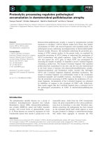
Fig. 7. AFP positively regulates formation of Apaf-1 apoptosome in cell-free extracts and promotes caspase activation and release of caspase 3 and 9
from the complex. Aliquots (1 mL) of S-100 extracts obtained from nonapoptotic HepG2 cells were left untreated (control) or activated by a 1-h
incubation at 30 °Cwith1m
M
dATP and 0.7 l
M
cyt-c in the presence or absence of 5.0 l
M
AFP. Subsequently, the extract aliquot (1 mg protein)
was fractionated by high-resolution chromatography on a Superose-6 HR 10/30 column. Fractions of 1 mL were collected and aliquots of 50 lL
were assayed fluorimetrically for IETDase (A) and DEVDase (B) activity. Caspase activity is given in arbitrary fluorescent units in the fraction per
minute. Arrowheads at the top of the patterns indicate sizes of calibration protein standards and their elution positions from the Superose-6 column.
Dot-blot analysis of the AFP distribution in the fractions for cell lysates treated with AFP/dATP/cyt-c is shown under the chromatographic pattern
(B). The corresponding fractions were concentrated, and aliquots of 20 lL were also resolved by SDS/PAGE and immunoblotting for caspase 9,
caspase 8 (A), caspase 3 (B), Apaf-1 (C) and anti-(cIAP-2) (D). The corresponding chromatography fraction numbers are indicated under the
patterns. The central line marked with an asterisk shows the blot of cell lysate with addition of cyt-c/dATP before chromatography. (E) AFP
displaces endogenous cIAP-2 from the complex with caspases 3 and 9. Recombinant His-tagged active caspase 9 and caspase 3 were immobilized
on the Ni/Sepharose beads and incubated with HepG2 S-100 extract with or without 5 l
M
AFP. Ni/Sepharose-bound proteins were analyzed by
SDS/PAGE/immunoblotting with polyclonal antibodies to cIAP-2.
Ó FEBS 2003 AFP amplifies cytochrome c-mediated caspase activation (Eur. J. Biochem. 270) 4395
Effect of AFP on the distribution of Apaf-1 and cIAP-2
proteins along the chromatographic pattern
of the apoptosome assembly
To determine possible mechanisms of the AFP-mediated
regulation of the apoptosome complex, we monitored the
distribution of Apaf-1 along the chromatographic pattern
of the apoptosome assembly, which was formed with and
without AFP (Fig. 7C). In cell extracts stimulated with low
cyt-c, Apaf-1 was recovered in two main peaks correspond-
ing to fractions 6–8 and 13–15, demonstrating that a low
suboptimal dose of cyt-c does not recruit all the available
Apaf-1 into the functional apoptosome and tends to form
the nonfunctional complex of molecular mass 1.4 MDa.
In the presence of AFP, Apaf-1 specificity was significantly
reduced in the biologically inactive 1.4-MDa complex
(fractions 6–7) [3,35], but notably increased in the region of
the 700-kDa apoptosome (fractions 8–10). These data
indicate that, at low cyt-c, AFP positively modulates
recruitment of Apaf-1 into the active 700-kDa apopto-
some complex.
Figure 7D shows that cIAP-2 distribution was not so
clearly affected by AFP addition as observed in the case of
Apaf-1. However, in the absence of AFP, full-length cIAP-2
was present in fractions 10–11, whereas fraction 9 mainly
contained fragmented IAP-2-specific material (Fig. 7D).
After the addition of AFP, the cIAP-2 specificity (including
full-length protein and its fragments) was distinctly reduced
in fractions 9–10 (Fig. 7D). The similar fragmentation
pattern for cIAP-1 and cIAP-2 has been described previ-
ously [36]. It was shown that fragmented cIAP-1 and cIAP-2
were more effective at protecting cells from apoptosis,
whereas full-length proteins lacked protective activity.
Removal of the RING domain by proteolysis restored the
antiapoptotic activity [36]. It was also shown that cIAP-1
was cleaved in vitro by pure caspase 3, producing similar
52-kDa and 35-kDa fragments. Our data allow us to suggest
that AFP may negatively regulate fragmentation of cIAP-2,
thus modulating its antiapoptotic activity. Alternatively,
AFP may stimulate release of active fragmented cIAP from
the apoptosome. From our data we proposed the possible
interaction of AFP with cIAP-2 and its partial removal from
the apoptosome complex. To confirm this, we studied the
direct interaction of AFP and cIAP-2 using a direct protein–
protein interaction assay.
Interaction between caspase 9, caspase 3, c-IAP-2
and AFP
To study further the interaction between AFP, cIAP-2 and
caspases 9 and 3, we precipitated pure recombinant active
caspases 3 and 9 (His-tagged) with nickel resin and then
incubated them with AFP and S-100 extract, as a source of
cIAP-2. A similar reaction mixture was also prepared
without AFP. The supernatants and pellets were probed
with antibodies against cIAP-2. As IAPs interact directly
with active caspases 3 and 9 [5], we speculated that AFP
may physically interact with one of these proteins to
displace cIAP-2 from the complex. Figure 7E shows that
cIAP-2 binds processed recombinant caspase 3 and/or 9.
Addition of the pure human AFP in the same reaction
system almost completely disrupts the interaction between
processed caspases and cIAP-2, demonstrating its release
from the complex (Fig. 7E). Additional experiments with
each protein member of the complex will be necessary to
clarify the exact molecular interactions involved in this
effect. Our data suggest that AFP may positively regulate
the activity of the apoptosome by negative modulation of
the cIAP-2 content, resulting in promotion of the release of
active caspases 3 and 9 from the complex.
Discussion
There is increasing evidence that AFP may selectively
induce activation of programmed cell death in tumor cells
[17–23], showing its potential for cancer treatment [10].
Various researchers have documented the tumor-selective
uptake of AFP by malignant cells [13–16], but the functional
significance of this phenomenon has not been clarified. The
exact molecular mechanisms of AFP-mediated apoptosis
also remain unclear. The present data explain some details
of the molecular interactions in this effect.
In this study we have investigated the ability of AFP to
directly activate the death program in a cell-free model of
apoptosis. Release of cyt-c into the cytoplasm of AFP-
treated cells suggests that a mitochondrion-dependent
mechanism of apoptosis signaling is involved. However,
these data do not exclude the possibility that another cyt-c-
independent pathway of AFP-mediated signaling of apop-
tosis is also involved in the sequential indirect induction of
cyt-c release with the onset of its activity. We found here
that AFP promotes low-dose cyt-c/dATP-mediated pro-
cessing and activation of procaspases 9 and 3 in a cell-free
system. These data show that AFP is directly involved in
regulating the mechanisms of caspase cascade activation
and suggest that it may be involved in regulating apopto-
some complex formation. We have demonstrated further
that AFP-mediated signaling of apoptosis requires the
presence of all the major members of the apoptosome
complex: cyt-c, dATP, caspases 9 and 3. To confirm that
AFP is involved in regulating activity of the apoptosome
complex, cell-free cytosolic extracts were activated in vitro
by addition of cyt-c/dATP or AFP/cyt-c/dATP, and, after
high-resolution gel filtration, the fractions from the column
were analysed by Western blotting. Our data clearly show
that AFP positively regulates cyt-c/dATP-mediated forma-
tion of the active 700-kDa Apaf-1–apoptosome complex
and stimulates release of the active caspase 3 from the
complex. The key was the finding that AFP negatively
regulates binding of cIAP-2 to active caspases 9 and 3. It
remains to be seen if AFP associates with and inhibits
interaction of other cIAPs with caspases, thus promoting
caspase activation within the apoptosome complex. Our
data suggest that AFP interacts with caspase 3, 9 and/or
cIAP-2 in a similar manner to DIABLO/Smac or Omi/Htr
[7,8], but the exact molecular determinants involved in these
interactions remain to be determined.
A similar effect of selective triggering of apoptosis in
tumor cells was observed for multimeric forms of human
a-lactalbumin, MAL [37,38]. It was shown that only
oligomerized forms of this protein are capable of inducing
apoptosis. Our recent data similarly showed that AFP
requires concentration-dependent oligomerization to
become apoptotically active [23]. The exact mechanism of
4396 L. Semenkova et al.(Eur. J. Biochem. 270) Ó FEBS 2003
MAL function has not yet been established, but it was
shown that MAL induces its proapoptotic effects by direct
activation of the caspase cascade independently of the
membrane-receptor signaling [38].
To prevent uncontrolled proliferation of rapidly growing
tissues, such as developing immature immune cells, embry-
onic cells or tumor cells, certain natural control mechanisms
have to exist that select and direct developing cells toward
maturation and prevent their neoplastic transformation.
This study describes a naturally occurring protein, the
expression of which is restricted by developing immature
embryonic cells or cells undergoing malignant transforma-
tion [9–12]. Proteins with quite mundane functions in
healthy cells often behave very differently during cell suicide.
The selective proapoptotic activity of AFP, targeting only
neoplastic [17–23] and activated immune cells [9,10], indi-
cates that it is a natural effector in a fetoembryonic defense
system to prevent malignant transformation of developing
cells. Our data allow us to propose that AFP helps cells to
overcome their resistance to apoptosis by significant ampli-
fication of the apoptotic signals induced by other factors,
such as drugs and oxidative stress. AFP may help cells,
which are resistant to apoptotic stimuli for any reason, to
overcome their resistance, which is induced, for example, by
overexpression of heat shock proteins, cIAPs or any other
defects of apoptosome-dependent apoptotic pathways.
Tumor cells are characterized by defects in expression of
apoptosis-promoting proteins, such as Apaf-1 and p53, and
simultaneous overexpression of the antiapoptotic proteins
Hsp70, Bcl-2 and Bcl-x
L
, resulting in tumor-specific sup-
pression of apoptosis and enhancement of the malignance
and therapy resistance of tumors [39–43]. The existence of a
high background level of antiapoptotic factors in the cytosol
of tumor cells often leads to their resistance to apoptosis
induced by weak stress stimuli. It has been demonstrated
that high levels of AFP in maternal serum during pregnancy
were associated with a low incidence of breast cancer [44,45].
It was proposed that AFP may delete immature breast
tissue cells that show the first signs of neoplastic transfor-
mation. Our results correlate with these data, indicating that
AFP may function to remove transformed neoplastic cells
by tumor-selective amplification of weak apoptotic signals.
Of special interest in cancer research are tumor-specific
factors that regulate apoptosis in tumor cells which function
at a common part of the apoptotic signaling pathway and
may cancel their resistance to apoptosis. There are several
hypotheses that may help to explain these findings. The
release of cyt-c from the mitochondria into the cytosol has
been shown to be one of the earliest apoptotic events, which
occurs before mitochondrial depolarization, caspase activa-
tion and DNA fragmentation. It has been documented that,
after apoptosis signaling, cyt-c is released from the mito-
chondria within 5 min [46]. The extramitochondrial cyt-c
has been shown to be a general apoptogen in cells with a
functional caspase system [47]. On the other hand, recent
papers have shown that cells can survive with reduced
mitochondrial membrane potential and released cytosolic
cyt-c given appropriate signals to suppress apoptosis [48,49].
It was observed that the amount of released cyt-c in K562
and CEM lines did not correlate with the extent of apoptosis
in response to UV light, showing reduced caspase 3 activa-
tion. The effect was explained by the reduced expression
of Apaf-1 protein in resistant leukemic cells [48]. A high
background level of cytosolic cyt-c has been shown in vivo in
the aging heart, with a significant decrease in the antiapop-
totic protein bcl-2 [49]. In certain types of cancer cell,
alterations in the regulation of apoptosis may contribute to
tumor malignancy and resistance to radiotherapy and
chemotherapy [50]. Sometimes dysfunctional apoptosome
activation in tumor cells is observed in the presence of the
required amount of cytosolic cyt-c, dATP, Apaf-1 and pro-
caspase 9, leading to significant enhancement of their
resistance to apoptotic stimuli including radiotherapy and
chemotherapy [51].
Our data indicate that AFP can be considered as a tumor-
specific regulator of cyt-c-mediated apoptotic signals.
In vivo, it may operate as a specific regulator of the
apoptosome dysfunction induced by the impaired release of
apoptogenic factors in the cytosol and/or the increased level
of cytosolic antiapoptotic proteins. It may operate to
amplify weak apoptotic signals induced by oxidative stress,
ionizing radiation or drugs to sensitize tumor cells to
chemotherapy. It seems to operate as a tumor-specific
regulator of apoptosis inhibitory proteins, but it remains to
be seen if it associates with and inhibits cIAPs other than
cIAP-2, and to determine the molecular mechanisms of
these interactions.
Acknowledgements
This work is supported in part by the International Science &
Technology Center, ISTC (grants Nos. 401-98 and 1878-01). We thank
Dr Alex Sazonov for the invaluable gift of recombinant caspase 3 and
caspase 9 and Dr Alex Chugunov for excellent assistance with FPLC
chromatography.
References
1. Earnshaw, W.C., Martins, L.M. & Kaufmann, S.H. (1999)
Mammalian caspases: structure, activation, substrates, and func-
tions during apoptosis. Annu.Rev.Biochem.68, 383–424.
2. Cain, K., Bratton, S.B. & Cohen, G.M. (2002) The Apaf-1
apoptosome: a large caspase-activating complex. Biochimie 84,
203–214.
3. Cain,K.,Bratton,S.B.,Langlais,C.,Walker,G.,Brown,D.G.,
Sun, X.M. & Cohen, G.M. (2000) Apaf-1 oligomerizes into bio-
logically active approximately 700-kDa and inactive approxi-
mately 1.4 MDa apoptosome complexes. J. Biol. Chem. 275,
6067–6070.
4. Bratton, S.B., Walker, G., Srinivasula, S.M., Sun, X.M., Butter-
worth, M., Alnemri, E.S. & Cohen, G.M. (2001) Recruitment,
activation and retention of caspases-9 and -3 by Apaf-1 apopto-
some and associated XIAP complexes. EMBO J. 5, 998–1009.
5. Deveraux, Q.L. & Reed, J.C. (1999) IAP family proteins: sup-
pressors of apoptosis. Genes Dev. 13, 239–252.
6. Shi, Y. (2002) Mechanisms of caspase activation and inhibition
during apoptosis. Mol. Cell 9, 459–470.
7. Shrinivasula, S.M., Saleh, A., Hedge, R., Datta, P., Shiozaki, E.,
Chai, J., Robbins, P.D., Fernandes-Alnemri, T., Shi, Y. &
Alnemri, E.S. (2001) A conserved XIAP interaction motif in cas-
pase-9 and Smac/DIABLO mediates opposing effects on caspase
activity and apoptosis. Nature (London) 409, 112–116.
8. Hedge, R., Shrinivasula, S.M., Zhang, Z., Wassel, R., Mukattash,
R., Cilenti, L., Lazebnik, Y., Zervos, A.S., Fernandes-Alnemri, T.
& Alnemri, E.S. (2002) Identification of Omi/HtrA2 as a mito-
chondrial apoptotic serine protease that disrupts inhibitor of
Ó FEBS 2003 AFP amplifies cytochrome c-mediated caspase activation (Eur. J. Biochem. 270) 4397
apoptosis protein–caspase interaction. J. Biol. Chem. 277, 432–
438.
9. Deutsch, H.F. (1991) Chemistry and biology of a-fetoprotein.
Adv. Cancer Res. 56, 253–312.
10. Mizejewsky, G.J. (2002) Biological role of a-fetoproteinincancer:
prospects for anticancer therapy. Expert Rev. Anticancer Ther. 2,
89–115.
11.Gabant,P.,Forrester,L.,Nichos,J.,Reeth,T.V.,Mees,C.,
Pajack,B.,Watt,A.,Smitz,J.,Alexandre,H.,Szpirer,C.&
Szpirer, J. (2002) Alpha-fetopretin, the major fetal serum protein,
is not essential for embrionic development but is required for
female fertility. Proc. Natl Acad. Sci. USA 99, 12865–12870.
12. Mizejewsky, G.J. (2001) Alpha-fetoprotein structure and function:
relevance to isoforms, epitopes, and conformational variants. Exp.
Biol. Med. 226, 377–408.
13. Laborda, J., Naval, J., Allouche, M., Calvo, M., Georgoulias, V.,
Mishai, Z. & Uriel, J. (1987) Specific uptake of alpha-fetoprotein
by malignant human lymphoid cells. Int. J. Cancer 40, 314–318.
14. Deutsch, H.F. (1994) The uptake of adriamycin-arachidonic acid
complexes by human tumor cells in the presence of a-fetoprotein.
J. Tumor Marker Oncol. 9, 11–15.
15. Geuskens, M., Dupressoir, T. & Uriel, J. (1992) A study,
by electron microscopy, of the specific uptake of alpha-fetoprotein
by mouse embryonic fibroblasts in relation to in vitro aging,
and by human mammary epithelial tumor cells in comparison
with normal donor’s cells. J. Submicrosc. Cytol. Pathol. 23,
59–66.
16. Alava, M.A., Iturralde, M., Lampreave, F. & Pineiro, A. (1999)
Specific uptake of alpha-fetoprotein and albumin by rat Mor
hepatoma cells. Tumour Biol. 20, 52–64.
17. Bennett, J.A., Semeniuk, D.J., Jacobson, H.I. & Murgita, R.A.
(1997) Similarity between natural and recombinant human alpha-
fetoprotein as inhibitors of estrogen-dependent breast cancer
growth. Breast Cancer Res. Treat. 45, 169–179.
18. Bennet, J.A., Zhu, S., Pagano-Mirarchi, A., Kellom, T.A. &
Jacobson, H.I. (1998) a-Fetoprotein derived from a human
hepatoma prevents growth of estrogen-dependent human breast
cancer xenografts. Clin. Cancer Res. 4, 2877–2884.
19. Sonnenschein, C., Ucci, A.A. & Soto, A.M. (1980) Inhibition of
growth of transplantable rat mammary tumors. Probable role of
a-fetoprotein. J. Natl Cancer Inst. 64, 1141–1146.
20. Dudich, E.I., Semenkova, L.N., Gorbatova, E.A., Dudich, I.V.,
Khromykh, L.M., Tatulov, E.B., Grechko, G.K. & Sukhikh, G.T.
(1998) Growth-regulative activity of alpha-fetoprotein for differ-
ent types of tumor and normal cells. Tumor Biol. 19, 30–40.
21. Dudich, E.I., Semenkova, L.N., Dudich, I.V., Gorbatova, E.A.,
Tokhtamysheva, N., Tatulov, E.B., Nikolaeva, M.A. &
Sukhikh, G.T. (1999) a-Fetoprotein causes apoptosis in tumor
cells via a pathway independent of CD95, TNFR1 and TNFR2
through activation of caspase-3-like proteases. Eur. J. Biochem.
266, 1–13.
22. Semenkova, L.N., Dudich, E.I. & Dudich, I.V. (1997) Induction
of apoptosis in human hepatoma cells by alpha-fetoprotein.
Tumor Biol. 18, 261–274.
23. Dudich, I.V., Tokhtamysheva, N., Semenkova, L., Dudich, E.,
Hellman, J. & Korpela, T. (1999) Isolation and structural and
functional characterization of two stable peptic fragments of
human alpha-fetoprotein. Biochemistry 38, 10406–10414.
24. Festin, S.M., Bennett, J.A., Fletcher, P.W., Jacobson, H.I., Shaye,
D.D. & Andersen, T.T. (1999) The recombinant third domain of
human alpha-fetoprotein retains the antiestrotrophic activity
found in the full-length molecule. Biochim. Biophys. Acta 1427,
307–314.
25. Mesfin, F.B., Bennet, J.A., Jacobson, H.I., Zhu, S. & Andersen,
T.T. (2000) Alpha-fetoprotein derived antiestrotrophic octa-
paptide. Biochim. Biophys. Acta 1501, 33–43.
26. Vakharia, D. & Mizejewsky, G.J. (2000) Human alpha-fetopro-
tein peptides bind estrogen receptor and estradiol and suppress
breast cancer. BreastCanc.Res.Treat.63, 41–52.
27. Eisele, L.E., Mesfin, F.B., Bennett, J.A., Andersen, T.T., Jacob-
son, H.I., Solwell, H., MacColl, R. & Mizejewski, G.J. (2001)
Studies on growth-inhibitory peptide derived from alpha-feto-
protein and some analogs. J. Peptide Res. 57, 29–38.
28. Liu, X., Kim, C.N., Yang, J., Jemmerson, R. & Wang, X. (1996)
Induction of apoptotic program in cell-free extracts: requirement
for dATP and cytochrome c. Cell 86, 147–157.
29. Slee, E.A., Harte, M.T., Kluck, R.M., Wolf, B.B., Casiano, C.A.,
Newmeyer,D.D.,Wang,H G.,Reed,J.C.,Nicholson,D.W.,
Alnemri,E.S.,Green,D.R.&Martin,S.J.(1999)Orderingthe
cytochrome c-initiated caspase cascade: hierarchical activation of
caspases-2-3-6-7-8, and -10 in a caspase-3-dependent manner.
J. Cell Biol. 144, 281–292.
30. Liu, X. & Wang, X. (2000) In vitro assays for caspase-3 activation
and DNA-fragmentation. Methods Enzymol. 322, 177–183.
31. Stennicke, H.R., Ju
ˆ
rgensmeier, J.M., Shin, H., Deveraux, Q.,
Wolf, B.B., Yang, X., Zhou, Q., Ellerby, M., Bredesen, D., Green,
D.,Reed,J.C.,Froelich,C.J.&Salvesen,G.S.(1998)Procaspase-
3 is a major physiologic target of caspase-8. J. Biol. Chem. 273,
27084–27090.
32. Verhagen, A.M., Silke, J., Ekert, P.G., Pakusch, M., Kaufmann,
H., Connolly, L.M., Day, C.L., Tikoo, A., Burke, R., Wrobel, C.,
Moritz, R.L., Simpson, R.J. & Vaux, D.L. (2002) Htr-A2 pro-
motes cell death through its serine protease activity and its ability
to antagonize inhibitor of apoptosis proteins. J. Biol. Chem. 277,
445–454.
33. Rodriguez, J. & Lazebnik, Y. (1999) Caspase-9 and APAF-1 form
and active holoenzyme. Genes Dev. 13, 3179–3184.
34. Thornberry, N.A., Chapman, K.T. & Nicholson, D.W. (2000)
Determination of caspase specificities using a peptide combina-
torial library. Methods Enzymol. 322, 100–110.
35.Bratton,S.B.,Walker,G.,Roberts,D.L.,Cain,K.&Cohen,
G.M. (2001) Caspase-3 cleaves Apaf-1 into a 30-kDa fragment
that associates with an inappropriately oligomerised and biologi-
cally inactive 1.4 MDa apoptosome complex. Cell Death Differ. 8,
425–433.
36. Clem, R.J., Sheu, T.T., Richter, B.W.M., He, W W., Thornberry,
N.A.,Duskett,C.S.&Hardwick,J.M.(2001)c-IAP1iscleavedby
caspases to produce a proapoptotic C-terminal fragment. J. Biol.
Chem. 276, 7602–7608.
37. Ha
˚
kansson, A., Andreasson, J., Zhivotovsky, B., Karpman, D.,
Orrenius, S. & Svanborg, C. (1999) Multimeric a-lactalbumin
from human milk induces apoptosis through a direct effect on cell
nuclei. Exp. Cell Res. 246, 451–460.
38. Ko
¨
hler, C., Ha
˚
kansson, A., Svanborg, C., Orrenius, S. & Zhivo-
tovsky, B. (1999) Protease activation in apoptosis induced by
MAL. Exp. Cell Res. 249, 260–268.
39. Ja
¨
a
¨
tela, M. (1999) Escaping cell death: survival proteins in cancer.
Exp. Cell Res. 248, 30–43.
40. Evan, G.I. & Vousden, K.H. (2001) Proliferation, cell cycle and
apoptosis in cancer. Nature (London) 411, 342–348.
41. Beere,H.M.,Wolf,B.B.,Cain,K.,Mosser,D.D.,Mahboubi,A.,
Kuwana, T., Tailor, P., Morimoto, R.I., Cohen, G.M. & Green,
D.R. (2000) Heat-shock protein 70 inhibits apoptosis by pre-
venting recruitment of procaspase-9 to the Apaf-1 apoptosome.
Nat. Cell Biol. 2, 469–475.
42. Pandey, P., Saleh, A., Nakazawa, A., Kumar, S., Srinivasula,
S.M., Kumar, V., Weichselbaum, R., Nalin, C., Alnemri, E.S.,
Kufe, D. & Kharabanda, S. (2000) Negative regulation of cyto-
chrome c mediated oligomerization of Apaf-1 and activation of
procaspase-9 by heat shock protein 90. EMBO J. 19, 4310–4322.
43. Samali,A.,Cai,J.,Zhivotovsky,B.,Jones,D.P.&Orrenius,S.
(1999) Presense of a pre-apoptotic complex of pro-caspase-3,
4398 L. Semenkova et al.(Eur. J. Biochem. 270) Ó FEBS 2003
Hsp60 and Hsp10 in the mitochondrial fraction of Jurkat cells.
EMBO J. 18, 2040–2047.
44. Melbue, M., Wohlfahrt, J., Norgard-Pedersen, B., Mouridsen,
H.T., Lambe, M. & Michels, K.B. (2000) Alpha-fetoprotein levels
in maternal serum during pregnancy and maternal breast cancer
incidence. J. Natl Cancer Inst. 92, 1001–1005.
45. Richardson, B.E., Huka, B.S., David Peck, J.L., Hughes, C.L.,
Berg, B.J., Christiansen, R.E. & Calvin, J.A. (1998) Levels of
maternal serum alpha-fetoprotein (AFP) in pregnant women and
subsequent breast cancer risk. Am.J.Epidemiol.148, 719–728.
46. Goldstein, J.C., Waterhouse, N.J., Juin, P., Evan, G.I. & Green,
D.R. (2000) The coordinate release of cytochrome c during
apoptosis is rapid, complete and kinetically invariant. Nat. Cell
Biol. 2, 156–162.
47. Brustugun, O.T., Fladmark, K.E., Doskeland, S.O., Orrenius, S.
& Zhivotovsky, B. (1998) Apoptosis induced by microinjection of
cytochrome c is caspase-dependent and is inhibited by Bcl-2. Cell
Death Differ. 5, 660–668.
48. Jia, L., Srinivasula, S.M., Liu, F T., Newland, A.C., Fernandes-
Alnemri, T., Alnemri, E.S. & Kesley, S.M. (2001) Apaf-1 protein
deficiency confers resistance to cytochrom c-dependent apoptosis
in human leukemic cells. Blood 98, 414–421.
49. Phaneuf, S. & Leeuwenburgh, C. (2002) Cytochrome c release
from the mitochondria in the aging heart: a possible mechanism
for apoptosis with age. Am. J. Physiol. Regulatory Integrative
Comp. Physiol. 282, 423–430.
50. Chen, Q., Takeyama, N., Brady, G., Watson, A.J.M. & Dive, C.
(1998) Blood cells with reduced mitochondrial membrane poten-
tial and cytosolic cytochrome c can survive and maintain clono-
genecity given appropriate signals to suppress apoptosis. Blood 92,
4545–4553.
51. Liu, R.J., Opipari, A.W., Tan, L., Jiang, Y., Zhang, Y., Tang, H.
& Nunes, G. (2002) Dysfunctional apoptososme activation in
ovarian cancer: implications for chemoresistance. Cancer Res. 62,
924–931.
Ó FEBS 2003 AFP amplifies cytochrome c-mediated caspase activation (Eur. J. Biochem. 270) 4399