Báo cáo Y học: Inhibition of hyaluronan synthesis in Streptococcus equi FM100 by 4-methylumbelliferone doc
Bạn đang xem bản rút gọn của tài liệu. Xem và tải ngay bản đầy đủ của tài liệu tại đây (344.59 KB, 10 trang )
Inhibition of hyaluronan synthesis in
Streptococcus equi
FM100
by 4-methylumbelliferone
Ikuko Kakizaki
1
, Keiichi Takagaki
1
, Yasufumi Endo
1
, Daisuke Kudo
1
, Hitoshi Ikeya
2
, Teruzo Miyoshi
2
,
Bruce A. Baggenstoss
3
, Valarie L. Tlapak-Simmons
3
, Kshama Kumari
3
, Akio Nakane
4
, Paul H. Weigel
3
and Masahiko Endo
1
Departments of
1
Biochemistry and
4
Bacteriology, Hirosaki University School of Medicine, Hirosaki;
2
Research Center Denki
Kagaku Kogyo Co. Ltd, Tokyo, Japan;
3
Department of Biochemistry and Molecular Biology, University of Oklahoma Health
Sciences Center, Oklahoma City, Oklahoma, USA
As observed previously in cultured human skin fibroblasts, a
decrease of hyaluronan production was also observed in
group C Streptococcus equi FM100 cells treated with
4-methylumbelliferone (MU), although there was no effect
on their growth. In this study, the inhibition mechanism of
hyaluronan synthesis by MU was examined using Strepto-
coccus equi FM100, as a model. When MU was added to a
reaction mixture containing the two sugar nucleotide donors
and a membrane-rich fraction as an enzyme source in a cell-
free hyaluronan synthesis experiment, there was no change
in the production of hyaluronan. On the contrary, when MU
was added to the culture medium of FM100 cells, hyaluro-
nan production in the isolated membranes was decreased in
a dose-dependent manner. However, when the effect of MU
on the expression level of hyaluronan synthase was exam-
ined, MU did not decrease either the mRNA level of the has
operon containing the hyaluronan synthase gene or the
protein level of hyaluronan synthase. Solubilization of the
enzyme from membranes of MU-treated cells and addition
of the exogenous phospholipid, cardiolipin, rescued hya-
luronan synthase activity. In the mass spectrometric analysis
of the membrane phospholipids from FM100 cells treated
with MU, changes were observed in the distribution of only
cardiolipin species but not of the other major phospholipid,
PtdGro. These results suggest that MU treatment may cause
a decrease in hyaluronan synthase activity by altering the
lipid environment of membranes, especially the distribution
of different cardiolipin species, surrounding hyaluronan
synthase.
Keywords: hyaluronan; synthesis; Streptococcus; 4-methyl-
umbelliferone; phospholipids.
Hyaluronan (HA) is a high molecular weight glycosamino-
glycan composed of repeating disaccharide units of
GlcNAc-b(1fi4)-GlcUA-b(1fi3) [1]. HA is one of the
major components of the extracellular matrix together with
proteoglycans and collagens, and is involved in many
biological processes, including tissue organization, wound
healing, tumor invasion and cancer metastasis, through its
interactions with other extracellular matrix components
[2,3].
It has long been suggested that HA may be implicated
in malignant transformation and tumor progression [4].
There are many reports that HA production is increased in
various tumor tissues including mesothelioma and Wilm’s
tumor. Recently, a direct correlation between HA and
tumorigenesis, and cancer metastasis was shown in studies
using genetic manipulations to create mutant cells that were
either overproducing HA or HA-deficient [5,6]. Overpro-
duction of HA is also observed in diseases associated with
inflammation and fibroses [3].
Many strains of group A and C Streptococci are able to
synthesize HA [7,8]. Their thick HA coats surrounding the
cell surfaces contribute to their pathogenicity by allowing
them to escape from the immune systems of their hosts.
The HA synthesized by Streptococci is not chemically
or structurally distinguishable from that synthesized in
mammalian cells.
HA is synthesized by a membrane-associated hyaluronan
synthase (HAS) from the precursors UDP-GlcUA and
UDP-GlcNAc in either mammalian cells or Streptococci [9].
In the last several years, three distinct mammalian genes and
three unique Streptococcal genes encoding the HASs have
been cloned and their properties have been examined [9–12].
From the genomic analysis, it has been clarified that the has
operon encodes for the HA synthesis system of Streptococci.
The has operon is composed of three genes, hasA (which
encodes the HA synthase), hasB (which encodes UDP-
glucose dehydrogenase), and hasC (which encodes UDP-
glucose pyrophosphorylase) [9]. Tlapak-Simmons et al.[13]
demonstrated that the functional sizes of both the group A
and the group C Streptococcus HASs are protein monomers
in association with about 16 phospholipid molecules, in
particular cardiolipin (CL), which was also shown to be
necessary for optimal enzymatic activity [14]. Due to the
Correspondence to M. Endo, Department of Biochemistry, Hirosaki
University School of Medicine, 5 Zaifu-cho, Hirosaki 036–8562,
Japan. Fax: + 81 172 39 5016, Tel.: + 81 172 39 5015,
E-mail:
Abbreviations: CL, cardiolipin; DDM, n-dodecyl-b-
D
-maltoside;
GlcNAc, N-acetylglucosamine; GlcUA, glucuronic acid; HA,
hyaluronan (hyaluronic acid); HABP, hyaluronan binding protein;
HAS (Has), hyaluronan synthase; MU, 4-methylumbelliferone;
spHAS, S. pyogenes HAS; seHAS, S. equisimilis HAS.
Note: A web site is available at />bioche1/test/Biochem-top/Biochem-top1.html
(Received 20 June 2002, revised 14 August 2002,
accepted 29 August 2002)
Eur. J. Biochem. 269, 5066–5075 (2002) Ó FEBS 2002 doi:10.1046/j.1432-1033.2002.03217.x
cloning of the HAS genes, it has been possible to genetically
manipulate the production of HA, and consequently,
correlations between HA production and various biological
processes have now been brought to light. For example, the
effects of antisense inhibition of HA production on the
organization of the extracellular matrix in human articular
chondrocytes has been examined [15]. Studies using targeted
deletion of HAS genes have also been made to investigate
theroleofHAin vivo.ItwasreportedthatHas2
+
embryos,
which lack HA production by Has2, exhibit severe cardiac
and vascular abnormalities and die during fetal develop-
ment [16]. However the details about the multiple functions
of HA have not been fully established.
We found that HA synthesis in cultured human skin
fibroblasts was inhibited by 4-methylumbelliferone (MU,
7-hydroxy-4-methyl-2H-1-benzopyran-2-one) with no effect
on the synthesis of any other glycosaminoglycan and that an
HA-deficient extracellular matrix was formed [17,18]. Some
agents have been reported to inhibit HA synthesis, however,
no clearly specific inhibitors for HA synthesis have been
found [19–23]. Although its biochemical mechanism of
inhibition is not well understood, MU has been used in
some studies on the function of HA [5,24,25]. For example,
it was used to prepare an HA-deficient transfected cell line
expressing a HAS gene in order to examine the role of HA
in tumorigenesis [5]. Recently, Endo et al. investigated the
correlation between HA and the other components of
extracellular matrices, using cultured human skin fibroblasts
in which HA production was inhibited by MU treatment
[24]. In order to elucidate the inhibition mechanism of HA
synthesis, in the present study we have examined the effect
of MU on prokaryotic cells, S. equi FM100, as a model. We
find that, as in cultured human skin fibroblasts [18], MU did
not directly inhibit HAS in vitro but did inhibit the
enzymatic activity of HAS in intact cells. Furthermore, we
show that MU does not directly inhibit the processes of
transcription or translation of HAS, but that a possible
novel mechanism of inhibition of HA synthesis by MU is
probably due to an alteration of the lipid environment of the
Streptococcal membranes.
MATERIALS AND METHODS
Materials
Lysozyme and MU were purchased from Wako Pure
Chemicals (Osaka, Japan). MU was dissolved in dimethyl-
sulfoxide, and the final concentration of dimethylsulfoxide
in the culture medium and reaction mixtures for HA syn-
thesis was 0.1%. UDP-[U-
14
C]GlcUA (270 mCiÆmmol
)1
)
was purchased from American Radiolabeled Chemicals
(St. Louis, MO, USA). UDP-GlcNAc, ATP, dithiothreitol,
bovine testicular hyaluronidase and bovine heart CL were
purchased from Sigma (St. Louis, MO, USA). HA from
human umbilical cords and Streptomyces hyaluronidase
were obtained from Seikagaku Corporation (Tokyo,
Japan). Actinase E was from Kaken Pharmaceutical
(Tokyo, Japan). Hyaluronic Acid ÔChugaiÕ quantitative test
kit for the sandwich binding protein assay was purchased
from Chugai Pharmaceutical (Tokyo, Japan) [26]. The
random-primed DNA labeling kit was from Amersham
Pharmacia Biotech (Tokyo, Japan) and [a-
32
P] dCTP was
from NEN Life Science Products (Boston, MA, USA).
Antiserum raised against the whole HAS from Streptococcus
pyogenes (spHAS) was described previously [10,27]. Anti-
rabbit Ig conjugated to horseradish peroxidase was from
Dako Japan (Kyoto, Japan) and n-dodecyl-b-
D
-maltoside
(DDM) was from Nakarai Tesque (Kyoto, Japan).
Culture of Streptococci and treatment with MU
Encapsulated group C S. equi FM100 was derived from
S. equi (ATCC9527) and maintained at 33 °C in a synthetic
solid medium (pH 8), which we have modified from the
medium reported by van de Rijn and Kessler [28]. Cells were
precultured in liquid medium (1.5% polypeptone-S/0.5%
yeast extract/0.2% dipotassium hydrogenphosphate/0.16%
sodium thiosulfate/0.02% sodium sulfate/2.0% glucose,
pH 8), and then added to 100 volumes of fresh medium and
grown with or without MU. Cell numbers were estimated
by measuring the absorbance at a wavelength of 660 nm.
Cells were stained with nigrosin and observed through a
microscope (OLYMPUS, model BHS). The detailed infor-
mation for the generation and characterization of S. equi
FM100 is described in Japanese patent (JP1750524, Japan
Patent Office).
Analysis of the HA released into the culture medium
Exponentially growing cells were cultured with or without
various concentrations of MU (0.2, 0.5, 1.0 and 2.0 m
M
). At
the various time points (0, 3, 5.5, 8 and 22 h), HA released
into the culture medium was measured by the sandwich
binding protein assay using hyaluronan binding protein
(HABP) according to the manufacture’s instructions for the
hyaluronic acid ÔChugaiÕ quantitative test kit [26].
The molecular size of HA released into the culture
medium was analyzed by gel filtration HPLC using a
Shodex OHpak KB-805 column (8 · 300 mm). Elution was
with 0.2
M
NaCl at a flow rate of 0.5 mLÆmin
)1
.Eluted
fractions were monitored by detecting absorbance at a
wavelength of 215 nm. The molecular sizes of standard HA
were 1.0, 3.0, 4.1, 8.0, 12 and 19 · 10
5
[29].
Preparation of a membrane-rich fraction
and solubilization
The membrane-rich fraction was prepared by following the
method of Sugahara et al. [30]. Briefly, exponential phase
cell cultures were harvested by centrifugation at 18 000 g for
30 min. Then, cells were suspended in 0.05
M
sodium and
potassium phosphate buffer, pH 7.4, containing 5 m
M
dithiothreitol. The cell suspension was sonicated on ice
using a Branson Sonifer (model 250) for 1 min. The
disrupted cells were centrifuged at 10 000 g for 10 min
and the supernatant fluid was withdrawn. Following
centrifugation of the 10 000 g supernatant fluid at
105 000 g for 60 min, the resultant pellet was washed with
fresh buffer by centrifugation at 229 000 g for 45 min. The
pellet was suspended in 0.033
M
sodium and potassium
phosphate buffer, pH 7.4 containing 5 m
M
dithiothreitol,
and used as an enzyme source for the cell-free HA synthesis
assay. Solubilization of membranes using DDM was
performed as previously reported by Tlapak-Simmons et al.
[14]. Each cell-free assay was standardized for the amount of
protein used in order to compare the activities between the
Ó FEBS 2002 Inhibition of HA synthesis in Streptococcus by MU (Eur. J. Biochem. 269) 5067
whole and partially solubilized membranes. Protein content
was determined by the method of Bradford [31], and the
results were confirmed by SDS/PAGE analysis of all the
samples, according to the method of Laemmli [32].
Assay of cell-free HA synthesis
Cell-free HA synthesis was performed by a modification of
the method reported by Nakamura et al. [18]. Analysis of
the transfer of UDP-[U-
14
C] GlcUA to HA was monitored
as follows. Assay mixtures contained the following compo-
nents in a final volume of 0.1 mL: 47 m
M
sodium and
potassium phosphate buffer (pH 7.1), 5 m
M
dithiothreitol,
5m
M
MgCl
2
,100l
M
UDP-GlcNAc, 3.7 l
M
UDP-[U-
14
C]
GlcUA (1.8 lCi), 5 m
M
ATP, and 12.5–250 lgofmem-
brane-rich fractions or DDM extracts as the enzyme source.
MU was added as indicated to the assay mixture or the
culture medium. For certain experiments, the enzyme was
preincubated with 2 m
M
bovine heart CL. The assay
mixture was incubated at 37 °Cfor1.5h,andthereaction
was terminated by boiling for 2 min. Because the reaction
was linear up to 1.5 h of incubation time, this time point
wasused.AfteractinaseEdigestion(2.5mgÆmL
)1
,16h
at 45 °C), 1/6 volume of 50% trichloroacetic acid was
added with mixing, the reaction mixture was cooled on
ice, and the supernatant was withdrawn after centrifugation.
In the presence of 50 lg of carrier HA, ethanol precipitation
was performed five times to remove the unincoporated,
free radioisotopes. The final precipitate was dissolved in
water, digested with Streptomyces hyaluronidase and then
precipitated with ethanol. The radioactivity remaining in
the supernatant following ethanol precipitation, which
represents the digestion products specifically derived from
HA, was then determined using a liquid scintillation
counter.
Isolation of
S. equi
FM100
hasA
gene
To obtain a probe for hybridization, the 1166 bp region of
the S. equi FM100 hasA gene was amplified with primers
based upon the nucleotide sequence of the group C
Streptococcus equisimilis hasA gene (seHAS gene, GenBank
Accession Number AF023876) [10], using genomic DNA
from FM100 cells as a template. Genomic DNA was
extracted following the method of Sambrook et al.[33]after
digestion with lysozyme and bovine testicular hyaluroni-
dase. The primer set used in the PCR had the following
sequence: forward, 5¢-ACTGTTGTGGCCTTTAGTA-3¢
and reverse, 5¢-AAGGGCTGTAGGACAAACAA-3¢.The
sequence of the amplified product completely matched the
region of nucleotides 25–1190 of AF023876.
RNA preparations and Northern hybridization
Total RNA was prepared from FM100 cells using a
RNeasy Plant Mini Kit (Qiagen Japan, Tokyo, Japan)
according to the manufacturer’s specifications. Five micro-
grams of RNA, denatured with formaldehyde and forma-
mide, was separated on a 1% (w/v) agarose gel containing
1.1
M
formaldehyde and transferred to a nylon membrane
(Hybond-N, Amersham, Buckinghamshire, UK) in 20·
NaCl/Cit (3.0
M
NaCl/0.3
M
sodium citrate, pH 7.0).
The hasA DNA probe was labeled by the random
priming method [34]. Hybridization was performed with
a
32
P-labeled probe at 42 °C for 18 h in 50% formamide,
3· NaCl/Cit, 0.05
M
Tris/HCl (pH 7.5), 1 m
M
EDTA,
0.02% BSA, 0.02% Ficoll, 0.02% polyvinylpyrrolidone,
20 lgÆmL
)1
tRNA, 20 lgÆmL
)1
herring sperm DNA. Then
the filters were washed twice with 3· NaCl/Cit, 0.1% SDS
at 37 °C for 30 min, and twice with 0.1· NaCl/Cit, 0.1%
SDS at 50–65 °C for 30 min. Autoradiography was carried
out by exposure to X-ray film (Kodak X-Omat AR) at
)80 °C using an intensifying screen. Results of autoradio-
graphy were quantified using
NIH IMAGE
(version 1.62)
software.
SDS/PAGE and immunoblotting
SDS/PAGE was performed in 10% acrylamide gels by the
method of Laemmli [32]. Protein was stained with the
Coomassie brilliant blue R-250. For immunoblotting,
proteins were transferred to a polyvinylidene fluoride
filter (Millipore Japan, Tokyo, Japan), and stained with
anti-spHAS antibody according to the method of Towbin
et al.[35]using3,3¢-diaminobenzidine tetrahydrochloride
(Dojindo Laboratories, Kumamoto, Japan) for detection.
Resulting bands were quantified using
NIH IMAGE
(version
1.62) software.
Extraction of lipids
Lipids were extracted using the method of Folch et al.with
minor modifications [36]. Membrane samples were dis-
persed in 100 lL of chloroform/methanol (2 : 1, v/v) per
milligram membranes by brief sonication and then shaken
gently for 40 min at room temperature. The sample was
then centrifuged at 3800 g for 10 min and the supernatant
fluid was drawn off, with care not to remove any pellet
material. A 0.2 · the volume of 0.9% NaCl was added to
the sample, which was mixed vigorously by vortexing twice
for 30 s. The mixture was centrifuged at 420 g and the top
aqueous layer was removed. The bottom chloroform layer
containing the phospholipids was analyzed directly or
saturatedwithnitrogenandstoredat)20 °C.
Mass spectrometric analysis
The MALDI-TOF mass spectrometer used was a Voyager
Elite (Applied Biosystems, Framingham, MA, USA)
equipped with an N
2
laser (337 nm) located in the NSF
EPSCoR Oklahoma Laser MS Facility (OUHSC). Samples
were analyzed in the reflector, negative ion mode using a
delayed extraction of 200 ns, a grid voltage of 79%, and
were subjected to a 20-kV accelerating voltage. An external
calibration was obtained using bovine heart CL, which has
a mass of 1448.97. The matrices used were 6-aza-2-
thiothymine or 2,4,6-trihydroxyacetophenone at 5 mgÆmL
)1
in chloroform/methanol (2 : 1, v/v) containing 10 m
M
dibasic ammonium citrate. Samples were diluted with two
volumes of chloroform/methanol (2 : 1, v/v) and then mixed
1 : 1 with the matrix solution prior to spotting on a sample
plate and air drying. Spectra are an average of 80–100 scans.
In some cases the identity of specific m/z species was
confirmed by post source decay analysis in both the positive
and negative ion modes. Total amounts of CL or PtdGro
recovered from FM100 cells were assessed based on their
5068 I. Kakizaki et al. (Eur. J. Biochem. 269) Ó FEBS 2002
signal intensities relative to appropriate phospholipids that
were used as standards.
RESULTS
Inhibition of HA production of FM100 cells
To examine the effect of MU on the growth of FM100 cells,
the cells were cultured in liquid medium with or without MU
for various periods, and cell numbers were estimated by
measuring absorbance at 660 nm at each time point. No
significant effect on the growth was observed in the range of
0–2.0 m
M
MU (data not shown). Absorbance at 660 nm was
measured in all subsequent experiments, however, inhibition
of growth of FM100 cells by MU was not observed.
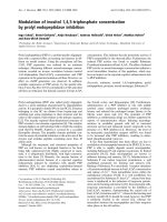
On microscopic examination, untreated control cells
formed HA coats on their cell surfaces, and the coat grew
thicker over the course of culture time (Fig. 1). However
the formation of the HA coating was dose-dependently
decreased, when the cells were cultured with 0.2–2.0 m
M
MU. The decrease in coat formation was observed as soon
as 3 h after culturing in the presence of 0.2 m
M
MU(data
not shown), and became more marked after longer incuba-
tion times.
When FM100 cells were cultured for 22 h, the HA coats
on cell surfaces essentially disappeared (Fig. 2A). Only a
very thin HA coating was observed at the highest concen-
tration of MU in the 22 h cultures (data not shown). On the
other hand, release of HA into the culture medium, as
assessed by HPLC analysis, was observed after 8 h, and
increased with continued incubation reaching a peak after
36 h (data not shown). The size distribution of HA released
into the culture medium was then analyzed by HPLC, after
the cells were cultured with various concentrations of MU
(0–2.0 m
M
) for 22 h (Fig. 2B). Regardless of the MU
concentration, the molecular sizes of the major peaks of HA
did not shift and were calculated at 1.2–1.9 · 10
6
.These
peaks disappeared after digestion with the very specific
Fig. 1. Effect of MU treatment on the HA coat formation of S. equi FM100 cells. Microscopic photograph of HA coats (arrowheads) on cell surfaces
of FM100. A–C, untreated cultures; D–F, treated with 0.5 m
M
MU; G–I, with 1.0 m
M
MU. A, D and G, cultured for 3 h; B, E and H, cultured for
5.5 h; C, F and I, cultured for 8 h. Original magnification, · 1000. Magnification bar represents 10 lm.
Fig. 2. Analysis of HA released into the culture medium. FM100 cells
were cultured with various concentrations of MU (0–2.0 m
M
)for22 h.
(A) Micrograph of the HA coats on cell surfaces cultured for 8 h (a)
andfor22h(b)withoutMU.(B)HPLCanalysisofHAreleasedinto
the culture medium. A Shodex OHpak KB-805 column (8 · 300 mm)
was used and eluted with 0.2
M
NaCl at a flow rate of 0.5 mLÆmin
)1
.
Eluted fractions were monitored at a wavelength of 215 nm. Arrow-
heads indicate the peak of HA.
Ó FEBS 2002 Inhibition of HA synthesis in Streptococcus by MU (Eur. J. Biochem. 269) 5069
Streptomyces hyaluronidase (data not shown). However,
when the secretion of HA into the culture medium by
FM100 cells was quantified by measuring the HA peak
areas, it was found that HA production was clearly
decreased by MU. To verify this effect and study it further,
FM100 cells were cultured in the presence of MU for
various periods (0, 3, 5.5, 8 and 22 h), and the HA
production in the culture medium was quantitated by a
sandwich binding protein assay using a very specific HABP
(Fig. 3). By 8 h of culturing, a small amount of HA released
into the culture medium was observed, but at longer times
HA production was markedly increased. After 22 h of
culturing, control cells treated with only dimethylsulfoxide
produced 725 ngÆmL
)1
of HA. However, HA production
and accumulation in the medium was dose-dependently
decreased by MU treatment. The HA production by the
cells treated with 1.0 m
M
MUwasdecreasedto
300 ngÆmL
)1
, about 40% of the control value. No signifi-
cant HA production was detected at a concentration of
2.0 m
M
. These inhibition effects by MU were reversed by
washing the cells after 22 h of MU treatment, resuspending
in fresh medium and allowing them to grow for 14 h
without or with diluting the cells (data not shown). The
effect of several analogues of MU on the HA synthesis in
FM100 cells was also examined. The inhibition of HA
production was not observed except in the case of the
sodium salt of MU (MU-Na), although the extent of
inhibition was less than with MU (data not shown).
Effect of MU on cell-free HA synthesis
In order to examine whether the addition of MU to a
membrane-rich fraction could inhibit HAS activity, a
cell-free HA synthesis experiment was performed. A mem-
brane-rich fraction was prepared from cultured FM100 cells
by sonication and ultracentrifugation, and used as an
enzyme source. UDP-[U-
14
C] GlcUA and UDP-GlcNAc
were used as donors, and the transfer of UDP-[U-
14
C]
GlcUA to newly synthesized HA was analyzed. The activity
in the membrane-rich fraction was hardly inhibited by MU
up to 1.0 m
M
(data not shown). This result suggest that the
inhibition of HA production was not caused by direct
inhibition of HAS activity.
Effect of MU on HAS activity in FM100 cells treated
with MU
The activity of the HAS in FM100 cells cultured with
various concentrations of MU for 12 h was also measured.
Membrane-rich fractions were prepared, and their ability to
support cell-free HA synthesis was determined. HA pro-
duction by these isolated membranes was decreased by MU
treatment of the live cells in a dose-dependent manner (data
not shown). At 2.0 m
M
, HA production was decreased to
about 10% of control value.
Effect of MU treatment on HAS expression level
in FM100 cells
To examine whether MU inhibits the expression of the has
operon, northern hybridization was performed using S. equi
FM100 hasA DNA as a probe. As has operon mRNA was
reported to be detected only at the exponential phase of
growth [37], which we also observed in a preliminary
experiment, a 4.5-h culture was used. The probe hybridized
to a 4.1-kb mRNA, corresponding to the has operon
(Fig. 4). Although treatment with 0.2 m
M
MUfor3h
resulted in inhibition of HA coat formation (data not
shown), has operon mRNA levels were hardly affected by
up to 2.0 m
M
MU.
The protein level of HAS was also examined after
the FM100 cells were incubated with MU (Fig. 5).
Fig. 4. Effect of MU on has operon mRNA level in S. equi FM100.
Total RNA was extracted from FM100 cells that had been treated with
various concentrations of MU (0–2.0 m
M
) for 4.5 h. Then, the level of
has operon mRNA was analyzed by northern hybridization using
S. equi FM100 hasA DNA as a probe. Lanes 1, 2, 3, 4 and 5 contained
mRNA from FM100 cells treated with 0, 0.2, 0.5, 1.0 and 2.0 m
M
MU,
respectively. Each lane contained 5 lgoftotalRNAandtheRNA
staining pattern is shown at the bottom.
Fig. 3. Effect of MU treatment of S. equi FM100 cells on HA accu-
mulation in culture medium. FM100 cells were cultured with or without
MU for various periods (0, 3, 5.5, 8 and 22 h). HA production and
release into culture medium of the FM100 cells was quantitated by the
sandwich binding protein assay. Time represents hours after addition
of a 1/100 volume of inoculum into fresh medium. Symbols are as
follows; d,0;h,0.2m
M
; j,0.5m
M
; n,1.0m
M
; m,2.0m
M
MU.
5070 I. Kakizaki et al. (Eur. J. Biochem. 269) Ó FEBS 2002
The anti-spHAS antibody used here is also strongly
cross-reactive with the seHAS, the HAS from group C
S. equisimilis [10]. In this experiment the antibody recog-
nized a protein of M
r
48 000, the size expected for the
calculated relative molecular mass (M
r
47 778) of seHAS
[10] (Fig. 5A). The HAS protein level, however, did not
change by treatment of cells with up to 1.0 m
M
MU. At
2.0 m
M
, however, a decrease in the level of total protein
containing HAS was observed (about 20% of the control
value of HAS content remained). These results are repro-
ducible. Two additional weak bands, which migrated at
higher molecular weights than HAS were also observed.
These appear to be nonspecific bands, unrelated to HAS
protein.
Effects of MU treatment on the phospholipid
composition of membranes
Streptococcal HAS, solubilized from membranes, is
dependent on the presence of exogenous phospholipids,
particularly CL, for optimal enzyme activity [14,38]. There-
fore, we examined the possibility that MU treatment of
FM100 cells results in a modified composition of membrane
phospholipids, which might in turn alter the environment of
lipids that surround and interact with the HAS. Membranes
were solubilized with DDM, and exogenous CL was added
tothereactionmixtureinthecellfreesystembeforethe
substrates were added (Fig. 6).
When whole membranes were preincubated with exo-
genous CL, the HAS activity was not affected. However,
when membranes were solubilized with DDM an increase in
HAS activity was then observed in each preparation of
membranes from cells treated with up to 1.0 m
M
MU
(Fig. 6, striped bars). The increased rate of HA synthesis
after solubilization with DDM was about 30% of the activ-
ity in whole membranes. The enhancement of HAS activity
when membranes are solubilized by DDM has already been
reported [14]. Further stimulation of HAS activity by
addition of exogenous CL to the DDM extract resulted in
recovery of HAS activity to about the level in samples not
treated with MU (Fig. 6, solid bars). Even when cells were
treatedwith2.0m
M
MU, subsequent stimulation of HAS
by CL was observed in DDM extracts, although the
stimulated activity did not reach that of MU-untreated
samples. This deficiency of reconstituting HA synthesis
activity at 2 m
M
MU can be explained by the decreased level
of HAS protein noted above. CL addition did not cause a
significant increase in HAS activity in the extracts from
untreated cells.
A key question in the experiment of Fig. 6 is whether
there might be a differential solubilization of some CL
species by the detergent DDM, so that the membrane
composition of CL is not reflected in the DDM extract. To
address this issue, Folch extractions were performed on
untreated membrane preparations and on the DDM
Fig. 6. Effects of detergent solubilization and addition of cardiolipin on
HAS activities in membranes from MU-treated cells. FM100 cells were
cultured with or without various concentrations of MU (0–2.0 m
M
)for
12 h. Then, the effect of solubilization with DDM and addition of
2.0 m
M
of exogenous cardiolipin (CL) on HAS activity of membranes
was examined in the cell-free system. Columns are as follows:
unshaded bars, whole membrane, CL(–); dotted bars, whole mem-
brane, CL(+); striped bars, solubilized membrane, CL(–); solid bars,
solubilized membrane, CL(+). Data shown are the mean of triplicate
assays and the bars represent SD.
Fig. 5. Effect of MU on HAS protein level in S. equi FM100. Mem-
brane-rich fractions were prepared from FM100 cells treated with
various concentrations of MU (0–2.0 m
M
) for 12 h. Then, they were
analyzed for HAS protein by SDS/PAGE and immunoblotting with
anti-spHAS antibody. (A) Immunoblotting. (B) Coomassie brilliant
blue R-250 staining pattern. Lanes 1, 2, 3, 4 and 5 in A and B contained
membrane-rich fractions (10 lg protein) derived from FM100 cells
treated with 0, 0.2, 0.5, 1.0 and 2.0 m
M
MU, respectively.
Ó FEBS 2002 Inhibition of HA synthesis in Streptococcus by MU (Eur. J. Biochem. 269) 5071
extracts made from membranes treated with DDM. The
samples were processed identically and analyzed by
MALDI-TOF MS as described in the Experimental proce-
dures. The average signals (peak heights) at a given CL mass
were calculated for a series of three samples, each analyzed
in duplicate (n ¼ 6). Within a given sample, the mass signals
for various CL species (e.g. as shown in Fig. 8, bottom
panel) were arbitrarily normalized to one of the largest
peaks, which was given a value of 1.0. The other mean
relative peak heights in each Folch extract typically varied
from 0.2 to 0.7 and their standard errors were ±5–15% of
these values. There were no statistically significant differ-
ences between the two Folch extracts for any of the CL
species detected (not shown). The pattern and relative
intensities of CL species was the same in membranes or in
the DDM extract derived from those membranes.
MALDI-TOF mass spectrometric analysis was then
performed to determine whether the phospholipid profile
of cell membranes was altered by MU treatment. Phos-
pholipids were extracted from the membranes of FM100
cells cultured with 0–2.0 m
M
MU for various times, and the
lipids present in the extracts were then analyzed. No change
in the total amount of CL was observed after MU treatment
(data not shown). Only two major classes of phospholipids
were detected by MALDI-TOF MS analysis of Folch
extracts, CL and PtdGro. Other phospholipids including
PtdEtn, PtdCho, PtdSer and PtdIns were not detected. As
reported by others, Gram-positive bacteria such as S. equi
typically have essentially only these two phospholipids [39].
Although only two phospholipids were present, their
diversity was striking, as at least 60 variants of CL and
PtdGro could be identified. The pattern and relative
abundance of the minor and major PtdGro species was
not altered by treatment with MU (data not shown).
Unexpectedly, there were no obvious or reproducible
changes in the composition of the multiple CL species, when
cells were treated with MU. One of these experiments is
shown in Fig. 7 for FM100 cells cultured with or without
MU for 12 h. The observed CL pattern (Fig. 7, bottom
panel) is a cluster of ‡5–7 m/z species, as each CL has
four fatty acyl chains, each of which can have a different
number of double bonds (e.g. 0–3) and carbons (e.g.
C16–C20). At least nine clusters of peaks were identified as
CL species, based on their characteristic m/z pattern [14]
and, in some cases, the specific fragments obtained by post
source decay analysis (not shown). These multiple clusters
of CL species were designated by the letters A–I. The same
CL species observed in the control (0 m
M
MU) were also
observed in membranes from the MU-treated cells. None-
theless, there was a potentially interesting effect of MU on
the distribution of CL species present in cells treated with
MU for increasing times. We observed in multiple spectra
that the relative amounts of CL species appeared to change
in treated cells. To assess this possibility more quantitatively,
the m/z signals for all the CL species, A–I, were integrated
and the percent of the total area represented by each CL
species was then calculated (Table 1). Exposure of cells to
MU caused a reproducible decrease in the relative amount
of the smaller mass CL species and a corresponding increase
in the larger mass CL species. Similar results were also
obtained with a second matrix molecule, 2,4,6-trihydroxy-
acetophenone. The distribution pattern of bovine heart CL
Fig. 7. MALDI-TOF mass spectrometric analysis of cardiolipin in the membranes of MU-treated S. equi FM100. Phospholipids were extracted from
the isolated membranes of FM100 cells, which had been cultured without or with 1.0 or 2.0 m
M
MU for 12 h. The phospholipid profile in the CL
mass region was then analyzed by MALDI-TOF MS. The matrix used was 6-aza-2-thiothymine. Multiple CL peaks, which are labeled A through I,
were detected. The distribution of these multiple CL species (as a percent of the total CL) is summarized in Table 1. Minor peaks that differ from a
more major peak by 1 m/z unit represent mass variants due to natural isotope abundance (e.g. one more
13
C present in the molecule instead of
12
C
as in a neighboring peak).
5072 I. Kakizaki et al. (Eur. J. Biochem. 269) Ó FEBS 2002
added in the cell-free HA synthesis experiment (Fig. 6) was
also analyzed, and was compared with that of the CL in the
FM100 cells. The bovine heart CL has the same compo-
sition as that reported in a previous paper (14). It contained
one major species with m/z-value of 1448.97. This bovine
CL species is relatively large and, although it may not be
present in FM100 cells, is intermediate in mass between two
of the major CL species (i.e. peaks E and F in Fig. 7).
DISCUSSION
As shown previously in fibroblasts, MU also did not affect
the molecular size distribution of HA produced by FM100
cells. This suggests that MU acts to inhibit the HA synthesis
pathway but not to stimulate the HA degradation pathway.
In the present study, we showed that MU did not directly
inhibit HAS activity even at a relatively high concentration,
and this result agrees with that obtained using cultured
human skin fibroblasts. It seems likely therefore that the
mechanisms of inhibition of HA synthesis by MU are very
similar if not identical between eukaryote and prokaryote
cells.
Cell-free experiments also showed that the decrease of
HA production by MU is not due to a decrease in the
intracellular concentration of the sugar-nucleotide precur-
sors, UDP-GlcUA and UDP-GlcNAc. Although the reac-
tion mixtures contained a very large molar excess of these
donors, well above their K
m
values [40], decreased HA
production was nonetheless still observed in the membrane-
rich fraction from the cells preincubated with MU.
Furthermore, the level either of transcription or translation
of HAS was hardly affected by MU. As Western analysis
revealed similar amounts of the HAS protein in the
MU-treated membranes, except at 2 m
M
, the delivery of
HAS to the Streptococcal cell membrane was not inhibited
byMUupto1m
M
. Because the level of total protein
decreased at 2 m
M
MU, MU must have nonspecific effects
on various proteins at very high concentration. Addition of
MU to a membrane-rich fraction did not inhibit its HAS
activity, whereas addition of MU to live cells did inhibit the
HAS activity of their membranes. In order to examine
whether possible metabolites of MU are involved in the
inhibition of HA synthesis in intact cells, we performed
some experiments using HPLC or ion-chromatography.
However, we did not detect any metabolites of MU either in
the culture supernatant or in the cell extract from FM100
cells cultured with MU (data not shown). We believe
therefore there is no or little involvement of MU-meta-
bolites in the inhibition of HA synthesis by MU in FM100
cells. These above results indicate that something required
for a fully functional HA synthesis system is down-regulated
or inhibited in intact cells exposed to MU. The presence
of post-translational modifications in the native enzyme
in Streptococci has not been addressed. Thus, it is likely
that MU may alter either a required event needed to
generate active HAS enzyme or the availability of a required
activator such as CL or some other event needed to generate
active enzyme.
It has been suggested by other investigators that HA
synthesis in mammalian cells and Streptococcal cells is
strictly controlled by complicated mechanisms, including
phosphoryl modification of HAS [21,41–43]. It should also
be noted that multiple consensus sequences for phosphory-
lation by some kinases are found in HASs [44,45]. Based on
protein motif analysis using the
PROSITE
database (release
16.22), multiple potential phosphorylation sites could also
be found in S. equi FM100 HAS. However, no change in
the phosphorylation-level of HAS protein by MU-treat-
ment was observed when it was examined by Western
analysis using anti-phosphoamino acid antibodies (data not
shown). Furthermore, previous MALDI-TOF analysis of
purified recombinant Streptococcal HAS demonstrated that
the enzyme contains no stoichiometric covalent modifica-
tions [13]. Thus, we conclude that there is no involvement of
phosphoryl control for the inhibition of HA synthesis by
MU.
The HAS is a transmembrane protein, and it has been
suggested that the monomer Streptococcal HAS forms a
pore-like structure with 14–18 molecules of CL, as an active
enzyme [13,14]. It has also been suggested that the HA chain
polymerized at the inner surface of the plasma membrane is
translocated to the outside of the cell through this intrinsic
enzyme pore [14]. Recently, the first topological organiza-
tion of the spHAS protein was determined experimentally,
not only by algorithms, and the requirement of lipid
association for the formation of the pore and for the
Table 1. Effect of MU on the distribution of CL in FM100 cells. Lipids were extracted from the isolated membranes of FM100 cells, which were
cultured without or with 1.0 or 2.0 m
M
MU for 12 h, and analysed by MALDI-TOF MS. Percent distribution of CL species (Fig. 7,A–I) was
summarized. The matrix used was 6-aza-2-thiothymine. Each value represents the mean of triplicate spectra ± SEM.
MU(m
M
)
0 1.0 2.0
Peak number Average % SEM Average % SEM Average % SEM
A 4.52 0.72 3.36 0.22 3.12 1.48
B 6.52 0.43 4.85 0.83 3.41 0.52
C 9.59 0.51 7.74 0.98 6.04 0.67
D 14.61 0.20 11.27 0.28 10.03 0.84
E 20.59 1.50 20.02 0.98 18.91 1.45
F 18.78 1.38 26.37 0.99 29.30 1.09
G 9.56 0.86 12.44 0.18 14.39 0.14
H 7.91 0.28 7.64 0.50 8.06 0.88
I 7.92 0.97 6.33 0.41 6.65 1.00
Ó FEBS 2002 Inhibition of HA synthesis in Streptococcus by MU (Eur. J. Biochem. 269) 5073
stabilization of the enzymatic activity was again suggested
[46]. If this pore model is correct, then another possibility for
how MU inhibits HA synthesis is that this HA translocation
process may be blocked by MU through subtle changes in
the steric conformation of the HAS protein or the mem-
brane bilayer. Because MU is very lipid soluble the
membrane-bound HAS or the organization of lipids in the
membrane itself may be very sensitive to this compound.
Alternatively, the glycosyltransferase activities of HAS or its
HA translocation activity, all of which are very dependent
on the proper conformation of this membrane protein, may
be adversely affected by MU in an indirect way.
Our results suggest that a possible mechanism of
inhibition is that MU alters the phospholipid distribution
of the cell membrane, which could then destabilize the HAS
activity. MALDI-TOF mass spectrometric analysis indi-
cates that FM100 cells contain predominantly only PtdGro
and CL as their major phopholipids. In fact, it may not be a
coincidence that CL is a major membrane lipid, as it is
required by HAS. Natural selection of cells able to
synthesize large HA coats may have resulted in a compo-
sition of membrane phopholipids compatible with high
HAS activity. A key finding in the present study is that HAS
inhibition, in membranes isolated from MU-treated cells, is
rescued by solubilizing the enzyme in DDM and then
providing endogenous CL. Even without MU treatment,
HAS activity is higher in DDM-solubilized membranes
than in whole membranes. Although we do not have a
complete explanation for this latter effect, it is not unusual
to find enhanced activity of membrane-bound enzymes after
detergent solubilization. The finding here is very reprodu-
cible and is consistent with the same observation made in an
earlier paper by one of our groups reporting the purification
of HAS [14]. This earlier study found that group C HAS
activity was enhanced 20% by solubilization of the
membranes in DDM and in the present study, using a
different group C strain, the stimulation was 30%.
One explanation for the enhancement with DDM may
simply be that the rate of HA synthesis by the DDM-
solubilized enzyme is not as diffusion-controlled in its ability
to encounter and utilize the substrates, as it might be when
membrane-bound. Another possibility is that DDM
micelles may reconstitute a membrane-like environment in
which the enzyme is intrinsically more active. For example,
the HA translocation function, in which the growing HA
chain traverses the bilayer in an intact membrane, may be
more efficient in the more flexible artificial environment of
micelles, thus enabling the enzyme to be less hindered and to
polymerize HA at a faster rate.
The ability of exogenous CL to rescue inhibited, DDM-
solubilized HAS suggests that the enzyme in the membranes
of MU-treated cells, is inactive because it is unable to
interact with CL species that are able to activate it more
optimally. The complexity of the natural pattern of CL in
FM100 cells is very impressive with well over 50 discrete,
identifiable species. In this regard, our most interesting
result indicated that with increasing time of MU treatment
there was a decrease in the proportion of smaller CL species
and an increase in the larger species. Although we do not
know exactly what this observation means for the activity of
HAS in membranes of MU-treated live cells, the results
provide a possible explanation for the inhibition of HAS by
MU and the rescue of solubilized inhibited HAS by CL,
because the enzyme is lipid-dependent and relatively
CL-specific for its activity. Our interpretation at this point
is that in order to be optimally active, the HAS may require
or prefer to interact with CL species containing fatty acids
with a particular chain length and unsaturation pattern, and
that the MU treatment of cells decreases the availability of
these favorable CL species. Additionally, the interaction of
HAS with CL species that have quite different fatty acid
components (e.g. larger or with a different number and
location of double bonds) may actually inhibit the enzyme,
so that in live cells the HAS activity could be decreased by
MU treatment as the cellular distribution of CL species
changed. The isolated membranes from the cells treated
with MU show a similar inhibition of HAS activity because
the enzyme is still associated with these ÔbadÕ CL species.
However, when these membranes are solubilized and
exogenous CL is added, the enzyme can then interact with
the CL species it prefers and become reactivated. Further
study will be required to confirm this interpretation and to
understand fully the mechanisms for inhibition of HA
synthesis by MU. This information may be useful in the
treatment of diseases involving excess production of HA.
ACKNOWLEDGMENTS
This work was supported by Grants-in Aid (Nos. 08457032, 09240202,
09358013, 11476029, 12680603 and 12793010) for Scientific Research
from the Ministry of Education, Culture, Sports, Science, and Tech-
nology of Japan and by National Institutes of Health grant GM35978
from the National Institute for General Medical Sciences, USA.
REFERENCES
1. Weissman, B. & Meyer, K. (1954) The structure of hyalobiuronic
acid and of hyaluronic acid from umbilical cord. J. Am. Chem.
Soc. 76, 1753–1757.
2. Laurent, T.C. & Fraser, J.R.E. (1992) Hyaluronan. FASEB J. 6,
2397–2404.
3. Knudson, C.B. & Knudson, W. (1993) Hyaluronan-binding pro-
teins in development, tissue homeostasis, and disease. FASEB J. 7,
1233–1241.
4. Knudson,W.,Biswas,C.,Li,X.Q.,Nemec,R.E.&Toole,B.P.
(1989) The role and regulation of tumour-associated hyaluronan.
CIBA Found. Symp 13, 150–159.
5. Kosaki, R., Watanabe, K. & Yamaguchi, Y. (1999) Over-
production of hyaluronan by expression of the hyaluronan
synthase Has2 enhances anchorage-independent growth and
tumorigenicity. Cancer Res. 59, 1141–1145.
6. Itano, N., Sawai, T., Miyaishi, O. & Kimata, K. (1999)
Relationship between hyaluronan production and metastatic
potential of mouse mammary carcinoma cells. Cancer Res. 59,
2499–2504.
7. Krause, R.M. (1972) The Streptococcal cell: relationship of
structure to function and pathogenesis. In Streptococci and
Streptococcal Diseases (Wannamaker, L.W. & Matsen, J.M., eds),
pp. 3–18. Academic Press, New York.
8. Wessels, M.R., Moses, A.E., Goldberg, J.B. & DiCesare, T.J.
(1991) Hyaluronic acid capsule is a virulence factor for mucoid
group A Streptococci. Proc. Natl Acad. Sci. USA 88, 8317–8321.
9. Weigel, P.H., Hascall, V.C. & Tammi, M. (1997) Hyaluronan
synthases. J. Biol. Chem. 272, 13997–14000.
10. Kumari, K. & Weigel, P.H. (1997) Molecular cloning, expression,
and characterization of the authentic hyaluronan synthase from
group C Streptococcus equisimilis. J. Biol. Chem. 272, 32539–
32546.
5074 I. Kakizaki et al. (Eur. J. Biochem. 269) Ó FEBS 2002
11. Ward, P.N., Field, T.R., Ditcham, W.G., Maguin, E. & Leigh,
J.A. (2001) Identification and disruption of two discrete
loci encoding hyaluronic acid capsule biosynthesis genes
hasA, hasB,andhasC. Streptococcus uberis. Infect. Immun. 69,
392–399.
12. Itano, N., Sawai, T., Yoshida, M., Lenas, P., Yamada, Y.,
Imagawa, M., Shinomura, T., Hamaguchi, M., Yoshida, Y.,
Ohnuki, Y., Miyauchi, S., Spicer, A.P., McDonald, J.A. &
Kimata, K. (1999) Three isoforms of mammalian hyaluronan
synthases have distinct enzymatic properties. J. Biol. Chem. 274,
25085–25092.
13. Tlapak-Simmons, V.L., Kempner, E.S., Baggenstoss, B.A. &
Weigel, P.H. (1998) The active Streptococcal hyaluronan syn-
thases (HASs) contain a single HAS monomer and multiple car-
diolipin molecules. J. Biol. Chem. 273, 26100–26109.
14. Tlapak-Simmons, V.L., Baggenstoss, B.A., Clyne, T. & Weigel,
P.H. (1999) Purification and lipid dependence of the recombinant
hyaluronan synthases from Streptococcus pyogenes and Strepto-
coccus equisimilis. J. Biol. Chem. 274, 4239–4245.
15. Nishida, Y., Knudson, C.B., Nietfeld, J.J., Margulis, A. &
Knudson, W. (1999) Antisense inhibition of hyaluronan synthase-
2 in human articular chondrocytes inhibits proteoglycan retention
and matrix assembly. J. Biol. Chem. 274, 21893–21899.
16. Camenisch, T.D., Spicer, A.P., Brehm-Gibson, T., Biesterfeldt, J.,
Augustine, M.L., Calabro, A. Jr, Kubalak, S., Klewer, S.E. &
McDonald, J.A. (2000) Disruption of hyaluronan synthase-2
abrogates normal cardiac morphogenesis and hyaluronan-
mediated transformation of epithelium to mesenchyme. J. Clin.
Invest. 106, 349–360.
17. Nakamura, T., Takagaki, K., Shibata, S., Tanaka, K., Higuchi, T.
& Endo, M. (1995) Hyaluronic-acid-deficient extracellular matrix
induced by addition of 4-methylumbelliferone to the medium of
cultured human skin fibroblasts. Biochem. Biophys. Res. Commun.
208, 470–475.
18. Nakamura, T., Funahashi, M., Takagaki, K., Munakata, H.,
Tanaka, K., Saito, Y. & Endo, M. (1997) Hyaluronic-acid-defi-
cient extracellular matrix induced by addition of 4-methyl-
umbelliferone to the medium of cultured human skin fibroblasts.
Biochem. Mol. Biol. Int. 43, 263–268.
19. Goldberg, R.L. & Toole, B.P. (1983) Monensin inhibition of
hyaluronate synthesis in rat fibrosarcoma cells. J. Biol. Chem. 258,
7041–7046.
20. Smith, T.J. (1990) Retinoic acid inhibition of hyaluronate syn-
thesis in cultured human skin fibroblasts. J. Clin. Endocrinol.
Metab. 70, 655–660.
21. Zaharevitz, D.W., Chisena, C.A., Duncan, K.L., August, E.M. &
Cysyk, R.L. (1993) Vanadate inhibition of hyaluronic acid
synthesis in Swiss 3T3 fibroblasts. Biochem. Mol. Biol. Int. 31, 627–
633.
22. August, E.M., Duncan, K.L., Malinowski, N.M. & Cysyk, R.L.
(1993) Inhibition of fibroblast hyaluronic acid production by
suramin. Oncol. Res. 5, 415–422.
23. Ueki, N., Taguchi, T., Takahashi, M., Adachi, M., Ohkawa, T.,
Amuro, Y., Hada, T. & Higashino, K. (2000) Inhibition of hya-
luronan synthesis by vesnarinone in cultured human myofibro-
blasts. Biochim. Biophys. Acta 1495, 160–167.
24. Endo, Y., Takagaki, K., Takahashi, G., Kakizaki, I., Funahashi,
M., Yokoyama, M. & Endo, M. (2000) Formation of hyaluronic
acid-knock-down extracellular matrix using 4-methylumbellifer-
one. In Progress in Transplantation (Munakata, A., ed.), pp. 1–7.
Elsevier Science B.V., Amsterdam, the Netherlands.
25. Sohara, Y., Ishiguro, N., Machida, K., Kurata, H., Thant, A.A.,
Senga, T., Matsuda, S., Kimata, K., Iwata, H. & Hamaguchi, M.
(2001) Hyaluronan activates cell motility of v-Src-transformed
cells via Ras-mitogen-activated protein kinase and phosphoinosi-
tide 3-kinase-Akt in a tumor-specific manner. Mol. Biol. Cell 12,
1859–1868.
26. Chichibu, K., Matsuura, T., Shichijo, S. & Yokoyama, M.M.
(1989) Assay of serum hyaluronic acid in clinical application. Clin.
Chim. Acta 181, 317–323.
27. DeAngelis, P.L. & Weigel, P.H. (1994) Immunochemical con-
firmation of the primary structure of Streptococcal hyaluronan
synthase and synthesis of high molecular weight product by the
recombinant enzyme. Biochemistry 33, 9033–9039.
28. van de Rijn, I. & Kessler, R.E. (1980) Growth characteristics of
group A Streptococci in a new chemically defined medium. Infect.
Immun. 27, 444–448.
29. Tanaka, K., Nakamura, T., Ikeya, H., Higuchi, T., Tanaka, A.,
Morikawa,A.,Saito,Y.,Takagaki,K.&Endo,M.(1994)Hya-
luronate depolymerization activity induced by progesterone in
cultured fibroblasts derived from human uterine cervix. FEBS
Lett. 347, 95–98.
30. Sugahara, K., Schwartz, N.B. & Dorfman, A. (1979) Biosynthesis
of hyaluronic acid by Streptococcus. J. Biol. Chem. 254, 6252–
6261.
31. Bradford, M.M. (1976) A rapid and sensitive method for the
quantitation of microgram quantities of protein utilizing the
principle of protein-dye binding. Anal. Biochem. 72, 248–254.
32. Laemmli, U.K. (1970) Cleavage of structural proteins during the
assembly of the head of bacteriophage T4. Nature 227, 680–685.
33. Sambrook, J., Fritsch, E.F. & Maniatis, T. (1989) Molecular
Cloning: a Laboratory Manual, 2nd edn. Cold Spring. Harbor
Laboratory Press, Cold Spring Harbor, New York.
34. Feinberg, A.P. & Vogelstein, B. (1984) ÔA technique for radio-
labeling DNA restriction endonuclease fragments to high specific
activityÕ. Addendum. Anal. Biochem. 137, 266–267.
35. Towbin,H.,Staehelin,T.&Gordon,J.(1979)Electrophoretic
transfer of proteins from polyacrylamide gels to nitrocellulose
sheets: procedure and some applications. Proc. Natl. Acad. Sci.
U.S.A. 76, 4350–4354.
36. Folch, J., Lees, M. & Stanley, G.H.S. (1957) A simple method for
the isolation and purification of total lipides from animal tissues.
J. Biol. Chem. 226, 497–509.
37. Crater, D.L. & van de Rijn, I. (1995) Hyaluronic acid synthesis
operon (has) expression in group A Streptococci. J. Biol. Chem.
270, 18452–18458.
38. Triscott, M.X. & van de Rijn, I. (1986) Solubilization of hya-
luronic acid synthetic activity from Streptococci and its activation
with phospholipids. J. Biol. Chem. 261, 6004–6009.
39. Goldfine, H. (1972) Comparative aspects of bacterial lipids. Adv.
Microb. Physiol. 8, 1–58.
40. Tlapak-Simmons, V.L., Baggenstoss, B.A., Kumari, K.,
Heldermon, C. & Weigel, P.H. (1999) Kinetic characterization of
the recombinant hyaluronan synthases from Streptococcus pyo-
genes and Streptococcus equisimilis. J. Biol. Chem. 274, 4246–4253.
41. Klewes, L. & Prehm, P. (1994) Intracellular signal transduction for
serum activation of the hyaluronan synthase in eukaryotic cell
lines. J. Cell. Physiol. 160, 539–544.
42. Nickel, V., Prehm, S., Lansing, M., Mausolf, A., Podbielski, A.,
Deutscher, J. & Prehm, P. (1998) An ectoprotein kinase of group
C Streptococci binds hyaluronan and regulates capsule formation.
J. Biol. Chem. 273, 23668–23673.
43. Jacobson, A., Brinck, J., Briskin, M.J., Spicer, A.P. & Heldin, P.
(2000) Expression of human hyaluronan synthases in response to
external stimuli. Biochem. J. 348, 29–35.
44. Spicer, A.P., Augustine, M.L. & McDonald, J.A. (1996) Mole-
cular cloning and characterization of a putative mouse hyaluronan
synthase. J. Biol. Chem. 271, 23400–23406.
45.Itano,N.&Kimata,K.(1996)Molecularcloningofhuman
hyaluronan synthase. Biochem. Biophys. Res. Commun. 222,
816–820.
46. Heldermon, C., DeAngelis, P.L. & Weigel, P.H. (2001) Topo-
logical organization of the hyaluronan synthase from Strepto-
coccus pyogenes. J. Biol. Chem. 276, 2037–2046.
Ó FEBS 2002 Inhibition of HA synthesis in Streptococcus by MU (Eur. J. Biochem. 269) 5075