Báo cáo Y học: Characterization of a low redox potential laccase from the basidiomycete C30 pptx
Bạn đang xem bản rút gọn của tài liệu. Xem và tải ngay bản đầy đủ của tài liệu tại đây (153.97 KB, 7 trang )
Characterization of a low redox potential laccase
from the basidiomycete C30
Agnieszka Klonowska
1
, Christian Gaudin
2,
*, Andre
´
Fournel
3
, Marcel Asso
3
, Jean Le Petit
2
,
Michel Giorgi
1
and Thierry Tron
1
1
Laboratoire de Bioinorganique Structurale CNRS UMR 6517 and
2
Laboratoire d’Ecologie Microbienne, CNRS UMR 6116,
Faculte
´
des Sciences de St Je
´
ro
ˆ
me, Marseille, France;
3
Laboratoire de Bioe
´
nerge
´
tique et Bioinge
´
nierie des Prote
´
ines,
CNRS UPR9036, Marseille, France
A new exocellular laccase was purified from the basidio-
mycete C30. LAC2 is an acidic protein (pI ¼ 3.2) prefer-
entially produced upon a combined induction by copper and
p-hydroxybenzoate. The spectroscopic signature (UV/visible
and EPR) of this isoform is typical of multicopper oxidases,
but its enzymatic and physico-chemical properties proved to
be markedly different from those of LAC1, the constitutive
laccase previously purified from the same organism. In
particular, the LAC2 k
cat
values observed for the oxidation
of the substrates syringaldazine (k
cat
¼ 65 600 min
)1
),
ABTS (2,2-azino-bis-[3-ethylthiazoline-6-sulfonate] (k
cat
¼
41 000 min
)1
) and guaiacol (k
cat
¼ 75 680 min
)1
) are 10–40
times those obtained with LAC1 and the redox potential of
its T1 copper is 0.17 V lower than that of LAC1 (E° ¼
0.73 V). This is the first report on a single organism produ-
cing simultaneously both a high and a low redox potential
laccase. The cDNA, clac2, was cloned and sequenced.
It encodes a protein of 528 amino acids that shares
69% identity (79% similarity) with LAC1 and 81% identity
(95% similarity) with Lcc3-2 from Polyporus ciliatus
(AF176321-1), its nearest neighbor in database. Possible
reasons for why this basidiomycete produces, in vivo,enzyme
forms with such different behaviors are discussed.
Keywords: metalloenzyme; copper; redox potential; laccase;
basidiomycete C30.
Laccases (EC 1.10.3.2) catalyze the oxidation of a large
spectrum of phenolic and nonphenolic substrates with a
concomitant reduction of dioxygen to water [1,2]. They
belong to the blue copper oxidase family and are charac-
terized by the presence of a type 1 copper acting as a primary
electron acceptor from reductant species, and a trinuclear
copper site (one type 2 and two type 3) responsible for the
four electron reduction of dioxygen [3]. These enzymes are
common in plants, fungi, insects and bacteria [1]. In plants,
they may mainly play a role in lignification [4] whereas in
fungi they probably play the opposite role, i.e. delignifica-
tion [5,6], among many others [1].
C30 is a white-rot basidiomycete that colonizes the
evergreen oak (Quercus ilex L.) leaf litter in the Mediterra-
nean area [7]. This fungus has previously been described as
Marasmius quercophilus [7,8], but recent phenotypic and
molecular evidence suggests that it may belong to another
taxon [9,10]. Considering the biotope from which C30 was
isolated, delignification is probably essential to this fungus
and laccases may be key enzymes involved in this process [7].
Evidence for the production of at least three different
laccases by C30 has previously been obtained [11]. The major
one, LAC1, was purified, characterized and the correspond-
ing gene sequenced [12]. However, under the conditions
of culture used, the fungus did not produce other forms in
sufficient quantities to allow their full characterization.
Laccases are generally encoded by gene families [13,14].
Expression of these genes can be either constitutive or
sensitive to the presence of inducers. Consequently, in
different fungi, the production of minor laccase forms can
be enhanced under appropriate culture conditions. This is
the case in C3D for which addition of both copper and
p-hydroxybenzoate to the culture broth greatly stimulated
the production of three enzyme forms in addition to LAC1
[15]. Under these conditions, the fungus was able to syn-
thesize the most active of these isoforms at a level similar
to that of LAC1, the constitutive enzyme [15]. Therefore,
using conditions of culture identical to those described in
[15], our objectives in the present work were: (a) to
produce enough material to purify and characterize this
new isoform named LAC2; (b) to clone the corresponding
coding sequence; and (c) to compare the properties of this
inducible laccase with those of the constitutive LAC1. The
great differences in properties observed between the two
enzymes are discussed in terms of structure–function
relationships.
MATERIALS AND METHODS
Enzyme production
Precultures were carried out on malt extract/Tween 80 as
previously described [12]. They were used to inoculate 3-L
Correspondence to T. Tron, Laboratoire de Bioinorganique
Structurale CNRS UMR 6517, case 432, Faculte
´
des sciences
Saint Je
´
roˆ me, 13397 Marseille cedex 20, France.
Tel.: 33 491 282856, Fax: + 33 491 983208,
E-mail:
Abbreviations: ABTS, 2,2-azino-bis-[3-ethylthiazoline-6-sulfonate].
*Present address: Laboratoire de Bioinorganique Structurale CNRS
UMR 6517.
Note: a web site is available at
(Received 25 June 2002, revised 4 October 2002,
accepted 22 October 2002)
Eur. J. Biochem. 269, 6119–6125 (2002) Ó FEBS 2002 doi:10.1046/j.1432-1033.2002.03324.x
conical flasks containing 600 mL of malt extract/Tween 80
containing 5 mgÆL
)1
of CuSO
4
and 250 mgÆL
)1
of
p-hydroxybenzoate as inducers (malt extract/Tween 80/
Cu/p-hydroxybenzoate) [15]. The fungus was cultivated at
30 °C on a reciprocal shaker (50 r.p.m.) for 5 days [15].
Laccase activity
The routine assay for laccase was based on syringaldazine
oxidation in 0.1
M
phosphate buffer (pH 5.7) at 30 °C.
2,2¢-Azinobis (3-ethylbenzothiazoline-6-sulfonate) (ABTS)
and guaiacol were also used as substrates under the same
conditions. Oxidation of syringaldazine, ABTS and guaia-
col was monitored spectroscopically by absorbance meas-
urements at 525 (e ¼ 6.5 · 10
4
M
)1
Æcm
)1
), 420 (e ¼ 3.6 ·
10
4
M
)1
Æcm
)1
) and 418 nm (e ¼ 1.6 · 10
3
M
)1
Æcm
)1
),
respectively. One unit of laccase oxidizes 1 lmol of substrate
per min.
Kinetic parameters (k
cat
and K
m
) were estimated using
Lineweaver–Burk plots over a large range of substrate
concentrations.
LAC2 purification
Culture supernatant (3.4 L) was filtered successively
through gauze, paper filter and glass microfiber filters
GFC and GFD (Whatman Ltd, Maidstone, UK), then
concentrated 10-fold by ultrafiltration using YM10 mem-
branes (Amicon, Millipore, Bedford, MA, USA), buffered
with 20 m
M
phosphate, pH 6.0 (buffer A) and finally
applied to an ion-exchange DEAE-Sepharose column
(2.5 · 20 cm, Amersham Pharmacia Biotech Europe
GmbH, Freiburg, Germany) equilibrated with the same
buffer. Proteins were eluted at a flow rate of 4 mLÆmin
)1
.
withastepgradientofNaCl:0.1
M
,0.15
M
,0.2
M
,0.25
M
,
0.3
M
,0.5
M
and 1
M
each for 25 min. More than 50% of
the total laccase activity eluted during the 0.3
M
step. The 0.3
M
NaCl active fractions were then concentrated and loaded
on a Sephacryl HR 100 column (1.2 · 80 cm, Amersham
Pharmacia Biotech Europe GmbH, Freiburg, Germany)
equilibrated with 20 m
M
phosphate buffer pH 6.0, 0.3
M
NaCl and eluted at a flow rate of 0.5 mLÆmin
)1
. Subse-
quently, fractions containing laccase activity were pooled,
concentrated and desalted. Enzyme purity was then con-
firmed by SDS/PAGE.
Enzyme characterization
Determination of protein concentration, syringaldazine
oxidation tests, native and denaturating PAGE and isoelec-
tric focusing analysis were as previously described [12].
Laccase activities were detected by incubating the gels
at 25 °Cin0.2
M
acetate buffer containing 0.2% (w/v)
p-phenylenediamine, at either pH 3.6 or pH 5.2. The
purified protein was subjected to cyanogen bromide treat-
ment as described in [16]; both N-terminal and internal
CNBr peptide sequences were determined by stepwise
Edman degradation. Dried gels were scanned with an Agfa
SnapscanÒ 1236 piloted with
FOTOLOOK
Ò 2.09.6. Legends
were added with
CANVAS
Ò 7.
Laccase absorption was determined on a Uvikon 930
spectrophotometer (Kontron Instruments, Milan, Italy).
X band EPR spectra were recorded on a Bruker (Wissem-
bourg, France) ESP 300 spectrophotometer at 9.3 GHz and
16 K in 20 m
M
phosphate buffer, pH 6.0. The optimum pH
for the enzyme was determined using 0.1
M
phosphate
buffer (pH 5.5–7.5), 0.1
M
glycine/HCl solution (pH 3.0–
5.0) or 50 m
M
acetate buffer (pH 4.0–5.5). Its temperature
tolerance was determined within the range of 30–85 °C
using phosphate buffer. The redox potential (E°)oftheT1
copper of the two laccases was measured by anaerobic
spectrophotometric titration. For both LAC1 and LAC2,
50–80 l
M
of enzyme in 0.1
M
phosphate buffer pH 5.7 were
titrated after adding 5–10 l
M
of the following mediators:
ferrocenecarboxylic acid, ferrocenedicarboxylic acid and
ferroin (Fluka). K
2
IrCl
6
and sodium dithionite were,
respectively, used as oxidant and reductant.
mRNA isolation and cDNA cloning
The fungus was cultivated with inducers as described in [15].
Total RNA was extracted from frozen mycelium as des-
cribed in [17]. Poly(A)
+
containing RNAs were purified
with magnetic oligo d(T) beads (PolyA Tract, Promega).
mRNA reverse transcription and cDNA library construc-
tion were performed under the experimental conditions
described in the Marathon cDNA construction kit manual
(Clontech). Specific cDNAs were amplified at annealing
temperature of 54 °C, using a degenerate forward PCR
primer (Eurogentec Seraing, Belgium) AK7 5¢CA(CT)TGG
CA(CT)GGNTT(CT)TT(CT)CA3¢ (identical to the Primer
I used in [18]). This primer is based on the consensus peptide
HWHGFFQ found in copper-binding region I. The univer-
sal Marathon cloning AP1 primer (Clontech) was used as
the reverse primer; nucleotides in parenthese indicate
minimal variations (degeneracy) for the same position.
Two amplicons of 1.9 (cDNA20) and 1.8 kb (cDNA19)
were separated and cloned. After sequencing of their 3¢
ends, specific primers were designed and used to clone full
length cDNAs using the Marathon AP1 primer as forward
primer. Their final sequencing (Genomexpress, Grenoble,
France) confirmed that only cDNA20, amplified with the
reverse primer AK20 5¢CAGAGAACGAACGTA
TGTGCTGG3¢ under the conditions described in the
Marathon cDNa cloning kit manual, encodes the peptides
sequenced from LAC2.
Nucleotide sequence accession no.
The sequence of the C30 laccase cDNA clac2 reported in
this paper has been submitted to GenBank under accession
no. AF491761.
Modeling of LAC1 and LAC2 enzymes
3D models for LAC1 and LAC2 were built on the basis of
the reported crystal structures of Coprinus cinereus laccase
(GenBank accession no. 1A65), using the
MODELLER
v4.0
[19]. Sequence identity with the templates was 57% for
LAC1 and 53% for LAC2. Sequences were first aligned
with
CLUSTAL
W [20]. Then, the alignments were manually
adjusted in order to minimize the impact of insertions/
deletions in the final calculations. Geometric parameters for
the copper coordinations were constrained according to
those of C. cinereus laccase. The top set of calculated
structures were finally analyzed with the
PROCHECK
v3.5 [21]
6120 A. Klonowska et al. (Eur. J. Biochem. 269) Ó FEBS 2002
and models visualized with the
SWISS
-
PDBVIEWER
v3.7b2
[22].
RESULTS
Induction of laccase production
When C30 was grown in shaken liquid culture amended
with both copper sulfate and p-hydroxybenzoic acid, total
laccase activity reached a value of 3.03 UÆmL
)1
within
5 days. As expected from earlier studies [15], when checked
on native gel electrophoresis, the pattern of laccase isoforms
present in the extracellular fluid showed a high production
of inducible laccases in addition to the constitutive LAC1
(data not shown).
LAC2 purification
Under these culture conditions, the most anionic laccase
isoform is by far the most active [15]. The purification of this
enzyme, named LAC2, was achieved in three steps, allowing
us to recover 5.5 mg of enzyme with a specific activity of
934 UÆmg
)1
, for a final yield of 50% (Table 1). The pure
LAC2 produced a single band both on a SDS/PAGE gel, at
a molecular mass of approximately 65 kDa (Fig. 1A), and
on a native PAGE gel (Fig. 1B).
Spectroscopic characterization
The three types of copper usually present in multicopper
oxidases were detected in purified LAC2. The intense blue
of the enzyme reflected the presence of a T1 copper (k
max
¼
608 nm) whereas that of the binuclear T3 pair was indicated
by a shoulder at 333 nm in the UV/visible spectrum (data
not shown). Values of the constants extracted from the X
band EPR spectrum for the T1 and the T2 coppers
(Table 2) were found to be very similar to those previously
obtained for LAC1 [12].
Redox potential of T1 copper
The progressive reduction of the fully oxidized T1 copper
was followed spectroscopically by the disappearance of
absorption at 608 nm. Under our experimental conditions,
the E° values determined for LAC1 and LAC2 were,
respectively, 0.73 and 0.56 V vs. normal hydrogen electrode
(Table 2). These values are consistent with those previously
reported for fungal laccases.
Physico-chemical properties and kinetic parameters
When LAC2 syringaldazine oxidation was monitored as a
function of pH (Table 2), the highest rates were obtained
between pH 5.5 and 6, an optimum pH zone 1 unit higher
than that previously determined for LAC1 [12]. The thermal
dependence of this reaction was therefore subsequently
tested in phosphate buffer pH 5.7 and maximal syringald-
azine oxidation rate was reached at a temperature around
55 °C (Table 2). This temperature is identical to that found
to be optimum for LAC1 when syringaldazine oxidation
was tested at pH 4.5–5.
Oxidation of syringaldazine, ABTS and guaiacol were
monitored spectroscopically under different pH conditions
for LAC1 and LAC2. Kinetic parameters, extracted from
Lineweaver–Burk plots, are reported in Table 3. LAC2
exhibited much greater apparent k
cat
than those observed
for LAC1 but with a lower affinity for all substrates
tested. The catalytic efficiency (k
cat
/K
m
)ofLAC2proved
to be 10 times that of LAC1 on syringaldazine, whereas
the two laccases exhibited roughly the same efficiency on
guaiacol. With the nonphenolic substrate ABTS, LAC1
efficiency was found to be two to three times that of
LAC2. Although the k
cat
values decreased by factor 3, the
ratio (k
cat
/K
m
) was not much affected when LAC1
kinetics were recorded at pH 5.7 instead of pH 5.0, the
optimum pH for LAC1.
Azide inhibition
Sodium azide inhibition of either syringaldazine or ABTS
oxidation was measured for LAC1 and LAC2. In both
cases, this inhibition was found of noncompetitive type. In
the reaction conditions of optimum pH and at 30 °C, the
observed I
50
for LAC2 (18 ± 5 l
M
) is approximately 10
times higher than that for LAC1 (1.5 ± 0.2 l
M
).
N-Terminal and CNBr peptides sequence analysis
Twenty lg of the purified protein were first reduced, then
carboxymethylated and subjected to Edman degradation.
Table 1. Purification of LAC2 from C30. Activities were measured
using SGZ as substrate.
Step
Protein
(mg)
Total
activity
(U)
Sp act
(U/mg)
Yield
(%)
Purification
(fold)
Supernatant 280 10314 37 100 1.0
Ultrafiltration 98 8505 87 83 2.4
DEAE-Sepharose 9 5580 642 54 17.5
Sephacryl S-100 5.5 5135 934 50 25.4
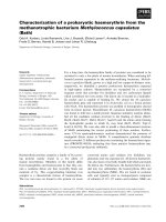
Fig. 1. Electrophoresis of purified LAC2. (A) Silver staining of a SDS/
7.5% PAGE; lanes 1 and 5: molecular mass standards; lane 2: 0.5 lgof
purified LAC1; lane 3: 0.5 lgofpurifiedLAC2;lane4:10lgoftotal
extra cellular proteins. (B) Native PAGE stained with p-phenylene
diamine; lane 1: total extra cellular proteins; lane 2: Lac1; lane 3, Lac2;
proteins corresponding to 0.005 U were deposited per lane. Dried gels
werescannedwithanAgfaSnapscanÒ 1236 using
FOTOLOOK
Ò 2.09.6.
Legends were added with
CANVAS
Ò 7.0.
Ó FEBS 2002 LAC2 from the basidiomycete C30 (Eur. J. Biochem. 269) 6121
The first 15 residues at the amino terminus are:
AIGPKADLTISNANI. The first six amino acids of this
sequence match perfectly the result obtained on the
laccase contained in fraction D in a preliminary study on
the enhancement of minor laccase production in C30
[15]. We also sequenced a 15-kDa internal peptide and
found that the first 15 residues from this peptide are
AIPNVGTINTDGGVN. A database search showed that
these peptides are closely related to those found in
laccase sequences from basidiomycete CECT 20197
(accession no. U65400), Trametes villosa (accession no.
L49376 and L78077) and Trametes versicolor (accession
no. Y18012).
clac2
cDNA cloning
The cDNA encoding LAC2 was cloned from a PCR
amplified cDNA library. It contains an open reading frame
1584 bp long coding for a protein 528 residue long, a 76-bp
5¢-untranslated region, a 220-bp 3¢-untranslated region and
a 31-bp poylA tail. The amino acid sequence deduced from
the open reading frame contains the peptide sequences
previously characterized from the LAC2 purified protein.
The clac2 ORF is 36 bp longer than the lac1 ORF
(AF162785) and the global identity between the two coding
sequences is 67%. At the protein level, the two enzymes are
69% identical but LAC2 possesses 12 extra amino acids,
seven of which constitute its carboxy terminus. The LAC2
nearest neighbors found in database are: Polyporus ciliatus
Lcc3-2 (AF176321-1, identity/homology: 81/95%), Tra-
metes versicolor LAC4 (Q12719, identity/homology: 71/
92%), Trametes pubescens LAC1A (AF414808-1, identity/
homology: 70/91%) and Trametes villosa LAC5 (Q99056,
identity/homology: 69/91).
LAC1 and LAC2 homology models
For each model, 10 calculated structures were fitted on the
reference structure, the C. cinereus laccase [23]. The general
organization of the two models was found to be very close
to that of the reference structure. The copper coordination
ligands are identical to those present in the reference and the
geometry of the T1 copper is basically preserved. However,
significant differences in folding were found in three regions
all located within 10 A
˚
around the T1 copper. All of these
differences are related to gaps present in the initial sequence
alignment and corresponding to either insertion or deletion
of one to four residues in the proteins considered (data
not shown). 3D representations of region the closest to the
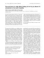
T1 copper are given in Fig. 2. Polypeptide chain length
variations of two residues for LAC1 (Fig. 2A) and three
residues for LAC2 (Fig. 2B) in the loop containing the T1
copper proximal ligand (H396 in C. cinereus) induce
noticeable structural changes around the copper.
DISCUSSION
The basidiomycete C30 secretes at least four different
laccases, the proportion of which depends on culture
conditions [12,15]. We have previously purified and
Fig. 2. Comparison of the backbone superposition at the T1 copper site
of the C. cinereus laccase and the C30 laccase models. The Ca trace of
C. cinereus (1A65) laccase is shown in red. For clarity, only segments
corresponding to loops L333–T341, V387–H399 and H451–A463 and
coordinating residues H396, C452 and H457 are represented. The Ca
traces of 10 calculated models of LAC1 (A) and LAC2 (B) are shown
in grey (coordinating residues, nearly superposable to those of the
C. cinereus laccase, have been omitted for clarity).
Table 3. LAC1 and LAC2 kinetic parameters. ND, not determined.
Substrate Enzymes pH
k
cat
(min
)1
)
K
m
(l
M
)
k
cat
/K
m
(min
)1
Æl
M
)1
)
SGZ LAC1 5.0 1800 1.8 1000
5.7 600 0.9 670
LAC2 5.7 65 600 6.8 9650
GUA LAC1 5.0 ND ND –
5.7 2300 71 30
LAC2 5.7 75 680 1006 76
ABTS LAC1 5.0 3350 10.7 310
5.7 610 2.9 210
LAC2 5.7 41 000 536 80
Table 2. Physico-chemical and EPR parameters of copper sites of laccases 1 and 2 from C30. A
//
values are in 10
)4
cm
)1
units; ND, not determined.
EPR parameters
T1 copper T2 copper
Enzymes opt pH
a
T (°C)
b
E° (V) A
//
g
//
g
^
A
//
g
//
g
^
Ref.
LAC1 4.5-5 55 0.73 96 ND ND > 140 ND ND 6
LAC2 5.5-6 55 0.56 88 2.165 2.025 172 2.25 2.027 This work
a
Values obtained with SGZ as substrate.
b
Temperature for which the main activity is reached with SGZ as substrate.
6122 A. Klonowska et al. (Eur. J. Biochem. 269) Ó FEBS 2002
characterized LAC1, the most abundant enzyme produced
by C30 [12]. The purification of a second laccase (LAC2)
from this fungus allows us to compare enzymes, the
synthesis of which is regulated differently. Indeed, LAC1
is produced under all the conditions we have tested so far
and thus is probably a constitutive form. On the other hand,
LAC2, which is almost absent in noninduced cultures,
becomes one of the most prominent laccases secreted when
the growth medium is supplemented with copper and
p-hydroxybenzoate; it can therefore be considered an
inducible enzyme [15]. Such differences in their patterns of
expression suggest a distinct physiological role for these two
isoforms and, although they share basic properties, the large
variation in catalytic activity for both phenolic and dyes
supports this idea.
The C30 laccase isoforms we have detected so far all have
an apparent molecular mass close to 65 kDa and, except for
a still-uncharacterized laccase, are all acidic proteins with pI
ranging from 3.2 (LAC2) to 3.6 (LAC1) [15]. The optimum
pH for syringaldazine oxidation is close to 4.5–5 for LAC1
and 5.5–6 for LAC2, values that, unlike the data obtained
for several laccases [24], do not correlate with their pI. On
the other hand, when tested at their respective optimum pH
values, the two isoforms are the most active at the same
temperature of 55 °C. The amino acid sequences deduced
from lac1, the gene encoding LAC1, and clac2, the cDNA
encoding LAC2, are 69% identical and the two proteins,
LAC1 and LAC2, present very similar optical and EPR
spectra. Generally speaking, all the above features of C30
enzymes are similar to those of other white-rot fungi. This is
expected as laccases form a group of highly homologous
proteins. However, several major physico-chemical features
distinguish LAC1 from LAC2. One is the 170 mV differ-
ence observed between their respective T1 redox poten-
tial measured at pH 5.7. Indeed, with a potential
E° ¼ 0.73 V, LAC1 belongs to the group of high redox
potential laccases (T. versicolor, T. villosa, Rhizoctonia sol-
ani, Pleurotus ostreatus POXC and POXA1b, Rigidosporus
lignosus B and D) whereas LAC2, with a potential
E° ¼ 0.56 V, belongs to the group of low redox potential
laccases (C. cinereus, Myceliophthora thermophila, Scytali-
dium thermophilum) [24–27]. To the best of our knowledge,
this difference in potential is the largest so far reported
between two laccases purified from the same organism.
Moreover, even though the T1 potentials of R. lignosus
laccases B and D and P. ostreatus POXC and POXA1b are,
respectively, 40 and 90 mV different [24], all these four
enzymes apparently belong to the high redox potential
group. C30 is therefore the first organism for which a
simultaneous production of high redox and low redox
potential laccases is reported.
Several attempts have been made to correlate the redox
potential variations found in laccases to the nature of the
specific amino acids present in their active site as their
oxidative capabilities appear tightly linked to this param-
eter [24,28]. The replacement F463M in a T. villosa
(accession no. AAC41686) high redox potential laccase
provides a fourth coordinating axial ligand to the T1
copper resulting in a 100-mV drop of its potential [27]. On
the other hand, the idea that the occurrence of a
phenylalanine residue might correlate with a high redox
potential in laccases was ruled out by the site directed
replacement of L fi F in two laccases [26]. The presence
of an F residue at the corresponding position in the
sequence of both the C30 high redox potential LAC1 and
low redox potential LAC2 sequences support this conclu-
sion. Similarly, the replacement of the LEA amino acid
triplet located immediately after the distal T1 coordinating
histidine (H456 in C. cinereus, accession no. 1A65) in the
high redox potential R. solani laccase (accession no.
Q02081), by a SVG amino acid triplet found in the low
redox potential M. thermophila (accession no. AAE35046)
and vice versa did not affect significantly the E° of the
recombinant enzymes [26]. In our study, the presence of a
LEA tripeptide both in the C30 LAC1 and LAC2
enzymes correlates well with these results. In an effort
to gain new insights into the factors influencing the
potential of the T1 copper in laccases, data on eight high
redox potential and four low redox potential enzymes
may not be enough to design new targets for mutagenesis.
On the other hand, models of our enzymes show
substantial structural variations close to the metal center
(Fig. 2) in a loop where main differences are found when
3D structures, including the recently published structures
of T. versicolor [30,31], and Melanocarpus albomyces [32]
laccases are compared (not shown). As folding of the
backbone around the metal center was already proposed
to be a major factor affecting the potential in iron–sulfur
proteins [29], it seems to us that it would make sense to
make mutants in this region.
A second distinction between C30 LAC1 and LAC2
enzymes can clearly be made on the basis of their kinetic
parameters for the oxidation of phenolic (syringaldazine
and guaiacol) as well as nonphenolic (ABTS) substrates
(Table 2). Depending on the pH conditions used, we found
that LAC2 k
cat
values are one to two orders of magnitude
higher than those of LAC1 whereas, the affinity for the three
substrates of the former enzyme is lower as reflected by
higher K
m
values. Strong differences in K
m
values are
commonly observed for laccases. As discussed in previous
studies [24,25], the differences in kinetics observed may be
the consequence of the variability of certain of the amino
acids involved in the substrates channel in specific enzymes.
A substantial variation in the folding of the T1 copper
pocket of the two enzymes, such as that mentioned above to
explain their difference in T1 potential, could also account
for their specific interaction with the substrates. In laccases,
enzyme efficiency (k
cat
/K
m
) correlates with the redox
potential of the substrates [24] and the two C30 laccases
behave more or less this way. In fact, like already observed
by Garzillo et al. in their study on T. trogii, R. lignosus and
P. ostreatus laccases [25], LAC1 and LAC2 activities on
phenolic compounds seem only partly related to their
specific redox capabilities. Indeed, LAC2 appears to be two
to 10 times more efficient than LAC1 on phenolic substrates
although with a 170-mV lower T1 copper redox potential.
As phenol oxidation involves release of a proton, factors
like hydrogen bonding or the extent of protonation of
ionizable residues in the vicinity of the T1 copper probably
have considerable effects on the overall efficiency.
Differences between LAC1 and LAC2 are not restricted
to the oxidation site as the two enzymes also react differently
toward sodium azide, an inhibitor known to bind to the
oxygen reduction site. It is likely that a channel governs the
accessibility to the T2/T3 cluster. Therefore, a LAC2/LAC1
I
50
ratio of 10 probably reflects a significant variation in the
Ó FEBS 2002 LAC2 from the basidiomycete C30 (Eur. J. Biochem. 269) 6123
size of the channel from LAC1 to LAC2. On the other
hand, in a recent study on laccases reactivity with dioxygen
[34], it is speculated from steady state analysis that laccases
have a conserved O
2
binding domain and that the rate of O
2
reduction is dependent on that of substrate oxidation. In
our case, this means that LAC2 should reduce dioxygen
much faster than LAC1 and a similar investigation on
oxygen reduction rates must be undertaken on C30 laccases
to further the description of their reactivity.
Generally speaking, when compared to kinetic data on
laccases from other fungi, it appears that while LAC1
activity is similar to other laccases for both phenolic and
nonphenolic substrates, LAC2 is a remarkably efficient
enzyme at least on the three substrates tested. A comparison
of enzyme efficiency restricted to the group of low redox
potential laccases reveals that, depending on the substrate,
LAC2 appears to be 2–100 times more efficient than its
C. cinereus, M. thermophila and S. thermophilum counter-
parts [24,33]. Again, as for their differences in T1 potential,
this is the first report on enzymes produced by a single
organism with such markedly different catalytic efficiency.
From a physiological point of view, the constitutive LAC1,
abundant in the culture supernatant [12], exhibits a relat-
ively high affinity for phenols but a relatively low capacity
for oxidation when compared to the inducible LAC2. The
consequences of these differences is not yet known but, by
analogy with permeation systems [34] for which such a
contrast between affinity and rate is often observed, we
could interpret the differences in laccase properties as a need
for the organism to maintain both a low capacity/high
specificity system when substrate level is low and a high
capacity/low specificity system when the substrate is abun-
dant.
In conclusion, we have demonstrated that LAC2, a laccase
produced by the basidiomycete C30 following copper and
p-hydroxybenzoate induction, is a low redox potential
enzyme with unusually high oxidative capabilities. The
kinetic data obtained both on phenolic and nonphenolic
substrates indicate that LAC2 might be a good catalyst for
the transformation of different substrates. As laccases are
generally produced as a number of isoenzymes encoded by
multigene families, the expression of which varies from
fungus to fungus, it is highly probable that other fungi
contain the equivalent of LAC2. A search for the appropri-
ate conditions of expression of a given activity being empirical
and time consuming, it will probably be more efficient to use
a heterologous expression system for laccase activities to
find other enzymes with high oxidative capacities.
ACKNOWLEDGEMENTS
A. K. is the recipient of an Agence de l’Environement et de la Maıˆtrise
de l’Energie (ADEME) fellowship. This work was in part supported by
a grant from the Conseil Ge
´
ne
´
ral 13. We thank Gilles Iacazio, Marius
Re
´
glier, Jalila Simaan and Marjorie Sweetko for their critical reading of
the manuscript.
REFERENCES
1. Gianfreda, L.F., Xu & Bollag, J M. (1999) Laccases: a useful
group of oxidoreductive enzymes. Bioremediation J. 3, 1–25.
2. Thurston, C.F. (1994) The structure and function of fungal lac-
cases. Microbiology 140, 19–26.
3. Solomon, E.I., Sundaram, U.M. & Machonkin, T.E. (1996)
Multicopper oxidases and oxygenases. Chem. Rev. 96, 2563–2605.
4. Mayer, A.M. (1987) Polyphenol oxidases in plants. Recent pro-
gress. Phytochem. 26, 11–20.
5. Hattaka, A. (1994) Lignin-modifying enzymes from selected
white-rot fungi: production and role in lignin degradation. FEMS
Microbiol. Rev. 13, 125–135.
6. Youn, H D., Hah, Y.C. & Kang, S O. (1995) Role of laccase in
lignin degradation by white-rot fungi. FEMS Microb. Lett. 132,
183–188.
7. Tagger, S., Pe
´
rissol,C.,Gil,G.,Vogt,G.&LePetit,J.(1998)
Phenoloxidases of the white-rot fungus Marasmius quercophilus
isolated from an evergreen oak litter (Quercus ilex L.). Enz.
Microb. Technol. 23, 372–379.
8. Farnet, A M., Tagger, S. & LePetit, J. (1999) Effects of copper
and aromatic inducers on the laccases of the white-rot fungus
Marasmius quercophilus. C. R. Acad. Sci. Paris/ Life Sci. 322, 1–5.
9. Farnet, A.M. (1998) Variabilite
´
phe
´
notypique et ge
´
ne
´
tique chez
Marasmius quercophilus basidiomyce
`
te colonisant une litie
`
re de
che
ˆ
ne vert. PhD Thesis. University of Aix-Marseille III, Marseille.
10. Klonowska, A., Gaudin, C., Ruzzi, M., Colao, M.C. & Tron, T.
(2002) Ribosomal DNA sequence analysis shows that the basi-
diomycete C30 belongs to the genus Trametes. Res. Microbiol,in
press.
11. Dedeyan, P. (1996) Purification et caracte
´
risation des laccases de
Marasmius quercophilus. PhD Thesis. University of Aix-Marseille
III, Marseille.
12. Dedeyan, B., Klonowska, A., Tagger, S., Tron, T., Iacazio, G.,
Gil, G. & LePetit, J. (2000) Biochemical and molecular char-
acterization of a laccase from Marasmius quercophilus. Appl. Env.
Microbiol. 66, 925–929.
13. Mansur, M., Suarez, T., Fernandez-Larrea, J.B., Brizuela, M.A. &
Gonzalez, A.E. (1997) Identification of a laccase gene family in the
new lignin-degrading basidiomycete CECT 20197. Appl. Env.
Microbiol. 63, 2637–2646.
14. Yaver, D.S. & Golightly, E.G. (1996) Cloning and characteriza-
tion of three laccase genes from the white-rot basidiomycete
Trametes villosa: genomic organization of the laccase gene family.
Gene 181, 95–102.
15. Klonowska, A., LePetit, J. & Tron, T. (2001) Enhancement of
minor laccases production in the basidiomycete Marasmius quer-
cophilus C30. FEMS Lett. 200, 25–30.
16. Matsudaira, P. (1990) Limited N-terminal sequence analysis. Meth
Enzymol. 182, 602–613.
17. Chirgwin, J.M., Przybyla, A.E., MacDonald, R.J. & Rutter, W.J.
(1979) Isolation of biologically active ribonucleic acid from sour-
ces enriched in ribonuclease. Biochemistry 18, 5294–5299.
18. D’souza, T.M., Boominathan, K. & Reddy, C.A. (1996) Isolation
of laccase gene-specific sequences from white-rot and brown-rot
fungi by PCR. Appl. Env. Microbiol. 62, 3739–3744.
19. Sali, A. & Blundell, T.L. (1993) Comparative protein modeling by
satisfaction of spatial restraints. J. Mol. Biol. 234, 779–815.
20. Thompson, J.D., Higgins, D.G. & Gibson, T.J. (1994) CLUSTAL
W: improving the sensitivity of progressive multiple sequence
alignment through sequence weighting, positions-specific gap
penalties and weight matrix choice. Nucl Acids Res. 22, 4673–4680.
21. Laskowski, A.R., MacArthur, M.W., Moss, D.S. & Thornton
(1993) PROCHECK: a program to check the stereochemical
quality of protein structures. J. Appl. Cryst. 26, 283–291.
22. Guex, N. & Peitsch, M.C. (1997) SWISS-MODEL and the Swiss-
PdbViewer: An environment for comparative protein modeling.
Electrophoresis 18, 2714–2723.
23. Ducros, V., Brzozowski, A.M., Wilson, K.S., Brown, S.H., Ost-
ergaard, P., Schneider, P., Yaver, D.S., Pedersen, A.H. & Davies,
G.J. (1998) Crystal structure of the type-2 Cu depleted laccase
from Coprinus cinereus at 2.2 A
˚
resolution. Nat. Struct. Biol. 5,
310–316.
6124 A. Klonowska et al. (Eur. J. Biochem. 269) Ó FEBS 2002
24. Xu, F., Shin, W., Brown, S.H., Wahleithner, J.A., Sundaram,
U.M. & Solomon, E.I. (1996) A study of a series of recombinant
fungal laccases and bilirubin oxidase that exhibit significant dif-
ferences in redox potential, substrate specificity, and stability.
Biochim. Biophys. Acta. 1292, 303–311.
25. Garzillo, A.M., Colao, M.C., Buonocore, V., Oliva, R., Falcigno,
L., Saviano, M., Santoro, A.M., Zappala, R., Bonomo, R.P.,
Bianco, C., Giardina, P., Palmieri, G. & Sannia, G. (2001)
Structural and kinetic characterization of native laccases from
Pleurotus ostreatus, Rigidoporus lignosus,andTrametes trogii.
J. Protein Chem. 20, 191–201.
26. Xu, F., Berka, R.M., Wahleitner, J.A., Nelson, B.A., Schuster,
J.R., Brown, S.H., Palmer, A.E. & Solomon, E.I. (1998) Site-
directed mutations in fungal laccases: effect on redox potential,
activity and pH profile. Biochem. J. 334, 63–70.
27. Xu,F.,Palmer,A.E.,Yaver,D.S.,Berka,R.M.,Gambetta,G.A.,
Brown, S.H. & Solomon, E.I. (1999) Targeted mutations in a
Trametes villosa laccase. Axial perturbations of the T1 copper.
J. Biol. Chem. 274, 12372–12375.
28. Messerschmidt, A. & Huber, R. (1990) The blue oxidases, ascor-
bate oxidases, laccase and ceruloplasmin. Eur. J. Biochem. 187,
341–352.
29. Stephens, P.J., Jollie, D.J. & Warshel, A. (1996) Protein control of
redox potentials of iron-sulfur proteins. Chem. Rev. 96, 2491–
2513.
30. Piontek, K., Antorini, M. & Choinowski, T. (2002) Crystal
Structure of a laccase from the fungus Trametes versicolor at 1.90-
A
˚
resolution containing a full complement of coppers. J. Biol.
Chem. 277, 37663–31669.
31. Bertrand, T., Jolivalt, C., Briozzo, P., Caminade, E., Joly, N.,
Madzak, C. & Mougin, C. (2002) Crystal structure of a four-
copper laccase complexed with an arylamine: insights into sub-
strate recognition and correlation with kinetics. Biochemistry 2002,
7325–7333.
32. Hakulinen, N., Kiiskinen, L.L., Kruus, K., Saloheimo, M.,
Paananen, A., Koivula, A. & Rouvinen, J. (2002) Crystal structure
of a laccase from Melanocarpus albomyces with an intact trinuclear
copper site. Nat Struct. Biol. 9, 601–605.
33. Xu, F. (2001) Dioxygen reactivity of laccase. Appl. Biochem.
Biotechn 95, 125–133.
34. Eide, D.J. (1998) The molecular biology of metal ion transport in
Saccharomyces cerevisiae. Annu. Rev. Nutr. 18, 441–469.
Ó FEBS 2002 LAC2 from the basidiomycete C30 (Eur. J. Biochem. 269) 6125